HAL Id: hal-03151601
https://hal.sorbonne-universite.fr/hal-03151601
Submitted on 24 Feb 2021HAL is a multi-disciplinary open access archive for the deposit and dissemination of sci-entific research documents, whether they are pub-lished or not. The documents may come from teaching and research institutions in France or abroad, or from public or private research centers.
L’archive ouverte pluridisciplinaire HAL, est destinée au dépôt et à la diffusion de documents scientifiques de niveau recherche, publiés ou non, émanant des établissements d’enseignement et de recherche français ou étrangers, des laboratoires publics ou privés.
Solid-State Phase Transformation and Self-Assembly of
Amorphous Nanoparticles into Higher-Order Mineral
Structures
Stanislas von Euw, Thierry Azaïs, Viacheslav Manichev, Guillaume Laurent,
Gérard Pehau-Arnaudet, Margarita Rivers, Nagarajan Murali, Daniel Kelly,
Paul Falkowski
To cite this version:
Stanislas von Euw, Thierry Azaïs, Viacheslav Manichev, Guillaume Laurent, Gérard Pehau-Arnaudet, et al.. Solid-State Phase Transformation and Self-Assembly of Amorphous Nanoparticles into Higher-Order Mineral Structures. Journal of the American Chemical Society, American Chemical Society, 2020, 142 (29), pp.12811-12825. �10.1021/jacs.0c05591�. �hal-03151601�
1
Title: Solid-state phase transformation and self-assembly of
1amorphous nanoparticles into higher-order mineral structures
2Authors: Stanislas Von Euw1,7*, Thierry Azaïs2, Viacheslav Manichev3,4, Guillaume
3
Laurent2, Gérard Pehau-Arnaudet5, Margarita Rivers4,6, Nagarajan Murali3, Daniel J. Kelly7,
4
Paul G. Falkowski1,3*
5 6
1Environmental Biophysics and Molecular Ecology Program, Department of Marine and
7
Coastal Sciences, Rutgers University, 71 Dudley Road, New Brunswick, New Jersey, 08901,
8
United States.
9
2Laboratoire de Chimie de la Matière Condensée de Paris,Sorbonne Université, CNRS, 4 place
10
Jussieu, F-75005, Paris, France.
11
3Department of Chemistry and Chemical Biology, Rutgers University, 123 Bevier Road,
12
Piscataway, New Jersey, 08854, United States.
13
4Institute of Advanced Materials, Devices, and Nanotechnology, Rutgers University, 607
14
Taylor Road, Piscataway, New Jersey, 08854, United States.
15
5UMR 3528 and UTech UBI, Institut Pasteur, 28 rue du Docteur Roux, F-75015 Paris, France.
16
6Department of Physics, Wellesley College, 106 Central Street, Wellesley, Massachusetts,
17
02481, United States.
18
7Trinity Centre for Bioengineering, Trinity Biomedical Sciences Institute, Trinity College
19
Dublin, Dublin 2, D02 R590, Ireland.
20 21
*correspondence to: [email protected]; [email protected]
22 23
2
Abstract:
24
Materials science has been informed by nonclassical pathways to crystallization based
25
on biological processes to fabricate damage-tolerant composite materials. Various
26
biomineralizing taxa, such as stony corals, deposit metastable, magnesium-rich, amorphous
27
calcium carbonate nanoparticles that further assemble and transform into higher-order mineral
28
structures. Here we examine a similar process in abiogenic conditions using synthetic,
29
amorphous calcium magnesium carbonate nanoparticles. Applying a combination of
high-30
resolution imaging and in situ solid-state nuclear magnetic resonance spectroscopy, we reveal
31
the underlying mechanism of the solid-state phase transformation of these amorphous
32
nanoparticles into crystals under aqueous conditions. These amorphous nanoparticles are
33
covered by a hydration shell of bound water molecules. Fast chemical exchanges occur: the
34
hydrogens present within the nanoparticles exchange with the hydrogens from the
surface-35
bound H2O molecules which, in turn, exchange with the hydrogens of the free H2O molecule
36
of the surrounding aqueous medium. This cascade of chemical exchanges is associated with
37
an enhanced mobility of the ions/molecules that compose the nanoparticles which, in turn,
38
allow for their rearrangement into crystalline domains via solid-state transformation.
39
Concurrently, the starting amorphous nanoparticles aggregate, and form ordered mineral
40
structures through crystal growth by particle attachment. Sphere-like aggregates and
spindle-41
shaped structures were respectively formed from relatively high or low weights per volume of
42
the same starting amorphous nanoparticles. These results offer promising prospects for
43
exerting control over such a non-classical pathway to crystallization to design mineral
44
structures that could not be achieved through classical ion-by-ion growth.
3
Main text:
46
Exerting control over non-classical pathways to crystallization to direct the growth,
47
polymorphism, and self-assembly of inorganic nanoparticles into higher-order structures is an
48
important goal of materials sciences, writ large 1–9. To achieve this, the dominant strategy is to
49
govern the initial nucleation stage in a multitude of precipitation reactions occurring far from
50
thermodynamic equilibrium 10. These reactions share the common purpose of converting
51
solution precursors into solid mineral materials 11 and use various approaches to overcome the
52
free-energy barrier to nucleation. In the case of calcium carbonate and calcium phosphate
53
minerals, these approaches were initially implemented to uncover the biomineralization
54
processes being used by marine calcifiers and vertebrates to allow support 12, mastication 13,
55
defense 14, attack 15, or optical 16 functions. They span from the utilization of (i)
56
supersaturated concentrations 17–19 sometimes combined with (ii) confinement effects 20–23,
57
the use of (iii) templates for epitaxial growth 24, and of (iv) various types of
mineralization-58
directing agents such as synthetic polyelectrolytes 25–29, proteins 30–34 and amino acids 35–37.
59
The future exploitation of stable pre-nucleation clusters 38–40 may offer additional prospects
60
for exerting some control over various non-classical crystallization processes 41,42.
61
Some of the above mentioned approaches were successfully applied to design and
62
manufacture different nature-inspired inorganic-organic composite materials 43–48; however,
63
they suffer from two main limitations. First, they often lead to final materials which are very
64
limited in terms of size (generally not exceeding a few millimeters along one dimension).
65
Second, the level of mineralization does not reach those of their natural analogues, principally
66
bone and nacre. The latter is the main limitation that prevents the fabrication of high strength
67
materials. Here we consider an alternative strategy that overcome these limitations and allow
68
the manufacturing of novel materials. Future approaches likely will skip over the initial
69
nucleation stage on which little control can be exercised, and focus on the conversion of solid,
4
metastable amorphous nanoparticles into their crystalline counterparts. Such metastable
71
amorphous nanoparticles that assemble and transform into higher-order, hierarchical mineral
72
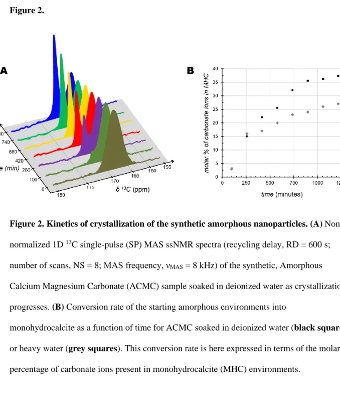
structures have been observed across various biomineralizing taxa 49–59, but their use in
73
synthetic systems remains extremely limited 60–64. Here we used Amorphous Calcium
74
Magnesium Carbonate (ACMC) nanoparticles as models since most biogenic deposits of
75
amorphous CaCO3 contain Mg2+ ions 49,65,66. Especially, we monitored their pathway to
76
crystallization in wet conditions to understand their potential use in the development of novel
77
materials. To this end, we applied a combination of high-resolution imaging and in situ
solid-78
state nuclear magnetic resonance (NMR) spectroscopy. In particular, the latter technique
79
provides unprecedented atomic-scale insights into the mechanism of phase transformation of
80
these amorphous nanoparticles over time.
81
Chemical structure and composition
82
A number of physical characterization techniques were applied to assess the structure
83
and composition of a 13C-labelled (98 atom% 13C) Amorphous Calcium Magnesium
84
Carbonate (ACMC) sample. This sample was first studied in dry conditions. Powder X-ray
85
diffraction observations confirm that this is a non-crystalline solid given the absence of Bragg
86
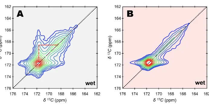
reflections (Fig. S1). According to TGA measurements (Fig. S2), the mass fraction of the
87
hydrous species associated with the particles of ACMC is in the range of 22 to 26 wt. %. An
88
average atomic Ca/Mg ratio ≈ 4.0 was estimated using energy-dispersive X-ray spectroscopy
89
(EDS). As such, the atom% of Mg in ACMC [defined as Mg/(Ca + Mg) x 100] is about 20.0.
90
This is similar to the value found in a number of biogenic deposits of amorphous calcium
91
carbonate such as those in the cuticles and gastroliths in crustaceans, along with those in the
92
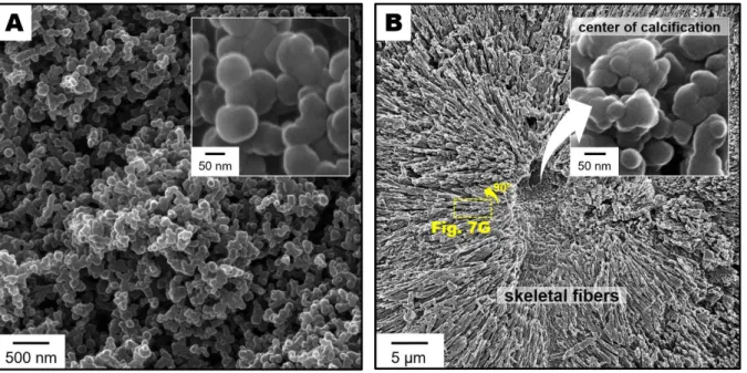
spicules in cnidarians 65. Scanning Helium Ion Microscopy (SHIM) observations show that
93
ACMC is in the form of spherical nanosized particles with a diameter of about 60 to 90 nm
5
(Fig. 1A). These features resemble those of biogenic, magnesium-rich, amorphous calcium
95
carbonate particles present at early stages of coral biomineralization (Fig. 1B).
96
Solid-state Nuclear Magnetic Resonance (ssNMR) spectroscopy was applied to
97
investigate the carbon and hydrogen chemical environments of ACMC. One dimensional (1D)
98
{1H}13C cross polarization (CP) (solid lines), and 13C single-pulse (SP) (doted lines) magic
99
angle spinning (MAS) ssNMR spectra of ACMC are shown in Fig. S3. Both spectra are in the
100
form of a single, symmetric resonance whose carbon chemical shift [δ(13C) = 168.2 ppm], full
101
width at half maximum (FWHM = 3.9 ppm) and Gaussian lineshape are characteristic of
102
carbonate ions (CO32-) present in amorphous environments. This carbon chemical shift is
103
similar with that of calcite 67 which suggests that the present sample may be considered as a
104
proto-calcite amorphous calcium carbonate 68. Further, the resonance in the 13C SP spectrum
105
is identical with the one in the {1H}13C CP spectrum in terms of lineshape and linewidth:
106
δ(13C) = 168.2 ppm and full FWHM = 3.9 ppm. This statement stands true regardless the CP
107
contact time (tCP) as shown in Fig. S4 where tCP was varied from 0.2 to 10 ms. The {1H}13C
108
CP spectrum selectively exposes 13C nuclei nearby 1H nuclei that belong to different rigid
109
hydrogen-bearing ions/molecules, whereas the 13C SP spectrum reveals all 13C nuclei since it
110
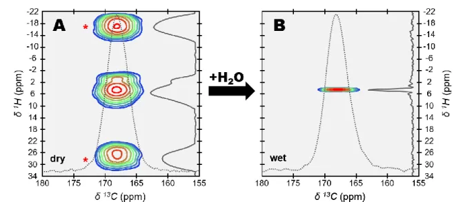
was recorded in quantitative conditions. Indeed, a long relaxation delay (RD) of 600 seconds
111
was chosen to allow for full relaxation of the longitudinal magnetization. According to their
112
similarity in terms of lineshape and linewidth, the {1H}13C CP spectrum exposes similar 13C
113
nuclei as those observed upon direct excitation in the quantitative 13C SP spectrum. These
114
observations provide important structural and chemical information. First, this is evidence that
115
the rigid hydrogen-bearing ions/molecules are homogenously distributed throughout the
116
amorphous calcium carbonate nanoparticles; and a similar conclusion was previously reached
117
by others 68,69. Second, this also rules out the presence of bicarbonate ions (HCO3-) given the
6
absence of a distinct upfield signal at short contact time (expected in the range of δ(13C) ≈
119
155-165 ppm 67,70,71).
120
To reveal the nature of the hydrogen-bearing ions/molecules present in the amorphous
121
nanoparticles, further 1H-based ssNMR experiments were applied. The 1D 1H direct
122
excitation (DE) MAS ssNMR spectrum of ACMC is shown in Fig. S5A. However, this
123
spectrum exposes a broad signal centered around δ(1H) = 4.9 ppm along with two narrow
124
resonances at δ(1H) = 1.2 and 3.6 ppm respectively, respectively due to the presence of water
125
and mobile ethanol molecules weakly adsorbed onto the surface of the particles. The latter
126
originate from anhydrous ethanol that was used post-precipitation to allow preservation of the
127
solid as an amorphous phase upon storage. As an alternative, we recorded a 1D {1H-13C}1H
128
double cross polarization (CP) MAS ssNMR experiment (Fig. S5B). It gives rise to a 13
C-129
filtered 1H spectrum whose signals correspond to structural hydrogen-bearing ions/molecules
130
present within the amorphous nanoparticles. This approach was successfully used to
131
investigate the hydrogen-bearing ions/molecules present in bone mineral in intact bone tissue
132
72. Here two main resonances are clearly observable. According to their respective chemical
133
shift, they reflect the presence of hydroxyl ions (OH-) [observable at δ(1H) = 1.0 ppm 73,74]
134
and structural water molecules [observable at δ(1H) = 5.6 ppm]. In addition, the apparent
135
dissymmetry of the main water resonance suggests the presence of a broad downfield signal
136
that spreads up to δ(1H) ≈ 13 ppm (black arrow). Further, the fact that the 13C-filtered 1H
137
spectrum cannot be satisfactory fitted with only two peaks at δ(1H) = 1.0 ppm (OH-) and 5.6
138
ppm (H2O) confirms the presence of this additional broad signal (Fig. S6A). In contrast, the
139
same 13C-filtered 1H spectrum can be properly fitted with three peaks at δ(1H) = 1.0 ppm (OH
-140
), 5.6 ppm (H2O) and 7.0 ppm (Fig. S6B). The use of a single peak centered at δ(1H) = 7.0
141
ppm to materialize the additional broad signal is somewhat arbitrary. Indeed, this broad signal
142
is probably composed of heterogeneous hydrogen environments leading to a wide distribution
7
of NMR chemical shifts. However, the fact that this broad signal is centered at δ(1H) = 7.0
144
ppm suggests the presence of structural water molecules engaged in stronger hydrogen bonds
145
than those observed at δ(1H) = 5.6 ppm 75.
146
Kinetics of crystallization
147
Time resolved, in situ ssNMR experiments were undertaken to investigate the kinetics
148
of crystallization of ACMC under aqueous conditions. To this end, the amorphous powder
149
was soaked in deionized water. Following this, 1D 13C single-pulse (SP) MAS ssNMR spectra
150
were recorded consecutively. After a period of approximately 1 h, a second carbon resonance
151
appears, and its intensity increases over time (Fig. 2A). This second carbon resonance is
152
narrow (FWHM = 0.70 ppm) and centered at δ(13C) = 171.7 ppm, and, hence, reflects the
153
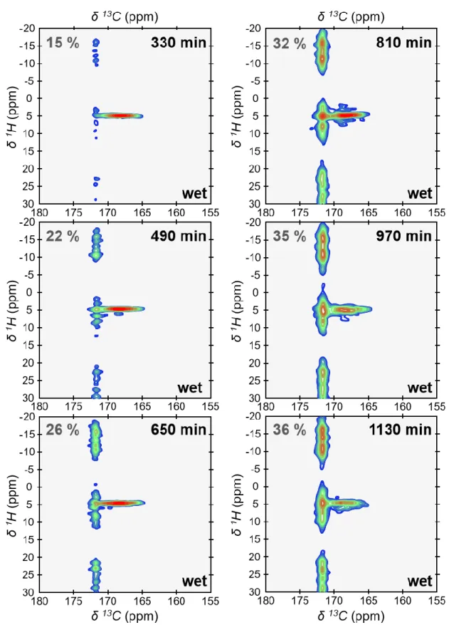
growth of monohydrocalcite (MHC, calcium carbonate monohydrate: CaCO3.H2O) 67.
154
Monohydrocalcite is one of the six crystalline forms of calcium carbonate. It is deposited in a
155
variety of sedimentary environments (e.g., lakes 76 and speleothems 77). Monohydrocalcite is
156
also the result of a number of biomineralization processes including otoliths of vertebrates 78,
157
calcareous corpuscles of certain flatworms 79 and guinea pig bladder stones 80. Further,
158
monohydrocalcite could also be a metastable intermediate phase in the formation of both
159
aragonite and calcite 81,82. It is well documented that amorphous calcium carbonates are
160
metastable and spontaneously crystallize in water. Especially if crystallization occurs via
161
solid-state transformation (which is discussed in the next section), one could expect here the
162
formation of calcite since ACMC may be considered as a proto-calcite amorphous calcium
163
carbonate (Fig. S3). However, it is also well documented that magnesium regulates the
164
crystallization of amorphous calcium carbonates 83,84 and, in certain conditions, favors the
165
formation of monohydrocalcite 81,82,85–87. Here the consecutive 13C SP MAS ssNMR spectra
166
were recorded under quantitative conditions (recycling delay, RD = 600s). As such, they
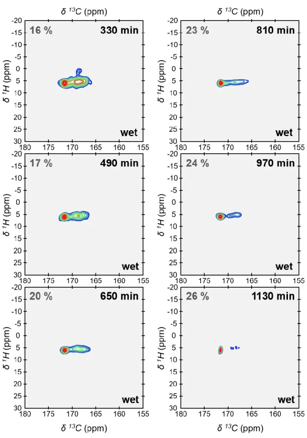
167
allow for determining the conversion rate of the starting amorphous environments into
8
monohydrocalcite as a function of time (Fig. 2B). This conversion rate is here expressed in
169
terms of the molar percentage of carbonate ions present in monohydrocalcite environments.
170
To achieve this, the consecutive 13C SP MAS ssNMR spectra were all fitted with two peaks as
171
shown in Fig. S7. The area under each peak was integrated to obtain the molar ratio of
172
carbonate ions present in crystalline and amorphous environments. As a result, here we show
173
that nearly 40% of the carbonate ions of ACMC were converted into monohydrocalcite after a
174
period of about 20 h.
175
Solid-state phase transformation versus dissolution-reprecipitation?
176
To shed light on the phase transformation process of ACMC into monohydrocalcite, a
177
two-dimensional (2D) 13C-13C Dipolar Assisted Rotational Resonance (DARR) MAS ssNMR
178
experiment was performed (Fig. 3A). To this end, the amorphous powder was soaked in water
179
until its conversion rate into monohydrocalcite reached about 30%, which took approximately
180
12 h (Fig. 2B). The 2D 13C-13C DARR MAS ssNMR spectrum is in the form of a 13C-13C
181
correlation map in which off-diagonal signals are due to magnetization exchange between
182
nearby 13C nuclei. Here a strong off-diagonal signal (red dotted lines) connects the carbonate
183
ions present in monohydrocalcite [observable at δ(13C) = 171.7 ppm] with those present in the
184
amorphous environments [observable at δ(13C) = 168.2 ppm]. As a consequence, these results
185
clearly suggest that the nascent crystalline environments together with the starting amorphous
186
environments belong to the same particles. This is strong evidence that the starting amorphous
187
nanoparticles transform into monohydrocalcite via a mechanism of solid-state phase
188
transformation.
189
An alternative scenario would see first the dissolution of the starting amorphous
190
nanoparticles followed by their reprecipitation into monohydrocalcite. Indeed, using various
191
methods [including isotopic labelling 88, in situ liquid cell transmission electron microscopy
192
(TEM) 89 and Raman spectroscopy 82, time resolved scanning electron microscopy (SEM) 86,
9
pH and supersaturation measurements 90, Energy Dispersive X-ray Diffraction (ED-XRD) 91,
194
small and wide angle X-ray scattering (SAXS/WAXS) 81,92, or the combination of several of
195
these techniques], various mechanisms including steps of dissolution-reprecipitation were
196
pointed out in the pathway to crystallization of different nanoparticles of synthetic amorphous
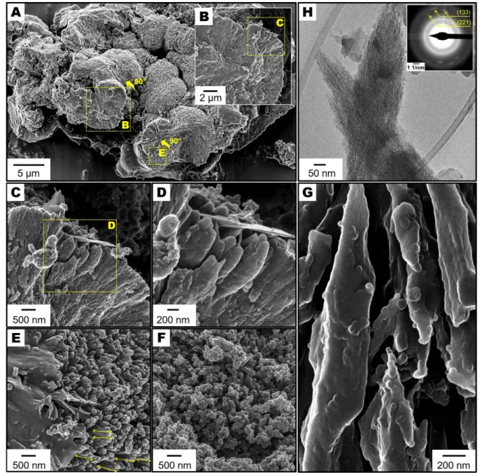
197
calcium carbonates into calcite, magnesian calcite, aragonite, vaterite and even
198
monohydrocalcite. Why under certain conditions some types of nanoparticles of amorphous
199
calcium carbonate crystalize via solid-state transformation while some others first dissolve
200
and then reprecipitate is still unclear. This major discrepancy is certainly multifactorial,
201
depending on the polymorph and the chemical composition of the starting amorphous
202
particles together the chemistry of the reaction solution. In this direction, a recent study
203
suggests that the presence of Mg2+ ions incorporated in the solid phase brings excess
204
structural water which, in turn, “weakens the ionic binding network” and favors a solid-state
205
transformation 89. In addition, it was also reported that an increased water content accelerates
206
the mechanism of solid-state transformation in the case of a temperature-induced
207
crystallization 93,94. As such, it is important to rule out the eventuality of a mechanism of
208
dissolution-reprecipitation during the crystallization of ACMC in the present study. To this
209
end, we simulated a mechanism of dissolution-reprecipitation using a physical mixture
210
containing 40 wt. % monohydrocalcite particles and 60 wt. % amorphous particles soaked in
211
water. The former particles originate from a new 13C-labelled (99 atom% 13C)
212
monohydrocalcite sample (MHC) (whose powder X-ray diffraction pattern is shown in Fig.
213
S8) that was prepared by a direct precipitation method 85, whereas the latter particles are those
214
of the amorphous ACMC sample. The 13C-13C DARR MAS ssNMR spectrum of this physical
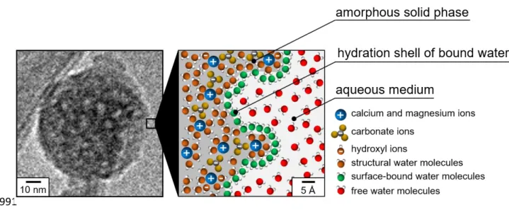
215
mixture soaked in water is shown in Fig. 3B. This spectrum was recorded within 90 minutes
216
following the wetting step so that the conversion rate of ACMC into monohydrocalcite
217
remains very low (must be below ≈ 2% - Fig. 2B). As expected, here the absence of
10
diagonal signal confirms that the carbonate ions present in the monohydrocalcite
219
environments of MHC are not in proximity with those present in the amorphous environments
220
of ACMC. As such, the observation of an off-diagonal signal in the 13C-13C DARR MAS
221
ssNMR spectrum of the ACMC sample partially converted into monohydrocalcite excludes a
222
mechanism of dissolution-reprecipitation (Fig. 3A).
223
Surface hydration shell and Hydrogen-Hydrogen chemical exchanges
224
The possible interactions of the amorphous nanoparticles of ACMC with water
225
molecules were examined. To this end, 2D {1H}13C HetCor MAS ssNMR experiments of
226
ACMC were performed both in dry and wet conditions (Fig. 4). In wet conditions, the sample
227
was soaked in water and the 2D {1H}13C HetCor MAS ssNMR experiment was performed
228
within one hour following the wetting step. In these conditions, monohydrocalcite has yet not
229
been formed, or rather remains below the detection threshold of ssNMR (Fig. 2B). These 2D
230
{1H}13C HetCor MAS ssNMR spectra are in the form of 1H-13C correlation maps in which the
231
different signals, named “correlation peaks”, reveal atomic-scale spatial proximities among
232
rigid hydrogen-bearing ions/molecules (displayed along the vertical, indirect 1H dimension)
233
and carbon-bearing ions (displayed along the horizontal, direct 13C dimension). In dry
234
conditions, a broad signal associated with two intense spinning sidebands are observable. This
235
signal results from the juxtaposition of two different correlation peaks which, similarly to the
236
{1H-13C}1H double cross polarization (CP) MAS ssNMR experiment, are due to the presence
237
of OH- ions [observable at δ(1H) ≈ 1.0 ppm] and structural water molecules [observable in the
238
range of δ(1H) ≈ 5-13 ppm]. In contrast, in wet conditions, only a single and sharp correlation
239
peak is observable while the two intense spinning sidebands are no longer observable. This
240
sharp correlation peak is at δ(1H) = 4.7 ppm (FWHM = 0.6 ppm) in the indirect 1H dimension,
241
and, hence, reflects the presence of water molecules. Since Brownian motion averages dipolar
242
couplings to zero, these water molecules must be adsorbed onto the particles’ surface to allow
11
CP magnetization transfer. We can infer from this that a hydration shell of bound water forms
244
around the particles of ACMC under aqueous conditions.
245
Further, the fact that the broad isotropic signal seen in dry conditions is no longer
246
observed in wet conditions where only a sharp water correlation peak is present (Fig. 4), is
247
diagnostic of a fast hydrogen exchange regime on the NMR time scale. Indeed, this is
248
evidence that fast chemical exchanges occur between hydrogens from the free water
249
molecules and those from the rigid hydrogen-bearing ions/molecules (i.e., OH- and H2O)
250
present in the amorphous solid. Due to the presence of the hydration shell of bound water,
251
these chemical exchanges are likely to occur in two steps: the hydrogens present in the
252
amorphous solid phase exchange with the hydrogens from the surface-bound H2O molecules
253
which, in turn, exchange with the hydrogens of the free H2O molecule of the surrounding
254
aqueous medium. Our TGA measurements (Fig. S2) show that, in dry conditions, the mass
255
fraction of the different populations of hydrogen-bearing ions/molecules (OH- and H2O)
256
associated with the particles of ACMC is in the range of 22 to 26 wt. %. As a result, we could
257
calculate the proportion of hydrogens originating from the particles over the total number of
258
hydrogens present in the MAS rotor following the wetting step: i.e., from about 5 to 10%. The
259
number of hydrogens from the free water molecules is therefore in large excess over the
260
number of hydrogens present in the particles and, hence, the latter are not detected in wet
261
conditions (fast exchange regime) and solely the excess water signal is observed at δ(1H) =
262
4.7 ppm. In addition, the fact that the two intense spinning sidebands observed in dry
263
conditions are no longer observable when the particles are soaked in water also advocate for
264
chemical exchanges. This is evidence that the hydrogens from the particles were mobilized in
265
wet conditions due to their exchanges with the hydrogens from the free water molecules.
266
Further, similar hydrogen-hydrogen exchanges were also pointed out not only for synthetic
12
particles of amorphous calcium phosphate (ACP) soaked in water, but also for the ACP-like
268
surface of bone mineral particles from a fresh and intact bone tissue sample 95.
269
For further evidence of the presence of a hydration shell of bound water, cryogenic
270
transmission electron microscopy (cryo-TEM) observations were obtained from the
271
amorphous nanoparticles of ACMC dispersed in water (Fig. S9). This dispersion was
272
cryofixed in liquid ethane within 10 min after its preparation and, hence, crystallization has
273
not yet started. A low magnification micrograph shows aggregates of nanoparticles that were
274
highlighted by yellow circles in Fig. S9A. Regions immediately around these aggregates
275
appear darker, indicating that the amorphous ice is thicker. This is evidence that the
276
nanoparticles (shown at higher magnification in Fig. S9B) retain water. Further, when these
277
nanoparticles are not “piled up” on top of each other as those pointed out with yellow arrows
278
in Fig. S9B, tiny lighter zones are visible and are certainly due to the presence of pores.
279
These pores are almost certainly not artifacts caused by electron beam irradiation since they
280
do not evolve upon prolonged observation. As a result, we suggest that the pores facilitate the
281
fast hydrogen-hydrogen exchanges that we observed in the 2D {1H}13C MAS HetCor ssNMR
282
experiments performed in wet conditions.
283
To better understand the origin of the hydrophilic properties of ACMC, we examined
284
whether a surface hydration shell can also form around the particles of a hydrated calcium
285
carbonate mineral soaked in water. To this end, the MHC sample was used since
286
monohydrocalcite (CaCO3.H2O, with one water molecule per calcium carbonate group) is a
287
suitable model mineral for amorphous calcium carbonates in terms of chemical composition
288
96. The 13C single-pulse (SP) MAS ssNMR spectrum of MHC is shown in Fig. S10A. This
289
sample is mainly composed of monohydrocalcite environments, but also contains residual
290
amorphous environments that were not converted into monohydrocalcite (Fig. S10B). Further,
291
the 1D {1H-13C}1H double cross polarization (CP) MAS ssNMR spectrum of MHC is shown
13
in Fig. S10C. This spectrum exposes a single, symmetric resonance [centered at δ(1H) = 6.0
293
ppm; FWHM = 8.9 ppm] attributed to structural water molecules in monohydrocalcite
294
environments. As for the possible interactions of the particles of MHC with water molecules,
295
they were investigated in a similar manner as for ACMC. Two-dimensional {1H}13C HetCor
296
MAS ssNMR experiments of MHC were performed both in dry and wet conditions and are
297
shown in Fig S11. The signal due to the presence of the residual amorphous environments
298
differs in dry and wet conditions. While this signal is broad and barely visible is dry
299
conditions (grey arrow), it is clearly detected in wet conditions where a sharp correlation peak
300
at δ(1H) = 4.7 ppm in the indirect 1H dimension shows the presence of surface-bound water
301
molecules. In contrast, the signal due to the presence of the monohydrocalcite environments
302
was unchanged after hydration. In both dry and wet conditions, this signal is in the form of a
303
broad correlation peak due to the presence of CO32- ions [observable at δ(13C) = 171.7 ppm in
304
the direct 13C dimension] near structural H2O molecules [centered at δ(1H) = 6.0 ppm in the
305
indirect 1H dimension]. As such, contrary to the amorphous environments of MHC but also
306
those of ACMC, the presence of an excess of free water in the MAS rotor does not cause the
307
formation of a hydration shell of bound water associated with the monohydrocalcite
308
environments. These results suggest that the hydrophilic properties of ACMC are not due to a
309
specific chemical composition including Ca2+ and CO32- ions along with structural H2O
310
molecules, but are rather the result of a definite amorphous structure.
311
Evolution of the hydrogen chemical environments as crystallization progresses
312
The evolution of the hydrogen chemical environments during the conversion of the
313
starting amorphous nanoparticles into monohydrocalcite was also scrutinized. To this end,
314
additional 2D {1H}13C HetCor MAS ssNMR experiments of ACMC soaked in water were
315
therefore undertaken consecutively following the initial wetting step (Fig. 5). The previously
316
mentioned, quantitative 1D 13C SP MAS ssNMR spectra, were recorded between each of
14
these 2D {1H}13C HetCor experiments so that the molar percentage of monohydrocalcite is
318
known. Here the growth of monohydrocalcite appears in the form of a broad, composite
319
signal along the vertical, indirect 1H dimension whose intensity progressively increases
320
[observable at δ(13C) = 171.7 ppm in the horizontal, direct 13C dimension]. This broad,
321
composite signal is also observable in the 1H slices taken at the monohydrocalcite position
322
(Fig. S12A). The signal arises from hydrogen-bearing ions/molecules present within the
323
nascent crystalline environments and spreads from δ(1H) ≈ -5 to 15 ppm; this is in agreement
324
with the 13C-filtered 1H MAS NMR spectrum of the reference sample of monohydrocalcite
325
shown in Fig. S10C. Moreover, intense spinning sidebands, signature of hydrogen-bearing
326
ions/molecules with restricted mobility, are also observed in agreement with the crystalline
327
nature of these nascent environments (Fig. S12A).
328
As for the starting amorphous environments, a sharp water correlation peak not
329
associated with any spinning sidebands (which is diagnostic of a fast hydrogen exchange
330
regime on the NMR time scale) is visible at the ACMC position[i.e., δ(13C) = 168.2 ppm in
331
the horizontal, direct 13C dimension](Fig. 5). As a result, the 1H slices taken at the ACMC
332
position only expose a single and narrow resonance [centered at δ(1H) = 4.7 ppm; FWHM in
333
the range of 0.6 to 1.3 ppm] attributed to water molecules bound to the particles' surface (Fig.
334
S12B). These observations are similar to what is observed for ACMC in wet conditions before
335
crystallization (Fig. 4B), and highlight that the starting amorphous environments remain
336
hydrated while crystallization progresses. They also imply that the hydrogen-bearing
337
ions/molecules remaining in the starting amorphous environments keep undergoing chemical
338
exchanges with the free water molecules as crystallization progresses.
339
Hydrogen-deuterium chemical exchanges
340
For further evidence of these chemical exchanges, 2D {1H}13C HetCor MAS ssNMR
341
experiments of ACMC soaked in heavy water (D2O - 99.99 atom% D) were undertaken
15
sequentially (Fig. 6). Quantitative 1D 13C SP MAS ssNMR spectra were recorded between
343
each of the 2D {1H}13C HetCor experiments so that the conversion rate of ACMC into
344
monohydrocalcite is known. Here this conversion rate reaches up 26% after 1130 minutes
345
which is therefore about 40% lower than in the case of ACMC soaked in H2O at the same
346
stage (conversion rate, 36%) (Fig. 2B); and this discrepancy is likely related to a kinetic
347
isotope effect. In contrast to the successive 2D {1H}13C HetCor MAS ssNMR spectra of
348
ACMC soaked in H2O where a broad, composite signal was observed along the indirect 1H
349
dimension, here the growth of monohydrocalcite appears in the form of a single, narrow
350
correlation peak [observable at δ(13C) = 171.7 ppm in the horizontal, direct 13C dimension].
351
The 1H slices taken at the monohydrocalcite position reveal a single, symmetric resonance
352
[centered at δ(1H) = 6.0 ppm; FWHM = 2.2 ppm] (Fig. S13A); this chemical shift is
353
characteristic of structural water molecules in monohydrocalcite environments (Fig. S10C).
354
However, the associated linewidth is much smaller due to a reduction of the 1H dipolar
355
couplings induced by a partial deuteration. Indeed, here 1H/2H chemical exchanges led to the
356
deuteration of the hydrogen-bearing ions/molecules present in the starting amorphous
357
nanoparticles before crystallization occurs (after about 1 h - Fig. 2B). As a result, the protium
358
isotopes, originating from ACMC, were diluted following the addition of the large excess of
359
deuterium isotopes originating from the D2O solution [2H/(2H + 1H) ≥ 90 atom% in the MAS
360
rotor]. This “isotopic dilution” has largely lowered the magnitude of the homonuclear 1H-1H
361
dipolar couplings within the particles which, in turn, gave rise to 1H spectra of the
362
monohydrocalcite environments with higher resolution (compared with H2O). The same
363
phenomenon also explains the presence of weaker spinning sidebands associated with the
364
nascent crystalline environments growing in D2O compared to H2O (Fig. S13A).
365
As for the starting amorphous environments, the sharp water correlation peak that was
366
previously seen at δ(1H) = 4.7 ppm (FWHM in the range of 0.6 to 1.3 ppm) along the indirect
16
1H dimension of the successive 2D {1H}13C HetCor MAS ssNMR spectra of ACMC soaked
368
in H2O was not observed. Instead, two narrow correlation peaks due to the presence of
369
structural OH- ions [δ(1H) = 1.0 ppm - no longer observed after 330 min] and structural water
370
molecules [δ(1H) = 5.6 ppm; FWHM ≈ 2.9 ppm] arise from the starting amorphous
371
environments (Fig. 6). The intensity of the water correlation peak progressively declines up to
372
970 minutes, after which the peak becomes almost no longer visible; this is also observable in
373
the 1H slices taken at the ACMC position [i.e., δ(13C) = 168.2 ppm in the direct 13C
374
dimension] (Fig. S13B). The number of hydrogens originating from the particles initially
375
represents almost 100% of the total number of hydrogens present in the MAS rotor following
376
the wetting step with D2O. The surface-bound water molecules (mostly in the form of D2O,
377
and in a less extent H2O but also HDO that were formed following the chemical exchanges)
378
are not detected in the 2D {1H}13C HetCor spectra due to the fast exchange regime where
379
solely the excess OH- / H2O and HDO signals arising from the particles are observed. Further.
380
the disappearance of these OH-/ H2O and HDO signals is due to the rearrangement of these
381
populations of hydrogen-bearing ions/molecules present in the starting amorphous
382
environments to form of monohydrocalcite via a solid-state phase transformation (Fig. 3A).
383
Crystal growth by accretion of amorphous nanoparticles
384
The mineral structures resulting from the crystallization of ACMC soaking in water in
385
the MAS rotor were scrutinized by Scanning Helium Ion Microscopy (SHIM). To this end,
386
the previously mentioned, consecutive 13C SP MAS ssNMR experiments of ACMC soaked in
387
water, have been run until the signal arising from the starting amorphous environments stops
388
evolving (after a period of approximately 2500 min). Hence, at this stage, the conversion rate
389
of ACMC into monohydrocalcite has reached its maximum (Fig. S14). Following this, the
390
wet powder was simply collected from the MAS rotor, washed with deionized water, and then
391
dried at ambient temperature. The resulting dry powder was analyzed by X-ray diffraction
17
that has confirmed the presence of monohydrocalcite (Fig S15). From the same powder, an
393
average atomic Ca/Mg ratio ≈ 5.0 was estimated using energy-dispersive X-ray spectroscopy
394
(EDS). It corresponds to an atom% of Mg in ACMC converted into monohydrocalcite
395
[defined as Mg/(Ca + Mg) x 100] of about 16.6 (against 20.0 for ACMC before
396
crystallization). It shows that a small proportion of Mg2+ have been expelled out of the
397
particles following crystallization. This suggests that the leading process of solid-state
398
transformation of ACMC into monohydrocalcite is here associated with a small loss of
399
magnesium that possibly occurs via a mechanism of Ostwald ripening 81. As for the SHIM
400
observations, a low magnification micrograph clearly shows that the crystallization process of
401
ACMC occurs via a spherulitic growth mechanism (Fig. 7A). Successive magnification
402
micrographs on an open spherulite reveal the presence of crystals that greatly differ from the
403
classical view of inorganic crystals with faceted surfaces (Fig. 7, B to D). Instead, acicular
404
crystals displaying a highly textured surface due to the apparent presence of spherical
405
nanoparticles “building-blocks” are here observed. Similar acicular crystals are also
406
observable at the surface of a spherulite (double yellow arrows in Fig. 7E), where the
407
spherical nanoparticles “building-blocks” match with the starting amorphous nanoparticles of
408
ACMC in terms of size (Fig. 7F). Such “nano-particulate” texture was initially observed for
409
biological aragonite in nacre 53,97 and more recently for biological aragonite in coral (Fig. 7G)
410
49 and across a broad range of biomineralizing taxa 98. This “nano-particulate” texture is
411
another evidence for a mechanism of solid-state transformation; this is also signature of
412
crystal growth by accretion of amorphous nanoparticles 99,100 which is one variety of
413
crystallization by particle attachment 101.
414
In addition, in order to assess the behavior of ACMC within in a higher volume of
415
water, the mineral structures resulting from the crystallization of ACMC soaking in a reaction
416
vial instead of in an NMR rotor were also scrutinized. To this end, the amorphous
18
nanoparticles of ACMC were dispersed in deionized water in a 10ml vial (mass concentration
418
≈ 2% w/v against about 30% w/v in the MAS rotor) and the resulting suspension was aged for
419
a period of 24 hours to allow crystallization. This dispersion was then cryofixed in liquid
420
ethane and imaged using cryogenic transmission electron microscopy (cryo-TEM). In the
421
present conditions, sphere-like aggregates were not observed but a low magnification
422
micrograph instead shows the presence of spindle-shaped, higher-order mineral structures
423
(Fig. S9C). These mineral structures have a length from 800 to 1700 nm and a thickness from
424
200 to 500 nm. At high magnification, it is clear that they are not monolithic but are rather in
425
the form of bundles of smaller parallel units (Figs. 7H and S9D). Selected-area
electron-426
diffraction (SAED) shows that these bundles of smaller parallel units are composed of
427
monohydrocalcite (inset in Fig. 7H). Similar spindle-shaped mineral structures were also
428
observed following the crystallization of amorphous calcium carbonate nanoparticles into
429
aragonite 102. In this study, nanoscale crystals formed within an “amorphous framework”
430
composed of aggregated amorphous nanoparticles. Our results support an analogous
431
mechanism for the crystallization of ACMC into monohydrocalcite: the amorphous
432
nanoparticles first aggregate (Fig. S9A) and then crystalize via solid-state transformation
433
(Figs. 3A, 7H and S9B).
434
Chemical and structural model of the surface region
435
The results presented in the present study allowed us to design a two-dimensional
436
chemical and structural model of the surface region of an amorphous particle of ACMC
437
soaked in water (Fig. 8B). The amorphous solid phase (grey area) is composed of
438
homogeneously distributed, structural OH- ions and H2O molecules close to CO32- ions. Here
439
the coordination number of the cations is arbitrary. But a 25Mg-based solid-state NMR study
440
suggests that, in Mg-stabilized amorphous calcium carbonates, each Mg2+ are surrounded by
441
4-4.5 CO32- ions in average along with at least one H2O molecule 103. The surface of the
19
nanoparticle is hydrophilic and, hence, they are covered by a hydration shell of bound water.
443
The assumption was made that the water molecules of the hydration shell form a second
444
sphere of coordination around the surface ions. Fast chemical exchanges continuously occur:
445
the hydrogens present in the amorphous solid phase exchange with the hydrogens from the
446
surface-bound H2O molecules which, in turn, exchange with the hydrogens of the free H2O
447
molecule of the surrounding aqueous medium. These exchanges may be facilitated due to the
448
presence of pores (size, 1 to 3 nm) seen in the cryo-TEM micrograph shown in Fig. 8A.
449
Conclusions
450
Our results reveal the underlying mechanism of the solid-state phase transformation of
451
Amorphous Calcium Magnesium Carbonate (ACMC) nanoparticles into crystals under
452
aqueous conditions. First, we showed that the 13C-13C Dipolar Assisted Rotational Resonance
453
(DARR) ssNMR technique can be used to assess whether crystallization occurs via
454
dissolution-reprecipitation or whether it occurs via solid-state transformation. Using this
455
technique, we show that the nascent crystalline environments in the form of monohyrocalcite
456
(MHC), together with the starting amorphous environments, belong to the same particles. This
457
is clear evidence of a solid-state phase transformation of the starting amorphous nanoparticles
458
into crystals. Second, we show that the surface of these amorphous nanoparticle is
459
hydrophilic. Indeed, when soaked in aqueous medium, these nanoparticles are covered by a
460
hydration shell of bound water (Fig. 8). As a result, when the particles come into contact
461
following aggregation or simple sedimentation, this hydration shell drives particle-particle
462
interactions. In a bigger picture, it is now acknowledged that “the role of hydration layers in
463
biogenic systems needs to be considered and may be responsible for phenomena seen in
464
biomineralization” 104. Our results also show that fast chemical exchanges continuously occur:
465
the hydrogens present in the particles exchange with the hydrogens from the hydration shell
466
of bound water which, in turn, exchange with the hydrogens of the free H2O molecule of the
20
surrounding aqueous medium. We also revealed that the starting amorphous nanoparticles
468
remain hydrated while crystallization progresses. And while the nanoparticles are partially
469
converted into monohydrocalcite, the domains part of the nanoparticles that remain
470
amorphous keep undergoing fast hydrogen-hydrogen chemical exchanges with the free H2O
471
molecules of the surrounding aqueous medium. Hence, our results question the role played by
472
these unceasing chemical exchanges towards crystallization.While hydrogens from the
473
amorphous nanoparticles are relocated into the surrounding aqueous medium, hydrogens from
474
the aqueous medium travel the reverse path. As a result, H-bonds, that presumably stabilizes
475
amorphous solids against crystallization 105, are being constantly broken and reformed
476
throughout the amorphous solid. Our results suggest that this process is associated with an
477
enhanced mobility of the ions/molecules that compose the amorphous nanoparticles which, in
478
turn, could allow for rearrangement of these ions/molecules into crystalline domains via
solid-479
state transformation. Further, we can hypothesize what sometimes triggers the total
480
dissolution of certain amorphous nanoparticles under aqueous conditions 81,86 (not observed in
481
the present study). We presume that an increased content of structural hydrous species will
482
escalate the hydrogen-hydrogen exchanges and, concomitantly, rise the mobility of the
483
ions/molecules that compose the amorphous nanoparticles. This mobility could reach a certain
484
level where the amorphous nanoparticles breaks down into solubilized ions that are now
485
available to reprecipitate into a new solid phase. From a wider perspective, our results shed
486
light on the means available to living organisms for directing crystallization into a process of
487
solid-state transformation rather than one of dissolution-precipitation and, as such, could help
488
to reconstruct the puzzle of various biomineralization processes.
489
Last, the results presented here and elsewhere 89,102,106,107 reveal the capability of
490
synthetic, amorphous inorganic nanoparticles to form higher-order mineral structures through
491
pathways to crystallization that combine solid-state phase transformation and particle
21
attachment. As a such, taking advantage of these pathways to crystallization opens new
493
avenues in materials science based on future strategies that will no longer be limited by the
494
initial nucleation stage. In addition, since these pathways to crystallization also offers the
495
opportunity to shape the resultinghigher-order mineral structures into different morphologies
496
63 (such as the spherulitic aggregates and the spindle-shaped mineral structures shown in the
497
present study), they also pave the way for future strategies that will no longer be “restricted by
498
the constraints of the crystal unit cell” 4.
22
Acknowledgments: We thank Aran Rafferty and Manuel Ruether for technical
500
support. We thank Prof. Leonard C. Feldman and Prof. Torgny Gustafsson for providing
501
access to the scanning helium ion microscope. We also thank M. Nilges and the Equipex
502
CACSICE for providing the Falcon II direct detector. SVE thanks Marie Albéric and Juan
503
Diego Rodriguez-Blanco for insightful discussions, and Kevin Wyman for daily assistance in
504
the lab. Our research was supported by the National Science Foundation under Grant No. EF
505
1416785 awarded to PF, the European Union’s Horizon 2020 research and innovation
506
program under the Marie Sklodowska-Curie grant agreement No. 793861 awarded to SVE,
507
and the Ulysses scheme of the Irish Research Council awarded to SVE and TA.
508 509
Supporting Information: X-ray diffraction analysis of ACMC (Figure S1); Weight
510
loss and heat flow measurements of ACMC (Figure S2); Carbon environments of ACMC
511
(Figure S3); Cross-polarization dynamics between 1H and 13C nuclei in ACMC (Figure S4);
512
Hydrogen environments of ACMC (Figure S5); Rigid hydrogen-bearing ions/molecules
513
present in ACMC (Figure S6); Evaluation of the conversion rate of ACMC into
514
monohydrocalcite (Figure S7); X-ray diffraction analysis of the monohydrocalcite sample
515
(Figure S8); Observations of the starting amorphous nanoparticles of ACMC and their
516
resulting higher-order mineral structures following crystallization (Figure S9); Carbon and
517
Hydrogen environments of the monohydrocalcite sample (Figure S10); Spatial proximities
518
among carbon-bearing ions and hydrogen-bearing ions/molecules in the particles of the
519
monohydrocalcite sample (Figure S11); Evolution of the hydrogen environments while the
520
amorphous nanoparticles of ACMC are crystalizing in water (Figure S12); Evolution of the
521
hydrogen environments while the amorphous nanoparticles of ACMC are crystalizing in
522
heavy water (Figure S13); Carbon environments of ACMC before and after crystallization
523
(Figure S14); X-ray diffraction analysis of ACMC following crystallization (Figure S15).
23
References
525
(1) Cölfen, H.; Mann, S. Higher-Order Organization by Mesoscale Self-Assembly and
526
Transformation of Hybrid Nanostructures. Angew. Chem. Int. Ed. 2003, 42 (21), 2350–
527
2365. https://doi.org/10.1002/anie.200200562.
528
(2) Meldrum, F. C.; Cölfen, H. Controlling Mineral Morphologies and Structures in
529
Biological and Synthetic Systems. Chem. Rev. 2008, 108 (11), 4332–4432.
530
https://doi.org/10.1021/cr8002856.
531
(3) Imai, H. Mesostructured Crystals: Growth Processes and Features. Prog. Cryst. Growth
532
Charact. Mater. 2016, 62 (2), 212–226. 533
https://doi.org/10.1016/j.pcrysgrow.2016.04.011.
534
(4) Jehannin, M.; Rao, A.; Cölfen, H. New Horizons of Nonclassical Crystallization. J. Am.
535
Chem. Soc. 2019, 141 (26), 10120–10136. https://doi.org/10.1021/jacs.9b01883. 536
(5) Rieger, J.; Kellermeier, M.; Nicoleau, L. Formation of Nanoparticles and
537
Nanostructures-An Industrial Perspective on CaCO3, Cement, and Polymers. Angew.
538
Chem. Int. Ed. 2014, n/a-n/a. https://doi.org/10.1002/anie.201402890. 539
(6) Imai, H. Self-Organized Formation of Hierarchical Structures. In Biomineralization I;
540
Naka, K., Ed.; Springer Berlin Heidelberg, 2007; Vol. 270, pp 43–72.
541
https://doi.org/10.1007/128_054.
542
(7) Cölfen, H.; Antonietti, M. Mesocrystals: Inorganic Superstructures Made by Highly
543
Parallel Crystallization and Controlled Alignment. Angew. Chem. Int. Ed. 2005, 44
544
(35), 5576–5591. https://doi.org/10.1002/anie.200500496.
545
(8) Lee, J.; Yang, J.; Kwon, S. G.; Hyeon, T. Nonclassical Nucleation and Growth of
546
Inorganic Nanoparticles. Nat. Rev. Mater. 2016, 1 (8), 16034.
547
https://doi.org/10.1038/natrevmats.2016.34.
548
(9) Navrotsky, A. Energetic Clues to Pathways to Biomineralization: Precursors, Clusters,
549
and Nanoparticles. Proc. Natl. Acad. Sci. 2004, 101 (33), 12096–12101.
550
https://doi.org/10.1073/pnas.0404778101.
551
(10) Nakouzi, E.; Steinbock, O. Self-Organization in Precipitation Reactions Far from the
552
Equilibrium. Sci. Adv. 2016, 2 (8), e1601144. https://doi.org/10.1126/sciadv.1601144.
553
(11) New Perspectives on Mineral Nucleation and Growth; Van Driessche, A. E. S.,
554
Kellermeier, M., Benning, L. G., Gebauer, D., Eds.; Springer International Publishing:
555
Cham, 2017. https://doi.org/10.1007/978-3-319-45669-0.
556
(12) Reznikov, N.; Bilton, M.; Lari, L.; Stevens, M. M.; Kröger, R. Fractal-like Hierarchical
557
Organization of Bone Begins at the Nanoscale. Science 2018, 360 (6388), eaao2189.
558
https://doi.org/10.1126/science.aao2189.
559
(13) Gordon, L. M.; Cohen, M. J.; MacRenaris, K. W.; Pasteris, J. D.; Seda, T.; Joester, D.
560
Amorphous Intergranular Phases Control the Properties of Rodent Tooth Enamel.
561
Science 2015, 347 (6223), 746–750. https://doi.org/10.1126/science.1258950. 562
(14) Macías-Sánchez, E.; Willinger, M. G.; Pina, C. M.; Checa, A. G. Transformation of
563
ACC into Aragonite and the Origin of the Nanogranular Structure of Nacre. Sci. Rep.
564
2017, 7 (1). https://doi.org/10.1038/s41598-017-12673-0.
565
(15) Weaver, J. C.; Milliron, G. W.; Miserez, A.; Evans-Lutterodt, K.; Herrera, S.; Gallana,
566
I.; Mershon, W. J.; Swanson, B.; Zavattieri, P.; DiMasi, E.; Kisailus, D. The
567
Stomatopod Dactyl Club: A Formidable Damage-Tolerant Biological Hammer. Science
568
2012, 336 (6086), 1275–1280. https://doi.org/10.1126/science.1218764.
569
(16) Aizenberg, J.; Tkachenko, A.; Weiner, S.; Addadi, L.; Hendler, G. Calcitic Microlenses
570
as Part of the Photoreceptor System in Brittlestars. Nature 2001, 412 (6849), 819–822.
571
https://doi.org/10.1038/35090573.
24
(17) Nassif, N.; Martineau, F.; Syzgantseva, O.; Gobeaux, F.; Willinger, M.; Coradin, T.;
573
Cassaignon, S.; Azaïs, T.; Giraud-Guille, M. M. In Vivo Inspired Conditions to
574
Synthesize Biomimetic Hydroxyapatite. Chem. Mater. 2010, 22 (12), 3653–3663.
575
https://doi.org/10.1021/cm903596q.
576
(18) Nebel, H.; Epple, M. Continuous Preparation of Calcite, Aragonite and Vaterite, and of
577
Magnesium-Substituted Amorphous Calcium Carbonate (Mg-ACC). Z. Für Anorg.
578
Allg. Chem. 2008, 634 (8), 1439–1443. https://doi.org/10.1002/zaac.200800134. 579
(19) Ma, M.; Wang, Y.; Cao, X.; Lu, W.; Guo, Y. Temperature and Supersaturation as Key
580
Parameters Controlling the Spontaneous Precipitation of Calcium Carbonate with
581
Distinct Physicochemical Properties from Pure Aqueous Solutions. Cryst. Growth Des.
582
2019, 19 (12), 6972–6988. https://doi.org/10.1021/acs.cgd.9b00758.
583
(20) Zeng, M.; Kim, Y.-Y.; Anduix-Canto, C.; Frontera, C.; Laundy, D.; Kapur, N.;
584
Christenson, H. K.; Meldrum, F. C. Confinement Generates Single-Crystal Aragonite
585
Rods at Room Temperature. Proc. Natl. Acad. Sci. 2018, 115 (30), 7670–7675.
586
https://doi.org/10.1073/pnas.1718926115.
587
(21) Wang, Y.; Zeng, M.; Meldrum, F. C.; Christenson, H. K. Using Confinement To Study
588
the Crystallization Pathway of Calcium Carbonate. Cryst. Growth Des. 2017, 17 (12),
589
6787–6792. https://doi.org/10.1021/acs.cgd.7b01359.
590
(22) Cantaert, B.; Beniash, E.; Meldrum, F. C. Nanoscale Confinement Controls the
591
Crystallization of Calcium Phosphate: Relevance to Bone Formation. Chem. - Eur. J.
592
2013, 19 (44), 14918–14924. https://doi.org/10.1002/chem.201302835.
593
(23) Wang, Y.; Von Euw, S.; Laurent, G.; Crevant, C.; Bonhomme-Coury, L.;
Giraud-594
Guille, M.-M.; Babonneau, F.; Nassif, N.; Azaïs, T. Impact of Collagen Confinement
595
vs. Ionic Substitutions on the Local Disorder in Bone and Biomimetic Apatites. Mater
596
Horiz 2014, 1 (2), 224–231. https://doi.org/10.1039/C3MH00071K. 597
(24) Shao, C.; Jin, B.; Mu, Z.; Lu, H.; Zhao, Y.; Wu, Z.; Yan, L.; Zhang, Z.; Zhou, Y.; Pan,
598
H.; Liu, Z.; Tang, R. Repair of Tooth Enamel by a Biomimetic Mineralization Frontier
599
Ensuring Epitaxial Growth. Sci. Adv. 2019, 5 (8), eaaw9569.
600
https://doi.org/10.1126/sciadv.aaw9569.
601
(25) Olszta, M. J.; Odom, D. J.; Douglas, E. P.; Gower, L. B. A New Paradigm for
602
Biomineral Formation: Mineralization via an Amorphous Liquid-Phase Precursor.
603
Connect. Tissue Res. 2003, 44 (1), 326–334. 604
https://doi.org/10.1080/03008200390181852.
605
(26) Yao, S.; Lin, X.; Xu, Y.; Chen, Y.; Qiu, P.; Shao, C.; Jin, B.; Mu, Z.; Sommerdijk, N.
606
A. J. M.; Tang, R. Osteoporotic Bone Recovery by a Highly Bone‐Inductive Calcium
607
Phosphate Polymer‐Induced Liquid‐Precursor. Adv. Sci. 2019, 6 (19), 1900683.
608
https://doi.org/10.1002/advs.201900683.
609
(27) Thula, T. T.; Svedlund, F.; Rodriguez, D. E.; Podschun, J.; Pendi, L.; Gower, L. B.
610
Mimicking the Nanostructure of Bone: Comparison of Polymeric Process-Directing
611
Agents. Polymers 2010, 3 (1), 10–35. https://doi.org/10.3390/polym3010010.
612
(28) Demmert, B.; Schinzel, F.; Schüßler, M.; Mondeshki, M.; Kaschta, J.; Schubert, D. W.;
613
Jacob, D. E.; Wolf, S. E. Polymer-Functionalised Nanograins of Mg-Doped
614
Amorphous Calcium Carbonate via a Flow-Chemistry Approach. Materials 2019, 12
615
(11), 1818. https://doi.org/10.3390/ma12111818.
616
(29) Zou, Z.; Bertinetti, L.; Politi, Y.; Fratzl, P.; Habraken, W. J. E. M. Control of
617
Polymorph Selection in Amorphous Calcium Carbonate Crystallization by
618
Poly(Aspartic Acid): Two Different Mechanisms. Small 2017, 13 (21), 1603100.
619
https://doi.org/10.1002/smll.201603100.
25
(30) Ruiz-Agudo, C.; Lutz, J.; Keckeis, P.; King, M.; Marx, A.; Gebauer, D. Ubiquitin
621
Designer Proteins as a New Additive Generation toward Controlling Crystallization. J.
622
Am. Chem. Soc. 2019, 141 (31), 12240–12245. https://doi.org/10.1021/jacs.9b06473. 623
(31) He, G.; Dahl, T.; Veis, A.; George, A. Nucleation of Apatite Crystals in Vitro by
Self-624
Assembled Dentin Matrix Protein 1. Nat. Mater. 2003, 2 (8), 552–558.
625
https://doi.org/10.1038/nmat945.
626
(32) Gavriel, R.; Nadav-Tsubery, M.; Glick, Y.; Yarmolenko, A.; Kofman, R.;
Keinan-627
Adamsky, K.; Berman, A.; Mass, T.; Goobes, G. The Coral Protein CARP3 Acts from
628
a Disordered Mineral Surface Film to Divert Aragonite Crystallization in Favor of
Mg-629
Calcite. Adv. Funct. Mater. 2018, 28 (21), 1707321.
630
https://doi.org/10.1002/adfm.201707321.
631
(33) Elsharkawy, S.; Al-Jawad, M.; Pantano, M. F.; Tejeda-Montes, E.; Mehta, K.; Jamal,
632
H.; Agarwal, S.; Shuturminska, K.; Rice, A.; Tarakina, N. V.; Wilson, R. M.; Bushby,
633
A. J.; Alonso, M.; Rodriguez-Cabello, J. C.; Barbieri, E.; del Río Hernández, A.;
634
Stevens, M. M.; Pugno, N. M.; Anderson, P.; Mata, A. Protein Disorder–Order
635
Interplay to Guide the Growth of Hierarchical Mineralized Structures. Nat. Commun.
636
2018, 9 (1), 2145. https://doi.org/10.1038/s41467-018-04319-0.
637
(34) Green, D. C.; Shida, Y.; Honma, N.; Holden, M. A.; Kim, Y.-Y.; Kulak, A. N.;
638
Ogasawara, W.; Meldrum, F. C. Skin-Deep Surface Patterning of Calcite. Chem.
639
Mater. 2019, 31 (21), 8725–8733. https://doi.org/10.1021/acs.chemmater.9b02421. 640
(35) Evans, D.; Webb, P. B.; Penkman, K.; Kröger, R.; Allison, N. The Characteristics and
641
Biological Relevance of Inorganic Amorphous Calcium Carbonate (ACC) Precipitated
642
from Seawater. Cryst. Growth Des. 2019, 19 (8), 4300–4313.
643
https://doi.org/10.1021/acs.cgd.9b00003.
644
(36) Štajner, L.; Kontrec, J.; Njegić Džakula, B.; Maltar-Strmečki, N.; Plodinec, M.; Lyons,
645
D. M.; Kralj, D. The Effect of Different Amino Acids on Spontaneous Precipitation of
646
Calcium Carbonate Polymorphs. J. Cryst. Growth 2018, 486, 71–81.
647
https://doi.org/10.1016/j.jcrysgro.2018.01.023.
648
(37) Tobler, D. J.; Blanco, J. D. R.; Dideriksen, K.; Sand, K. K.; Bovet, N.; Benning, L. G.;
649
Stipp, S. L. S. The Effect of Aspartic Acid and Glycine on Amorphous Calcium
650
Carbonate (ACC) Structure, Stability and Crystallization. Procedia Earth Planet. Sci.
651
2014, 10, 143–148. https://doi.org/10.1016/j.proeps.2014.08.047.
652
(38) Pouget, E. M.; Bomans, P. H. H.; Goos, J. A. C. M.; Frederik, P. M.; de With, G.;
653
Sommerdijk, N. A. J. M. The Initial Stages of Template-Controlled CaCO3 Formation
654
Revealed by Cryo-TEM. Science 2009, 323 (5920), 1455–1458.
655
https://doi.org/10.1126/science.1169434.
656
(39) Gebauer, D.; Völkel, A.; Cölfen, H. Stable Prenucleation Calcium Carbonate Clusters.
657
Science 2008, 322 (5909), 1819–1822. https://doi.org/10.1126/science.1164271. 658
(40) Dey, A.; Bomans, P. H. H.; Müller, F. A.; Will, J.; Frederik, P. M.; de With, G.;
659
Sommerdijk, N. A. J. M. The Role of Prenucleation Clusters in Surface-Induced
660
Calcium Phosphate Crystallization. Nat. Mater. 2010, 9 (12), 1010–1014.
661
https://doi.org/10.1038/nmat2900.
662
(41) Gebauer, D.; Kellermeier, M.; Gale, J. D.; Bergström, L.; Cölfen, H. Pre-Nucleation
663
Clusters as Solute Precursors in Crystallisation. Chem Soc Rev 2014, 43 (7), 2348–
664
2371. https://doi.org/10.1039/C3CS60451A.
665
(42) Gebauer, D.; Wolf, S. E. Designing Solid Materials from Their Solute State: A Shift in
666
Paradigms toward a Holistic Approach in Functional Materials Chemistry. J. Am.
667
Chem. Soc. 2019, 141 (11), 4490–4504. https://doi.org/10.1021/jacs.8b13231. 668
(43) Wang, Y.; Azaïs, T.; Robin, M.; Vallée, A.; Catania, C.; Legriel, P.; Pehau-Arnaudet,
669
G.; Babonneau, F.; Giraud-Guille, M.-M.; Nassif, N. The Predominant Role of