Cyanophycin Mediates the Accumulation and
Storage of Fixed Carbon in Non-Heterocystous
Filamentous Cyanobacteria from Coniform Mats
The MIT Faculty has made this article openly available.
Please share
how this access benefits you. Your story matters.
Citation
Liang, Biqing, Ting-Di Wu, Hao-Jhe Sun, Hojatollah Vali, Jean-Luc
Guerquin-Kern, Chung-Ho Wang, and Tanja Bosak. “Cyanophycin
Mediates the Accumulation and Storage of Fixed Carbon in
Non-Heterocystous Filamentous Cyanobacteria from Coniform Mats.”
Edited by Brett Neilan. PLoS ONE 9, no. 2 (February 7, 2014):
e88142.
As Published
http://dx.doi.org/10.1371/journal.pone.0088142
Publisher
Public Library of Science
Version
Final published version
Citable link
http://hdl.handle.net/1721.1/86175
Terms of Use
Creative Commons Attribution
Fixed Carbon in Non-Heterocystous Filamentous
Cyanobacteria from Coniform Mats
Biqing Liang1,2,3*, Ting-Di Wu4,5, Hao-Jhe Sun6, Hojatollah Vali7,8, Jean-Luc Guerquin-Kern4,5, Chung-Ho Wang1, Tanja Bosak2*
1 Institute of Earth Sciences, Academia Sinica, Nangang, Taipei, Taiwan, ROC, 2 Department of Earth, Atmospheric and Planetary Sciences, Massachusetts Institute of Technology, Cambridge, Massachusetts, United States of America,3 Department of Earth Sciences, National Cheng Kung University, Tainan 701, Taiwan, ROC, 4 INSERM, U759, Orsay, France,5 Institut Curie, Laboratoire de Microscopie Ionique, Orsay, France, 6 Department of Life Sciences, National Central University, Jhongli City, Taiwan, ROC,7 Facility for Electron Microscopy Research, McGill University, Montreal, Canada, 8 Department of Anatomy and Cell Biology, McGill University, Montreal, Canada
Abstract
Thin, filamentous, non-heterocystous, benthic cyanobacteria (Subsection III) from some marine, lacustrine and thermal environments aggregate into macroscopic cones and conical stromatolites. We investigate the uptake and storage of inorganic carbon by cone-forming cyanobacteria from Yellowstone National Park using high-resolution stable isotope mapping of labeled carbon (H13CO32) and immunoassays. Observations and incubation experiments in actively
photosynthesizing enrichment cultures and field samples reveal the presence of abundant cyanophycin granules in the active growth layer of cones. These ultrastructurally heterogeneous granules rapidly accumulate newly fixed carbon and store 18% of the total particulate labeled carbon after 120 mins of incubation. The intracellular distribution of labeled carbon during the incubation experiment demonstrates an unexpectedly large contribution of PEP carboxylase to carbon fixation, and a large flow of carbon and nitrogen toward cyanophycin in thin filamentous, non-heterocystous cyanobacteria. This pattern does not occur in obvious response to a changing N or C status. Instead, it may suggest an unusual interplay between the regulation of carbon concentration mechanisms and accumulation of photorespiratory products that facilitates uptake of inorganic C and reduces photorespiration in the dense, surface-attached communities of cyanobacteria from Subsection III.
Citation: Liang B, Wu T-D, Sun H-J, Vali H, Guerquin-Kern J-L, et al. (2014) Cyanophycin Mediates the Accumulation and Storage of Fixed Carbon in Non-Heterocystous Filamentous Cyanobacteria from Coniform Mats. PLoS ONE 9(2): e88142. doi:10.1371/journal.pone.0088142
Editor: Brett Neilan, University of New South Wales, Australia
Received August 30, 2013; Accepted January 6, 2014; Published February 7, 2014
Copyright: ß 2014 Liang et al. This is an open-access article distributed under the terms of the Creative Commons Attribution License, which permits unrestricted use, distribution, and reproduction in any medium, provided the original author and source are credited.
Funding: BL acknowledged a distinguished postdoctoral scholar award from Academia Sinica. BL and CHW acknowledged the National Science Council of Taiwan ROC (NSC 102-2116-M-006-018-MY2; NSC 101-2116-M-001-007). TB thanked the Solomon Buchsbaum fund (MIT). HV acknowledged the financial support by National Science and Engineering. The funders had no role in study design, data collection and analysis, decision to publish, or preparation of the manuscript. Competing Interests: The authors have declared that no competing interests exist.
* E-mail: liangbq@mail.ncku.edu.tw (BL); tbosak@mit.edu (TB)
Introduction
Cyanobacteria fix inorganic carbon using the enzyme ribulose 1,5 bi-phosphate carboxylase oxygenase (RuBisCO) [1] and commonly encapsulate this enzyme in polyhedral inclusions called carboxysomes to improve the carboxylation efficiency [2–5]. Cyanobacterial cells can also contain other carbon- and nitrogen-rich inclusions, including cyanophycin, but the roles of these inclusions are less well understood. Cyanophycin, a non-protein, non-ribosomally produced branched polypeptide is composed of an aspartic acid backbone and (L)-arginine side chains [6–8] and is commonly found in stationary phase cells [9–11]. Some studies suggest that cyanophycin is a dynamic buffer for fixed nitrogen in cyanobacterial heterocysts and unicellular cyanobacteria [11,12] and in some filamentous, non-heterocystous, nitrogen-fixing cyanobacteria [13]. However, less is known about cyanophycin and other cellular inclusions in non-heterocystous cyanobacteria [14] and filamentous, non-heterocystous benthic cyanobacteria (Subsection III) [15], although the latter are common in various marine and terrestrial benthic environments [16–19]. For exam-ple, our recent study reported the accumulation of particulate
organic carbon in the abundant, 150–750 nm wide, dark intracellular granules within photosynthetically active, thin, cone-forming filamentous cyanobacteria from Yellowstone Na-tional Park (YNP) [19]. These cyanobacteria are classified into filamentous, non-heterocystous Subsection III, based on the analysis of 16S rRNA gene sequences and microscopic observa-tions of both field and laboratory samples [19]. The large, dark granules are common in modern coniform mats and visible by light microscopy, so much so that they inspired the name Phormidium tenue var. granuliferum [20] for cone-forming cyanobac-teria. The composition and function of these granules have not been determined to date.
Here, we analyze the composition and ultrastructure of these intracellular inclusions by transmission electron microscopy (TEM) and immunogold labeling. We also track the flow of labeled carbon in cone-forming cyanobacteria by incubation experiments, isotopic labeling and nano-scale secondary ion mass spectrometry (SIMS). These experiments, performed in the laboratory enrich-ment cultures of cyanobacteria from YNP and in alkaline hot springs in YNP, identify the large inclusions within cyanobacterial
cells as cyanophycin and demonstrate ultrastructural and compo-sitional heterogeneity within individual inclusions. Labeling experiments coupled with nano-scale secondary ion mass spec-trometry (SIMS) and transmission electron microscopy (TEM) reveal a rapid flow of fixed carbon toward cyanophycin, and the accumulation of a substantial fraction of particulate organic carbon in this reservoir. This surprising pattern, which involves the channeling of carbon toward phosphoenolpyruvate (PEP) carbox-ylase and the synthesis of cyanophycin, may be an important adaptation of modern, cone-forming cyanobacteria, and other Subsection III cyanobacteria to life in biofilms and chemically stratified environments.
Results and Discussion
Detection and Characterization of Cyanophycin Granules
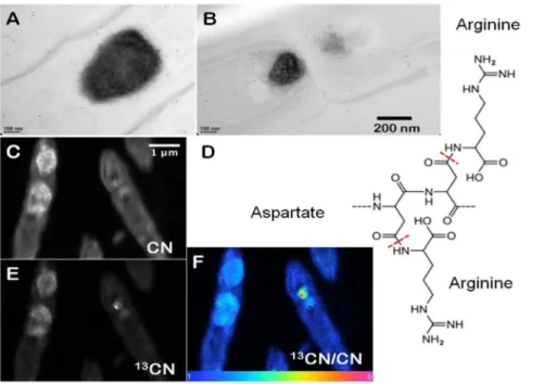
Cyanobacteria that form cones and pinnacles in YNP and laboratory cultures contain prominent, 150–750 nm wide, carbon-and nitrogen-rich inclusions that appear dark in TEM images [19]. These granules rapidly concentrate new organic carbon during active photosynthesis and are localized close to the septa that separate cells in filaments (Fig. 1). To test for the presence of RuBisCO in these granules and elsewhere in the cells, we used Rabbit anti-RuBisCO for immunogold labeling. Antibodies for RuBisCO were present primarily in the cytoplasm (Fig. 2A) and much more rarely in the ,100 nm wide inclusions (Fig. 2C), but were absent from the large granules (Fig. 2B). Instead, immuno-gold labeling using Rabbit anti-L-arginine demonstrated abundant L-arginine in the large, dark granules (Figs. 1A and B, Fig. S1; see Fig. S2 for images of negative controls). Together, these experiments identified the large, ,150–750 nm wide, dark granules in thin filamentous cyanobacteria as cyanophycin.
High resolution nano-scale SIMS mapping of the CN- ion species, used to track proteinaceous compounds [21], revealed the ultrastructure of cyanophycin granules at an unprecedented scale. Each large, ,150–750 nm wide granule contained densely clustered, rimmed, ,100 nm wide subcompartments with a distinct outer rim (Fig. 1C). The contrast between the interiors and rims of subcompartments stems from a higher intensity of the CN-in the rims, suggesting compositional heterogeneity. A higher intensity of CN-in the rims is consistent with an enrichment of nitrogen-rich L-arginine in the rims and a higher abundance of nitrogen-poor L-aspartate in the interiors of the subcompartments (Fig. 1D). Alternatively, the heterogeneous distribution of CN -within the bright rims and the duller interiors of cyanophycin granules, respectively, may track cyanophycin and another peptide-rich compound, respectively.
Carbon Accumulation and Storage in Cyanophycin
Studies of carbon flux through cone-forming communities in YNP and laboratory enrichment cultures suggested a central role for cyanophycin granules in the accumulation and storage of fixed carbon in the active growth layer of cones (Fig. 1 E, F) [19,22]. To track the flow of newly fixed carbon through cyanobacterial cells in time, we grew coniform mats in the laboratory, in media containing a high concentration of inorganic carbon (Ci). Small
cones were cut out, and incubated in a medium that contained
13
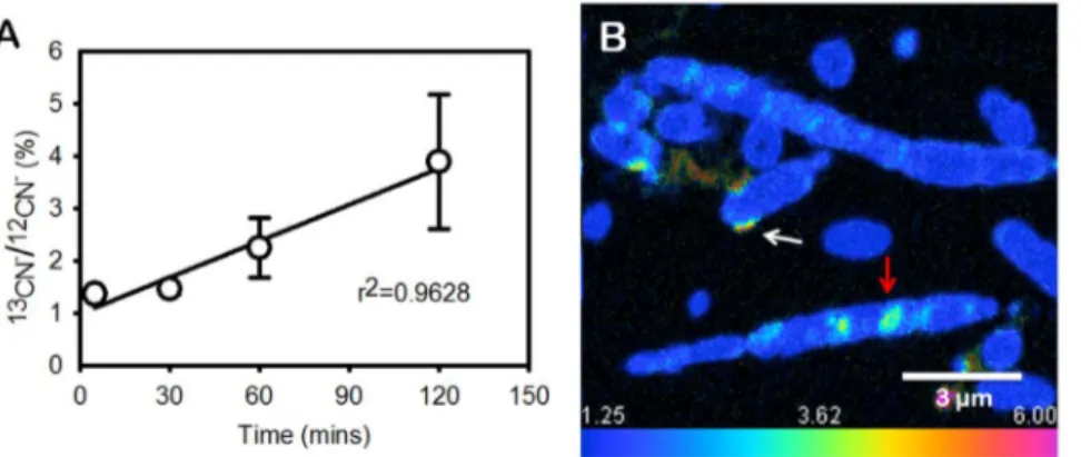
C-labeled bicarbonate in the presence of a light source (Methods). Cones labeled in this manner were sampled at 5, 30, 60 and 120 mins after the start of the incubation. High resolution nano-scale SIMS mapping of 12CN2 ions and13CN2 ions (see Methods) of thin sections through cones showed an increasing accumulation of labeled particulate carbon in time (Fig. 3A). After 5 minutes of incubation, labeled carbon was only detectable in the
Figure 1. Composition and appearance of cyanophycin granules. (A) TEM image of cyanophycin granules within filamentous cyanobacterial cells from field samples (YNP). The sample was immunogold labelled by Rabbit anti-L-Arginine at a dilution of 200X. (B) TEM image of filamentous cyanobacterial cells from a lab cone. The sample was immunogold labelled by Rabbit anti-L-Arginine at a dilution of 400X. Scale bar is 200 nm for (A andB). (C) High resolution nano-scale SIMS imaging of CN-, showing rimmed globular clusters within cyanophycin granules. (D) The suggested chemical structure of cyanophycin, with aspartate as the backbone and arginine as the side chain. (E, F) High resolution nano-scale SIMS map of
13
CN2in the granules of cyanophycin from cyanobacteria grown in the laboratory. (F) Ratio map of13CN2to12CN2in cyanophycin granules. Scale bar is 1mm for (C, E, F).
doi:10.1371/journal.pone.0088142.g001
envelopes of filamentous cyanobacteria (Fig. S3). After 30 minutes, labeled carbon was present primarily in the cyanophycin granules (Fig. 3B), where the13CN2/12CN2ratio was twice higher than the background (1.25%, in resin). After an additional 30 minutes (60 min total), cyanophycin granules occupied 3.9% of the cell area and accounted for 13.9% of the particulate labeled carbon. The isotopic enrichment, expressed as13CN2/12CN2 ratio, was 2.0% (60.6%) and 7.2% (61.0%) in the cytosol and cyanophycin, respectively. After 120 mins, the large cyanophycin granules were enriched isotopically by 11.5% (62.9%) relative to the 3.6% (61.3%) enrichment of the cytosol (Fig. 4A). At this time, cyanophycin granules accounted for about 5.7% of the cell area and for 18% of the labeled carbon in cyanobacterial cells (Fig. 4B, C). The inflow of13C into cyanophycin requires that cone-forming
cyanobacteria rapidly channel fixed carbon via phosphoenolpyr-uvate (PEP) carboxylase to PEP pool and aspartate (Fig. 5). These fluxes of fixed carbon resemble the reported rapid production of labeled aspartate and PEP in the unicellular cyanobacterium Synechocystis sp. strain PCC 6803 acclimated to low Ciand subject
to a pulse of13C-labeled medium with very high Ci[23]. Longer
term labeling experiments, pulse-chase experiments, and the tracking of soluble metabolites could evaluate fluxes of carbon through different pools of carbon in filamentous, cone-forming cyanobacteria, and in other cells within the community.
Photosynthetically active cone-forming cyanobacteria from untreated YNP samples and laboratory cultures contain RuBisCO (Fig. 2) and rare carboxysomes [19]. These organisms also excrete glycolate in the presence of high Ci under our laboratory
conditions [24]. Therefore, cone-forming cyanobacteria use RuBisCO to fix inorganic carbon under all tested experimental and field conditions (Fig. 4). Surprisingly, the intracellular distribution of labeled particulate organic carbon in cone-forming cyanobacteria also provides evidence for a substantial flow of fixed carbon toward PEP carboxylase, and the synthesis of aspartate, arginine and cyanophycin (Fig. 5). This occurs both in laboratory enrichment cultures (Figs. 1, 4) and in field cones from YNP [19]. PEP carboxylase contributes to carbon fixation in the stationary cultures of unicellular cyanobacteria Aphanocapsa 6308 [25] and in the cultures of Synechocystis sp. PCC 6803 acclimated to low concentration of inorganic carbon (, 60 mM Ci) but experiencing
a pulse of high Ci [23]. In contrast, we speculate that
cyanobacterial filaments in modern coniform mats channel fixed carbon toward PEP and synthesize cyanophycin even when growing actively and in the presence of both low and high Ci. We
infer this from the following: 1) cyanophycin granules are common in the actively growing tips of laboratory and field cones even before the transfer to the labeling medium [19,22] (Methods); 2) cyanobacteria in the tips of laboratory cones grow in solutions containing , 25 mM Ci, i.e., two order of magnitude higher
concentration than low Ci conditions in Synechocystis cultures
(0.18 mM); and 3) cyanobacterial cones were transferred to labeling solutions with , 25 mM Ci(Methods).
Studies of unicellular cyanobacteria that do not fix nitrogen and studies of N- and C-fixing cyanobacteria show that cyanophycin is typically synthesized by cyanobacteria that transition between a nitrogen-replete to a nitrogen-poor regime, and may be degraded during the transition from low to high Ciconcentrations to balance
the internal C/N ratio [11–13,23,26,27]. The observed flux of carbon toward PEP carboxylase and cyanophycin under our laboratory conditions did not occur in response to a changing N regime because both the growth and the incubation media contained comparable concentrations of Ciand Ni(,25 mM DIC
and 1.8 mM nitrate). Cells grown and incubated under these conditions contained rare carboxysomes and abundant cyanophy-cin granules. Cyanophycyanophy-cin is also synthesized during typical growth conditions in coniform mats in YNP, because cyanobac-teria in the field cones contain abundant cyanophycin granules and rare carboxysomes [19]. The field conditions are likely characterized by less than 5 mM DIC and 0.05 mM Niin the
solution above the coniform mats [28,29]. Because the field structures grew acclimated to low Ni, but were labeled in the
medium that contained 1.8 mM nitrate [19], it is possible that our
13
C-labeling experiment in the field probed the response to a changing N regime. Notably, we observed similar patterns of C flow through cyanobacterial cells both in the laboratory and in the field [19].
Actively growing cone-forming cyanobacteria from cone tips do not appear to synthesize cyanophycin in response to a changing N
Figure 2. Immunogold TEM images of cyanobacterial cells from YNP samples. All cells were immunogold labelled by Rabbit anti-RuBisCO (large subunit, form I and II, AS03 037) at a dilution of 1000X. The gold particles are uniformly sized and dark, two examples are marked by red arrows in (A, B, C). (A) The distribution of immunogold particles tagging free RuBisCO in a cell lacking cyanophycin or carboxysomes. SiO2around the cell is marked by a black arrow, these
granules are bigger than gold particles. (B) The absence of immunogold particles in cyanophycin granules indicates the absence of RuBisCO. (C) Immunogold particles tagging RuBisCO in small inclusions suggest the presence of morphologically atypical carboxysomes. These inclusions are rare and are found in the same areas where large cyanophycin granules typically occur. Few gold particles outside the cell in (A, B, C) indicate a low level backgroung. Scale bars: 200 nm in (A, B), and 100 nm in (C).
regime. Instead, morphological, chemical and isotopic patterns observed in the macroscopic, surface-attached, diffusion-limited [22,30] or even carbon-limited cones [28] suggest that cone-forming cyanobacteria may synthesize cyanophycin in response to the low or changing C/O ratios. Mutants of Synechococcus sp. strain
PCC 6803 that lack carboxysomes and are impaired in the ability to concentrate carbon and those that accumulate the products of RuBisCO oxygenation such as glycolate (Fig. 5) channel larger portions of fixed carbon toward PEP carboxylase [23,31]. Wild-type cone-forming cyanobacteria exhibit similar trends under our
Figure 3. Temporal dynamics of13CN2in cyanophycin. (A) Average accumulation of labeled carbon within the cells (N.10) at different time points. (B) Accumulation of labeled carbon within cyanophycin after 30 min of incubation. The13CN2/12CN2ratio was 2.5% (i.e., twice above the
1.25% background). Cyanophycin is marked by a red arrow. Labeled carbon is also present in or close to cell envelopes (white arrow). Scale bar is 3mm.
doi:10.1371/journal.pone.0088142.g003
Figure 4. Carbon accumulation and storage within cyanophycin over time. (A) Isotopic enrichment in cyanophycin granules and cell cytosol after 60 and 120 mins of incubation (see images ‘‘60 mins’’ and ‘‘120 mins’’). (B) Labeled carbon in cyanophycin granules expressed as the percentage of total labeled carbon in samples shown in (60 mins, 120 mins). (C) Percentage of cellular area that is occupied by cyanophycin granules in (60 mins, 120 mins). (60 mins) Nano-scale SIMS image of large cyanophycin granules in filamentous cyanobacteria ,120mm below the surface of the cone after 60 min of incubation in the labeling medium. (120 mins) Nano-scale SIMS image of cyanophycin granules in filamentous cyanobacteria ,120mm below the surface of the cone after 120 mins of incubation in the labeling medium. Cyanophycin granules in (II) contain more labeled carbon and are larger. Scale bar is 5mm for (D, E).
doi:10.1371/journal.pone.0088142.g004
experimental conditions (,25 mM Ciin the bulk medium): they
excrete glycolate, and incorporate aspartate derived from the PEP carboxylase pathway into cyanophycin granules (Fig. 5). Given that phosphorespiratory products, including glycolate, inhibit enzymes of the Calvin Benson Basham pathway [32,33], and that the clumping behavior of cone-forming cyanobacteria promotes the removal of glycolate and does not occur at low oxygen concentrations [24], both clumping and the increased flux of carbon through PEP carboxylase may compensate for some of the inhibitory effects of photorespiration. This pathway may be of particular importance in dense, surface-attached cyanobacterial communities where the transport of metabolites is limited by diffusion [22]. These hypotheses can be explored by quantifying the production of glycolate and cyanophycin in coniform mats and dispersed mats exposed to different C/O ratios and by further exploring the dynamics of carbon cycling through cyanophycin granules.
Cyanophycin seems to play an extraordinary role in the cycling of carbon in thin filamentous cyanobacteria from modern cone-forming aggregates. The previously unrecognized flux of fixed carbon toward PEP carboxylase and the incorporation of this carbon into cyanophycin directly links the cycles of carbon, nitrogen and oxygen, and may be one of many unusual adaptations of thin, filamentous cyanobacteria to life in macro-scopic aggregates in the presence of low flow [19,20,22,24,30,34]. Cyanobacteria from Subsection III that live in environments other than hot springs also have the capacity to synthesize cyanophycin and often do so [16–18,35], under conditions that remain to be elucidated.
Materials and Methods Culturing and Isotope Labeling
Field work was performed in August 2008 and 2009 in Mound Spring at Sentinel Meadows, Yellowstone National Park (YNP) under the permit YELL-2008-SCI-5758 and coniform mats were
sampled and transported as described previously [19,34]. Labo-ratory enrichment cultures were grown as described previously [36], in 200 ml glass beakers at 45uC, in a modified Castenholz D medium, on silica or aragonite sand as solid substrate. Genomic DNA was extracted from cones grown in laboratory enrichment cultures. Primer sets of 27F/1492R for universal bacterial 16s rRNA sequences, and cyanobacteria-specific primers 338F/781R, 359F/781R were used for PCR and nested PCR as described in references [19,34]. The cultures were grown at a light intensity of 180 mE/m2/s using cold fluorescent light with a day-night cycle of 12–12 hours. Three weeks after the inoculation, and 3–7 days after the appearance of , 5 mm tall and , 2 mm wide cyanobacterial cones in laboratory enrichment cultures, the liquid medium above the surface-attached cyanobacteria was replaced by the fresh, modified Castenholz D medium [36,37] that contained 1.8 mM NO32 and ,25 mM sodium bicarbonate enriched in 13C (98
atom%, Isotec, Miamisburg, Ohio) and equilibrated with 5%
13
CO2in the culture headspace. Cones were incubated for 5, 30,
60 and 120 mins in separate containers.
Isotopic Mapping of Labeled Carbon by Nano-scale SIMS
After the incubation with isotopically labeled inorganic carbon, the samples were fixed with 2.5% glutaraldehyde in 0.1 M sodium cacodylate buffer (pH 7.4), washed with the same buffer, and post-fixed in 1% osmium tetroxide and 1.5% potassium ferrocyanide. The fixed samples were dehydrated in a gradient series (30–100%) of acetone/water solutions and embedded in epoxy resin (Epon). Thin sections (300 nm) were obtained using a Reichert-Jung Ultracut E ultramicrotome with a diamond knife. The sections were collected and transferred onto a clean Si chip for SIMS imaging.
Nano-scale Secondary Ion Spectrometry (Nano-scale SIMS) analyses were carried out at the Institut Curie, Orsay, France using a NanoSIMS-50 ion microprobe (Cameca, Gennevillieres, France). Measurement conditions were described in details in
Figure 5. Proposed scheme of central carbon and nitrogen metabolism in cone-forming cyanobacteria. Adapted from Huege et al., 2011 and Hackenberg et al., 2012. Key metabolites including arginine, cyanophycin and glycolate and enzyme (RuBisCO) detected in this study are marked in dark.
previous studies [19,22]. The optical system in the microprobe was adapted to ensure high transmission with a high mass resolution. The primary beam was operated in the probe scanning mode over the sample surface to create elemental and isotopic images. To obtain high resolution images of individual cell details, the raster was reduced to 6, 12 and 20 mm, with the probe size smaller than 100 nm and an intensity of about 1 pA. Images were created for ion species12C2,13C2,12C14N2,13C14N2and32S2. The dwell time per pixel was 30 ms for high resolution images. High mass resolution setup was used to separate13C14N2from the isobaric
12
C15N2 species on one hand [21], and on the other hand, to prevent the mass interference from11B16O2species that was likely present in the embedding resin.
Nano-scale SIMS Data Processing
Ion species12C14N2and13C14N2were used to detect carbon associated with biomass, as they have higher intensity and are more statistically reliable than 12C2 and 13C2. The ratio (%) of
13
C14N2/12C14N2 was used to quantify the uptake of carbon (enrichment over the background). Color ratio image was generated by using OpenMIMS, an ImageJ plugin [38] developed by Claude Lechene’s Laboratory. OpenMIMS allows the visual-ization of ratio value in a HSI (Hue-Saturation-Intensity) image [39,40]. To compute the average13C14N2/12C14N2ratio in the cells in each image, the information pertaining to microbial biomass (enriched in 13C) was separated from the background resin (natural abundance of13C) by a mask. The binary mask was generated by adjusting the threshold level on the sum image of
13
CN2and12CN2. Each pixel containing biomass was assigned a value of 1, other pixels were assigned a value of 0. A 13CN2/
12
CN2 ratio map was obtained by multiplying the measured
13
CN2/12CN2 ratio image with the binary mask image to eliminate the resin background. The ratio characteristic for the unlabeled controls (N = 3, numerical value 1.25) was first subtracted from each pixel value, and pixel values in the corrected ratio of 13CN2/12CN2 were integrated over all pixels. The isotopic enrichment in carbon concentrating granules was determined using ultra-high resolution images.
Immunohistochemistry and Transmission Electron Microscopy Imaging
Samples of cyanobacterial cones were frozen using a high pressure freezer (Leica EMPACT2) at pressure 2000–2050 bar. Freeze substitution was performed in anhydrous ethanol (contain-ing 0.2% uranyl acetate and 0.2% glutaraldehyde) us(contain-ing a Leica EM AFS2 (automatic freeze substitution). The samples were kept at 290uC for 3 days, at 260uC for 1 day, at 220uC for 1 day, at 0uC for 1 day and were then warmed to room temperature. The LR Gold resin was used for infiltration and embedding. Ultrathin sections (90–120 nm) were obtained using a Reichert Ultracut S or Leica EM UC6ultramicrotome (Leica, Vienna, Austria) with a diamond knife. The sections were collected and transferred onto 100-mesh nickel grids.
In immunogold labeling experiments, the individual grids were floated on Tris-buffered saline (TBS) for 15 mins, and 1% bovine serum albumen (BSA) in TBS for 15 mins. To determine whether L-Arginine was present in the large intracellular granules, the grids were incubated with Rabbit anti-L-Arginine (Acris, AP31587SU-S) diluted 200X, 400X and 800X in TBS and 1% BSA for 1 hour (Fig. S1). Normal Rabbit Immunoglobulin G (IgG) (Millipore, 12–
370), diluted 200X, 500X and 800X, was used as a control (Fig. S2). After 4 washes with TBS, the grids were floated on an excess (1:20 dilution) of 12 or 18 nm colloidal Donkey anti-rabbit IgG (Jackson Immuno Research, West Grove, PA, USA) at room temperature for 1 hour. The grids were washed sequentially with 3 drops of TBS, followed by three washes with ddH2O. After
immunogold labeling, the sections were stained with 5% uranyl acetate in water for 10 mins and 0.4% lead citrate in water for 6 mins. Sections were imaged using a Philips CM 100 Transmis-sion Electron Microscope at 80 kV and recorded by a GatanOrius CCD camera at the Plant Cell Biology Core Lab, Institute of Plant and Microbial Biology, Academia Sinica. To determine the distribution of RuBisCO in thin filamentous cyanobacteria, immunogold labeling was also performed using Rabbit anti-RuBisCO large subunit (1000 dilution in TBS and 1% BSA, Agrisera, AS03 037) following the protocol described above (Fig. 2).
Supporting Information
Figure S1 (A, B) TEM image of cyanophycin granules within
filamentous cyanobacterial cells from YNP cones. The samples were immunogold labelled by Rabbit anti-L-Arginine at 200X and 400X dilutions. (C) TEM image of cyanobacteria from a lab culture. The samples were immunogold labelled by Rabbit anti-L-Arginine at a dilution of 800X. Scale bar for (A, B, C) is 100 nm. Small amount of L-Arginine may be present in the cell, accounting for a certain level of background.
(TIF)
Figure S2 TEM image of controls for immunogold labelling
using Normal Rabbit IgG antibody (Millipore, 12–370). (A, B, and C) show samples that used the antibody at dilutions of 200X, 500X and 800X, respectively.(A and C) are field samples from YNP,B shows cells from a laboratory culture. The 200X dilution generates a low background cross-reaction, 500X and greater dilutions show zero cross-reaction. The scale bars are 0.5 mm for (A and C), and 0.2 mm for (B).
(TIF)
Figure S3 Nano-scale SIMS isotopic ratio map of 13CN- to
12
CN2for filamentous cyanobacteria from the tip of a cone after a 5 min incubation. Labeled carbon scattered around cell envelopes, suggesting that the initial carbon incorporation possibly occurs there. Scale bar is 5 mm.
(TIF)
Acknowledgments
We thank the PICT-IBiSA imaging facility in the institut Curie, Dr. G. Geesey (Univ. Montana) and S. Gunther (NPS) for help in YNP, Dr. L. Casillas (Univ. PR) and members of the Newman Lab (MIT) for advice and help, Dr. W-N. Jane, Dr. C-Y. Chiu and Ms. C-M. Hsu (Academia Sinica) for help with immunogold labeling, J. Mui and Dr. S.K. Sears (McGill Univ.) for sample preparation for Nano-scale SIMS, and former and current members of the MIT Laboratory for Geomicrobiology and Microbial Sedimentology for ideas, lab help and support. A. Maheras, A. Evans and M.S. Sim helped with the culturing experiments.
Author Contributions
Conceived and designed the experiments: BL CHW TB. Performed the experiments: BL TDW HV HJS. Analyzed the data: BL TDW JLGK TB. Contributed reagents/materials/analysis tools: TDW HV JLGK CHW TB. Wrote the paper: BL CHW TB.
References
1. Portis AR, Parry MA (2007). Discoveries in Rubisco (Ribulose 1, 5-bisphosphate carboxylase/oxygenase): a historical perspective. Photosyn Res 94: 121–43. 2. Cannon GC, Heinhorst S, Bradburne CE, Shively JM (2001) Carboxysome
genomics: a status report. Funct Plant Biol 29: 179–182.
3. Badger MR, Hanson DT, Price GD (2002) Evolution and diversity of CO2
concentrating mechanisms in cyanobacteria. Funct Plant Biol 29: 407–416. 4. Badger MR, Price GD (2003) CO2 concentrating mechanisms in cyanobacteria:
molecular components, their diversity and evolution. J Exp Bot 54: 609–622. 5. Price GD, Badger MR, Woodger FJ, Long BM (2008) Advances in
understanding the cyanobacterial CO2-concentrating-mechanism (CCM): functional components, Ci transporters, diversity, genetic regulation and prospects for engineering into plants. J Exp Bot 59: 1441–1461.
6. Simon RD (1971) Cyanophycin granules from the blue-Green algae Anabaena cylindrica: a reserve material consisting of copolymers of aspartic acid and arginine. Proc Natl Acad Sci USA 68: 265–267.
7. Simon RD (1973) Measurement of cyanophycin granule polypeptide content in the blue-green alga Anabaena cylindrica. J Bacteriol 114: 1213–1216.
8. Simon RD, Weathers P (1976) Determination of the structure of the novel polypeptide containing aspartic acid and arginine which is found in cyanobacteria. Biochim Biophys Acta 420: 165–176.
9. Allen MM, Hutchison F, Weathers PJ (1980) Cyanophycin granule polypeptide formation and degradation in the cyanobacterium Aphanocapsa 6308. J Bacteriol 141: 687–693.
10. Allen MM, Hawley AM (1983) Protein degradation and synthesis of cyanophycin granule polypeptide in Aphanocapsa sp. J Bacteriol 154: 1480–1484. 11. Allen MM (1984) Cyanobacterial cell inclusions. Ann Rev Microbiol 38: 1–25. 12. Obst M, Steinbuchel A (2006) Cyanophycin–an ideal bacterial nitrogen storage material with unique chemical properties. Shively JM (ed): Inclusions in Prokaryotes, Volume 1, Microbiology Monographs. Springer–Verlag Berlin Heidelberg. 168–187.
13. Finzi-Hart JA, Pett-Ridge J, Weber PK, Popa R, Fallon SJ et al. (2009) Fixation and fate of C and N in the cyanobacterium Trichodesmium using nanometer-scale secondary ion mass spectrometry. Proc Natl Acad Sci USA 106: 6345–6350. 14. Li H, Sherman DM, Bao S, Sherman LA (2001) Pattern of cyanophycin
accumulation in nitrogen-fixing and non-nitrogen-fixing cyanobacteria: Arch Microb 176: 9–18.
15. Castenholz RW (2001) Cyanobacteria. In: Bergey’s Manual of Systematic Bacteriology, 2nd ed (eds Boone DR, Castenholz RW, Garrity GM) The Archaea and the deeply branching and phototrophic bacteria (Springer Verlag, NY), 473–599.
16. Paerl HW, Prufert LE, Ambrose WW (1991) Contemporaneous N2fixation and
oxygenic photosynthesis in the nonheterocystous mat-forming cyanobacterium Lyngbya aestuarii: Appl Environ Microbiol 57: 3086–3092.
17. Congestri R, Sangiorgi VC, Albertano P (2003) Cytomorphology and distribution of periphytic cyanobacteria in one Italian waste water treatment plant: Algol Stud 109: 185–195.
18. Jones AC, Monroe EA, Podell S, Hess WR, Klages S et al. (2011) Genomic insights into the physiology and ecology of the marine filamentous cyanobac-terium Lyngbya majuscula. Proc Natl Acad Sci USA 108: 8815–8820. 19. Bosak T, Liang BQ, Wu TD, Templer S, Evans A et al. (2012) Cyanobacterial
composition and activity in modern conical microbialites. Geobiology 10: 384– 401.
20. Walter MR, Bauld J, Brock TD (1976) Microbiology and morphogenesis of columnar stromatolites (Conophyton, Vacerrilla) from hot springs in Yellow-stone National Park in Developments in Sedimentology, Stromatolites, eds Walter MR (Elsevier, Amsterdam), 273–310.
21. Guerquin-Kern JL, Wu TD, Quintana C, Croisy A (2005) Progress in analytical imaging of the cell by dynamic secondary ion mass spectrometry (SIMS microscopy). Biochim et Biophys Acta 1724: 228–238.
22. Petroff A, Wu TD, Liang BQ, Mui J, Guerquin-Kern JL et al. (2011) Reaction-diffusion model of nutrient uptake in a biofilm: theory and experiment. J Theoret Biol 289: 90–95.
23. Huege J, Goetze J, Schwarz D, Bauwe H, Hagemann M et al. (2011) Modulation of the major paths of carbon in photorespiratory mutants of Synechocystis. PLoS ONE 6: e16278.
24. Sim MS, Liang BQ, Petroff AP, Evans A, Klepac-Ceraj V et al. (2012) Oxygen-dependent morphogenesis of modern clumped photosynthetic mats and implications for the Archean stromatolite record. Geosciences 2: 235–259. 25. Weathers PJ, Allen MM (1978) Variations in short term products of inorganic
carbon fixation in exponential and stationary phase cultures of Aphanocapsa 6308. Arch Microbiol 116: 231–234.
26. Mackerras AH, De Chazal NM, Smith GD (1990) Transient accumulations of cyanophycin in Anabaena cylindrica and Synechocystis 6308. J Gen Microbiol 136: 2057–2065.
27. Maheswaran M, Ziegler K, Lockau W, Hagemann M, Forchhammer K (2006) PII-regulated arginine synthesis controls accumulation of cyanophycin in Synechocystis sp. strain PCC 6803. J Bact 188: 2730–2734.
28. Jahnke L, Embaye T, Hope J, Turk KA, Van Zuilen M et al. (2004) Lipid biomarker and carbon isotopic signatures for stromatolite-forming, microbial mat communities and Phormidium cultures from Yellowstone National Park. Geobiology 2: 31–47.
29. Loiacono ST, Meyer-Dombard DR, Havig JR, Poret-Peterson AT, Hartnett HE et al. (2012) Evidence for high-temperature in situ nifH transcription in an alkaline hot spring of Lower Geyser Basin, Yellowstone National Park. Environ Microbiol. 14: 1272–83.
30. Petroff AP, Sim MS, Maslov A, Krupenin M, Rothman DH et al. (2010) Biophysical basis for the geometry of conical stromatolites. Proc Natl Acad Sci USA 107: 9956–9961.
31. Hackenberg C, Huege J, Engelhardt A, Wittink F, Laue M et al. (2012) Low-carbon acclimation in carboxysome-less and photorespiratory mutants of the cyanobacterium Synechocystis sp. strain PCC 6803. Microbiology 158: 398–413. 32. Kelly GJ, Latzko E (1977) Chloroplast phosphofructokinase II. Partial
purification, kinetic and regulatory properties. Plant Physiol 60: 295–299. 33. Norman EG, Colman B (1991) Purification and characterization of
phospho-glycolate phosphatase from the cyanobacterium Coccochloris peniocystis. Plant physiol 95: 693–698.
34. Bosak T, Liang BQ, Sim MS, Petroff AP (2009) Morphological record of oxygenic photosynthesis in conical stromatolites. Proc Natl Acad Sci USA 106: 10939–10943.
35. Kruskopf M, Du Plessis S (2006) Growth and filament length of the bloom forming Oscillatoria simplicissima (Oscillatoriales, Cyanophyta) in varying N and P concentrations: Hydrobiol 556: 357–362.
36. Bosak T, Bush JWM, Flynn MR, Liang BQ, Ono S et al. (2010) Formation and stability of oxygen-rich bubbles that shape photosynthetic mats. Geobiology 8: 45–55.
37. Castenholz RW (1988) in Methods of Enzymology, eds Packer L and Glazer AN (Academic, San Diego), 68–93.
38. Rasband WS (1997–2006) ImageJ, U. S. National Institutes of Health (Bethesda, Maryland, USA). Available: http://rsb.info.nih.gov/ij/.
39. Benson D (2003) HSI Ratio Method. Available: http://www.nrims.hms. harvard.edu/slides/NRIMS_-_BensonWorkshop2003.pdf.
40. Poczatek C, Kaufman Z, Lechene C (2009) OpenMIMS ImageJ Plugin Guide. Harvard Medical School (Boston, Massachusetts, USA).

