Antibody Engineering for Cancer Therapy
By
Yik Andy Yeung
B.S., University of Wisconsin-Madison, 1999 M.S.CEP., Massachusetts Institute Technology 2002
Submitted to the Department of Chemical Engineering in Partial Fulfillment of the Requirements for the Degree of
DOCTOR OF PHILOSOPHY in Chemical Engineering
at the
Massachusetts Institute of Technology May, 2005 Je 2.o03
© 2005 Massachusetts Institute of Technology All rights reserved
Signature of Author
Certified by_ 6
OF TECHNO1OGY
JUN 0 12005
LIBRARIES
l bepktmenof Chemical Engineering May 2005
IE IF -.1 Io M)
K. Dane Wittrup J.R. Mares Professor of Chemical Engineering and Bioengineering Thesis Supervisor
Accepted b
Daniel Blankschtein Professor of Chemical Engineering
Antibody Engineering for Cancer Therapy
By
Yik Andy Yeung
Submitted to the Department of Chemical Engineering on May, 2005 in Partial Fulfillment of the Requirements for the
Degree of Doctor of Philosophy in Chemical Engineering
ABSTRACT
Antibodies targeting various tumor-associated antigens have been developed
successfully to treat cancer. In this Thesis, novel antibodies and antibody-conjugate against two tumor antigens, AF-20 antigen and human aspartyl (asparaginyl) 3-hydroxylase (HAAH), were developed. Previously, these two tumor antigens have been shown to be present on a variety of tumor cells, while they have minimal expression on
normal tissues, rendering them excellent targets for antibody therapy.
For the AF-20 work, the variable region (V) gene of a previously isolated mouse monoclonal antibody (mAb) AF-20 was cloned from hybridoma mRNA and used to construct an AF-20 single-chain Fv (scFv). The AF-20 scFv was shown to bind
specifically to the same epitope as mAb 20 with a binding affinity of 4nM. The
AF-20 scFv was also internalized into tumor cells in a manner identical to that of the original mAb AF-20. The scFv was later employed for cellular internalization of virus-sized
fluorescent quantum dots. In addition, to demonstrate the versatility of this antibody, an
immunotoxin composed of AF-20 scFv fused to the highly cytotoxic recombinant toxin
gelonin was constructed, and its in-vitro efficacy against three different tumor cell lines
were evaluated. The IC50 of the AF-20 scFv-gelonin fusion was consistently one to two logs lower than the IC5 0 of free gelonin on FOCUS (liver), L3.6pl (pancreas) and PC3 (prostate) cells, further demonstrating the capability of the AF-20 scFv as a targeting module. Therefore, this AF-20 scFv is a potential internalization vector for toxins, enzymes, radionuclides and virus for targeted therapy of AF-20-antigen expressing tumor cells.
For the HAAH study, twelve novel human scFv against HAAH were isolated from a human non-immune scFv library displayed on the surface of yeast. Five of the twelve scFv were reformatted as human IgG 1. One of the reformatted IgG, 6-22, showed
significant binding to recombinant HAAH protein in ELISA, tumor cell lines, and tumor
tissues. 6-22 IgG was also shown to target the catalytic domain of HAAH, and its apparent dissociation constant was determined to be 1.OnM. 6-22 IgG alone does not
exhibit significant cytotoxicity toward the tumor cells. However, 6-22 IgG internalizes into tumor cells and can therefore be employed to deliver cytotoxic moieties into tumor
cells. A goat anti-human IgG-saporin conjugate was delivered into tumor cells by 6-22 IgG and hence elicited cytotoxicity toward the tumor cells in vitro. Meanwhile, the monovalent affinity of 6-22 scFv was too low for therapeutic or diagnostic application, so 6-22 scFv was affinity matured using directed evolution and yeast surface display. After
two rounds of mutagenesis, a mutant, C4-18, with an affinity of 0.6nM was isolated.
Overall, these human ax-HAAH scFv and IgG can potentially be used in the diagnosis and
therapeutic treatment of HAAH-expressing tumor cells. Thesis Supervisor: K. Dane Wittrup
Dedicated to my parents Biao and Yuk-yin. Thanks for your unconditional support.
Acknowledgements
I would like to thank my thesis advisor, Dane Wittrup, for his advice, encouragement and support over the course of my research study. I am very thankful for having such a wonderful experience working with him. I would like to appreciate him for providing exceptional insights on my research, and a great working environment
where I can share openly, try freely, and learn greatly
I would also like to thank Jack Wands, my thesis committee member and collaborator, with numerous advice, discussions and reagents. I would not be able to work on this project without him starting the groundwork. In addition, I want to give
thanks to the other member of my committee, Linda Griffith, for her advice. I am
grateful for the financial support from the Biotechnology Process Engineering Center (BPEC) and Panacea Pharmaceuticals. Many thanks to Michael Lebowitz and Angela Finney of Panacea for all the discussions, ideas and reagents they have provided.
I would like to express gratitude to the past and present members of the Wittrup
Lab for being my terrific colleagues and making the lab such a pleasant place to work. I truly enjoy all the scientific and non-scientific discussions with them. And I especially appreciate all the help and encouragements from them. They are Brenda Kellogg,
Katarina Midelfort, Jason Burbank, Christilyn Graff, Sarah Bannister, Jeffrey Swers,
Balaji Rao, Jennifer Cochran, Mark Olsen, Yongsung Kim, Andy Rakestraw, David Colby, Stefan Zajic, Shaun Lippow, Ginger Chao, Stephen Sazinsky, Wai Lau, Dasa
Lipovsek, Andrea Piatesi, Shanshan Howland, and Greg Thurber. Thanks to my
undergraduate assistants, Terence Dobrowsky and Inna Koyrakh, for providing help on my research.
I would also like to acknowledge and thank the people who have made my stay in MIT and Boston so wonderful. I first want to thank brothers and sisters from MIT Hong
Kong Students Bible Study Group, they have been tremendous in encouraging, learning,
playing, praying and sharing with me. I especially thank the pastors, brothers and sisters
from Boston Chinese Evangelical Church for their constant support and nurturing.
Most importantly, I want to give the biggest thanks to my parents, Biao and Yuk-yin. Without their unconditional support and love, I would not be able to achieve this degree. It was their influence for having the initiative and plan that I should study in the United States. To this end, they have made huge sacrifice and I am forever grateful to them. I am extremely thankful for their care, love, teaching, and for bringing me up in a
Table of Contents
Chapter 1: Introduction and Background ... 8
1.1 Antibody Structure and Function ... 8
1.2 Antibody Engineering ... ... 10
1.2.1 Yeast Surface Display ... 10
1.2.2 Mutagenesis and Library Screening . . ... 13
1.3 Antibody Therapeutics for Cancer Therapy ... 16
1.3.1 Tumor Antigens ... ... 16
1.3.2 Mechanism of Tumor Killing ... 17
1.3.3 Current Antibody Therapeutics ... 18
1.4 Thesis Overview ... ... 19
Chapter 2: Characterization of a Single-chain Fv for Delivery of Cytotoxic Moieties to Hepatocellular Carcinoma Cells ... 21
2.1 Introduction ... 21
2.2 Materials and Methods ... 23
2.2.1 Cloning of the AF-20 V Genes ... 23
2.2.2 Construction of the AF-20 scFv . . ... 24
2.2.3 Soluble Expression of AF-20 scFv ... 25
2.2.4 Coomassie Gel Analysis and Western Blot ... 25
2.2.5 Binding of the scFv to FOCUS Cells . . ... 26
2.2.6 Internalization of the AF-20 scFv into FOCUS Cells ... 27
2.2.7 Construction of AF-20 scFv-Gelonin Immunotoxin ... 27
2.2.8 In-vitro Cytotoxicity of the Immunotoxin .. ... 28
2.2.9 Display of AF-20 scFv and Binding of AF-20 scFv Displaying Yeasts .. 28
2.3 R esults ... 29
2.3.1 Construction of AF-20 scFv ... 29
2.3.2 Expression of the AF-20 scFv in Saccharomyces Cerevisiae ... 29
2.3.3 Binding Epitope and Affinity of the AF-20 scFv against FOCUS Cells.. 34
2.3.4 Internalization of the AF-20 scFv into Tumor Cells ... 36
2.3.5 Facilitated Internalization of Nano-scale Particles ...39
2.3.6 In-vitro Cytotoxicity of AF-20 Gelonin Fusion Construct ...40
2.3.7 Display of AF-20 scFv on the Yeast Surface . ... 42
2.3.8 Binding of AF-20 Displaying Yeasts against FOCUS Cells ...42
2.4 Discussion ... 46
Chapter 3: Isolation and Engineering of Human Antibodies against Human Aspartyl (Asparaginyl) -Hydroxylase ... 54
3.1 Introduction ... 54
3.2 Materials and Methods ... 56
3.2.1 Cell Lines and Materials ... 56
3.2.2 Isolation of Anti-HAAH Leads ... 57
3.2.3 Conversion of scFv to IgG ... 58
3.2.5 Binding of the IgGs to Tumor Cells ... 60
3.2.6 Immunohistochemistry ... 60
3.2.7 Internalization Studies of 6-22 IgG ... 61
3.2.8 Cytotoxicity of 6-22 Immunotoxins ... 61
3.2.9 Construction of Random Mutagenesis Library ... 62
3.2.10 Construction of CDR Domain Shuffling Library of 6-22 scFv ... 63
3.2.11 Construction of Heavy Chain Shuffling Library of 6-22 scFv ... 64
3.2.12 Fluorescence Activated Cell Sorting of Mutant Library ... 64
3.3 Results ... 65
3.3.1 Isolation of Human Antibody Fragments against HAAH ... 65
3.3.2 Conversion of scFv to IgG and ELISA . ... 66
3.3.3 IgG Binding against Tumor Cell Lines . ... 69
3.3.4 Domain Mapping of Clone 6-22 and 6-23 ... 72
3.3.5 Immunohistochemistry using 6-22 IgG ... 74
3.3.6 Internalization of 6-22 IgG into the Tumor Cells ... 74
3.3.7 Cytotoxicity of 6-22 IgG Immunotoxin on Tumor Cell Lines ... 79
3.3.8 Random Mutagenesis of 6-22 scFv ... 81
3.3.9 CDR Domain Shuffling of 6-22 scFv ... 83
3.3.10 Heavy Chain Shuffling of 6-22 scFv ... 83
3.3.11 Conversion of C4 scFv to C4 IgG ... 85
3.3.12 Random Mutagenesis of C4 scFv ... ... 85
3.4 Discussion ... 88
Chapter 4: Quantitative Screening of Yeast Surface-Displayed Polypeptide Libraries by Magnetic Bead Capture ... 95
4.1 Introduction ... 95
4.2 Materials and Methods ... 97
4.2.1 Yeast Strains and Plasmids ... 97
4.2.2 Materials and Media ... 98
4.2.3 Growth and Induction ... 99
4.2.4 Fluorescence Labeling and Measurements ... 99
4.2.5 Binder Identification from Magnetic Bead Capture ... 100
4.2.6 Dissociation Kinetics by Fluorescent Measurement and Magnetic BeadlOl 4.2.7 Kinetic Screening by Magnetic Bead Capture ... 102
4.3 R esults ... 103
4.3.1 Model System Validation ... 103
4.3.2 Isolation of binders from nonbinders ... 106
4.3.3 Affinity Maturation ... 111
4.4 Discussion ... 118
4.5 Conclusion ... 123
Appendix... 125
Chapter 1: Introduction and Background
1.1 Antibody Structure and Function
Antibody is involved in the humoral branch of the adaptive immunity. Antibodies, produced by B cells, recognize pathogens or foreign molecules through specific binding
to the antigen. This specific interaction can neutralize the antigen or trigger effector
functions of the immune system to eliminate the antigen. Examples of the effector functions are opsonization, activation of complement and antibody-dependent cell-mediated cytotoxicity (ADCC).
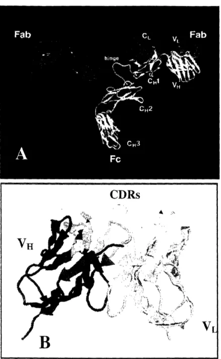
Antibody consists of two identical light (L) chains and two identical heavy (H)
chains, as shown in Figure 1.1. The molecular weight of the light and heavy chain are
about 25 and 50OkDa respectively. Each light chain is bound to a heavy chain by a
disulfide bond and a combination of noncovalent interactions such as salt bridges, hydrogen bonds and hydrophobic interaction. The amino-terminal regions of light and heavy chains, which vary greatly among antibodies with different specificities, are called variable (V) regions, VL for light chain and VH for heavy chain. The regions of relatively constant sequences beyond the variable regions are called constant (C) regions, CL for light chain and CH for heavy chain. Within the V regions, sequence variability is concentrated in several hypervariable regions. These hypervariable regions, which constitute the antigen-binding site of an antibody, are called complementarity-determining regions (CDRs). The remaining domains of VL and VH, which exhibit far
Figure 1.1 Schematics of Antibody Structure. (A) Antibody consists of two identical
light (red) and heavy (blue & yellow) chains. The amino-terminal regions of the light and
heavy chain, which are responsible for antigen binding, are called VH and VL. The Fc region is involved in activating the effector function of immune system. The IgG picture
is adapted from www.antibodyresource.com intactab.htnil. (B) Antibody mediates
specific interaction with its antigen through the CDRs of the V.1(blue and green) and VL (red and yellow). The CDRs of VH and VL are shown in green and yellow respectively, whereas the framework regions are shown in blue and red colors respectively for VaH and VL.
CDRs
VL
D
1.2 Antibody Engineering
Protein engineering has been employed extensively to modify the functions and properties of antibodies (Hudson and Souriau, 2001). Examples of protein engineering
include reducing the size of an antibody from IgG format to single-chain Fv (scFv),
modifying the valency of an antibody, changing the affinity of Fc receptors, and more
frequently, improving the affinity and stability of an antibody. To date, given the
inconsistent results from rational protein design, directed evolution is still the most
effective method available for exploration and engineering of antibodies. Directed
evolution involves displaying a diverse array of antibody mutants on a genetic package
(Shusta, et al., 1999), and then selecting favorable mutants from the pool. Such packages,
which link the phenotype to its genotype, include ribosome (Hanes, et al., 1998; He and
Taussig, 1997; Mattheakis, et al., 1994), bacteriophage (Griffiths and Duncan, 1998;
McCafferty, et al., 1990; Smith, 1985), bacteria (Francisco, et al., 1992; Georgiou, et al.,
1997)and yeast (Boder and Wittrup, 1997; Boder and Wittrup, 2000).
1.2.1 Yeast Surface Display
Yeast surface display was previously developed in our lab for directed evolution
of protein. One of the advantages of using yeast for displaying protein is that as a
eukaryote, yeast contains similar protein-processing machinery to a mammalian cell.
Thus yeast is more likely than prokaryotes to correctly express and display mammalian
surface or secreted proteins, in this case antibodies. Yeast surface display involves
displaying the antibody of interest through fusion with a two-unit cell wall glycoprotein
a-cc'q"~
Yeast
Cell:
I/
VI I iI e iAb N /labeled lig hns Nail I I I / / / "I N J9 NFigure 1.2 Schematic of Yeast Surface Display. The antibody of interest (scFv) is
displayed on the yeast surface through fusion to Agal-Aga2 protein. Two epitopes tags,
HA and c-myc, are fused to the N-terminus and C-terminus of the antibody respectively. The displayed antibody is able to bind ligands on the yeast surface, and the presence of
type and -type haploid cells (Boder and Wittrup, 1997) (Figure 1.2). A-agglutinin consists of two distinct domains, Agalp and Aga2p, connected by two disulfide bonds. Agalp subunit anchors the assembly to the cell wall via a 3-glucan covalent linkage. The
antibody is fused to the C-terminus of Aga2p, where the native a-agglutinin binding
activity localizes. The antibody in the display construct is flanked by two epitope tags,
with hemagglutinin (HA) and c-myc on the N- and C-terminus of the antibody
respectively. Expression of the full-length antibody on the cell surface can be simply confirmed by detecting the presence of c-myc tag on the cell surface using commercially available antibodies. Additionally, due to the intrinsic quality control of the endoplasmic
reticulum (ER), the presence of c-myc tag on the cell surface strongly implies that the
surface displayed antibody is folded properly (Hammond and Helenius, 1995).
The number of displayed antibodies is on the order of 104 copies per cell. The
displayed antibody is able to bind ligands on the yeast surface. Specifically,
surface-displayed antibodies are incubated with soluble fluorophore-conjugated ligands. The
presence of fluorescence on the yeast cells, which indicates the binding of ligands, is
detected using a flow cytometer. The fluorescence from surface-displayed antibody binding can be normalized with the simultaneous labeling of the epitope tags (HA or
c-myc), preventing any expression bias on the antibody binding fluorescence level.
Previously, yeast displaying anti-FITC scFv has been shown to bind simultaneously two macromolecules, a 200kDa-size FITC-dextran and a 150kDa-size mouse antibody 9e10 (anti-c-myc IgG) (Boder and Wittrup, 1997). This demonstrates that the
surface-displayed antibodies are readily accessible to their antigens and there is minimal steric
addition, the dissociation constant of the antibody-antigen interaction can be measured directly on the yeast surface by labeling the antibody-displaying yeast with different concentrations of the antigen, eliminating the need for soluble antibody expression. The
dissociation constants measured from yeast surface were shown to be highly similar to
the ones measured using soluble antibodies (Boder, et al., 2000).
1.2.2 Mutagenesis and Library Screening
Antibody engineering using yeast surface display involves mutagenizing the
antibody gene, displaying the library of mutant antibodies on the yeast surface and
subsequently screening for the desired mutants. Multiple methods have been used to
mutagenize an antibody gene: random mutagenesis (Boder, et al., 2000; Daugherty, et
al., 2000a; Graff, et al., 2004; Leung, 1989), hot-spot site directed mutagenesis
(Chowdhury and Pastan, 1999; Goyenechea and Milstein, 1996), targeted mutagenesis of
complementary determined region (CDR) residues (Schier, et al., 1996b; Wu, et al.,
1998; Yang, et al., 1995), DNA shuffling (Crameri, et al., 1996; Graff, et al., 2004; van
den Beucken, et al., 2001; Zhao, et al., 1998), CDR shuffling (Ellmark, et al., 2002;
Jirholt, et al., 1998; Marks, et al., 1992; Schier, et al., 1996a), and chain shuffling (Marks,
et al., 1992; Schier, et al., 1996a).
Random mutagenesis introduces amino acid changes throughout the entire
antibody sequence. One of the most frequently used random mutagenesis techniques is
error-prone PCR. In error-prone PCR, mutations are intentionally introduced during PCR through the use of error-prone DNA polymerases and reaction conditions (Cadwell and
Joyce, 1992; Leung, 1989). Taq DNA polymerase, which lacks proofreading ability, is
pair. To attain practical mutation frequencies, the error rate of Taq DNA polymerase is further increased by altering PCR reaction buffers, such as by using unbalanced dNTP concentrations (Cadwell and Joyce, 1992) during the reaction, by addition of manganese
(Leung, 1989), or by addition of nucleoside analogues (Zaccolo and Gherardi, 1999;
Zaccolo, et al., 1996). The desired range of mutation frequencies is usually achieved by controlling the amount of the different reagents added and the number of PCR cycles. For example, in the case of random mutagenesis using nucleoside analogues, concentration of the nucleoside analogues and number of PCR cycles can both be used to control mutation rate. Typical. mutation frequency for an antibody in random mutagenesis is from 0.5% to
3%.
On the other hand, other mutagenesis techniques, which target particular regions of an antibody, have also been used to improve the affinity of an antibody. One of the most commonly targeted regions is the CDRs of an antibody, as the CDRs are responsible for the direct interaction with the antigen. Mutagenesis targeting antibody
CDRs would minimize the structural change in the antibody framework, as mutations are
being concentrated in the CDRs only. Minimal framework residue alteration is particular
important in the engineering of human antibodies, since changes in the framework
residues may affect the immunogenicity of an antibody.
Overall, different mutagenesis methods have different advantages and each one
achieves a different objective. Considerations such as ease and rate of mutagenesis,
extent of affinity improvement per round of mutagenesis, and immunogenicity of the
method. However, these mutagenesis methods can also be applied in a sequential manner to affinity mature an antibody over several rounds of mutagenesis.
After mutagenesis, improved mutants are screened using yeast surface display.
These mutant genes, along with the restriction enzyme-digested display plasmid
backbone, are co-transformed into yeast. Through homologous recombination in yeast,
full-length plasmids containing the mutant genes are created (Gunyuzlu, et al., 2001). A
typical number of transformants in a random mutagenesis library is approximately 107
per 10 jgg of mutant DNA. In general, if the antigen is available in soluble form,
fluorescence-activated cell sorting (FACS) can be employed to isolate the improved
mutants from the library. The mutant library is first labeled at the desired equilibrium or
kinetic binding conditions to provide maximum fluorescence differentiation between the improved mutants and the wild-type scFvs (Boder and Wittrup, 1998). Then FACS is
used to isolate the improved mutants, which have higher fluorescence intensity. Optimal
equilibrium and kinetic binding conditions for the cell labeling have been calculated from mathematical models (Boder and Wittrup, 1998). Screening using optimal labeling and FACS allow for separation of mutants based on a specified quantitative improvement in
affinity. With optimal labeling, FACS has been shown to be capable of discriminating
with precision and reproducibility minimally-improved mutant over wild type yeast (VanAntwerp and Wittrup, 2000). Current FACS instrumentation is capable of sorting up
to 108 cells per hour. With a library of 107 yeast transformants, a ten-fold over-sampling
of the library requires about I hour to sort. Typically, four to five rounds of screening
excess of 108, magnetic beads can be used to enrich the populations, so that the library
size can be manageable by FACS (Siegel, et al., 2004).
Previously, yeast surface display has been used to affinity mature an anti-fluorescein antibody from InM to 50fM in 4 rounds of mutagenesis and screening
(Boder, et al., 2000). In addition, the dissociation rate of an anti-carcinoembryonic
antigen (CEA) antibody has also been improved over 1000 fold (from a half-life of 10
minutes to 4-7 days at 370C) after 2 rounds of mutagenesis and screening using yeast
surface display (Graff, et al., 2004). Therefore, yeast surface display has been shown to be a robust screening method, and is used in this study to isolate novel binder and to engineer antibodies.
1.3 Antibody Therapeutics for Cancer Therapy
1.3.1 Tumor Antigens
Malignant transformation of the cell is usually associated with alteration in the surface antigenic composition. Tumor antigens can result from genetic mutation in tumor cells that generate altered cellular proteins; these kinds of antigens are unique to the
tumor cells. These genetic mutations can be induced chemically, physically or virally (i.e.
Epstein-Barr virus and Human Papilloma virus). In addition, tumor cell surface antigens can also be aberrantly glycosylated (Tag-72 and Mucins), rendering them different from
those on the normal cells (Urban and Schreiber, 1992). However, there are also tumor
antigens that are not unique to the cancer cells; these tumor antigens are normally present on healthy cells. When the cells become malignant, the expression profile or level of these antigens change dramatically. One example of aberrant expression profiles is carcinoembryonic antigen (CEA). Tumor cells express CEA over the entire surface of the
cells, while in normal cells, CEA is only present on the apical surface of the cells
(Hammarstrom, 1999). Meanwhile, tumor antigens can also be over-expressed when cells become cancerous. Examples are CD20 overexpression in non-Hodgkin lymphoma and CD44 overexpression in lung cancer. When these tumor-associated antigens are expressed at a level (higher density) that can significantly distinguish their malignant phenotypes from the normal healthy phenotypes, this makes them (antigens) excellent targets for antibody-targeted therapy.
1.3.2 Mechanism of Tumor Killing
Antibody can block tumor growth or mediate the killing of tumor cell by several
different mechanisms (Houghton and Scheinberg, 2000). First, antibody can bind to the
growth factor receptors or other signaling molecules on the cancer cells, leading to
apoptosis or inhibition of the cell growth (Groner, et al., 2004). Examples are
Bevacizumab, which binds vascular endothelial growth factor (VEGF), and Cetuximab, which block the binding of EGF or TGF-a against EGFR. Another way to mediate killing is to recruit the natural immune system to kill tumor cells. Antibody bound to the cancer
antigen can activate the complement components, leading to opsonization of cancer cells
by complement receptors-expressing phagocytic cells, direct lysis of tumor cells and inflammation with recruitment of inflammatory cells. In addition, the bound antibody
can bind to the activating Fc receptors on the effector cells like macrophages and NK
cells, leading to antibody-dependent cellular cytotoxicity (ADCC) or release of cytokines. Examples are Rituxan® in the treatment of non-Hodgkin's lymphoma and Herceptin in the treatment of metastatic breast cancer. Besides using naked antibody to cure cancer, antibodies or antibody fragments can be conjugated with different cytotoxic moieties to
kill the tumor cells. These cytotoxic moieties include a variety of entities, ranging from radionuclide molecules to a virus carrying therapeutic genes or a liposome carrying loads of drugs, toxins, or enzymes (Trail and Bianchi, 1999). For example, antibody BR96-doxorubicin conjugate was used to target LeY-related tumor-associated antigen expressed
on most human carcinoma (Trail, et al., 1993). Antibody-directed drug delivery can
improve the therapeutic efficacy of cytotoxic moieties by targeting tumor cells specifically while reducing the potential systemic toxicities of the drugs. However, the success of a targeted cell-killing function is predicated on the existence of tumor-associated antigens.
1.3.3 Current Antibody Therapeutics
Antibodies targeting various tumor-associated antigens have been developed successfully to treat cancer. Currently, there are eight monoclonal antibodies approved by the Food and Drug Administration (FDA) for cancer therapy. Table .1 summarizes these FDA-approved antibodies and their targets. These approved antibodies can be divided into two types, naked and conjugated. Naked antibodies are those without any moiety attached to it; while conjugated antibodies have either toxin, radioactive material or cytotoxic drug attached to them. Most of the approved antibodies, except Bexxar, are either chimaeric or humanized, minimizing the neutralization effect of HAMA (human
anti-murine antibody). In addition to these approved antibodies, there are hundreds of
clinical trials worldwide involving the use of antibodies to treat cancer
(www.clinicaltrials.gov). In general, antibody has been proven to be a successful
Table 1.1 FDA Approved Antibodies for Cancer Therapy
Antibody Type Target Condition Approved
Rituxan Chimaeric CD20 Non-Hodgkin's Nov, 1997
Lymphoma
Herceptin Humanized HER2 Metastatic Breast Caner Sept, 1998
Mylotarg Humanized CD33 Acute Myelogenous May 2000
(Toxin: calicheamicin) Leukemia
Campath Humanized CD52 Chronic Lymphocytic May, 2001
Leukemia
Zevalin Chimaeric Non-Hodgkin's
Zevalin (In-l I or Y-90 linked) CD20 Lymphoma Feb, 2002
Bexxar Murine Non-Hodgkin's Jun,2003
(I-131 linked) CD- - Lymphoma
Erbitux Chimaeric EGFR Metastatic Colorectal Feb, 2004 Cancer
Metastatic Colorectal
Avastin Humanized VEGF Metastatic Colorectal Feb, 2004 Cancer
1.4 Thesis Overview
In this work, antibodies and antibody-conjugates against two tumor antigens,
AF-20 antigen and human aspartyl (asparaginyl) 3-hydroxylase (HAAH), were developed.
These two antigens have been previously shown to be present on a variety of tumor cells; while they have minimal expression on normal tissues, rendering them good targets for antibody therapy.
Chapter 2 of this thesis describes the development of an antibody fragment and an antibody toxin conjugate against AF-20 antigen. Previously, a mouse monoclonal
antibody (AF-20) was raised against this antigen; however, the gene of this antibody was
not available. In this work, the gene of AF-20 antibody was cloned and used to construct
an antibody fragment (scFv). The antibody fragment was characterized and shown to
constructed to illustrate that the antibody fragment can be a targeting domain for immunotoxin to treat a variety of cancers.
Chapter 3 of the thesis gives details about the isolation and engineering of novel human antibodies against HAAH using yeast surface display. Novel human antibody
fragments (scFv) were isolated against HAAH, and then converted into IgG formats.
These human IgGs were shown to bind specifically to the tumor cells, illustrating the
potential use of them in cancer diagnosis and therapy. One of the antibodies was also affinity matured using directed evolution and yeast surface display.
Chapter 4 focuses on the development of a quantitative screening tool, magnetic bead capture, for cell-based polypeptide library screening. Magnetic bead capture was used as an alternative tool to the flow cytometric sorting for screening of favorable mutants from a library. Results showed that magnetic bead capture probability of labeled cells correlated closely with the surface ligand density, and magnetic beads capture was capable of quantitatively screening for both novel binders from an excess of non-binders and high-affinity binders from an excess of low-affinity binders.
Chapter 2: Characterization of a Single-chain Fv for Delivery
of Cytotoxic Moieties to Hepatocellular Carcinoma Cells
2.1 Introduction
Antibodies have been employed extensively in recent years for the treatment of
various diseases, in particular cancer (Carter, 2001; Gura, 2002). Tumor cell killing can
be achieved by blocking the biological function of the antigen, through recruitment of immune effector functions or through delivery of attached toxins or radionuclides. Previously, monoclonal antibody (Mab) AF-20 was obtained by immunizing BALB/c
mice with the FOCUS cell line, isolated from a poorly differentiated hepatitis B virus
positive hepatocellular carcinoma (HCC) (He, et al., 1984; Wilson, et al., 1988). The antigen to which AF-20 IgG binds has been partially characterized (Moradpour, et al.,
1995). The antigen is a 180kDa homodimeric glycoprotein found on a variety of cancers
including liver, pancreatic, prostate, colon and breast tumor cell lines, while it has
minimal expression on normal tissues. However, full identification of this antigen is still
in progress.
AF-20 antigen expression is particularly prevalent in hepatocellular carcinoma,
where it is expressed on 75 of 75 primary HCC tumors and on distant metastases. HCC
comprises about 90% of the primary liver cancer in the United States, and the incidence
for this tumor type has increased recently (El-Serag, et al., 2003; El-Serag and Mason,
1999). Common cancer therapies like chemotherapy and radiotherapy are relatively ineffective in treating HCC and prognosis remains poor despite a variety of other treatment options including surgical resection, chemoembolization, and percutaneous
injection of ethanol. Immunotherapy using AF-20 IgG would present an attractive
alternative option for the treatment of HCC.
Previously, an iodinated form of AF-20 IgG showed excellent localization toward target tumor cells in vivo, and the highly specific and sensitive interaction rendered this
antigen to be a potential target for immunotherapy (Takahashi, et al., 1989). One
interesting property of this antigen is that once AF-20 IgG is bound, the complex is
rapidly internalized at 370C (Moradpour, et al., 1995). Utilizing this internalization
property of the antigen, AF-20 IgG has been conjugated chemically to different moieties
to specifically deliver both detection and therapeutic DNA to tumor cells. Examples of
the moieties conjugated include liposomes (Moradpour, et al., 1995), DNA-binding
cholesteryl-spermine (Mohr, et al., 1999) and adenovirus (Mohr, et al., 2000; Yoon, et al.,
2000). Overall, AF-20 IgG has been proven to be an effective antibody for targeting tumor cells in mice. However for targeting solid tumors, whole intact IgG has poor tumor penetration properties (Jain and Baxter, 1988). One possible way to improve the tumor targeting properties of AF-20 IgG is to reduce its size. Fragments of antibodies like single-chain Fv (scFv) have better penetration abilities and faster whole body clearances
than the intact IgG (Adams, et al., 1993; Yokota, et al., 1992; Yokota, et al., 1993),
exhibiting potentially better pharmacokinetics for tumor targeting. Genetic fusion of AF-20 antibody fragments with protein toxins or enzymes or conjugates with virus particles or toxins are also of interest for targeted therapy of cancer.
In this study, we have cloned the V gene of AF-20 IgG and use it to construct a
scFv. The scFv was displayed on the yeast surface and secreted as soluble form from
a binding affinity of 4nM. The AF-20 scFv also retained the internalization ability of the AF-20 IgG, and was shown to facilitate the internalization of virus-sized particles, Quantum Dots, into the tumor cells. These studies demonstrate the potential of using the
AF20 scFv to target virus particles or liposome vehicles to HCC and other tumor cells
expressing AF-20 antigen. Also, the potential of immunotherapy using AF-20 scFv as the targeting domain of an immunotoxin was explored. The toxin used in this study was the recombinant plant toxin gelonin (Falasca, et al., 1982). Our results show that AF-20 scFv-gelonin fusions consistently gave higher cytotoxicities than native gelonin on different tumor cell lines. Overall, this study demonstrates the promise of using engineered antibody fragments derived from Mab AF-20 for targeted therapy of HCC and other cancers.
2.2 Materials and Methods
2.2.1 Cloning of the AF-20 V Genes
Hybridoma cells expressing AF-20 IgG were cultured in media consisting of Dulbecco's modified eagle media (Sigma, St Louis, MO), 20% fetal calf serum (Hyclone,
Logan, UT), 2% glutamine, l00U/ml penicillin and 0.1mg/ml streptomycin (Sigma).
Cells were grown in T-flasks at 370C in a 5% CO2 atmosphere. About 108 cells were
grown up for RNA isolation, and total RNA was isolated using guanidinium thiocyanate
precipitation as described in the Ig Prime Kit protocol (Novagen, Madison, WI). First
strand cDNA synthesis was carried out as described in the Ig Prime Kit protocol. The
PCR amplification reactions were then setup respectively for the heavy and light chains
were run on a 1% agarose gel and stained. DNA bands in the region of 400 to 500 bp
were excised, purified and ligated into pSTBluel using the Perfectly Blunt Cloning Kit (Novagen). The ligated products were transformed into Nova Blue E. coli cells (Novagen), and the resulting plasmids were isolated from the E. coli and sequenced to determine the identity of the AF-20 heavy and light chain variable regions.
2.2.2 Construction of the AF-20 scFv
The scFv was constructed in the configuration of VH-Linker-VL using the splicing
by overlap extension (SOE) PCR method (Horton, et al., 1989; Krebber, et al., 1997). A
20-amino acid linker with four repeats of (Gly)4-Ser was used. The VH chains were
amplified using VH 5'primer: gatcgatcgagctagc-caggtccaactgcagcagtc, and VH 3'primer: ccactctcacagtctcctcaggtggtgg-tggttctggtggtggtggttctggcggcggcggctcc. The VL chains were amplified using VL 5'primer: ggcggcggcggctccggaggaggaggatcggacatcttgctgactcag
and VL 3'primer: gggacaaagttggaaataaaaaatggctgatgctggtggatcccatcatcatcatcatcattgataactc
gaggctcgatcgatc. The VL 3'primer incorporated an arginine to asparagine mutation at a lysine-arginine site near the c-terminus in order to remove a potential Kex2 cleavage site.
PCR reactions were carried out on a Perkin Elmer DNA Thermal Cycler 480 in a 100 gL
volume using 100 ng of template, I uM of primers, 0.2 mM of dNTP, 5 U Pfu
Polymerase (Invitrogen, Carlsbad, CA). The cycling conditions used were I cycle of
96C for 2 minute followed by 30 cycles of 96°C for I minute, 53°C for I minute and 72°C for 2 minutes, and finally 1 cycle of 72°C for 10 minutes. The VH and VL PCR fragments were separately gel-purified. The SOE reaction contained 400RM of each
dNTP, 2g each of the VH and VL PCR products and 5U of Pfil Polymerase in 100 L
for 1 minute, 53C for 1 minute, 72°C for 2 minutes and then followed by 1 cycle of 72°C
for 10 minutes. Outside primers (VH 5'primer and VL 3'primer) were then added to the
reaction for a further 15 cycles. The PCR product was gel purified, restriction-digested
with NheI and Xhol, and ligated into the corresponding sites in the pRS based secretion
vector. The ligated product was then transformed into XL1-Blue E. coli cells (Stratagene,
La Jolla, CA) for amplification and sequencing.
2.2.3 Soluble Expression of AF-20 scFv
The AF-20 scFv secretion plasmid along with a plasmid contain a Trp marker
were co-transformed into yeast strain YVHIO using lithium acetate method (Gietz and
Schiestl, 1991). The pRS based yeast secretion vector and the yeast strain have been
previously described (Parekh, et al., 1995; Robinson, et al., 1994). Yeast culturing and
induction of scFv secretion were performed as previously described (Shusta, et al., 1998).
The yeast supernatant containing soluble scFv was concentrated using stirred cell protein
concentrator series 8000 from Millipore (Billerica, MA). The concentrated protein solution was then purified using Nickel-NTA resin according to the manufacturing
protocol from Qiagen (Valencia, CA). Nickel-column-purified AF-20 scFv was dialyzed
in PBS (pH 7.4) for later experiments.
2.2.4 Coomassie Gel Analysis and Western Blot
Various amount of protein samples were resolved by SDS-PAGE gel electrophoresis using a 4% stacking and 12.5% resolving gel. For Coomassie analysis,
the gel was stained with Coomassie staining solution (0.4g/L stain, 7% acetic acid, 35%
methanol) for I hour and destained overnight in destain buffer (7% acetic acid, 35%
CA). For western blot, after electrophoresis, protein was transferred to a nitrocellulose membrane using an Xcell II transfer apparatus (Invitrogen, Carlsbad, CA). The membrane was then probed with 0.5pg/ml of tetra-HIS antibody (Qiagen) in Tris-buffered saline with 0. I1% Tween-20 and followed by 1:2000 dilution of goat anti-mouse IgG horseradish peroxidase conjugate (Sigma). Detection was performed with SuperSignal® substrate from Pierce (Rockford, IL) and Fluor-S imager. In this work, purified proteins were de-glycosylated with N-glycosidase Endo Hf and PNGase F
according to the manufacturing protocol from New England Biolabs (Beverly, MA)
2.2.5 Binding of the scFv to FOCUS Cells
FOCUS cells were cultured as previously described (Yoon, et al., 2000). FOCUS
cells were detached from the culture plates using versene solution (Gibco, Carlsbad, CA), and then resuspended in PBS supplemented with lg/L bovine serum albumin (BSA). ScFv was allowed to bind to the FOCUS cells at 40C for 4 to 6 hours. The cells were then washed with PBS/BSA and stained with 1:50 dilution of anti-FLAG-IgG-FITC conjugate
(Sigma) at 40C for 30min. Stained cells were then analyzed by flow cytometry. The scFv
epitope mapping experiment was similarly performed with the exception that excess
amount (10 fold) of AF-20 IgG was added to the initial scFv incubation step. The titration
of AF-20 scFv against FOCUS cells was also performed similarly but with different
secondary detection antibodies. Different concentrations of scFv were incubated with
FOCUS cells at 4"C for 4 to 6 hours, and then the cells were stained with 100nM of
mouse anti-FLAG IgG (Sigma) and followed by 1:50 dilution of goat anti-mouse
IgG-phycoerythrin conjugate (Sigma) for 20min at 40C. Cell fluorescence was then detected
2.2.6 Internalization of the AF-20 scFv into FOCUS Cells
Vitrogen from Cohesion Technologies (Palo Alto, CA) was added to a clean coverslip and subsequently FOCUS cells were cultured on these collagen-coated coverslips. 200nM of AF-20 scFv or control scFv was pre-incubated with 50nM of mouse anti-FLAG IgG. After overnight culturing, FOCUS cells were washed, and allowed to
bind the pre-incubated materials in serum-free media at 370C for 1 hour. After the
incubation, cells were fixed with 4% cold formaldehyde (Sigma) for 20min and then
permeabilized with 0.1% saponin (Sigma). The permeabilized cells were labeled with
1:100 dilution of goat anti-mouse IgG-FITC conjugate (Sigma) and 1:10000 dilution of Hoechst dye (Molecular Probe) at room temperature for 30min. Fluorescence inside the cells was detected using a confocal microscope. For the internalization study of Quantum dot, 250nM of AF-20 scFv or control scFv was pre-incubated with 75nM of biotinylated mouse anti-FLAG IgG and 2L of Qdot 565-streptavidin conjugate (Quantum Dot,
Hayward, CA) in a total volume of 50OL of serum-free media. After I hr of
pre-incubation, the pre-incubated mixture were diluted with 300gL of serum-free media and
presented to the cells grown on coverslip at 37°C for 3hour. Cells were then analyzed
directly using a confocal microscope.
2.2.7 Construction of AF-20 scFv-Gelonin Immunotoxin
20 scFv was fused to the amino terminus of the gelonin to construct the
AF-20 scFv-gelonin immunotoxin. The immunotoxin was constructed and expressed according to the previous report (Rosenblum, et al., 2003). Briefly, overlapping PCR products containing the AF-20 scFv/rGel gene were cloned into the bacterial expression plasmid pET-32a(Novagen). An epitope tag, FLAG, was inserted at the amino terminus
of the immunotoxin for detection. The AF-20scFv/rGel protein was expressed using the E. coli strain AD494(DE3)pLysS by induction with 100 jgM IPTG at 230C for 16 hrs. Soluble protein was purified using Blue Sepharose 6 Fast Flow and stored in PBD at 40C.
2.2.8 In-vitro Cytotoxicity of the Immunotoxin
A 72-hr cell proliferation assay with log-phase (5000 to 10000 cells/well) AF-20
antigen-positive cells were performed as described previously (Nishikawa, et al., 1992).
The cells used in this studied were FOCUS cells (HCC), L3.6pl cells (pancreatic tumor)
and PC3 cells (prostate tumor). Cell viability was assessed using either alamarBlueTM
(Biosource, Camarillo, CA) or the previously described crystal violet staining procedures
(Nishikawa, et al., 1992). Cell viability detection using alamarBlueT M was performed
according to the manufacturing protocol.
2.2.9 Display of AF-20 scFv and Binding of AF-20 scFv Displaying Yeasts
For yeast surface display, AF-20 scFv was restriction digested and ligated into
pCTCON vector. The vector was then transformed into EBY100. EBY100 cells carrying
the plasmid was grown in SD-CAA for 24hr and then induced in SG-CAA+0.2% glucose
media for another 24hr. The display level was detected with mouse monoclonal antibody
9e 10 (Covance, Berkeley, CA) and goat anti mouse-phycoerythrin conjugates (Sigma).
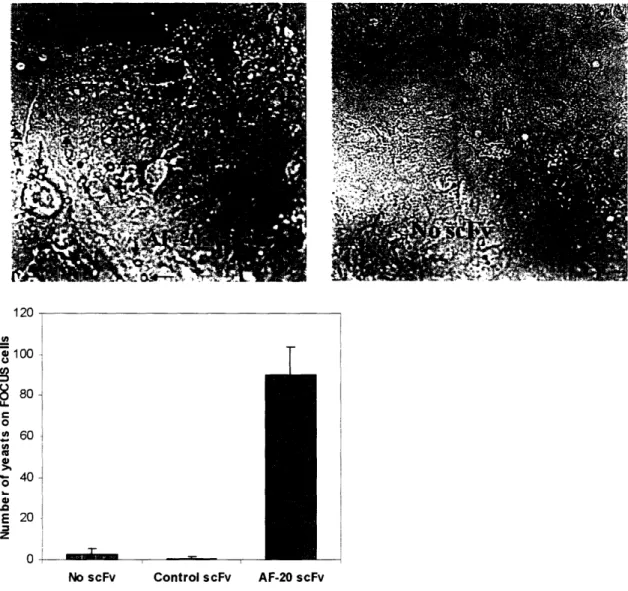
FOCUS cells were grown as a monolayer on a coverslip-bottom petri dish
(Mattek, Ashland, MA). 107 yeast cells displaying either no scFv, control scFv or AF-20
scFv were allowed to roll and bind to the monolayer of FOCUS cells respectively at room temperature for lhr in Dulbecco's PBS (Gibco). Four washes with Dulbecco's PBS were performed to remove the yeast cells that non-specifically stick to the FOCUS cells. Then
then washed four times with room temperature PBS. The complex was later imaged using a light microscope and the number of yeast cells on the FOCUS cells was counted.
2.3 Results
2.3.1 Construction of AF-20 scFv
The variable region genes of the mouse AF-20 IgG were cloned from AF-20
hybridoma cDNA with degenerate PCR primers from the Ig Prime kit (Novagen), and
were sequenced separately. The translated amino acid sequences of the variable regions of both heavy and light chains are shown in Figure 2.1. The AF-20 scFv was constructed in the configuration of VH-linker-VL using the splicing by overlap extension (SOE) PCR
method (Horton, et al., 1989; Krebber, et al., 1997), with four repeats of (Gly4-Ser) as
the linker. Two epitope tags were added to the flanking regions of the scFv, a FLAG tag
on the N-terminus and a 6xHis tag at the C-terminus. The epitope tags were later used for
both scFv detection and purification. DNA sequencing of the whole AF-20 scFv gene
construct confirmed that the scFv gene sequence containing both epitope tags was in
frame and contained no mutations. A schematic of the AF-20 scFv is shown in the top
panel of Figure 2.2.
2.3.2 Expression of the AF-20 scFv in Saccharomyces Cerevisiae
The AF-20 scFv gene was subcloned into the pRS based secretion vector (Parekh,
et al., 1995), and the AF-20 scFv was expressed in Sccharomyces cerevisiae strain
YVH10 (Robinson, et al., 1994). Supernatant from the AF-20 scFv secreting yeast was
A S Q V Q L Q Q S G P D L V K P G A S V 1 gctagccaggtccaactgcagcagtctggacctgacctggtgaagcctggggcttcagtg 60 R I S C K A S G Y T F A G H Y V H W V K 61 aggatatcctgcaaggcttctggctacaccttcgcaggccactatgtacactgggtgaag 120 Q R P G R G L E W I G W I F P G K V N T 121 cagaggcctggacggggacttgagtggattggatggattttccctggaaaggtaaatact 180 K Y N E K F K G K A T L T A D K S S S T 181 aagtacaatgagaagttcaagggcaaggccacattgactgcagacaaatcctccagcaca 240 A Y M Q L S S L T S E D S A V Y F C A R 241 gcctacatgcagctcagcagcctgacctctgaggactctgcggtctatttctgtgcaaga 300 V G Y D Y P Y Y F D Y W G Q G T T L T V 301 gttggatatgattacccgtactactttgactactggggccaaggcaccactctcacagtc 360 S S G G G G S G G G G S G G G G S G G G 361 tcctcaggtggtggtggttctggtggtggtggttctggcggcggcggctccggaggagga 420 G S D I L L T Q S P A I L S V S P G D R 421 ggatcggacatcttgctgactcagtctccagccatcctgtctgtgagtccaggagacaga 480 V S F S C R A S Q S I G T S I H W Y Q Q 481 gtcagtttctcctgcagggccagtcagagcattggcacaagcatacactggtatcagcaa 540 R T N G S P R L L I K Y A S E S I S G I 541 agaacaaatggttctccaaggcttctcataaagtatgcttctgagtCtatCtctggtatc 600 P S R F S G S G S G T D F T L S I N S V 601 ccttccaggtttagtggcagtggatcagggacagattttactcttagcattaacagtgtg 660 E S E D V A D Y Y C Q Q S S S W P F T F 661 gagtctgaagatgttgcagattattactgtcaacaaagtagtagctggccattcacgttc 720 G S G T K L E I K N A D A G G S 721 ggctcggggacaaagttggaaataaaaaatgctgatgctggtggatcc
Protein Sequence: variable domain of heavy chain
QVQLQQSGPDLVKPGASVRISCKASGYTFAGHYVHWVKQRPGRGLEWIGWIFP GKVNTKYNEKFKGKATLTADKSSSTAYMQLSSLTSEDSAVYFCARVGYDYPYY FDYWGQGTTLTVSS
Protein Sequence: variable domain of light chain
DILLTQSPAILSVSPGDRVSFSCRASOSIGTSIHWYQQRTNGSPRLLIKYASESISGI PSRFSGSGSGTDFTLSINSVESEDVADYYCQQSSSWPFTFGSGTKLEIKNA
Figure 2.1 The nucleic acid and amino acid sequences of the variable domains of AF-20 IgG. Genes of the variable domains were cloned from the AF-20 IgG secreting hybridoma cells and sequenced. Underlined regions are the corresponding
complementarity-determining regions (CDRs) of the variable domains. N-Glycosylation occurred on the italic/red asparagine residue during the expression of antibody fragment in Saccharonyces cerevisiae.
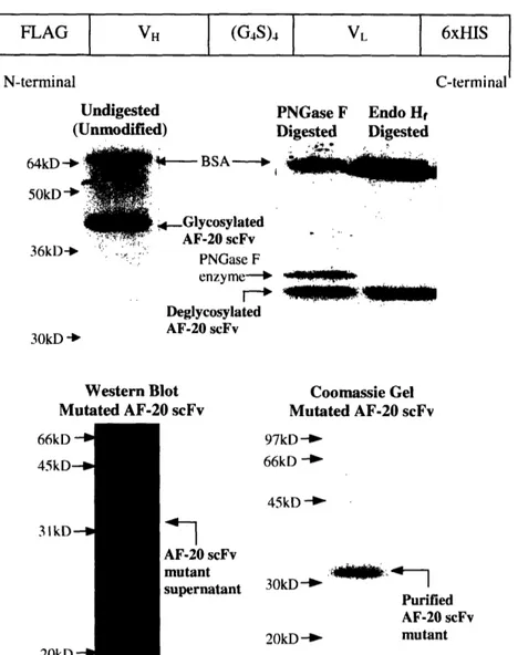
FLAG VH (G4S)4 VL 6xHIS
l l l l l l(G4)4
N-terminal C-terminal
Undigested PNGase F Endo Hf (Unmodified) Digested Digested
64kD 50kD -%- BSA- No 4 Glycosylated AF-20 scFv 36k1) --: PNGase F enzyme-- i Deglycosylated AF-20 scFv 30kD 'b
Western Blot Coomassie Gel
Mutated AF-20 scFv Mutated AF-20 scFv 66kD - 45kD-3IkD--I 20kD 97kD -66kD -45kD -AF-20 scFv mutant 3 -- :kD 4 supernatant 30kD -Purified AF-20 scFv 20kD _ mutant
Figure 2.2 Secretion and Purification of AF-20 scFv. The top panel illustrated the
schematic of the secreted AF-20scFv. Variable domains of heavy chain and light chain
were connected through a (Gly)4-Ser linker, forming a single-chain Fv. Two epitope tags, n-terminal FLAG and c-terminal 6xHis, were inserted for detection and purification.
Soluble AF-20 scFv was purified using Nickel-NTA resin. Native AF-20 scFv was
glycosylated in Saccharomyces cerevisiae as shown in the middle panel. Purified proteins were run on SDS-PAGE gel and stained with Coomassie Blue-stain. Unmodified
AF-20scFv migrated at about 40 to 45kDa, which was much higher than the expected size of
30kDa (middle left panel). After being digested in N-glycosidase (PNGase F and Endo
Hf), AF-20 scFv migrated at a smaller size (about 33kDa), which was closer to the expected size (middle right panel). The asparagine on light chain, where N-glycosylation occurred (Figure 1), was mutated to aspartic acid. Western blot of the asn-to-asp mutant
supernatant using mouse anti-FLAG IgG-HRP conjugate showed that the AF-20scFv mutant migrated at about 30kDa (lower left panel). The Coomassie Blue-stained gel on the lower right panel showed that purified AF-20 scFv mutant (N-,D) also migrated at about 30kDa and was without any significant contamination from yeast proteins.
analyzed by SDS-PAGE. The middle left panel of Figure 2.2 shows that purified AF-20
scFv migrated at a size about 40 to 45kDa, which was larger than the expected size of
30kDa. Western blot analysis of the 40kDa band using tetra-HIS antibody (Qiagen)
revealed that the band contained a 6xHis tag (data not shown). The reason for the
aberrant size of the AF-20 scFv was determined to be due to glycosylation in S.
cerevisiae. A consensus N-glycosylation site (asn-gly-ser) is present on the light chain sequence, and this particular region of the antibody was predicted to be surface exposed
by homology modeling. To investigate the glycosylation status of the scFv, the purified antibody fragment was incubated with two different N-glycosidases (PNGase F and Endo
Hf) and then examined by SDS-PAGE (Bretthauer and Castellino, 1999). The middle
right panel of Figure 2.2 showed that the N-glycosidase-digested scFv bands migrated at
about 33kDa, which was smaller than the undigested band and closer to the theoretical
size (30kDa). This confirmed that the AF-20 scFv was N-glycosylated during secretion in
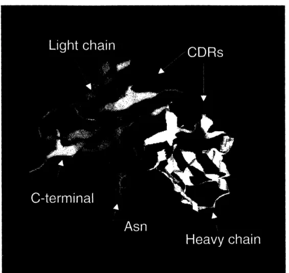
Saccharomyces cerevisiae. The Fv model in Figure 2.3 also indicated that the
glycosylation site was close to the junction between variable and constant domains of the
light chain, and on the opposite side from the CDRs. Coomassie gel analysis of the
purified AF-20 IgG showed that the light chain migrated at slightly less than 30kDa, thus free of glycosylation (data not shown). Therefore, this glycosylation site is likely unique
to the scFv format of the antibody, due to the exposure of this interfacial site upon
construction of the scFv format. This would suggest that removing the glycosylation site should not significantly alter the binding affinity of AF-20 scFv. In order to prevent the potential interference of glycosylation on protein expression in Saccharomyces cerevisiae
Figure 2.3 Homology Model of AF-20 Fv. The AF-20 Fv model was computed using web antibody modeling (http://antibody.bath.ac.uk). The red and white ribbons are the
framework regions of the light and heavy chains respectively. The CDRs of the light and
heavy chains are shown in blue. The asparagine residue where N-glycosylation occurs is shown in green, and it is located far away from the CDRs.
and downstream purification, the asparagine of the Asn-Gly-Ser consensus sequence
(Figure 2.1), was changed to an aspartic acid by site directed mutagenesis. Western blot
analysis of the supernatant of mutated AF-20 scFv (N41D) showed that the scFv
migrated at the expected size of 30kDa (bottom left panel of Figure 2.2). This was further
confirmed by SDS-PAGE of the purified, mutant AF-20 scFv (bottom right panel of Figure 2.2). In addition, purified AF-20 scFv was free of any major contaminants from
yeast native proteins. This N41D non-glycosylated mutant was used in subsequent
studies.
2.3.3 Binding Epitope and Affinity of the AF-20 scFv against FOCUS Cells
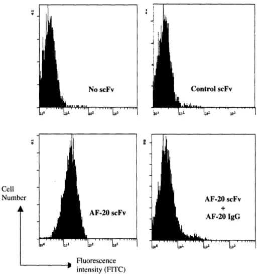
FOCUS cells which overexpress AF-20 antigen were used as a model system to
determine the specificity of the AF20 scFv (Wands, et al., 1997). FOCUS cells were
detached from culture plates using versene solution to protect the integrity of the cell membrane antigens. FOCUS cells were incubated with buffer alone, nonspecific control
scFv (anti-fluorescein scFv) or AF-20 scFv at 40C. Then the cells were stained with
mouse anti-FLAG IgG-FITC conjugate and analyzed by flow cytometry (FACS). The top
panels of Figure 2.4 showed that the FACS histograms of control (anti-fluorescein) scFv
and no scFv (background) were virtually identical. The bottom left panel of Figure 2.4 shows that AF-20 scFv binds to FOCUS cells. To demonstrate that this binding is
specific, 20 scFv was incubated with FOCUS cells in the presence of an excess
AF-20 IgG. Cells were then stained with mouse anti-FLAG IgG-FITC conjugate, which only recognizes the 20 scFv. Figure 2.4 bottom right panel showed that the binding of
Cell Number
I
XD th ii VF-20 scFv AF-20 IgG r | | Tn1 r r T re , Fluorescence intensity (FITC)Figure 2.4 In-vitro Specific Binding of AF-20 scFv on the Surfaces of FOCUS Cells.
FOCUS cells were detached from the plates and were allowed to bind buffer, control
scFv (anti-fluorescein scFv) or AF-20scFv at 4°C. The cells were then stained with
mouse anti-FLAG IgG-FITC conjugate, which only recognized the FLAG tag on control and AF-20 scFv, and stained cell were analyzed using FACS. The top left and right panels showed that control scFv did not bind FOCUS cells, whereas the AF-20 scFv showed binding against FOCUS cells (lower left panels). With the addition of AF-20 IgG, the binding of AF-20scFv on the surface of FOCUS cells were significantly lowered
(lower right panel). This indicated that the AF-20 scFv bind to the same epitope as the AF-20 IgG.
Vn r
I
illustrates that AF-20 scFv binds specifically to FOCUS cells, with a binding epitope competitive with and likely identical to AF-20 IgG. The dissociation constant of AF-20
scFv against AF-20 antigen at 40C was determined. FOCUS cells were allowed to bind to
different concentrations of AF-20 scFv solubly at 40C. The scFv bound cells were then
incubated with mouse anti-FLAG IgG, and followed by staining with goat anti-mouse
IgG-phycoerythrin conjugate. The cell surface fluorescence was analyzed using FACS.
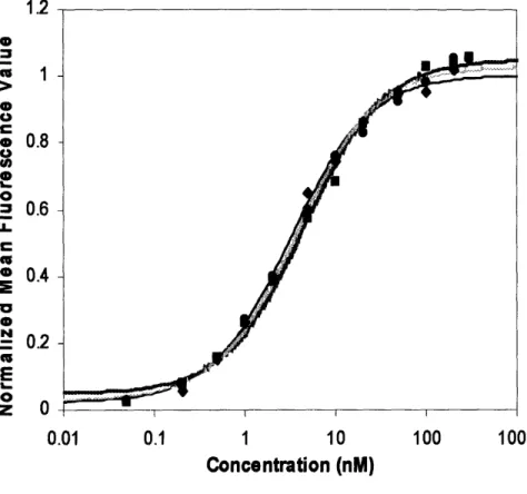
Figure 2.5 shows the titration curves of AF-20 scFv against FOCUS cells at 40C. The
dissociation constant was determined to be 3.8 +0.7nM at 40C.
2.3.4 Internalization of the AF-20 scFv into Tumor Cells
One of the salient properties of AF-20 antigen is that once AF-20 IgG binds, the
complex is internalized readily (Moradpour, et al., 1995). The ability of the AF-20 scFv
to internalize similarly was studied. FOCUS cells were seeded on collagen plated
coverslips overnight. Control (nonspecific) scFv or AF-20 scFv were pre-incubated with
mouse anti-FLAG IgG, so that the scFv would be presented in a bivalent form to the
FOCUS cells, similar to the AF-20 IgG. Conjugated bivalent scFv were then incubated
with the adhered FOCUS cells for hr at 370C in serum free media. The cells were then
washed, fixed and permeablized. The presence of mouse antibodies inside the cells, which implied the internalization of scFv, was detected with goat anti-mouse IgG-FITC
conjugate. Cells were also co-stained with Hoechst, a nuclear dye. Stained cells were then
analyzed using a confocal microscope. Figure 2.6 top, left panel showed negligible internalized green fluorescence signal (FITC) for the control scFv, indicating the absence
of any substantial internalization of control scFv into the FOCUS cells. On the other
1.2 0 1 0 e 0.8 0 l) e 0
o
= 0.6 E 0.4 E0
N = 0.2 Ez O
0.01 0.1 1 10 100 1000 Concentration (nM)Figure 2.5 Titration Curve of AF-20 scFv against FOCUS Cells. FOCUS cells were
first resuspended in solution, and incubated with various concentrations of AF-20 scFv at 4°C. AF-20 scFv bound cells were then stained with mouse anti-FLAG IgG and followed by goat anti-mouse IgG-PE conjugate. Cell fluorescence was detected with FACS. Triplicate trials were performed. The data was fit respectively for each trial and the average dissociation constant of the AF-20 scFv against FOCUS cell at 40C was determined to be 3.8 ± 0.7nM.
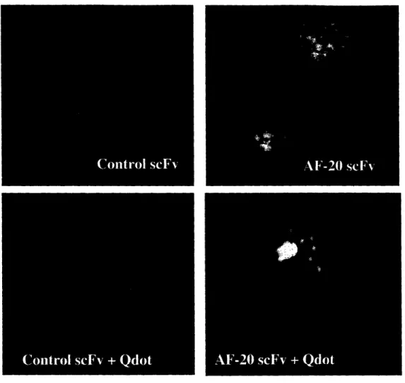
Figure 2.6 In-vitro Internalization Study of AF-20 scFv into FOCUS Cells. The
abilities of the AF-20scFv to internalize by itself and to facilitate the internalization of
other particles into the tumors cells were studied.. FOCUS cells were used in both studies. For the self-internalization study, mouse anti FLAG 1gG conjugated AF-20 scFv or control scFv were incubated with the FOCUS cells at 370C in serum-free media for 1 hr. Cells were then fixed, permeabilized, and stained with goat anti-mouse gG-FITC conjugate and Hoechst dye. The green fluorescence beside the nuclei in the top right
panel showed that conjugated AF-20 scFv internalized readily into the FOCUS cells in vitro. There was no significant green fluorescence signal in the control scFv experiment (top left panel), indicating the lack of internalization of control scFv. For the facilitated internalization study, quantum dot Qdot565 was used as the model particle. The AF-20 scFv or control scFv was pre-incubated with biotinylated mouse anti-FLAG IgG and Qdot 565-streptavidin conjugate. Adhered FOCUS cells were then incubated with the pre-incubated materials and Hoechst dye at 370C in serum-free media for I hr. The absence of red fluorescence in the control scFv experiment (bottom left panel) indicated that there was no facilitated internalization of quantum dot into the FOCUS cells by control scFv. Whereas the red fluorescence in the bottom right panel showed that AF-20 scFv was able to facilitate the internalization of nano-scale particles, quantum dots, into FOCUS cells. The blue fluorescence was from the Hoechst staining of the nuclei.
the nuclei of the cells, suggesting that the bivalent AF-20 scFv internalized readily into
the FOCUS cells, just as AF-20 IgG did. However, when AF-20 scFv was presented
monovalently to the FOCUS cells, the extent of internalization was significantly reduced, signifying the importance of bivalency in the rapid internalization of the antibody.
2.3.5 Facilitated Internalization of Nano-scale Particles
The ability of the AF-20 scFv to internalize into the tumor cells could be
employed to deliver virus or liposome particles carrying suicide gene into tumor cells for
gene therapy (Haisma, et al., 2000; Kashentseva, et al., 2002; Mohr, et al., 2000;
Nettelbeck, et al., 2004; Yoon, et al., 2000). Virus or liposome particles in this case are
considerably larger than the AF-20 scFv protein. The ability of the AF-20 scFv to
facilitate internalization of much larger particles into tumor cells was examined. Quantum
dots conjugated to streptavidin (Qdot 565) were used as model particles in this study.
This particle is about 30nm in diameter and comparable in size to a viral particle. AF-20
scFv or control scFv (fluorescein) was pre-incubated with biotinylated mouse
anti-FLAG IgG and Qdot 565-streptavidin conjugate. The pre-incubated materials were then
incubated with FOCUS cells at 37°C in serum free media for 3 hr. Cells were also
co-incubated with the Hoechst dye. Internalized quantum dots, which emit red fluorescence
upon excitation at 565nm, were detected by confocal microscopy. The control scFv did
not facilitate the internalization of quantum dots (bottom left panel of Figure 2.6), as
there was negligible red fluorescence inside the cells. However in the AF20 treated
samples there was considerable red fluorescence inside the cells (bottom right panel
Figure 2.6). These results indicate that the AF-20 scFv is able to facilitate internalization
2.3.6 In-vitro Cytotoxicity of AF-20 Gelonin Fusion Construct
The capability of AF-20 scFv to serve as the targeting domain in an immunotoxin construct was studied. The toxin used in this study was recombinant gelonin, a highly cytotoxic ribosome-inactivating protein (Falasca, et al., 1982). It was previously shown
that fusing a targeting scFv or a growth factor to gelonin greatly improved the cell-killing
efficacy of gelonin both in vivo and in vitro (Rosenblum, et al., 2003). Therefore, AF-20 scFv-gelonin fusion were constructed as previously reported to determine if the addition of AF-20 scFv to gelonin would similarly improve the efficacy of gelonin (Rosenblum, et
al., 2003). The AF-20 scFv/rGel purified protein was confirmed to have the same binding
affinity as AF-20 scFv at 40C, indicating that the AF-20 scFv domain was properly folded
and fully functional (data not shown). The in vitro cytotoxic effects of the AF-20 scFv/rGel fusion toxin and free rGel were assessed and compared on three different
tumor cell line. The tumor cells lines employed in this study were FOCUS cells, L3.6pl
cells and PC3 cells, which are liver, pancreatic and prostate cancer cell lines respectively.
Log-phase cells (5000 to 10000 cells per well) were seeded in 96-well plates overnight,
and then exposed to various concentrations of AF-20 scFv/rGel fusion and free rGel
respectively. After the treatment, cell viabilities of the treated cells and the untreated positive control cells were determined either using Alamar Blue stain or crystal violet
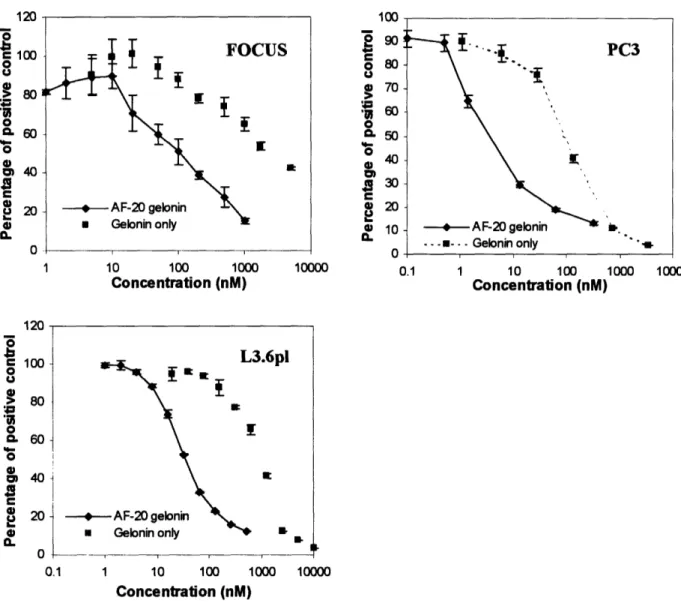
stain. Figure 2.7 showed that in the case of FOCUS cells, the IC50 value for AF-20
scFv/rGel was approximately 100 to 200nM; whereas the IC50 value for free rGel was
about 2gM, one log higher than the IC50 of AF-20 scFv-gelonin fusion. As for the L3.6pl
cells, the IC50 value for AF-20 scFv-gelonin was approximately 35nM; meanwhile the
AF-I U o o e 100 o 0 U 20 0 IUJ 2 90 o 80 U g 70 0 CL 50
40
a
3 0 4 20 0 10 10 10 10 100 o0 10000 0. 1 10 100 1000 10000 Concentration (nM) Concentration (nM) I4 n/ I I . 1 4j 100o
> 80 0. 60-o E 40 a 4) 0 1,3 nl -J I _ A~~~~~~~~~. - -d 1--- T- M --- --I. 0.1 1 10 100 1000 10000 Concentration (nM)Figure 2.7 Comparative In-vitro Cytotoxicity of the Free Gelonin and AF-20 scFv/rGel Fusion Construct on FOCUS, L3.6pl and PC3 Cells. Different tumor cell
lines (FOCUS, L3.6pl and PC3) were plated and treated with various concentrations of free rGel or AF-20 scFv/rGel fusion construct respectively. Cell viabilities were detected using Alamar blue stain (FOCUS) or using crystal violet dye (L3.6pl and PC3). For the FOCUS cells, the IC50 values for AF-20 scFv/rGel and free rGel were about 100nM and
2jiM respectively. For the L3.6pl cells, the IC5o values for AF-20 scFv/rGel and free rGel were about 35nM and ltM respectively. As for the PC3 cells, the IC50values for AF-20 scFv/rGel and free rGel were about 3.5nM and 100nM respectively. The IC5o value for AF-20 scFv/rGel was consistently shown to be one to two logs lower than the IC5o value
for free rGel on three different cell lines.
-- A
4 'in4 fv