Dopamine transporter (DAT) knockdown in the nucleus accumbens
improves anxiety- and depression-related behaviors in adult mice
Amine Bahi
a,⁎, Jean-Luc Dreyer
baDepartment of Anatomy, Tawam Medical Campus, United Arab Emirates University, Al Ain, United Arab Emirates bDivision of Biochemistry, Department of Medicine, University of Fribourg, CH-1700 Fribourg, Switzerland
Many epidemiological and clinical studies have demonstrated a strong comorbidity between anxiety and de-pression, and a number of experimental studies indicates that the dopamine transporter (DAT) is involved in the pathophysiology of anxiety and depression. However, studies using laboratory animals have yielded incon-clusive results. The aim of the present study was to examine the effects of DAT manipulation on anxiety- and depression-like behaviors in mice. For this purpose, animals were stereotaxically injected with DAT siRNA-expressing lentiviral vectors (siDAT) in the caudate putamen (CPu) or in the nucleus accumbens (Nacc) and the behavioral outcomes were assessed using the open-field (OF), elevated-plus maze (EPM), light-dark box (LDB), sucrose preference (SPT), novelty suppressed feeding (NSF), and forced-swim (FST) tests. The results showed that in the Nacc, but not in the CPu, siDAT increased the time spent at the center of the arena and decreased the number of fecal boli in the OF test. In the EPM and LDB tests, Nacc siDAT injection increased the entries and time spent on open arms, and increased the time spent in the light side of the box, respectively, suggesting an an-xiolytic-like activity. In addition, siDAT, in the Nacc, induced significant antidepressant-like effects, evidenced by increased sucrose preference, shorter latency to feed in the NSF test, and decreased immobility time in the FST. Most importantly, Pearson’s test clearly showed significant correlations between DAT mRNA in the Nacc with anxiety and depression parameters. Overall, these results suggest that low DAT levels, in the Nacc, might act as protective factors against anxiety and depression. Therefore, targeting DAT activity might be a very attractive approach to tackle affective disorders.
1. Introduction
According to the World Health Organization, the proportion of the global population with anxiety and depression in 2015 was estimated to
be 3.6 and 4.4% respectively [1]. In addition, the total estimated
number of people living with anxiety and depression increased by 14.9% and 18.4% respectively between 2005 and 2015, as a result of
population growth and ageing [2]. Although generalized anxiety and
major depressive disorders (GAD and MDD respectively) are associated with high disease burden, the molecular mechanisms involved in these psychiatric disorders are not fully understood. Therefore, elucidating the precise nature of these comorbidities could help understanding the pathophysiology of anxiety- and depression-related disorders.
The dopaminergic system plays a crucial role in both the periphery
and the central nervous system (CNS). Centrally, dopamine is a neu-rotransmitter distributed in the brain stem in pons and medulla
ob-longata [3] as well as in the forebrain (covering hippocampus,
amyg-dala, nucleus accumbens, putamen, and caudate) [4]. It regulates
emotional and behavioral conditions along with other neuro-transmitters. In fact, in patients with GAD and MDD, neuroreceptor imaging and anatomical/pharmacological studies showed abnormal
central dopamine function [5,6]. For example, using SPECT
neuror-eceptor imaging, Tiihonen and co-workers found, when measuring striatal presynaptic dopaminergic innervation, that DAT binding was significantly lower in the patients with social anxiety disorder than in
the age- and gender-matched comparison subjects [7]. Furthermore,
when subjects with generalized social phobia, according to the DSM-IV, were tested using a fMRI study while executing the implicit sequence
Abbreviations: CPu, caudate putamen; DAT, dopamine transporter; EPM, elevated-plus maze; FST, forced-swim test; LDB, light-dark box; LV, lentiviral vectors; NSF, novelty supressed feeding; Nacc, nucleus accumbens; OF, open-field; shRNA, short herpin RNA; siRNA, small interfering RNA; SPT, sucrose preference test; SNc, substantia nigra pars compacta; TST, tail suspension test; VTA, ventral tegmental area
⁎Corresponding author.
E-mail address:[email protected](A. Bahi).
http://doc.rero.ch
Published in "Behavioural Brain Research 359(): 104–115, 2019"
which should be cited to refer to this work.
learning task, results have shown significant striatal abnormalities especially in the left caudate head, left inferior parietal lobe, and
bi-lateral insula [8].
The dopamine transporter (DAT) is involved in the transport of dopamine, either into or out of the neuron, to control its levels at the synapse. Indeed, in the dorsal striatum and Nacc, it has been shown that the removal of dopamine from the synapse is predominantly performed
by the neuronal DAT and not metabolism or diffusion [9]. However, in
the prefrontal cortex (PFC), it’s important to clarify that the
nor-epinephrine transporter (NET) is present in much greater concentra-tions than DAT. Thus, dopamine uptake in the PFC is thought to depend
primarily on NET [10].
The rat DAT cDNA, encoding a protein of 620 amino acid, was cloned more than 25 years ago. The analysis of the protein sequence suggested the presence of twelve putative transmembrane domains
[11]. Apart from thefive intracellular and six extracellular loops, DAT
exhibits a cytoplasmic amino and carboxyl-termini that were shown to
interact with the Ca2+/calmodulin-dependent protein kinase II
(CaMKII) to regulate the efflux of dopamine through DAT. In fact, CaMKIIα binds to the distal carboxyl-terminal of DAT and colocalize
with DAT in dopaminergic neurons [12]. DAT is a target for the
de-velopment of pharmacotherapies for a number of disorders including
Parkinson’s disease [13], Alzheimer’s disease [14], schizophrenia [15],
Tourette’s syndrome [16], Lesch-Nyhan’s disease [17], attention deficit
hyperactivity disorder [18], obesity [19], depression [20], and
stimu-lant abuse [21] as well as normal aging [22]. Therefore, and because
DAT can influence the re-uptake of dopamine, we suggest that it may have a role in the pathophysiological mechanisms of anxiety and de-pression-like behaviors.
Although the exact mechanisms by which DAT affects dopamine-mediated neurotransmission in anxiety- and depression-like behaviors remain to be fully defined, previous studies have provided evidence that DAT might be involved in the pathophysiology of mood disorders. For example, neuroimaging studies suggested a dysfunction of the
striatal presynaptic DAT in social anxiety disorder [23]. Also, PET
imaging revealed a significant reduction in DAT binding potential (BP)
in the striatum of unmedicated patients with MDD [24], and decreased
BP in the caudate, but not the putamen of patients relative to healthy
controls [25].Therefore, understanding these processes will not only
help elucidate the complex mechanisms of anxiety and depression
co-morbidities, but also help in the development of effective therapies to
counteract these disorders.
Although a previous study showed that DAT over-expression in adult Wistar rats' Nacc leads to impulsive and risk prone phenotype
[26], and rats with intra-accumbal delivery of DAT shRNA-expressing
lentiviral vectors, showed a trend, (0.05 < p < 0.10) toward an
an-xiogenic-like phenotype [27], few experimental studies have directly
and systematically examined the role of DAT in anxiety and depression, and little is known about the specific contribution of accumbal DAT. In fact, there is evidence that emotional disorders such as anxiety and
depression are associated with increased impulsivity [28–31].
How-ever, other studies revealed that reduced anxiety- and
depression-re-lated responses corredepression-re-lated with increased impulsivity [32,33].
There-fore, the current experiments were designed to test the hypothesis that DAT knockdown in the Nacc can elicit dysregulated behaviors in adult C57BL/6 mice, including anxiety- and depressive-like symptoms. In first, we choose a battery of the most commonly used tests (OF, EPM and LDB) to study the effects of DAT knock-down on anxiety-like be-havior. The second goal was to examine the consequences of DAT knock-down on depression-like behavior in adult C57BL/6 mice using the SPT, NSFT, and FST. We hypothesized that DAT knock-down in the Nacc will ameliorate anxiety-like behaviors, and consequently DAT anxiolytic prone phenotype will be associated to an antidepressant-like activity.
2. Materials & methods 2.1. Animals
C57BL/6 J mice were originally obtained from Jackson Laboratory (Bar Harbor, ME) and bred in the local animal facility of the College of Medicine & Health Sciences of the UAE university. All mice were of identical genetic background and back-crossed to the parental inbreed mice each ten generations. For the current study, adult male C57BL/6 J (approximately 22–26 g) were maintained in a temperature-controlled (∼22 °C) colony room with a 12-hour light–dark cycle (0600 to 1800 light on). A large number of published reports used the same lighting conditions with the housing room kept under a regular light/dark
schedule [34,35]. The mice (44 in total) were group-housed until they
underwent the stereotaxic injections yielding 4 experimental groups (n = 11 each). Mice were singly housed after the surgery, and during subsequent behavioral testing to minimize confounding effects of group
housing and establishment of social hierarchies [36]. Bedding was
produced locally and autoclaved before use. Water and food were available ad libitum throughout the experiment. Standard rodents’ chow diet was obtained from the National Feed and Flour Production and Marketing Company LLC (Abu Dhabi, UAE). The local Animal Research Ethics Committee approved the procedures (Application Re-ference No. A27-12).
2.2. Design and cloning of small hairpin RNAs and lentivirus production Plasmid construction was described in previously published work
from our laboratory [26,27,37]. In brief, the following target within
DAT sequence 5’-AGC CAT GGA TGG CAT CAG AGC ATA CCT-3’ was
selected based on Hannon’s design criterion (http://katahdin.cshl.org:
9331/RNAi/html/rnai.html). The sequence was subjected to a BLAST search to verify its specificity and the pairwise sequence similarity search yielded a 96% homology between mouse and rat sequences. A
stem-loop structure incorporating the nucleotides’ target sequence was
created so that small hairpin RNAs (shRNAs) could be produced from the lentiviral vector pTK431 (graciously provided by Dr. Tal Kafri, UNC Gene Therapy Center). The siRNA target was synthesized and added to the mouse U6 promoter by PCR, using following PCR program: 120 s at 94 °C (initial denaturation) followed by 35 cycles (45 s at 94 °C, 45 s at 64 °C and 45 s at 72 °C) in 4% dimethyl sulfoxide (DMSO). The PCR product was digested with BamHI and XhoI, cloned into similar sites into pTK431. The control vector (Mock) consisted of an empty pTK431. For lentiviral production, a triple transfection protocol was used as
described previously [38–40]. Briefly, the pTK431-transfer plasmids
together with the packaging pΔNRF and the envelope pMDG-VSV-G plasmids were co-transfected into HEK293 T cells using calcium phos-phate. Medium was collected at 24 and 48 h post-transfection and virus concentrated by centrifugation. The supernatant was removed, and
virus was suspended in sterile cold phosphate-buffered saline (PBS)
supplemented with 1% bovine serum albumin (BSA). Viral titers
(ap-proximately 108-109 unit/mL) were determined using a p24 antigen
ELISA kit.
2.3. Stereotaxic injection of lentiviral vectors
For viral injection, mice werefirst anesthetized with a cocktail of
ketamine (100 mg/kg, i.p.) and xylazine (10 mg/kg, i.p.) and placed in
a stereotaxic frame. Using ear bars, the head wasfixed, and a midline
incision was made to measure the bregma from the skull surface. A
craniotomy was drilled, and a Hamilton micro-syringefilled with a viral
solution was lowered using the following coordinates for the Nacc [ + 1.7 mm AP, ± 0.75 mm ML, and 4.5 mm ventral from the dural surface]. Because the CPu is part of the dorsal striatum and is sig-nificantly large and functionally heterotopic, two separate injections were performed in each hemisphere using the following coordinates:
[1stinjection: + 1 mm AP, ± 1.5 mm ML, and 3.5 mm ventral], [2nd
injection: -0.10 mm AP, ± 2.5 mm ML, and 3.8 mm DV] [41–44]. For
each injection, 0.5μL of viral particles were infused at a rate of 0.1 μL/
min. Following vector administration, the Hamilton micro-syringe was left in situ for an additional 5 min to permit time for the vector to diffuse from the needle tip and minimize upward flow of viral solution after raising the needle before being slowly retracted from the brain. The skin was stitched, and mice were monitored until recovery from the surgery before returned to their home cage and left to recover 10 days before behavioral experiments started to ensure good levels of expres-sion. None of the injected animals were excluded from the statistical analysis.
2.4. Anxiety- and depression-related behavior tests
All anxiety-like behavior tests were conducted between 10:00 AM and 4:00 PM in a brightly lit room that was illuminated with four fluorescent overhead bulb lights, which produce consistent illumination within the room (approximately 100 lx). The mice were habituated to the room for 1 h before starting the respective test. The general design of the study is depicted in Supp. Fig. 1.
2.4.1. Open-field (OF)
10 days following viral injection, spontaneous and exploratory lo-comotor activity and anxiety were tested in an OF test as reported in
our previous studies [45–47]. In brief, each mouse was placed in the
corner of the OF apparatus (32 × 32 × 20 cm3) (L × W × H) and
al-lowed to freely explore the arena for 15 min. The time spent in the
center of the arena (8 × 8 cm2), the number of line crossings, and the
number of fecal boli were hand scored. After each session, fecal boli were counted and removed, and the arena was cleaned with a 70% pure ethanol solution to remove odor trails.
2.4.2. Elevated-plus maze (EPM)
This test is an ethologically relevant assessment of anxiety levels in
rodents [48]. Two days after the OF test, animals were tested in the
EPM as previously performed in our laboratory [47,49,50]. The EPM
apparatus consisted of two open arms (OA) (40 × 6 cm2) and two
closed arms (CA) (40 × 6 × 20 cm3), connected by a central platform
(6 × 6 cm2) and elevated 40 cm off the floor. Although, differences
were observed when rodents were placed facing toward an OA versus
toward the CA [48], at the beginning of the session each mouse was
placed into the center of the maze, facing one of the OA and behavior
was monitored live for 5 min as described by others [51,52]. The
number of entries and the time spent in each arm of the maze were hand scored. After each EPM testing, animals were immediately re-turned to their home cages, and the maze was cleaned with a 70% pure ethanol solution to remove odor trails.
2.4.3. Light-dark box (LDB)
The original method described by [53] was used with minor
mod-ifications as in our recent study [46]. In brief, the apparatus consisted of
two wood chambers (24 × 30 × 30 cm3) connected by a 6-cm wide
guillotine door. One chamber had black walls covered by the lid, and the other had white walls with a 60 W light bulb located 30 cm above the chamber. For the experiment, each mouse was placed in the light chamber and could explore the two chambers for 5 min as described by
others [54–56]. The following parameters were hand scored: i) latency
to enter the dark chamber (sec), ii) latency to re-enter the light chamber (sec), iii) total time spent in the light chamber (sec), and iv) number of crossings/transitions between the two chambers (n). The two chambers were cleaned with 70% ethyl alcohol and dried between trials. 2.4.4. Sucrose preference test (SPT)
The SPT was performed as described previously [57]. Briefly, a
two-bottle choice procedure was used to test for differences between the
groups for their relative preference for sucrose over water. The mice were given free choice access to 2 pipettes: one containing tap water and the other containing a 2% sucrose-solution for 24 h. The pipettes were switched (left/right) after 12 h to eliminate side preference. We monitored the latency to initiate drinking, and the amount of water and sucrose consumed in milliliters during the 24 h was measured and the percent preference for sucrose consumption was calculated as a
per-centage of sucrose solution consumed in relation to the totalfluid
in-take. The total fluid intake (water + sucrose) was measured and
ex-pressed in milliliters per kilogram of body weight. 2.4.5. Novelty suppressed feeding test (NSFT)
The NSFT was performed as described previously [57]. In brief, 24 h
before the test all the mice were food deprived. The novel environment
consisted of a Plexiglas cage (45 × 28 × 13 cm3) brightly lit using a
60 W light bulb located 50 cm above the cage lid, directly above the food dish. The food dish was a 5 cm‐diameter filter paper containing a previously weighed piece of standard lab chow. Each mouse was placed into the novel cage for 15 min and the following parameters were hand scored: i) latency to begin eating (defined as chewing of food), ii) total time spent eating in the test box, iii) amount of food consumed in the test box. At the end of each session, the mouse was immediately re-turned to its home cage and, as a control, food consumption was as-sessed by weighing each cage's food at the beginning and end of a 24-h period.
2.4.6. Forced-swim test (FST)
The FST originally described by Porsolt and co-workers [58] was
performed as we described in our previous studies [47,59]. Although,
the FST for mice consists of a single 6-min exposure, some investigators
have developed two session procedures as well [60]. In the current
study, each mouse was individually forced to swim in a 1-liter glass beaker (Height: 158 mm; Width: 108 mm) containing water at a tem-perature of approximately 25 °C for 6 min as described by others
[61–64]. The mice were unable to escape or rest by touching the bottom
of the beaker. The time taken by the animal to start struggling and swimming (latency time) as well as the duration of immobility were hand scored. After each trial, the water was changed.
2.5. Total RNA isolation and quantitative RT-PCR analysis
After the completion of behavioral testing, the mice were killed by rapid decapitation. The brains were quickly removed, Nacc and CPu samples (used as a control region) were dissected out and stored at −80 °C. Total RNA was extracted using the TRIzol reagent and pre-cipitated with isopropanol according to the manufacturer instructions.
To verify DAT mRNA (Accession No.NM_012694) knockdown,
single-stranded cDNA was synthesized from total RNA using the SuperScript III reverse transcriptase procedure. Following reverse transcription, quantitative RT-PCR was performed in triplicate using SyberGreen purchased from Applied Biosystems, per the manufacturer instructions. The temperature cycling parameters consisted of initial denaturation at 95 °C for 4 min followed by 40 cycles of denaturation at 94 °C for 30 s, annealing and extension at 60 °C for 45 s. PCR for the endogenous housekeeping gene, cyclophilin, was run with the same cycling
para-meters. The template (2μl) was amplified by PCR in 20 μl total reaction
volume containing 0.5μmol of each specific PCR primer. Gene
ex-pression levels were analyzed by theΔΔCt method using cyclophilin as
a reference gene because of its low variability between samples. 2.6. Statistical analyses
For statistical comparisons, the software package IBM SPSS Statistics 16.0 was used. Data were expressed as means ± SEM. The data representing the effects of DAT knockdown on anxiety- and de-pression-like behaviors were analyzed using a two-way analysis of
variance (ANOVA) with viral-injection“Mock vs. siDAT” and brain
re-gion“Nacc vs. CPu” as the between-subject factors. Simple linear
re-gression (Pearson's test) analysis was performed to examine the corre-lation between DAT mRNA levels in the CPu or the Nacc and measures of anxiety- and depression-like behaviors. The relationship is expressed as the correlation coefficient (r). Levene's test was used to inspect the homogeneity of variance of all data. The acceptable level of significance was 5% for each analysis.
3. Results
3.1. siDAT injection reduced DAT mRNA in the Nacc and CPu
To knock-down DAT expression, we expressed DAT siRNAs (siDAT) using a lentiviral vector that was stereotaxically injected into the Nacc or into the CPu (used as a control region), of adult mice (for each group, n = 11).
To determine the persistence of infection in the Nacc and CPu, mice were injected bilaterally with siDAT and examined for mRNA expres-sion after completion of the behavioral experiment. In fact, to de-termine the efficacy of siDAT, we measured levels of DAT mRNA, using RT-PCR, of infected Nacc and CPu, tissues and the results are
sum-marized inTable 1. The two-way ANOVA revealed a significant effect of
viral-injection (F(1,40)= 254.713, p < 0.0001). However, and as
ex-pected there was no significant effect of brain region (F(1,40)= 0.271,
p = 0.605), and the interaction between the two factors was not found
significant (F(1,40)= 0.263, p = 0.611). Bonferroni post hoc evaluations
indicated that, regardless of the brain region, DAT mRNA levels were decreased by approximately 70%, indicating that the siDAT was func-tional. We also measured DAT mRNA expression in both areas for
vector inoculated in either area, and the results are depicted inTable 1.
The two-way ANOVA revealed no effect of viral-injection
(F(1,40)= 0.051, p = 0.822), or brain region (F(1,40)= 1.125, p =
0.295), with no significant interaction (F(1,40)= 1.056, p = 0.310),
clearly indicating that the injections were site specific and mRNA
ex-pression alterations could be found in a non-targeted region. Thus, the effects were limited to the region of the injection. Taken together, these results suggested that we could consistently infect both the CPu and Nacc, the infection lasted for the time required to complete behavioral experiments, and siDAT were effective in downregulating DAT ex-pression in infected neurons in vivo.
3.2. DAT knockdown in the Nacc altered anxiety-like behavior
3.2.1. Open-field (OF)
Wefirst tested the mice in an OF test that was performed 10 days
after viral injections as depicted in Suppl. Fig. 1. During this assay, in which the effects of stimulation on spontaneous behaviors could be characterized, the Levene's test for equality of variances was found to
be statistically non-significant (F(3,40)= 0.930, p = 0.435). Therefore,
and as depicted in Fig. 1A, the two-way ANOVA test revealed a
significant effect of viral-injection (F(1,40)= 8.925, p = 0.005), and
brain region (F(1,40)= 4.212, p = 0.047). Interestingly, the interaction
between the two factors was also found significant (F(1,40)= 7.509,
p = 0.009). Bonferroni post hoc evaluations indicated that, compared to Mock controls, lentiviral-mediated knockdown of DAT (siDAT) in the Nacc, but not in the CPu, increased the time spent in the center of the
arena (p = 0.001 and p = 1.000 respectively). Thesefindings indicate
that siDAT-injected mice in the Nacc show less aversion than Mock controls to the center of the OF, which is consistent with decreased
anxiety-like behavior. Also, and as depicted inFig. 1B, the Levene's test
was not significant (F(3,40)= 0.558, p = 0.646) for the number of fecal
boli. However, the two-way analysis indicated that there was a
sig-nificant effect of viral-injection (F(1,40)= 10.851, p = 0.002), and
brain region (F(1,40)= 8.756, p = 0.005) on the number of fecal boli
left in the arena with a significant interaction (F(1,40)= 10.128, p =
0.003). Pairwise evaluations revealed that, compared to Mock controls, siDAT injections in the Nacc, but not in the CPu, significantly decreased the number of fecal boli (p < 0.0001 and p = 1.000 respectively). Finally, to verify whether DAT manipulation in the Nacc influenced spontaneous exploratory behavior, which may influence the perfor-mance on the OF test, general locomotor activity was determined over the 15-min test. The statistical analysis indicated that the number of line crossings did not differ significantly between the four experimental
groups as the main effect of viral-injection (F(1,40)= 0.0001, p =
0.991) and brain region (F(1,40)= 0.107, p = 0.746) were not found
significant. Consequently the viral-injection × brain interaction was
not significant (F(1,40)= 0.526, p = 0.473) (Levene’s test:
(F(3,40)= 0.812, p = 0.495) (Fig. 1C) suggesting that the potential
anxiolytic effect of DAT knockdown in the Nacc, observed in the OF test was not confounded by changes or deficits in overall spontaneous motor activity.
3.2.2. Elevated-plus maze (EPM)
Two days after the OF test, the mice were tested in the EPM test which exploits the conflict between the desire to explore a novel area
and aversion to open areas and height [65]. For the percentage of time
spent into the open arms (OA), the two-way ANOVA revealed a
sig-nificant effect of viral injection (F(1,40)= 6.405, p = 0.015), and brain
region (F(1,40)= 7.240, p = 0.010), with significant interaction
(F(1,40)= 8.242, p = 0.007) (Levene’s test: F(3,40)= 1.503, p = 0.229)
(Fig. 1D). Post hoc evaluations indicated that siDAT-injected mice in the Nacc, but not in the CPu, spent more time in the OA of the maze than Mock controls (p = 0.003 and p = 1.000 respectively). Similarly, there
was a significant effect of viral injection (F(1,40)= 10.295, p = 0.003),
and brain region (F(1,40)= 9.573, p = 0.004) on the number of entries
into the OA with a statistically significant interaction (F(1,40)= 8.877,
p = 0.005) (Levene’s test: F(3,40)= 0.843, p = 0.479). Post hoc tests
indicated that the number of entries into the OA was significantly in-creased in Nacc mice injected with siDAT compared to Mock (p =
0.001). However, in the CPu group, there was no difference between
Mock and siDAT conditions (p = 1.000, Bonferroni post hoc test) (Fig. 1E). Significant differences between the CPu and Nacc groups
were revealed when OA entries were expressed as a percentage of total
entries to account for differences in locomotion (main effect of brain
region: F(1,40)= 4.613, p = 0.038). siDAT injection in the Nacc
sig-nificantly increased the percentage of OA entries (main effect of
viral-injection: F(1,40)= 9.346, p = 0.004) yielding significant interaction
between the two factors (F(1,40)= 5.311, p = 0.026) (Levene’s test:
F(3,40)= 0.857, p = 0.471) (Data not shown). It should be mentioned
that the siDAT effects on the parameters measured in the EPM test were
mainly driven by the Nacc group, which differentially and significantly
affected the Mock- and siDAT-injected mice (p = 0.001, Bonferroni post hoc test) (Data not shown). The overall activity was estimated by the number of entries performed into the closed arms (CA) during the 5-min
observation period. Interestingly, as depicted inFig. 1F this parameter
was not affected by viral-injection (F(1,40)= 0.963, p = 0.332), nor by
Table 1
DAT mRNA expression in Mock- and siDAT-injected mice.
Injection site mRNA level in Mock siDAT
CPu CPu 1.000 ± 0.176 0.322 ± 0.105*,#
Nacc 1.000 ± 0.157 1.072 ± 0.221
Nacc CPu 1.000 ± 0.182 0.951 ± 0.202
Nacc 1.000 ± 0.191 0.277 ± 0.079*,#
The data are expressed as mean ± SEM for the relative mRNA expression in the Nacc and CPu with or without lentiviral injection.
* p < 0.0001 indicate significant differences between Mock- and siDAT (Injection site).
# p < 0.0001 indicate significant differences between Nacc and the CPu.
For each group n = 11.
brain region (F(1,40)= 0.076, p = 0.785), with no significant
interac-tion (F(1,40)= 0.125, p = 0.726) (Levene’s test: F(3,40)= 0.082, p =
0.969) indicating no change in overall ambulatory behavior during the
EPM test. Thesefindings further indicate that lower levels of DAT in the
Nacc reduced anxiety-like behavior. 3.2.3. Light-dark box (LDB)
Next, we assessed anxiety-like behavior using a LDB test, which examines the conflict between the drive to explore and aversion to a brightly lit space. The two-way ANOVA revealed that the latency for the
mice to make thefirst move to the dark box was significantly affected
by viral-injection (F(1,40)= 4.786, p = 0.035), and brain region
(F(1,40)= 5.403, p = 0.025), with a significant interaction between the
two factors (F(1,40)= 5.090, p = 0.030) (Levene’s test: F(3,40)= 2.175,
p = 0.106) (Fig. 2A). Post hoc tests indicated that when injected in the
Nacc, siDAT-injected mice showed a significantly increased latency to enter the dark side of the box compared to the Mock controls (p =
0.019). However, and as depicted inFig. 2B, once they have entered the
dark box, siDAT-injected mice displayed a significantly shorter latency
to re-enter the light box; main effect of viral-injection: (F(1,40)= 8.860,
p = 0.005), main effect of brain region (F(1,40)= 10.486, p = 0.002),
interaction (F(1,40)= 12.021, p = 0.001) (Levene’s test:
F(3,40)= 1.495, p = 0.231). Although there was a main effect of brain
region across both groups, Bonferroni post hoc tests revealed that this
effect was driven by the Nacc group. In fact, the latency to re-enter the
light box was significantly lower in Nacc siDAT-injected mice compared
to Mock controls (p < 0.0001). In contrast, no significant differences
were found in the CPu group between Mock-and siDAT-injected mice (p = 1.000). In addition, there were main effects of viral-injection (F(1,40)= 6.464, p = 0.015) and brain region (F(1,40)= 5.618, p =
0.023) on time spent in the light side of the chamber. Also, there was a
significant interaction between the two variables (F(1,40)= 7.535, p =
0.009) (Levene’s test: F(3,40)= 1.050, p = 0.381) (Fig. 2C). Bonferroni
post hoc tests revealed that the only comparison which differed
sig-nificantly was in the Nacc group in which siDAT-injected mice spent more time in the light side of the box compared with Mock controls
(p = 0.003). Thesefindings further indicate that anxiety-like behavior
is reduced in Nacc siDAT-injected mice. Interestingly, there was no
significant effect of viral-injection (F(1,40)= 0.044, p = 0.836), brain
region (F(1,40)= 0.006, p = 0.939) or an interaction of the independent
variables (F(1,40)= 0.245, p = 0.623) in the number of crossings/
transitions between light and dark sides of the box, again confirming
that locomotor activity is similar in both groups (Levene’s test:
F(3,40)= 0.940, p = 0.430) (Fig. 2D).
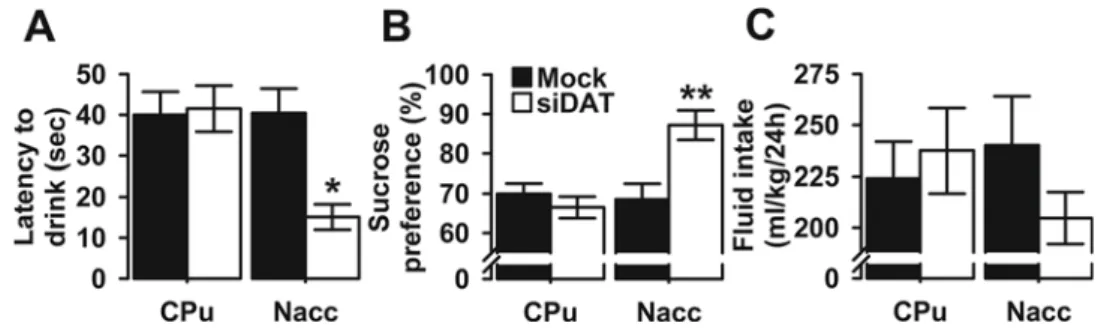
3.3. DAT knockdown in the Nacc altered depression-like behavior 3.3.1. Sucrose preference test (SPT)
During the SPT, the mice could drink either tap water or a 2%
su-crose solution and the results are shown inFig. 3. The latency to drink
after the bottles were presented was analyzed using a two-way ANOVA and the Levene's test for equality of variances was found to be
statis-tically non-significant (F(3,40)= 1.998, p = 0.130), but there was a
significant main effect of viral injection (F(1,40)= 5.151, p = 0.029)
and brain region (F(1,40)= 6.138, p = 0.018). Also, the viral-injection
× brain region interaction term was significant (F(1,40)= 6.575, p =
0.014) (Fig. 3A). Bonferroni post hoc testing indicated that Nacc
siDAT-Fig. 1. Anxiety-like behavior in Mock- and siDAT-injected mice in the OF and EPM tests. The data are expressed as mean ± SEM for the A) time spent in the center of the arena, B) number of fecal boli, C) number of line cross-ings in the OF test, D) percentage of time spent in open arms (OA), E) number of entries into the OA, and F) number of entries into the closed arms (CA) of the EPM. *p < 0.005, and **p < 0.001 indicate significant differences between Mock- and siDAT-injected mice in the Nacc. For each group n = 11.
Fig. 2. Anxiety-like behavior in Mock- and siDAT-injected mice in the LDB test. The data are expressed as mean ± SEM for the A) latency to enter the dark box, B) latency to reenter the light box, C) total time spent in the light box, and D) number of crossings between the light and dark boxes. *p < 0.05, **p < 0.005 and ***p < 0.001 indicate significant differences between Mock- and siDAT-injected mice in the Nacc. For each group n = 11.
injected mice, but not CPu, showed a significantly shorter latency time than Mock controls (p = 0.009 and p = 1.000 respectively). In
addi-tion, sucrose preference was significantly affected by viral-injection
(F(1,40)= 5.305, p = 0.027), and brain region (F(1,40)= 8.433, p =
0.006) with a significant interaction between the two factors (F(1,40)= 11.050, p = 0.002) (Levene’s test: F(3,40)= 1.082, p =
0.368) (Fig. 3B). Pairwise comparisons revealed that, in the Nacc group,
siDAT-injected mice showed a significantly higher preference for 2% sucrose (p = 0.002, Bonferroni post hoc) which confirmed an increased hedonic response as a consequence of Nacc DAT knock-down.
Im-portantly, the mean daily fluid consumption (sucrose + water) per
body weight was not significantly different between the
four-experi-mental groups; main effect of viral injection (F(1,40)= 0.324, p =
0.572), main effect of brain region (F(1,40)= 0.190, p = 0.665),
in-teraction (F(1,40)= 1.600, p = 0.213) (Levene’s test: F(3,40)= 2.125,
p = 0.112) (Fig. 3C).
3.3.2. Novelty-suppressed feeding (NSF)
After 24 h of food deprivation, the changes in body weight were not significantly different in the four-experimental groups (main effect of
viral-injection: F(1,40)= 0.375, p = 0.544) (Data not shown). However,
in the test box the latency to initiate eating was affected by
viral-in-jection (F(1,40)= 6.616, p = 0.014), and brain region (F(1,40)= 8.234,
p = 0.007). Consequently, the interaction between the two
in-dependent variables was found significant (F(1,40)= 13.125, p =
0.001), but the Levene's test for equality of variances was found to be
statistically non-significant (F(3,40)= 1.550, p = 0.216) (Fig. 4A). Post
hoc analyses revealed that, when injected in the Nacc, siDAT mice
showed a significantly shorter latency to bite into pellets as compared
to Mock controls (p < 0.0001) (Fig. 4A). In addition, and during the
15-min test period, siDAT-injected mice spent significantly more time
eating; main effect of viral injection (F(1,40)= 6.682, p = 0.013), main
effect of brain region (F(1,40)= 7.116, p = 0.011), interaction
(F(1,40)= 8.078, p = 0.007) (Levene’s test: F(3,40)= 2.084, p = 0.118)
(Fig. 4B). Again, the siDAT effects were driven by the Nacc group, which was significantly different between the viral-injection conditions (p = 0.003, Bonferroni post hoc), whereas the CPu group did not differ between Mock and siDAT-injected rats (p = 1.000, Bonferroni post hoc). Similarly, DAT knockdown yielded a greater food intake during
the test; main effect of viral injection (F(1,40)= 8.288, p = 0.006), main
effect of brain region (F(1,40)= 4.570, p = 0.039), interaction
(F(1,40)= 8.288, p = 0.006) (Levene’s test: F(3,40)= 0.254, p = 0.858)
(Fig. 4C). It should be mentioned that the siDAT effects on food intake
during the NSF test were mainly driven by the Nacc group, which
dif-ferentially and significantly affected the Mock- and siDAT-injected
groups (p = 0.001, Bonferroni post hoc). However, after returning to
their home cage, the daily total food consumption was not significantly
different between the four experimental groups; main effect of viral
injection (F(1,40)= 0.041, p = 0.840), main effect of brain region
(F(1,40)= 0.623, p = 0.435), interaction (F(1,40)= 0.006, p = 0.940)
(Levene’s test: F(3,40)= 0.392, p = 0.760) (Fig. 4D).
3.3.3. Forced-swim test (FST)
In this last test, the two-way ANOVA analysis revealed that for la-tency to immobility, a significant effect was seen of both viral-injection (F(1,40)= 4.484, p = 0.040), brain region (F(1,40)= 5.551, p = 0.023)
and of the interaction between the two factors (F(1,40)= 7.825, p =
0.008) (Levene’s test: F(3,40)= 0.991, p = 0.407) (Fig. 4E). Post hoc
evaluations indicated that Nacc siDAT-injected mice showed prolonged latency to immobility compared to Mock controls (p = 0.007) which is also a sign of anti-depression-like behavior in the rodent FST. Finally,
and as showed in Fig. 4F, the two-way ANOVA showed significant
Fig. 3. Depression-like behavior in Mock- and siDAT-injected mice in the SPT. The data are expressed as mean ± SEM for the A) latency to initiate sucrose drinking, B) sucrose pre-ference, and C) totalfluid intake in milliliters per kilogram of body weight. *p < 0.01 and **p < 0.005 indicate significant differences between Mock- and siDAT-injected mice in the Nacc. For each group n = 11.
Fig. 4. Depression-like behavior in Mock- and siDAT-injected mice in the NSFT and FST. The data are expressed as mean ± SEM for the A) latency to feed, B) feeding time in the test box in (sec/15 min), C) food consumed in the test box (g/15 min), and D) food consumed in the home cage expressed as grams per kilogram of body weight per 24 h (g/kg/24 h) in the NSFT, E) latency to initiate immobility, and F) total immobility time in the FST. *p < 0.01, **p < 0.005, and ***p < 0.001 indicate significant differences between Mock- and siDAT-injected mice in the Nacc. For each group n = 11.
effects of both viral-injection (F(1,40)= 5.186, p = 0.028) and brain
region (F(1,40)= 4.587, p = 0.038), and of their interaction on
im-mobility (F(1,40)= 6.890, p = 0.012) (Levene’s test: F(3,40)= 0.218,
p = 0.883). Although there was a main effect of brain region across
both groups, post hoc tests revealed that this effect was driven by the
Nacc group. Indeed, immobility duration was significantly lower in
Nacc mice injected with siDAT compared to Mock controls (p = 0.008, Bonferroni post hoc test). In contrast, no significant differences were found in the CPu group between Mock-and siDAT-injected mice (p = 1.000, Bonferroni post hoc test). These results indicate that the knock-down of DAT in the Nacc, but not in the CPu, attenuated depression-like behavior.
3.4. DAT mRNA in the Nacc correlated with anxiety- and depression-like behaviors
Pearson’s correlation was used to establish the relationship between DAT mRNA levels in the Nacc, and measures of anxiety- and
depression-like behaviors andfindings are displayed inFig. 5and Suppl. Fig. 2. In
the OF test, the results have shown that DAT mRNA correlated nega-tively with the time spent in the center of the arena (r = -0.486, p =
0.022;Fig. 5A), and positively with number of fecal boli recovered from
the OF test (r = 0.714, p < 0.0001;Fig. 5B). However, there was no
correlation between DAT mRNA levels and the number of line crossings (r = 0.112, p = 0.619; Suppl. Fig. 2A). In the EPM test, the Pearson's
correlation coefficients revealed a negative correlation between DAT
mRNA levels with the proportion of time spent in the OA (Pearson’s
test: r = -0.474, p = 0.026;Fig. 5C). Also, we found a negative
cor-relation between DAT mRNA with the number (r = −0.565, p =
0.006;Fig. 5D), and the percentage of entries into the OA (r =−0.596,
p = 0.003; Suppl. Fig. 2B), but not with the number of CA entries (r = 0.272, p = 0.221; Suppl. Fig. 2C). In addition, the Pearson
cor-relation coefficient was used to determine any relationship between
DAT mRNA expression and the parameters of the LDB test and the re-sults indicated that DAT mRNA correlated negatively with the time
spent in the light side of the box (r =−0.474, p = 0.026;Fig. 5E) and
with the latency to enter the dark side of the box (r =−0.563, p =
0.006; Suppl. Fig. 2D). However, there was a strong positive correlation
between DAT mRNA with the latency to re-enter the light side of the box (r = 0.579, p = 0.005; Suppl. Fig. 2E), but not with the number of
crossings between light and dark sides of the box (r =−0.059, p =
0.795; Suppl. Fig. 2F). For depression-like behavior measures, DAT mRNAs correlated positively with the latency to drink sucrose
(r = 0.523, p = 0.013;Fig. 5F), and negatively with the sucrose
pre-ference (r =−0.556, p = 0.007;Fig. 5G), but not with mean daily
fluid consumption (sucrose + water) per body weight (r = 0.206, p =
0.357; Suppl. Fig. 2G). Similarly, the Pearson’s correlation coefficients
indicate matching between DAT mRNA expression with the latency to
bite into pellets (r = 0.582, p = 0.004;Fig. 5H). However, DAT mRNA
correlated negatively with the time spent feeding (r =−0.515, p =
0.014; Fig. 5I), and food intake (r = −0.549, p = 0.008; Suppl.
Fig. 2H) in the test box during the test period, but not with the home cage daily total food consumption (r = -0.034, p = 0.882; Suppl. Fig. 2I). Finally, DAT mRNA in the Nacc correlated negatively with the
latency to thefirst immobility period (r = −0.424, p = 0.049; Suppl.
Fig. 2J), and positively with the immobility time (r = 0.628, p =
0.002;Fig. 5J). Taken together, thesefindings suggest that mice with
low DAT mRNA expression levels in the Nacc are most likely to cor-relate with reduced anxiety- and depression-like phenotypes.
3.5. DAT mRNA in the CPu did not correlate with anxiety- and depression-like behaviors
We also used Pearson’s correlation to establish the relationship
between DAT mRNA levels in the CPu, and measures of anxiety- and
depression-like behaviors. The findings are displayed in Fig. 6 and
Suppl. Fig. 3. In the OF test, the results have shown that DAT mRNA had no correlation with the time spent in the center of the arena (r =
−0.119, p = 0.596;Fig. 6A), the number of fecal boli (r =−0.072,
p = 0.750;Fig. 6B), or the number of line crossings (r =−0.051, p =
0.821; Suppl. Fig. 3A). Also, in the EPM test the Pearson's correlation coefficients revealed no correlation between DAT mRNA with the time
spent in the OA (r =−0.061, p = 0.788;Fig. 6C), the number (r =
−0.037, p = 0.870;Fig. 6D), and the percentage of entries in to the OA
(r = 0.001, p = 0.998; Suppl. Fig. 3B), or the number of CA entries (r =-0.019, p = 0.935; Suppl. Fig. 2C). In the LDB test, DAT mRNA did
Fig. 5. Pearson’s correlations. The data represent simple scatter regression between DAT mRNA levels in the Nacc with A) time spent in the center of the arena, B) number of fecal boli of the OF test, C) percentage of time into the OA, D) number of entries into the OA of the EPM test, E) total time spent in the light box in the LDB test, F) latency time to initiate sucrose drinking, G) sucrose preference of the SPT, H) latency time to initiate eating, I) time spent feeding in the test box during the 15-min test period of the NSFT, and J) total time spent immobile in the FST.
not correlate with the time spent in the light side of the box (r = 0.033,
p = 0.883; Fig. 6E), the latency to enter the dark side of the box
(r = 0.010, p = 0.963; Suppl. Fig. 3D), the latency to re-enter the light side of the box (r = -0.160, p = 0.476; Suppl. Fig. 3E), or the number of transitions (r = 0.048, p = 0.831; Suppl. Fig. 3F). For depression-like behavior measures, DAT mRNAs did not correlate with the drinking
latency (r = −0.057, p = 0.801; Fig. 6F), the sucrose preference
(r = 0.151, p = 0.502;Fig. 6G), or with the totalfluid intake (r =
−0.313, p = 0.156; Suppl. Fig. 3G). For the NSF test, there was no
correlation between DAT mRNA with feeding latency (r =−0.165,
p = 0.462;Fig. 6H), feeding time (r = 0.032, p = 0.888;Fig. 6I), food
intake (r =−0.121, p = 0.592; Suppl. Fig. 3H) in the test box during
the test period, or with food intake in the home cage (r = 0.015, p = 0.948; Suppl. Fig. 3I). Finally, DAT mRNA in the CPu did not correlate
with the latency to thefirst immobility period (r = 0.059, p = 0.795;
Suppl. Fig. 3J), or with the immobility time (r =−0.251, p = 0.260;
Fig. 6J). Taken together, thesefindings suggest that low DAT mRNA expression levels in the CPu had no effect on measures of anxiety- and depression-like behaviors.
4. Discussion
In the present study we examined the effects of DAT manipulation
on anxiety- and depression-like behaviors in adult mice. The major findings are that lentiviral-mediated knock down of DAT in the Nacc,
but not in the CPu, triggered an anxiolytic-like effect, demonstrated,
consistently, by significant behavioral alterations in the OF test (central area entry, and number of fecal boli), the EPM test (time spent and entries into the open arms), and LDB test (latency to enter the dark side and time spent in the light side of the box). In addition, DAT knock down also altered depression-like behavior in the SPT (latency and sucrose preference), the NSF (latency to initiate eating and food intake in the test box), and the FST (latency to initiate immobility and im-mobility time). We also showed that stereotaxic injection of DAT siRNA-expressing lentiviral vectors stably reduced DAT mRNA expres-sion in both regions. However, measures of anxiety and depresexpres-sion parameters only correlated with DAT mRNA alterations in the Nacc. DAT has been found to be highly expressed in brain regions associated
with mood disorders. For example, Richtand and colleagues reported more than 20 years ago that DAT mRNA was differentially expressed in the brain and was detected solely in cell bodies of dopaminergic
neu-rons with highest localization patterns in the SNc/VTA [66]. Also, using
immunohistochemistry, DAT was found in areas with well-known do-paminergic circuitry such as the mesostriatal, mesolimbic, and meso-cortical pathways, DAT-immunoreactivity was enhanced in the SNc and VTA with dense and heterogeneous staining in the striatum and Nacc
[67]. Given its anatomical distribution, many studies have implicated
DAT in the pathophysiology of psychiatric disorders [23–25].
To further investigate the physiological implication of DAT in mood disorders, we explored the effect of DAT manipulation on anxiety- and depression-like behaviors in adult mice using DAT shRNA-expressing lentiviral vectors. These vectors have been successfully used in our
previous studies [26,27,37]. Using OF, EPM and LDB tests we found
that anxiety-like behavior was reduced following DAT knock-down in the Nacc. More importantly, these effects were region specific as DAT manipulation in the CPu had no effect. Our findings are perfectly in line
with previously published studies using DAT deficient mice in which
anxiety-like behavior seemed to be reduced, compared to WT controls,
in the EPM and LDB assays [68]. Also, and compared to WT, DAT KO
mice spent more time in the OA, suggesting that they were less anxious in the EPM test, with no indication of hyperactivity, as both genotypes
showed the same number of total entries [69]. In addition, transgenic
mice lacking the limbic system-associated membrane protein (Lsamp) gene displayed reduced anxiety that was associated with lower level of
DAT mRNA in the mesencephalon as measured by qRT-PCR [70].
However, other results showed that DAT ablation-induced alterations,
affected locomotor activity in a novel as well as in a familiar
environ-ment because, when repetitively exposed to the same environenviron-ment for a long duration (up to 10 h), DAT KO showed no locomotor adaptation
[71]. In the mice striatum, disruption of DAT gene leads to a 50%
re-duction or complete ablation of DAT expression of heterozygous (HET)
and KO [72]. Using quantitative RT-PCR we showed that the reduction
in DAT expression is approximately 70%. In terms of comparisons to
genetically modified mice this places this degree of reduction, using
siRNA-expressing lentiviral vectors, somewhere between HET and KO mice (e.g. between a 50% and 100% reduction in DAT expression), and
Fig. 6. Pearson’s correlations. The data represent simple scatter regression between DAT mRNA levels in the CPu with A) time spent in the center of the arena, B) number of fecal boli of the OF test, C) percentage of time into the OA, D) number of entries into the OA of the EPM test, E) total time spent in the light box in the LDB test, F) latency time to initiate sucrose drinking, G) sucrose preference of the SPT, H) latency time to initiate eating, I) time spent feeding in the test box during the 15-min test period of the NSFT, and J) total time spent immobile in the FST.
ourfindings are somehow in line of those of Pogorelov and colleagues. In fact, and compared to other genotypes, HET mice are less anxious they are actively involved in novelty-seeking behaviors that include increased time spent in the center of the OF, enhanced investigation of
objects, and increased free exploration of a novel environment [73]. In
contrast, KO mice exhibited neophobia when originally exposed to novel conditions. Over time the anxiety-like response acclimatizes and behaviors become activated and stereotyped, these behaviors are
dis-tinct from exploration or novelty seeking [73]. Eventhough increased
activity in the center zone across days is suggestive of cocaine-induced
behavioral sensitization in naive animals [74], it is legitimate to
spec-ulate that they are associated to exploration. One possible explanation is that the formation of a regular exploratory behavior of the novel environment was affected by the significant hyperactivity of DAT KO, which in turn prevents the locomotor adaptation during subsequent introduction to the environment. In the contrary, the anxiolytic-like phenotype observed in the current study following DAT knockdown in the Nacc was not associated to locomotor impairment, suggesting that, compared to genetically engineered transgenic mice, viral vectors’ technology was a relatively reliable method for in vivo assessment of DAT in the brain. Regardless, the manifestation of anxiety disorders required a functional DAT because in a previous report, Yorgason and colleagues found a significant negative correlation between EPM
open-arm time and both dopamine release and uptake [75] regulated by
increased transporter activity, supporting the idea that elevated ac-cumbal dopaminergic transmission may be correlated with increased anxiety-like behavior.
The relationship between anxiety and DAT/dopamine activity is rather inconsistent and may vary by brain region. For example, al-though systemic injection of apomorphine in rats (dopaminergic
ago-nist) produced an anxiolytic-like response [76], intra-basolateral
amygdala (BLA) infusion of either SKF38393 or quinpirole, D1 and D2
agonists respectively, increased anxiety-like behaviors [77]. More
sur-prisingly, a potential differential and opposing role of dopamine in the
central versus the basolateral amygdala was reported [78]. Taken
to-gether, although, the exact mechanism is not clear, ourfinding of
sig-nificant correlations between OF and EPM measures and DAT mRNA
expression supports the conception that lower accumbal DAT levels may be related with decreased anxiety-like behavior.
We also investigated whether DAT manipulation is involved in
de-pression-like behavior using SPT, NSFT and FST. Ourfindings suggested
that depression-like behavior was attenuated in siDAT-expressing mice. In fact, sucrose preference, latency to feed and immobility time were
significantly reduced following DAT mRNA knockdown using
shRNA-expressing viral vectors, in the Nacc, compared with control mice (Mock vector), suggesting that DAT activity in the Nacc, but not in the CPu, plays a crucial role in depression-like behavior in mice. Our findings agree with those of Dutta and colleagues who showed that the monoamine transporter inhibitor D-161, showed significant activity in reducing immobility in the FST and TST with no effect on motor
acti-vation [79]. Also, DAT blocker bupropion (2 and 4 mg/kg), tested using
the FST, decreased the immobility time of the inbred mouse strain
C57BL/6 J Rj [80], and produced clinically effective anti-depressant
actions, with in vivo brain microdialysis studies demonstrating that, accumbal extracellular dopamine increased following chronic
admin-istration of bupropion [81]. More recently, it was found that depressed
patients had greater DAT expression on both sides of the striatum and that bupropion’s treatment reduced significantly DAT binding in the
striatum [82]. Also, the triple reuptake inhibitors JZAD-IV-22, TP1, and
D-473 that block the dopamine, norepinephrine, and serotonin trans-porters exhibited antidepressant-like efficacy in the FST and/or TST
without locomotor stimulant or sensitization properties [83–85].
However, deep brain stimulation of the medial forebrain bundle in male
Wistar rats showed a significant increase in swimming duration, that
was associated to a significant increase in DAT protein expression in the
hippocampus [86].
To examine the role of DAT in models of depression, KO mice were studied in the FST and TST and results indicated that DAT transgenics exhibited less immobility. In the same study, DAT KO showed a sig-nificant higher consumption of sucrose solution, in the SPT, compared to their WT controls, indicating decreased anhedonia in DAT
trans-genics [87], suggesting an overall anti-depressant-like phenotype. One
can speculate that alterations in behavior in the FST between DAT transgenics and their WT littermates were due to increase hyperactivity
(in DAT KO) as concluded in a previous study [71]. However, Perona
and colleagues clearly showed that swimming, and immobility were almost abolished in DAT KO, replaced with tenacious and continuous
climbing (e.g. escape attempts) [87], indicating an anti-depressant-like
behavior.
For DAT constitutive KO mice, it is virtually impossible to assign
behavioral alterations to a specific brain structure or pathway, or even
to the nervous system as the mutation is present in all cells of the brain as well as many peripheral organs. Also, because the mutation is pre-sent from the earliest stages of development, it is generally uncertain whether the observed phenotypic changes are the result of DAT loss or whether it is due to functional compensatory properties that the animal has adapted throughout development. To overcome these limitations, we strongly believe that, compared to DAT constitutive KO mice, the lentiviral-mediated gene transfer approach, used in the current study, is a relatively rapid and reliable method for in vivo assessment of DAT in the brain. In fact, the stereotaxic injection is believed to be tissue-spe-cific providing a spatial control. In addition, DAT knockdown occurred in a temporally-selective manner in the adult mouse and does not in-clude any possibility of compensatory adaptations during development.
In support of our findings, optogenetic stimulation of VTA
dopami-nergic neurons, using adeno-associated virus, relieves chronic
stress-induced depressive behavior in FST [88] suggesting that
hypodopami-nergia may be implicated in depressive-like behaviors.
Although, the detailed neuronal mechanisms underlying the
anti-depressant effects of DAT knockdown are not clear, the outcome from
the current study indicates that enhancing dopaminergic signaling may be sufficient to observe antidepressant-like effects in three behavioral models. In fact, reduced dopamine activity in the mesocortical circuit is suggested to play a critical role in expressing anhedonia, a core
symptom of depression in humans [89]. Therefore, we speculate that
DAT reductions in the Nacc would certainly be associated with in-creased dopamine levels and that the increase in sucrose preference and decrease immobility in the current study may reflect this mechanism. In fact, drugs that increase dopamine release/function have an
anti-depressant-like profile in patients and animal models of depression
[90,91]. One could argue that the molecular measurements were
per-formed following the behavioral tests. This raises the possibility that the changes of DAT expression levels observed in the Nacc may be the result of the lentiviral injection, the alterations in behavior, or both. Further studies are needed to a better understanding of this issue.
Despite the fact that studies using imaging techniques yielded
conflicting results, there is substantial evidence for a role of DAT in
depression in humans. For example, using SPECT with a high-affinity DAT radioligand, studies indicated that, in patients with MDD, DAT
density was significantly higher comparted to controls [92,93].
How-ever, when the density of DAT was assessed in other studies, the data indicated decreases in striatal DAT binding in depressed patients
[24,94]. Likewise, in Parkinsonian patients, measures for anxiety and
depression were associated with diminished left anterior putamen DAT
binding [95], and striatal DAT binding was significantly lower in
de-pressed versus non-dede-pressed cervical dystonia patients [96]. The
sig-nificant flaw in the results reported above is the absence of repeated cross-sectional studies that examine variations during phases of illness. Preferably future studies should focus on clarifying phase-related DAT alterations by studying patients in depressive episodes to determine the direction of causality.
5. Conclusion
In Summary, DAT knockdown in the Nacc, but not in the CPu, im-prove anxiety- and depression-like symptoms in adult mice and the data illustrate the complex way DAT contributes to emotional behavior. Although dopamine/DAT interactions with affective disorders are still being clarified, our findings support the possibility that developing new selective DAT inhibitors would be of great therapeutic value against anxiety and depression disorders.
Authors’ contribution
AB designed the study and wrote the protocol. AB and JLD managed the literature searches and analyses. AB undertook the statistical
ana-lysis, and AB and JLD wrote thefirst draft of the manuscript. All authors
contributed to and have approved thefinal manuscript.
Role of the funding source
AB was supported by grants from the United Arab Emirates University (No. NP/13/05) and the National Research foundation (No. 31M082). JLD received grants from the Swiss National Science Foundation3100-059350 and 3100AO-100686. The funders had no further role in study design, analysis, writing of the report, or in the decision to submit the paper for publication.
Disclosure/conflict of interest
The authors have nofinancial interests that might be perceived to
influence the results, or the discussion reported in this article. Acknowledgments
The authors would like to acknowledge Mrs. Christine Deforel-Poncet and Dr. Frederic Boyer for their technical assistance with the lentiviral vectors’ preparation. The authors are also grateful to Mr.
Mohamed Shafiullah and Dr. Mahmoud Hag Ali from the Central
Animal Facility for their advice on animal care and welfare. Appendix A. Supplementary data
Supplementary data associated with this article can be found, in the
online version, athttps://doi.org/10.1016/j.bbr.2018.10.028.
References
[1] WHO, Depression and Other Common Mental Disorders: Global Health Estimates, (2017).
[2] T. Lancet, GBD 2015: from big data to meaningful change, Lancet 388 (10053) (2016) 1447.
[3] K. Kitahama, I. Nagatsu, M. Geffard, T. Maeda, Distribution of dopamine-im-munoreactivefibers in the rat brainstem, J. Chem. Neuroanat. 18 (1-2) (2000) 1–9. [4] R. Adolfsson, C.G. Gottfries, B.E. Roos, B. Winblad, Post-mortem distribution of
dopamine and homovanillic acid in human brain, variations related to age, and a review of the literature, J. Neural Transm. 45 (2) (1979) 81–105.
[5] S. Hama, T. Murakami, H. Yamashita, K. Onoda, S. Yamawaki, K. Kurisu, Neuroanatomic pathways associated with monoaminergic dysregulation after stroke, Int. J. Geriatr. Psychiatry (2016).
[6] K.A. Raczka, M.L. Mechias, N. Gartmann, A. Reif, J. Deckert, M. Pessiglione, R. Kalisch, Empirical support for an involvement of the mesostriatal dopamine system in human fear extinction, Transl. Psychiatry 1 (2011) e12.
[7] J. Tiihonen, J. Kuikka, K. Bergstrom, U. Lepola, H. Koponen, E. Leinonen, Dopamine reuptake site densities in patients with social phobia, Am. J. Psychiatry 154 (2) (1997) 239–242.
[8] J. Sareen, D.W. Campbell, W.D. Leslie, K.L. Malisza, M.B. Stein, M.P. Paulus, L.B. Kravetsky, K.D. Kjernisted, J.R. Walker, J.P. Reiss, Striatal function in gen-eralized social phobia: a functional magnetic resonance imaging study, Biol. Psychiatry 61 (3) (2007) 396–404.
[9] W.A. Cass, N.R. Zahniser, K.A. Flach, G.A. Gerhardt, Clearance of exogenous do-pamine in rat dorsal striatum and nucleus accumbens: role of metabolism and ef-fects of locally applied uptake inhibitors, J. Neurochem. 61 (6) (1993) 2269–2278.
[10] J.A. Moron, A. Brockington, R.A. Wise, B.A. Rocha, B.T. Hope, Dopamine uptake through the norepinephrine transporter in brain regions with low levels of the dopamine transporter: evidence from knock-out mouse lines, J. Neurosci. 22 (2) (2002) 389–395.
[11] B. Giros, S. el Mestikawy, L. Bertrand, M.G. Caron, Cloning and functional char-acterization of a cocaine-sensitive dopamine transporter, FEBS Lett. 295 (1-3) (1991) 149–154.
[12] J.U. Fog, H. Khoshbouei, M. Holy, W.A. Owens, C.B. Vaegter, N. Sen, Y. Nikandrova, E. Bowton, D.G. McMahon, R.J. Colbran, L.C. Daws, H.H. Sitte, J.A. Javitch, A. Galli, U. Gether, Calmodulin kinase II interacts with the dopamine transporter C terminus to regulate amphetamine-induced reverse transport, Neuron 51 (4) (2006) 417–429.
[13] M.C. Shih, M.Q. Hoexter, L.A. Andrade, R.A. Bressan, Parkinson’s disease and do-pamine transporter neuroimaging: a critical review, Sao Paulo Med. J. 124 (3) (2006) 168–175.
[14] N. Sinha, M. Firbank, J.T. O’Brien, Biomarkers in dementia with Lewy bodies: a review, Int. J. Geriatr. Psychiatry 27 (5) (2012) 443–453.
[15] P. Seeman, H.B. Niznik, Dopamine receptors and transporters in Parkinson’s disease and schizophrenia, FASEB J. 4 (10) (1990) 2737–2744.
[16] S. Kasper, J. Tauscher, M. Willeit, M. Stamenkovic, A. Neumeister, B. Kufferle, C. Barnas, J. Stastny, N. Praschak-Rieder, L. Pezawas, M. de Zwaan, S. Quiner, W. Pirker, S. Asenbaum, I. Podreka, T. Brucke, Receptor and transporter imaging studies in schizophrenia, depression, bulimia and Tourette’s disorder–implications for psychopharmacology, World J. Biol. Psychiatry 3 (3) (2002) 133–146. [17] W.L. Nyhan, Dopamine function in Lesch-Nyhan disease, Environ. Health Perspect.
108 (Suppl 3) (2000) 409–411.
[18] D. Leo, R.R. Gainetdinov, Transgenic mouse models for ADHD, Cell Tissue Res. 354 (1) (2013) 259–271.
[19] V. Narayanaswami, A.C. Thompson, L.A. Cassis, M.T. Bardo, L.P. Dwoskin, Diet-induced obesity: dopamine transporter function, impulsivity and motivation, Int. J. Obes. (Lond.) 37 (8) (2013) 1095–1103.
[20] G. Camardese, D. Di Giuda, M. Di Nicola, F. Cocciolillo, A. Giordano, L. Janiri, R. Guglielmo, Imaging studies on dopamine transporter and depression: a review of literature and suggestions for future research, J. Psychiatr. Res. 51 (2014) 7–18. [21] C.N. Haile, T.R. Kosten, T.A. Kosten, Pharmacogenetic treatments for drug
addic-tion: cocaine, amphetamine and methamphetamine, Am. J. Drug Alcohol Abuse 35 (3) (2009) 161–177.
[22] S.P. Runyon, F.I. Carroll, Dopamine transporter ligands: recent developments and therapeutic potential, Curr. Top. Med. Chem. 6 (17) (2006) 1825–1843. [23] J.A. den Boer, Social anxiety disorder/social phobia: epidemiology, diagnosis,
neurobiology, and treatment, Compr. Psychiatry 41 (6) (2000) 405–415. [24] J.H. Meyer, S. Kruger, A.A. Wilson, B.K. Christensen, V.S. Goulding, A. Schaffer,
C. Minifie, S. Houle, D. Hussey, S.H. Kennedy, Lower dopamine transporter binding potential in striatum during depression, Neuroreport 12 (18) (2001) 4121–4125. [25] A. Anand, G. Barkay, M. Dzemidzic, D. Albrecht, H. Karne, Q.H. Zheng,
G.D. Hutchins, M.D. Normandin, K.K. Yoder, Striatal dopamine transporter avail-ability in unmedicated bipolar disorder, Bipolar Disord. 13 (4) (2011) 406–413. [26] W. Adriani, F. Boyer, L. Gioiosa, S. Macri, J.L. Dreyer, G. Laviola, Increased
im-pulsive behavior and risk proneness following lentivirus-mediated dopamine transporter over-expression in rats’ nucleus accumbens, Neuroscience 159 (1) (2009) 47–58.
[27] W. Adriani, F. Boyer, D. Leo, R. Canese, F. Podo, C. Perrone-Capano, J.L. Dreyer, G. Laviola, Social withdrawal and gambling-like profile after lentiviral manipula-tion of DAT expression in the rat accumbens, Int. J. Neuropsychopharmacol. 13 (10) (2010) 1329–1342.
[28] L. Booij, C.A. Swenne, J.F. Brosschot, P.M. Haffmans, J.F. Thayer, A.J. Van der Does, Tryptophan depletion affects heart rate variability and impulsivity in re-mitted depressed patients with a history of suicidal ideation, Biol. Psychiatry 60 (5) (2006) 507–514.
[29] J.L. Hudson, M. Gradisar, A. Gamble, C.A. Schniering, I. Rebelo, The sleep patterns and problems of clinically anxious children, Behav. Res. Ther. 47 (4) (2009) 339–344.
[30] K. Laas, D. Eensoo, M. Paaver, K.P. Lesch, A. Reif, J. Harro, Further evidence for the association of the NPSR1 gene A/T polymorphism (Asn107Ile) with impulsivity and hyperactivity, J. Psychopharmacol. 29 (8) (2015) 878–883.
[31] G.K. O’Malley, L. McHugh, N. Mac Giollabhui, J. Bramham, Characterizing adult attention-deficit/hyperactivity-disorder and comorbid borderline personality dis-order: ADHD symptoms, psychopathology, cognitive functioning and psychosocial factors, Eur. Psychiatry 31 (2016) 29–36.
[32] C. Lahmann, R.H. Clark, M. Iberl, F.M. Ashcroft, A mutation causing increased KATP channel activity leads to reduced anxiety in mice, Physiol. Behav. 129 (2014) 79–84.
[33] L. Gutknecht, S. Popp, J. Waider, F.M. Sommerlandt, C. Goppner, A. Post, A. Reif, D. van den Hove, T. Strekalova, A. Schmitt, M.B. Colavarsigmao, C. Sommer, R. Palme, K.P. Lesch, Interaction of brain 5-HT synthesis deficiency, chronic stress and sex differentially impact emotional behavior in Tph2 knockout mice, Psychopharmacology (Berl.) 232 (14) (2015) 2429–2441.
[34] F. Vahid-Ansari, M. Daigle, M.C. Manzini, K.F. Tanaka, R. Hen, S.D. Geddes, J.C. Beique, J. James, Z. Merali, P.R. Albert, Abrogated Freud-1/Cc2d1a repression of 5-HT1A autoreceptors inducesfluoxetine-resistant anxiety/depression-like be-havior, J. Neurosci. 37 (49) (2017) 11967–11978.
[35] O. Lopatina, T. Yoshihara, T. Nishimura, J. Zhong, S. Akther, A.A. Fakhrul, M. Liang, C. Higashida, K. Sumi, K. Furuhara, Y. Inahata, J.J. Huang, K. Koizumi, S. Yokoyama, T. Tsuji, Y. Petugina, A. Sumarokov, A.B. Salmina, K. Hashida, Y. Kitao, O. Hori, M. Asano, Y. Kitamura, T. Kozaka, K. Shiba, F. Zhong, M.J. Xie, M. Sato, K. Ishihara, H. Higashida, Anxiety- and depression-like behavior in mice