0066-4804/01/$04.00⫹0 DOI: 10.1128/AAC.45.7.2075–2081.2001
Copyright © 2001, American Society for Microbiology. All Rights Reserved.
All Detectable High-Molecular-Mass Penicillin-Binding Proteins
Are Modified in a High-Level
-Lactam-Resistant
Clinical Isolate of Streptococcus mitis
ANA AMOROSO,1DIEGO DEMARES,1MARTA MOLLERACH,1GABRIEL GUTKIND,1* ANDJACQUES COYETTE2
Laboratorio de resistencia microbiana, Ca´tedra de Microbiologı´a. Departamento de Microbiologı´a, Inmunologı´a y Biotecnologı´a, Facultad de Farmacia y Bioquı´mica, Universidad de Buenos Aires, Buenos Aires, Argentina,1and
Centre d’ingenierie des Prote´ines, Institut de Chimie, Universite´ de Lie`ge, Sart Tilman, Liege, Belgium2
Received 9 October 2000/Returned for modification 29 January 2001/Accepted 6 April 2001
All detectable high-molecular-mass penicillin-binding proteins (HMM PBPs) are altered in a clinical isolate
of Streptococcus mitis for which the-lactam MICs are increased from those previously reported in our region
(cefotaxime MIC, 64g/ml). These proteins were hardly detected at concentrations that saturate all PBPs in
clinical isolates and showed, after densitometric analysis, 50-fold-lower radiotracer binding. Resistance was related to mosaic structure in all HMM PBP-coding genes, where critical region replacement was comple-mented not only by substitutions already reported for the closely related Streptococcus pneumoniae but also by other specific replacements that are presumably close to the active-site serine. Mosaic structure was also presumed in a pbp1a-sensitive strain used for comparison, confirming that these structures do not unambig-uously imply, by themselves, detectable critical changes in the kinetic properties of these proteins.
Although viridans streptococci are recognized as a common cause of endocarditis (4, 9, 25) and are an increasingly re-ported cause of bacteremia in neutropenic patients (2, 3, 5, 19, 25), the scientific community has lacked a clear delineation of the pathogenic potential of individual species for viridans streptococci, probably due to past and present difficulties in their taxonomy and identification (7, 25, 45, 51, 52).
However, a rising incidence of-lactam resistance in clinical isolates of this group has often compromised patient survival (4, 5, 8, 19, 30, 49), while occurrence of pneumococcal viru-lence factor-encoding genes within them may imply that their true pathogenic potential should be reevaluated (51).
Interspecies gene transfer of a variety of resistance markers between different but closely related streptococcal species, all of them naturally transformable and included in the mitis group, is well documented (6, 10, 12, 13, 22). Frequently the result of this interspecific recombination is mosaic genes, com-posed of alternating blocks of nucleotides with different de-grees of relatedness derived from a particular donor, which in turn may have been affected by the same event. The recipient may encode a protein with altered catalytic and/or immuno-logical activity (21).
PBPs (penicillin-binding proteins), the -lactam bacterial targets, are ubiquitous enzymes involved in late steps of pep-tidoglycan biosynthesis, a bacterium-specific pathway. These proteins include the monofunctional DD-carboxypeptidases,
which are not essential for viability, low-molecular-mass PBPs, and multidomain, generally essential, high-molecular-mass
(HMM) PBPs, in which a transmembrane spanner is linked to a non-PB module, which in turn is linked to the amino end of a PB module. It is assumed that all PBPs retain the same basic tertiary structure in their PB module, with three conserved motives forming the catalytic center: *SxxK, SxN, and KTG (x denotes a variable amino acid residue). Each organism con-tains a complete set of these enzymes, which are all targeted by -lactams. PBPs interact with the antibiotic by forming a rel-atively stable, covalent complex via the active-site serine (15– 17).
-Lactam resistance in clinically important viridans strepto-cocci is mostly mediated by the presence of mosaic PBP genes encoding altered PBPs with decreased affinity to antibiotics (11, 12, 30). Higher concentrations of drugs are thus both required to inhibit altered PBPs and to confer in vivo activity. During 1994, a resistant strain (penicillin MIC, 16 g/ml; cefotaxime MIC, 64g/ml) tentatively identified as
Streptococ-cus mitis by biochemical methods, which formerly was
misiden-tified as Streptococcus pneumoniae, was isolated in an Argen-tinian pediatric hospital. The misidentification was related to phenotypic characteristics (colony morphology, Gram stain, hemolytic pattern, and optochin sensitivity). Such resistance levels had never been reported before for any member of the mitis group in our area.
The aim of this work was to investigate and describe the mechanism of resistance responsible for these high MICs.
All experiments were performed simultaneously with an-other clinical isolate which was biochemically identical but sensitive.
In order to solve any biochemical pitfall in the identification of both isolates, a more reliable genetic identification was attempted by sequencing the gene coding for 16S rRNA and the manganese-dependent superoxide dismutase (sodA).
Their PBP pattern, relative affinity, and codifying genes, as well as their deduced amino acid sequences, were analyzed. * Corresponding author. Mailing address: Laboratorio de
resisten-cia microbiana, Ca´tedra de Microbiologı´a. Departamento de Micro-biologı´a, Inmunologı´a y Biotecnologı´a, Facultad de Farmacia y Bio-quı´mica, Universidad de Buenos Aires, Junin 956 (1113) Buenos Aires, Argentina. Phone: 54 11 4964 8285. Fax: 54 11 4962. 5341. E-mail: [email protected].
2075
on February 29, 2016 by UNIV DE LIEGE
http://aac.asm.org/
Comparative analysis of PBP amino acid sequences coming from sensitive and resistant strains of S. mitis was done to search for particular amino acid alterations present in resistant strains of this species.
Although the participation of S. mitis in homologous recom-bination events leading to mosaic genes in pneumococci is well documented (11, 13, 22), reports of PBP patterns and PBP genes of sensitive and resistant strains of S. mitis are still rare. They are rare even when the appearance and spread of S. mitis clinical isolates resistant to penicillin are a serious problem, not only in areas where penicillin-resistant S. pneumoniae is common but also in those with a low incidence of resistant pneumococci (8, 19, 26, 30, 46).
(Part of this work was presented at the 100th General Meet-ing of the American Society for Microbiology, Los Angeles, Calif., May 2000.)
MATERIALS AND METHODS
Bacterial strains and growth conditions.Strains 127R and 209S were isolated in close temporal proximity (3 months) in a pediatric hospital in Buenos Aires, Argentina, during 1994 and were preliminarily identified by biochemical methods as S. mitis. Identifications were confirmed by R. Facklam (Centers for Disease Control and Prevention, Atlanta, Ga.). For most purposes both strains were grown in brain heart infusion broth (Difco) at 37°C. The microorganisms were kept frozen in 20% skim milk (Difco)–20% glycerol at⫺20, ⫺70, and ⫺196°C (the lowest temperature was for long-term storage). Escherichia coli INV␣F⬘ and
E. coli TOP 10F⬘ (Invitrogen, Leek, The Netherlands) were used for cloning experiments. SOC medium (1) was used during the cloning procedures.
Susceptibility tests.-Lactam antibiotics were tested by the agar dilution method on brain heart infusion agar (Difco) supplemented with 2.5% sheep blood. Inocula were prepared by growing the strains overnight in liquid media and diluting the cells 100-fold before plating. Incubation was performed at 37°C for at least 48 h. Antibiotics were provided by various companies: penicillin G (Eli Lilly Laboratorios, Buenos Aires, Argentina), dicloxacillin, oxacillin, and cephaloridin (Sigma Chemical Co., St. Louis, Mo.), ampicillin (Laboratorios Roemmers, Buenos Aires, Argentina), cefotaxime (Merck, Sharp and Dohme, Buenos Aires, Argentina), cefuroxime and ceftazidime (Glaxo Wellcome, Buenos Aires, Argentina), cephalotin (Roussel, Buenos Aires, Argentina), ceftri-axone and cefalexin (Laboratorios Argentina, Buenos Aires, Argentina), ce-foperazone (Pfizer, Buenos Aires, Argentina), and piperacillin and aztreonam (Bristol-Myers, Buenos Aires, Argentina).
Sequencing of 16S rRNA coding and sodA genes.Chromosomal DNA was obtained by conventional methods (43). fD1 and rD1 primers were used for amplifying 16S rRNA genes (35, 50) in a Trio-thermoblock (Biometra thermo-cycler). The reaction mixture (100l) contained 80 pmol of each primer and 2.5 mol of each dNTP (Pharmacia Biotech, Uppsala, Sweden). PCR was per-formed as follows: 5 min of denaturation at 95°C and 15 min of annealing at 50°C, with 2.5 U of Taq polymerase (Pharmacia Biotech) added 6 min after the annealing temperature was reached. The reaction was followed by 30 cycles of denaturation at 95°C for 1 min, annealing at 50°C for 2 min, and extension at 72°C for 2 min, concluding with a final extension for 20 min at 72°C. The DNA fragments (1.5 kb) were purified with the Gene Clean purification kit (Bio 101, La Jolla, Calif.), digested with EcoRI and BamHI (New England Bio Labs, Beverly, Mass.), and cloned into digested pUC18.
An internal region of the sodA gene, coding for almost 85% of the manganese-dependent superoxide dismutase, was amplified by PCR using degenerate prim-ers (40) as described above but with the annealing temperature changed to 39°C. The amplified 0.45-kb fragments were cloned into the PCR II vector (TA Clon-ing kit; Invitrogen) followClon-ing the manufacturer’s instructions.
DNA sequences were determined in at least two different clones using the method described by Sanger et al. (44) in an Alf automatic sequencer (Pharmacia Biotech).
Preparation of membranes.Cells of exponentially growing cultures were har-vested by centrifugation and washed twice in 50 mM phosphate buffer (pH 7.0). After resuspension in a minimum volume of the same buffer containing 50g of DNase I (Sigma Chemical Co.)/ml, cells were disrupted by five passages through a French press (SLM Instruments, Urbana, Ill.). The cell lysate was clarified by centrifugation (10 min at 5,000 rpm; SS34 rotor in a Sorvall RC5 centrifuge), and the clear supernatant was then centrifuged at 40,000 rpm for 50 min in a 50 Ti
rotor (Beckman RT ultracentrifuge). The pellet obtained was resuspended in a minimum volume of 40 mM phosphate, 1 mM MgCl2, pH 7.0, and 5% glycerol
buffer and was conserved at⫺20°C until use. All steps were performed at 4°C. Protein concentrations were determined by a modification of the method pio-neered by Lowry (1).
Detection of PBPs.Membrane samples containing approximately 200g of protein were labeled with the indicated concentrations of125I-penicillin X (38)
and were incubated for 15 min at 37°C. The reaction was stopped by adding an excess of nonradiolabeled antibiotic and by chilling on ice. Denaturing buffer (0.25 M Tris-HCl, 8% sodium dodecyl sulfate, 20%-mercaptoethanol, 0.008% bromophenol blue, and 50% glycerol) was added, and the samples were imme-diately boiled for 5 min and centrifuged for 2 min at 14,000 rpm (Bench Eppen-dorf microcentrifuge).
Proteins were separated by sodium dodecyl sulfate-polyacrylamide gel elec-trophoresis in 20-cm-long gels. The final acrylamide concentration of the sepa-ration gel was 7% with an acrylamide/bisacrylamide ratio of 29.2:0.8. PBPs were visualized after 10 days’ exposure at⫺20°C of an X-Omat Kodak film.
Amplification and sequencing of PBP genes.The genes homologous to pneu-mococcal pbp2x and pbp2b from 127R and 209S were amplified with 2XUP and 2XDW primers (32) (corresponding to a region of the pneumococcal pbpX gene including motives 2 to 7 of class B HMM PBPs) and with 2BUP and 2BDW primers (14) (corresponding to a region coding for the transpeptidase domain). The amplified DNA fragments were purified from agarose with the Gene Clean II purification kit. Their ends were filled with T4 DNA polymerase. The frag-ments were then phosphorylated with T4 DNA kinase (Pharmacia Biotech), repurified, and cloned into SmaI/BAP pUC18 (Pharmacia Biotech). DNA se-quences were determined in two clones, with oligonucleotides that primed at intervals along each strand of the sequence.
Only a small portion of the gene homologous to pneumococcal pbp1a could be amplified after several PCR attempts with published primers. A fragment of 0.8 kb could be obtained with primers 1AUp786(5⬘-CGGCATTCGATTTGAT
TCGCTTCT-3⬘) (36) and 1A2Dw1598(5⬘-GGGTCATATTGGTTTGGTGC-3⬘)
(37). Based on the known fragment sequence, the LA PCR cloning kit (Takara Shuzo Co., Kyoto, Japan) was used following the manufacturer’s instructions. Briefly, after preliminary previous hybridization experiments using the 0.8-kb fragment as the probe, genomic DNA was completely digested with PstI and was ligated with a PstI cassette (a double-stranded, synthetic oligonucleotide pro-vided with the kit) and the corresponding PstI restriction site. A first PCR with this ligation as DNA matrix was performed by using a cassette primer (provided with the kit) and primer 1AS1 (5⬘-CGAACTGATTGCTGACCTTGGATCTG AAC-3⬘), the design of which was based upon the known external region of 0.8 kb. A second PCR was performed after dilution of the first PCR product by using a second inner cassette primer (provided) and primer 1AS2 (5⬘-ACTTGGTCA AGGCAATTGTGTCTATCGAAG-3⬘), the design of which was based on the inner 0.8 kb (an already-known region). A unique band of 2.4 kb was obtained, cloned into pUC18, and sequenced as noted above.
Nucleotide sequence accession number.The EMBL database accession num-bers for each gene are given for 127R and 209S, respectively: 16S rRNA gene, AJ295848 and AJ295853; sodA, AJ295849 and AJ295854; pen2X, AJ295850 and AJ295855; pen2b, AJ295851 and AJ295857; pen1a, AJ295852 and AJ295856.
RESULTS
Amplification and sequencing of the 16S rRNA gene.Single
bands of 1.5 kb were obtained for both strains. Their nucleo-tide sequence had 99% identity with homologous genes of S.
mitis (GenBank accession no. D38482) (27), S. pneumoniae
(GenBank accession no. AF003930) (S. Emler, N. Liassine, J. Pawlowsky, B. Hirschel, P. Rohner, and R. Auckenthaler, Ab-str. 97th Gen. Meet. Am. Soc. Microbiol., abAb-str. D-155, p. 235, 1997), Streptococcus oralis (GenBank accession no. AF003932) (Emler et al., Abstr. 97th Gen. Meet. Am. Soc. Microbiol.), and Streptococcus sanguinis (GenBank accession no. AF 003928) (Emler et al., Abstr. 97th Gen. Meet. Am. Soc. Mi-crobiol.). The sequences obtained from 127R and 209S differ by only 2 nucleotides (99.94% of identity), clearly indicating that both strains belong to the same species.
Amplification and sequencing of the sodA gene. Searching
for a more discriminative target sequence to differentiate our
on February 29, 2016 by UNIV DE LIEGE
http://aac.asm.org/
strains from closely related bacterial species, we amplified by PCR an internal fragment of the sodA gene, obtaining a single product of 0.45 kb. Nucleotide sequences from 127R and 209S showed 97% identity with the homologous gene of S. mitis NCTC 12261 (GenBank accession no. Z95909) (40) and 94% identity with those of S. pneumoniae (GenBank accession no. 95914) (40) and S. oralis (GenBank accession no. Z99195) (40). In all cases and for both strains, only 2 of 143 amino acids changed in the analyzed fragment; showing 98% similarity to the S. mitis protein, 97% similarity to the corresponding pneu-mococcal protein, and 96% similarity to that of S. oralis. sodA from S. sanguinis ATCC 10556 (GenBank accession no. Z95918) (40) was 82% identical. Alignment of 127R and 209S sequences revealed a 97% identity, pointing out that the sodA heterogeneity was higher than in the 16S rRNA. Nevertheless, this identity confirms that both strains belong to the same bacterial species.
Antimicrobial susceptibility.MICs of the various-lactams
tested, including early- and later-generation cephalosporins (Table 1) showed high-level resistance in 127R, with values far above those reported for this group of microorganisms. No correlation between chemical structure and MICs could be detected, suggesting that more than one PBP should be al-tered.
This strain also showed, by agar diffusion tests, resistance to erythromycin, rifampin, chloramphenicol, and trimetoprim-sulfamethoxazole.
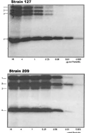
PBP pattern in sensitive and resistant strains.When PBPs
were labeled with125I-penicillin at concentrations that usually
saturate PBPs of penicillin-resistant S. pneumoniae (23), three HMM PBPs and one low-molecular-mass PBP could be de-tected in both strains, but they did not show identical electro-phoretic mobility (Fig. 1).
At a concentration that could saturate all the PBPs in the sensitive strain (125I-penicillin X, 0.25g/ml), HMM PBPs in
the resistant strain were hardly visualized and showed, by means of densitometry, an almost 50-fold-lower binding capac-ity of the radiotracer (data not shown). Several PBPs are present at the same positions in S. mitis NCTC 10712 and S.
pneumoniae R6 as well (30), but the relationship to these
proteins is not known.
HMM PBP genes in sensitive and resistant strains. The
unusually high resistance levels measured for strain 127R and the fact that all the HMM PBPs of this strain showed a lower affinity than did those of the sensitive strain suggested that sequences of the PBP-encoding genes could give some insights about changes probably responsible for sensitivity alterations. Portions of the genes homologous to pbp2x and pbp2b of S.
pneumoniae could be amplified with primers designed
accord-ing to known sequences of penicillin-sensitive and -resistant
S. pneumoniae strains. In the case of the pbp1a homologous
genes, a different strategy was followed, as described in Mate-rials and Methods.
pbp2x.In the resistant strain, this gene had a mosaic struc-ture. When compared with the homologous gene of S. mitis NCTC 10712 (the type strain for the species; sensitive) (46) the C- and N-terminal coding portions (positions 1 to 400 and 1600 to 2000, respectively) showed 95% identity but diverged by 20% between positions 400 and 1000 and also between 1200 and 1600, with a short region of 90% identity between posi-tions 1000 and 1200. Furthermore, the block between posiposi-tions 540 and 780 had almost the same sequence as that of a peni-cillin-resistant S. pneumoniae mutant (GenBank accession no. X65132) (34), with 98% identity. At the C-terminal end en-coding sequence, another 400-bp block, which started at posi-tion 1620, was identical (99% identity) to that of a highly TABLE 1. In vitro-lactam antibiotic susceptibilities of strains
127R and 209Sa
Antibiotic MIC (g/ml) for strain:
127R 209S Penicillin G 16 0.003 Ampicillin 16 0.003 Cefotoxime 64 0.003 Ceftriaxone 32 0.003 Cefoperazone 16 0.003 Piperacillin 16 0.006 Cefuroxime 32 0.003 Cephalotin 64 0.012 Ceftazidime 32 0.012 Cefalexin 32 0.012 Cephaloridin 16 0.006 Dicloxacillin 64 0.06 Oxacillin 64 ND Aztreonam ⬎64 ND
aMICs were determined by the agar dilution method. ND, not determined.
FIG. 1. Radioactive detection of PBPs from strains 127R and 209S. Membranes containing approximately 200g of proteins were labeled with the indicated concentration of125I-penicillin X and were
incu-bated for 15 min at 37°C. Penicillin concentrations are given in micro-grams per milliliter.
-LACTAM RESISTANCE IN S. MITIS
on February 29, 2016 by UNIV DE LIEGE
http://aac.asm.org/
penicillin-resistant S. pneumoniae isolate (GenBank accession no. U87092) (53). Another short sequence between positions 1260 and 1500 showed 98% identity with a penicillin-resistant strain of S. mitis (GenBank accession no. Y10535) (42).
pbp2x from 209S was 91% identical to the homologous gene
of S. mitis NCTC 10712 (46) without any evidence of mosaic structure
pbp2b. For this gene, the portion coding for the complete C-terminal transpeptidase domain was amplified. In 127R it also showed a mosaic structure. When compared to pbp2b of
S. mitis NCTC 10712 (13), only a small portion of the gene,
between positions 960 and 1260, showed 98% identity. The first 350-bp block diverged by 5% from that of the sensitive type strain, and blocks from positions 350 to 960 and 1260 to 1500 differed by 17 and 21%, respectively. In addition, the first block (positions 1 to 350) also showed similar percentages of identity with those of several genes of penicillin-resistant clinical iso-lates of S. pneumoniae (GenBank accession nos. U20076 and U20074) (46) and of a recently described (22), highly cefo-taxime-resistant clinical isolate of S. mitis (GenBank accession no. AJ002289). Between positions 350 and 660 several homol-ogous genes of penicillin-resistant S. pneumoniae strains showed 94 to 96% identity. Two blocks of 300 bases (positions 660 to 960 and 1200 to 1500) showed no divergence with those of penicillin-resistant isolates of S. pneumoniae (GenBank ac-cession no. Z21981) (6) and S. oralis (GenBank acac-cession no. M32228) (11).
pbp2b from 209S is 94% identical to the homologous gene of S. mitis NCTC 10712 (13), showing no evident mosaic
struc-ture.
pbp1a.For this gene, we encountered several difficulties in the sequence analysis. First, the number of sequences available in data banks was quite low compared to those for other PBP genes, probably because PBP1A is not the primarily affected -lactam target in clinical isolates (18). Only a few sensitive genes belonging to S. pneumoniae were available for compar-ison. Furthermore, to our knowledge, only a portion of a highly cefotaxime-resistant S. mitis was recently sequenced. However, analysis of both sensitive and resistant genes strongly suggests that intragenic recombination events may have occurred, since polymorphism distribution was not at random. When com-pared with homologous genes of S. pneumoniae, a very closely related species, blocks with clearly different rates of divergence could be detected in both resistant and sensitive genes as well.
Amino acid changes in resistant PBPs.The deduced amino
acid sequences were analyzed and aligned with homologous sequences of sensitive and resistant S. mitis strains (Table 2). Although changes already described to be involved in penicillin and cefotaxime resistance in S. pneumoniae were also found in 127R, a few extra amino acid changes highlight new interesting positions near the active site of the enzyme that are likely to lead to changes in the affinity of the PBP (Fig. 2).
PBP2X.For this protein sequence, alignments from resistant
isolates of S. mitis showed that immediately after the active S337, T338was changed to P, A, or G. In S. pneumoniae these
changes reduced the acylation efficiency of the enzyme by cefotaxime (39) (T to A) and by penicillins (T to P or G). These changes were frequently found in clinical isolates with unusually high levels of-lactam resistance (33, 34). Accord-ingly, strain 127R showed the T-to-P change, but strain 209S
possessed the same T residue as the type strain. Other resistant
S. mitis PBP2X analyzed also showed changes from T338to P, A, or G, but no correlation could be established with the resistance level for any particular amino acid change. In all resistant S. mitis strains, between the conserved SxxK and SxN motives, I371changed to T and downstream the conserved SxN
motif I398replaced V, as observed in S. pneumoniae mutants
(33). In addition, we found substitutions at positions 510, 523, 531, 535, 538, and 574 in all resistant S. mitis strains, not previously reported, which could point out interesting posi-tions influencing the affinity of the PBP.
On the other hand, as opposed to changes described for resistant pneumococci (22, 34, 46), positions 571 and 586 were not changed in resistant strains of S. mitis.
PBP2B.In resistant S. mitis strains we found the T446-to-A
and A619-to-G changes described previously in
penicillin-resis-tant pneumococci and supposedly responsible for low-level penicillin resistance, lower lysis rate, and piperacillin resistance as well (18, 21, 24, 47). However, alignments showed additional substitutions, which were present only in resistant S. mitis strains. Upstream of the SxN motif, N421was changed to Y and
Q426to L. Downstream of the SxN motif, the following
mod-ifications were observed: S472to T, S489to A, I502to V, N507to
S, G594to T, A596to P, N605to D, A608to T, and finally, just
behind the KxG triad, A618to G. In the C-terminal portion of
the protein, SD640and NG659were replaced by TE and KN,
respectively.
PBP1A. For this protein, the alignment includes only the
sequences obtained from 127R and 209S and a partial se-quence belonging to a S. mitis clinical isolate, highly resistant to cefotaxime, from Germany (22). Analyzing the PB domain of PBP1A of the resistant strains, we found that at position 358, an I changed to T in the resistant strains. Several addi-tional substitutions were also observed in the resistant strains: V306was changed to I, immediately after the active S370; T371
was changed to S, SIVH395to YMLK; D405to N and L425to I;
N438to E; V443to D; N455to D; SIH460to TMV; Q475to K;
E512to S; A518to Q; AY541to YS; and finally, LP550to IS 6
residues upstream of the KTG conserved motif. Part of the amino acid changes is present in positions already described for this and other PBPs in resistant pneumococci, but we also detected other alterations in positions not previously de-scribed.
TABLE 2. Strains used for PBP sequence alignmentsa Accession no. S. mitisstrain Origin Penicillin MIC(g/ml) Reference(s)
Y10535 Hu8 Hungary, 1990–92 6–12 22
Y10532 B5 Germany, 1994 8 22, 42 Y10533 197 Spain, 1990 16 42 Y10534 476 Spain, 1992 10 42 AJ002288 B6 Germany, 1994 50 22 AJ002289 B6 Germany, 1994 50 22 Z22183 11189 England, 1977 0.016 14 Z22231 NS51 Sweden, 1965 0.03 14 Z22186 K208 United States, 1932 0.03 14
aFigure 2 depicts alignments.
on February 29, 2016 by UNIV DE LIEGE
http://aac.asm.org/
DISCUSSION
The extensive and relatively rapid changes that have taken place in the classification of the oral streptococci and the increase in the number of species recognized in this group of bacteria have not been accompanied by the development of comprehensive species identification schemes for the ordinary laboratory (28, 29, 45, 52). However, striking species-specific variations in susceptibility, especially to-lactams, are evident from different studies (3, 19, 41, 49), suggesting that speciation of oral streptococci must be warranted in order to evaluate results reported by different studies. Up to date, no single phenotypic or genotypic approach has demonstrated by itself its universal usefulness (28, 29, 52). Intraspecies heterogeneity and both lack of biochemical activity and genetic exchange between different species have contributed to this scenario.
The identification of our strains could be achieved only when biochemical and genetic results were taken together. Even considered as a “gold standard” for several purposes, estab-lishment of the complete coding sequence for the 16S rRNA did not allow us to differentiate among S. mitis, S. sanguinis, S.
oralis, and S. pneumoniae species, since they showed 99%
iden-tity in this gene (they differ by only 10 to 12 nucleotides). We concluded, thus, that the 16S rRNA sequence could not be applied unambiguously to the identification of this group of microorganisms.
Analysis of partial sequence of sodA has been proposed as a reliable and accurate method for members of the mitis group (28, 40). Kawamura et al. indicated that the evolutionary rate
of the sodA partial gene was much higher than that of the 16S rRNA gene and that the sodA partial sequencing would be useful for differentiating closely genetically related organisms (28). In our case, sequences indicated that a higher interspe-cies heterogeneity than in the 16S rRNA could exist in this gene, but the identity with S. pneumoniae and S. oralis re-mained high (up to 95%).
The difficulties encountered for the identification of our strains could be due, in part, to the genetic exchanges that they have undergone. Further efforts should be made to improve the available methods or to find new powerful approaches in order to identify oral streptococci and particularly those of the mitis group, which, because of genetic exchange, seem to con-stitute a complex mosaic group more than clearly separated entities (12, 45). When studying an isolate of this group of organisms, and before defining the species, all available bio-chemical and genetic features must be examined and analyzed as a whole.
The -lactam MICs determined for strain 127R are ex-tremely high for this group of microorganisms, which are usu-ally susceptible to penicillin concentrations as low as a few nanograms per milliliter. Such MICs, at least in pneumococci, could only be reached when more than one-lactam target is altered (21, 22, 24). In the resistant strain all the HMM PBPs (detected with a radiotracer with a high specific activity) showed a much lower binding of the radioactive antibiotic than did those of the sensitive strain, indicating that they all had a reduced affinity for penicillin. Precompetition with other non-FIG. 2. Protein sequence alignments from sensitive and resistant strains of S. mitis. All amino acid sequences belong to different strains of S.
mitis and are named under their accession number (GenBank). 127R results are from this study. Shown in boldface are conserved motifs and
selected amino acid positions where we detected variation along the complete protein sequence of sensitive strains 209S (this study) and S. mitis NCTC 10712 (there were no amino acid differences between them).R,-lactam-resistant strain;S,-lactam-sensitive strain; ⴱⴱ, changes not
previously reported in pneumococci; #, changes found only in strain 127R. The indicated numbers correspond to positions in PBPs from S.
pneumoniae R6.
-LACTAM RESISTANCE IN S. MITIS
on February 29, 2016 by UNIV DE LIEGE
http://aac.asm.org/
radioactive-lactam antibiotics and postlabeling assays (data not shown) confirmed the observation that a reduced affinity existed in all the HMM PBPs of strain 127R. Since all of the -lactam targets had altered affinity, higher concentrations of drugs are thus required to inhibit these enzymes.
In 127R the sequences of the HMM PBP-encoding genes display a mosaic structure, with blocks diverging up to 20% from the sensitive homologous genes.
However, this feature does not seem to be restricted to the resistant phenotype: a mosaic structure could be inferred when analyzing pbp1a from the sensitive strain 209S. Sibold et al. and Dowson et al. described this phenomenon earlier in PBP2B genes of penicillin-sensitive S. mitis (13, 46), as did Spratt et al. for penicillin-sensitive isolates of Neisseria lactamica and
Neis-seria polisaccharea (48), but mosaic genes, even coding for
primarily affected-lactam targets, have no selective advan-tage for penicillin resistance. The observation that, in pneu-mococci, mosaic genes are found only in penicillin-resistant isolates but not in penicillin-sensitive strains (24) seems not to be the case for S. mitis. In this species, genes displaying mosaic structures do not unequivocally code for proteins with detect-able reduced affinity, and the same could be true also for pneumococci, considering that most of the analyzed PBP genes come from resistant strains. Nevertheless, PBP1A is not a primarily affected target in resistant pneumococci (18, 31), as it presumably is in the mitis group. Consequently, it can be ex-pected that alterations in kinetic properties in this protein, if present, should not be by themselves responsible for changes in susceptibility.
With those results, we assume that precise amino acid changes should simultaneously be present along with a mosaic structure, at least in primarily affected-lactam targets (PBP2B and PBP2X), to yield highly resistant isolates. Protein se-quence alignments showed that the amino acid changes found in resistant isolates are not present in sensitive strains, so both mosaic structures and amino acid changes seem to be indis-pensable for-lactam resistance in the viridans streptococci.
Furthermore, the amino acid changes that we found after protein alignment occur at positions common to all the ana-lyzed PBPs. A few additional substitutions are seen in all re-sistant strains. They were not reported before in the closely related pneumococci; perhaps they could be pointing out in-teresting positions leading to reduced affinity of each PBP. Most of these modifications are probably responsible for in-creasing MICs. Those for example in the vicinity of the cavity forming the active site of the PBPs could be important for the interaction of the antibiotic with the enzyme. More informa-tion about the three-dimensional structure of these proteins is needed to validate these observations.
Further studies should be encouraged to enlarge our knowl-edge about particular resistance mechanisms in this species, since reports of resistant isolates are rapidly increasing. Per-haps most important is the fact that these resistant strains constitute a potential pool of genes for the development of high-level -lactam resistance in S. pneumoniae strains and other pathogens of the mitis group, in which genetic compe-tence has provided a powerful tool for the uptake and incor-poration of resistance determinants.
ACKNOWLEDGMENTS
We are grateful to Horacio Lopardo (Hosp. Nac. de Pediatrı´a) and to P. Garrahan and Marta Altschuler (Hosp. Sor Marı´a Ludovica, Buenos Aires, Argentina) for providing clinical isolates; to Carlos Vay for helpful discussion related to bacterial identification; and to Richard Facklam, Centers for Disease Control and Prevention, Atlanta, Ga., for biochemical identification of strains. We thank Iris Thamm for technical support with automated DNA sequencing.
This work was supported in part by grants from the University of Buenos Aires, (Buenos Aires, Argentina) (TB 039) and from the CONICET (PID 4413) to G.G. and in part by the Belgian Programme on Interuniversity Poles of Attraction initiated by the Belgian govern-ment, Prime Minister’s Office, Services fe´de´raux des affaires scienti-fiques, techniques et culturelles (PAI P4/03). A.A. was a recipient of a fellowship from the European Community, Program ALFA, Project 5.0111.9. G.G. is a member of the “Carrera de Investigador Cientı´-fico,” CONICET, Buenos Aires, Argentina.
REFERENCES
1. Ausubel, F., R. Brent, R. Kingston, D. Moore, J. Seidman, J. Smith, and J. Struhl (ed.).1990. Current protocols in molecular biology. Greene Publish-ing Associates and Wiley Interscience, New York, N.Y.
2. Avada, A., P. van der Auwera, F. Meunier, D. Daneau, and J. Klastersky. 1992. Streptococcal and enterococcal bacteremia in patients with cancer. Clin. Infect. Dis. 15:33–48.
3. Bochud, P. Y., P. Engiman, T. Calandra, G. van Melle, L. Saghafi, and P. Francioli.1994. Bacteremia due to viridans streptococci in neutropenic pa-tients with cancer: clinical spectrum and risk factors. Clin. Infect. Dis. 18: 25–31.
4. Bouvet, A., A. Durand, C. Devine, J. Etienne, and C. Leport. 1994. In vitro susceptibility to antibiotics of 200 strains of streptococci and enterococci isolated during infective endocarditis, p. 72–73. In A. Totolian (ed.), Patho-genic streptococci: present and future. Lancer Publications, St. Petersburg, Russia.
5. Carratala, J., F. Alcaide, A. Fernandez-Sevilla, X. Corbella, J. Lin˜ares, and F. Gudiol.1995. Bacteremia due to viridans streptococci that are highly resistant to penicillin: increase among neutropenic patients with cancer. Clin. Infect. Dis. 20:1169–1173.
6. Coffey, T. J., C. G. Dowson, M. Daniels, and B. G. Spratt. 1993. Horizontal spread of an altered penicillin-binding protein 2B gene between
Streptococ-cus pneumoniae and StreptococStreptococ-cus oralis. FEMS Microbiol. Lett. 110:335–
339.
7. Coykendall, A. 1989. Classification and identification of the viridans strep-tococci. Clin. Microbiol. Rev. 2:315–328.
8. Doern, G., M. Ferraro, A. Brueggemann, and K. Ruoff. 1996. Emergence of high rates of antimicrobial resistance among viridans group streptococci in the United States. Antimicrob. Agents Chemother. 40:891–894.
9. Douglas, C. W. I., J. Heath, K. K. Hampton, and F. E. Preston. 1993. Identity of viridans streptococci isolated from cases of infective endocarditis. J. Med. Microbiol. 39:179–182.
10. Dowson, C., A. Hutchison, J. Brannigan, R. George, D. Hansman, J. Lin˜ares, A. Tomasz, J. Smith, and B. Spratt.1989. Horizonal transfer of penicillin-binding protein genes in penicillin-resistant clinical isolates of Streptococcus
pneumoniae. Proc. Natl. Acad. Sci. USA 86:8842–8846.
11. Dowson, C., A. Hutchison, N. Woodford, A. Johnson, R. George, and B. Spratt.1990. Penicillin resistant viridans streptococci have obtained altered penicillin-binding protein genes from penicillin-resistant strains of
Strepto-coccus pneumoniae. Proc. Natl. Acad. Sci. USA 87:5858–5862.
12. Dowson, C., V. Barcus, S. King, P. Pickerill, A. Whatmore, and M. Yeo. 1997. Horizontal gene transfer and the evolution of resistance and virulence de-terminants in Streptococcus. J. Appl. Microbiol. Symp. Ser. 83:42S–51S. 13. Dowson, C., T. Coffey, C. Kell, and R. Whiley. 1993. Evolution of penicillin
resistance in Streptococcus pneumoniae: the role of Streptococcus mitis in the formation of a low affinity PBP 2B in Streptococcus pneumoniae. Mol. Mi-crobiol. 9:635–643.
14. Dowson, C., A. Hutchison, and B. Spratt. 1989. Extensive remodelling of the transpeptidase domain of penicillin binding protein 2B of a penicillin resis-tant South African isolate of Streptococcus pneumoniae. Mol. Microbiol. 3:95–102.
15. Ghuysen, J. M. 1997. Penicillin binding proteins, wall peptidoglycan assem-bly and resistance to penicillin: facts, doubts and hopes. Int. J. Antimicrob. Agents 8:45–60.
16. Ghuysen, J. M., P. Charlier, J. Coyette, C. Duez, E. Fonze´, C. Fraipont, C. Goffin, B. Joris, and M. Nguyen-Diste`che.1996. Penicillin and beyond: protein fold, multimodular polypeptides and multiprotein complexes. Mi-crob. Drug Resist. 2:163–175.
17. Goffin, C., and J. M. Ghuysen. 1998. The multimodular penicillin-binding proteins: an enigmatic family of orthologs and paralogs. Microbiol. Mol. Biol. Rev. 62:1079–1093.
on February 29, 2016 by UNIV DE LIEGE
http://aac.asm.org/
18. Grebe, T., and R. Hakenbeck. 1996. Penicillin-binding proteins 2b and 2x of
Streptococcus pneumoniae are primary resistance determinants for different
classes of-lactam antibiotics. Antimicrob. Agents Chemother. 40:829–834. 19. Guiot, H. F. L., L. J. K. Corel, and J. M. J. J. Vossen. 1994. Prevalence of penicillin-resistant viridans streptococci in healthy children and in patients with malignant haematological disorders. Eur. J. Clin. Microbiol. Infect. Dis. 13:645–650.
20. Hakenbeck, R. 1995. Target-mediated resistance to -lactam antibiotics. Biochem. Pharmacol. 50:1121–1127.
21. Hakenbeck, R. 1998. Mosaic genes and their role in penicillin-resistant
Strep-tococcus pneumoniae. Electrophoresis 19:597–601.
22. Hakenbeck, R., A. Ko¨nig, I. Kern, M. van der Linden, W. Keck, D. Billot-Klein, R. Legrand, B. Schoot, and L. Gutmann.1998. Acquisition of five high-Mr penicillin-binding protein variants during transfer of high-level
-lactam resistance from Streptococcus mitis to Streptococcus pneumoniae. J. Bacteriol. 180:1831–1840.
23. Hakenbeck, R., C. Martin, C. Dowson, and T. Grebe. 1994. Penicillin-binding protein 2b of Streptococcus pneumoniae in piperacillin-resistant laboratory mutants. J. Bacteriol. 176:5574–5577.
24. Hakenbeck, R., T. Grebe, D. Za¨hner, and J. Stock. 1999.-Lactam resistance in Streptococcus pneumoniae: penicillin-binding proteins and non-penicillin-binding proteins. Mol. Microbiol. 33:673–678.
25. Hardie, J., and R. Whiley. 1994. Recent developments in streptococcal tax-onomy: their relation to infections. Rev. Med. Microbiol. 3:151–162. 26. Jacobs, J., H. Schouten, E. Stobberingh, and P. Soeters. 1995. Viridans
streptococci isolated from the bloodstream. Diagn. Microbiol. Infect. Dis. 22:267–273.
27. Kawamura, Y., X. G. Hou, F. Sultana, H. Miura, and T. Ezaki. 1995. De-termination of 16S rRNA sequences of Streptococcus mitis and Streptococcus
gordonii and phylogenetic relationships among members of genus Strepto-coccus. Int. J. Syst. Bacteriol. 45:406–408.
28. Kawamura, Y., R. Whiley, S. Shu, T. Ezaki, and J. Hardie. 1999. Genetic approaches to the identification of the mitis group within the genus
Strep-tococcus. Microbiology 145:2605–2613.
29. Kikuchi, K., T. Ezaki, K.-I. Totsuka, and K. Shimizu. 1995. Comparison of phenotypic characteristics, DNA-DNA hybridization results, and results with a commercial rapid biochemical and enzymatic reaction system for identifi-cation of viridans group streptococci. J. Clin. Microbiol. 33:1215–1222. 30. Ko¨nig, A., R. Reinert, and R. Hakenbeck. 1998. Streptococcus mitis with
unusually high-level resistance to-lactam antibiotics. Microb. Drug Resist. 4:45–49.
31. Kraub, J., M. van der Linden, T. Grebe, and R. Hakenbeck. 1996. Penicillin-binding proteins 2x and 2b as primary PBP targets in Streptococcus
pneu-moniae. Microb. Drug Resist. 2:183–186.
32. Laible, G., R. Hakenbeck, M. A. Sicard, B. Joris, and J. M. Ghuysen. 1989. Nucleotide sequence of the pbpX gene encoding the penicillin-binding pro-tein 2X from Streptococcus pneumoniae R6 and a cefotaxime-resistant mu-tant, C506. Mol. Microbiol. 3:1337–1348.
33. Laible, G., and R. Hackenbeck. 1991. Five independent combinations of mutations can result in low-affinity penicillin-binding protein 2x of
Strepto-coccus pneumoniae. J. Bacteriol. 173:6986–6990.
34. Laible, G., B. G. Spratt, and R. Hakenbeck. 1991. Interspecies recombina-tional events during the evolution of altered PBP 2x genes in penicillin-resistant clinical isolates of Streptococcus pneumoniae. Mol. Microbiol. 5:1993–2002.
35. Lane, D., B. Pace, G. Olsen, D. Stahl, M. Sogin, and N. Pace. 1985. Rapid determination of 16S ribosomal RNA segments for phylogenetic analyses. Proc. Natl. Acad. Sci. USA 82:6955–6959.
36. Martin, C., T. R. Briese, and R. Hakenbeck. 1992. Nucleotide sequences of genes encoding penicillin-binding proteins from Streptococcus pneumoniae
and Streptococcus oralis with high homology to Escherichia coli penicillin-binding proteins 1A and 2B. J. Bacteriol. 174:4517–4523.
37. Martin, C., C. Sibold, and R. Hakenbeck. 1992. Relatedness of penicillin-binding protein 1A genes from different clones of penicillin-resistant
Strep-tococcus pneumoniae isolated in South Africa and Spain. EMBO J. 11:3831–
3836.
38. Masson, J. M., and R. Labia. 1983. Synthesis of a125I-radiolabeled penicillin
for penicillin-binding protein studies. Anal. Biochem. 128:164–168. 39. Mouz, N., E. Gordon, A. M. Di Guilmi, I. Petit, Y. Petillot, Y. Dupont, R.
Hakenbeck, T. Vernet, and O. Dideberg.1998. Identification of a structural determinant for resistance to-lactam antibiotics in Gram-positive bacteria. Proc. Natl. Acad. Sci. USA 95:13403–13406.
40. Poyart, C., G. Quesne, S. Caelon, P. Berche, and P. Trieu-Cuot. 1998. Identification of streptococci to species levels by sequencing the gene en-coding the manganese-dependent superoxide dismutase. J. Clin Microbiol. 36:41–47.
41. Quinn, J. P., C. Di Vicenzo, D. Lucks, R. Luskin, K. Shatzer, and S. Lerner. 1988. Serious infections due to penicillin-resistant strains of viridans strep-tococci with altered penicillin-binding proteins. J. Infect. Dis. 157:764–769. 42. Reichmann, P., A. Ko¨nig, J. Lin˜ares, F. Alcaide, F. Tenover, L. McDugal, S. Swidsinski, and R. Hakenbeck.1997. A global gene pool for high-level cephalosporin resistance in commensal Streptococcus species and
Streptococ-cus pneumoniae. J. Infect. Dis. 176:1001–1012.
43. Sambrook, J., E. F. Fritsch, and T. Maniatis. 1989. Molecular cloning: a laboratory manual, 2nd ed. Cold Spring Harbor Laboratory Press, Cold Spring Harbor, N.Y.
44. Sanger, I., S. Nicklen, and A. Coulson. 1977. DNA sequencing with chain-terminating inhibitors. Proc. Natl. Acad. Sci. USA 74:5463–5467. 45. Schlegel, L., and A. Bouvet. 1998. Streptocoques et genres apparente´s:
abio-trophes et enterocoques. Bull. Soc. Fr. Microbiol. 13:7–17.
46. Sibold, C., J. Henrichsen, A. Ko¨nig, C. Martin, L. Chalkley, and R. Haken-beck.1994. Mosaic pbpX genes of major clones of penicillin-resistant
Strep-tococcus pneumoniae have evolved from pbpX genes of a penicillin-sensitive Streptococcus oralis. Mol. Microbiol. 12:1013–1023.
47. Smith, A. M., and K. P. Klugman. 1995. Alterations in penicillin-binding protein 2B from penicillin-resistant wild-type strains of Streptococcus
pneu-moniae. Antimicrob. Agents Chemother. 39:859–867.
48. Spratt, B. G., L. D. Bowler, Q.-Y. Zhang, J. Zhou, and J. M. Smith. 1992. Role of interspecies transfer of chromosomal genes in the evolution of penicillin resistance in pathogenic and commensal Neisseria species. J. Mol. Evol. 34:115–125.
49. Tuohy, M., and J. Washington. 1997. Antimicrobial susceptibility of viridans group streptococci. Diagn. Microbiol. Infect. Dis. 29:277–280.
50. Weisburg, W., S. Barns, D. Pelletier, and D. Lane. 1991. 16S ribosomal DNA amplification for phylogenetic study. J. Bacteriol. 173:697–703.
51. Whatmore, A. M., A. Efstratiou, A. P. Pickerill, K. Broughton, G. Woodward, D. Sturgeon, R. George, and C. G. Dowson.2000. Genetic relationships between clinical isolates of Streptococcus pneumoniae, Streptococcus oralis, and Streptococcus mitis: characterization of “atypical” pneumococci and or-ganisms allied to S. mitis harboring S. pneumoniae virulence factor-encoding genes. Infect. Immun. 68:1374–1382.
52. Whiley, R. A., and D. Beighton. 1998. Current classification of the oral streptococci. Oral Microbiol. Immunol. 13:195–216.
53. Zhao, G., W.-K. Yeh, R. H. Carnahan, J. Flokowitsch, T. I. Meier, W. E. Alborn, Jr., G. W. Becker, and S. R. Jaskunas.1997. Biochemical charac-terization of penicillin-resistant and -sensitive penicillin-binding protein 2x transpeptidase activities of Streptococcus pneumoniae and mechanistic im-plications in bacterial resistance to-lactam antibiotics. J. Bacteriol. 179: 4901–4908.
-LACTAM RESISTANCE IN S. MITIS