i
Université de Montréal
Rôle de la méthylation des histones dans la
régulation de l’expression des gènes de la COX-2,
iNOS, et mPGES-1 dans les chondrocytes humains:
Implication pour l’arthrose
Par
Fatima ezzahra El Mansouri
Département de Pharmacologie
Faculté de Médecine
Thèse présentée à la Faculté des études supérieures et postdoctorales
en vue de l’obtention du grade de Philosophae Doctor (Ph.D.)
en Pharmacologie
Avril 2015
ii
Université de Montréal
Faculté des études supérieures et postdoctorales
Cette thèse intitulée:
Rôle de la méthylation des histones dans la régulation de l’expression des gènes de
la COX-2, iNOS, et mPGES-1 dans les chondrocytes humains:
Implication pour l’arthrose
Présentée par
Fatima ezzahra El Mansouri
a été évaluée par un jury composé des personnes suivantes :
Dr Christopher Rose, président-rapporteur
Dr Hassan Fahmi, directeur de recherche
Dr Mohamed Benderdour, co-directeur
Dr Walid Mourad, membre du jury
Dr Fawzi Aoudjit, examinateur externe
iii
University of Montreal
Role of histone methylation in the regulation of
COX-2, iNOS, and mPGES-1 gene expression in
human chondrocytes: Implication for Osteoarthritis
By
Fatima ezzahra El Mansouri
Departement of Pharmacology
Faculty of Medecine
Thesis presented to the faculty of Medicine
to obtain the distinction of Philosophae Doctor (P.hD.)
in Pharmacology
April 2015
iv
University of Montreal
Faculty of Graduate and Postdoctoral Studies
This thesis entitled:
Role of histone methylation in the regulation of COX-2, iNOS, and mPGES-1 gene
expression in human chondrocytes: Implication for Osteoarthritis
Presented by
Fatima ezzahra El Mansouri
Evaluated by a jury composed of the following people:
Dr Christopher Rose, president-reporter
Dr Hassan Fahmi, research director
Dr Mohamed Benderdour, co-director
Dr Walid mourad, member of the jury
Dr Fawzi Aoudjit, external member of the jury
Dr Emmanuelle Brochiero, dean’s representative
v
L'arthrose (OA) est une maladie articulaire dégénérative, classée comme la forme la plus fréquente au monde. Elle est caractérisée par la dégénérescence du cartilage articulaire, l’inflammation de la membrane synoviale, et le remodelage de l’os sous-chondral. Ces changements structurels et fonctionnels sont dues à de nombreux facteurs.
Les cytokines, les prostaglandines (PG), et les espèces réactives de l'oxygène sont les principaux médiateurs impliqués dans la pathophysiologie de l'OA. L'interleukine-1β (IL-1β) est une cytokine pro-inflammatoire majeure qui joue un rôle crucial dans l'OA. L'IL-1β induit l'expression de la cyclooxygénase-2 (COX-2), la microsomale prostaglandine E synthase-1 (mPGES-1), la synthase inductible de l'oxyde nitrique (iNOS), ainsi que leurs produits la
prostaglandine E2 (PGE2) et l'oxyde nitrique (NO). Ce sont des médiateurs essentiels de la
réponse inflammatoire au cours de l'OA qui contribuent aux mécanismes des douleurs, de gonflement, et de destruction des tissus articulaires.
Les modifications épigénétiques jouent un rôle très important dans la régulation de l’expression de ces gènes pro-inflammatoires. Parmi ces modifications, la méthylation/ déméthylation des histones joue un rôle critique dans la régulation des gènes. La méthylation/ déméthylation des histones est médiée par deux types d'enzymes: les histones méthyltransférases (HMT) et les histones déméthylases (HDM) qui favorisent l’activation et/ou la répression de la transcription. Il est donc nécessaire de comprendre les mécanismes moléculaires qui contrôlent l’expression des gènes de la COX-2, la mPGES-1, et l’iNOS.
vi
contribute à la régulation de l’expression des gènes COX-2, mPGES-1, et iNOS dans des chondrocytes OA humains induits par l'IL-1β.
Nous avons montré que la méthylation de la lysine K4 de l'histone H3 (H3K4) par SET-1A contribue à l’activation des gènes COX-2 et iNOS dans les chondrocytes humains OA induite par l'IL-1β. Nous avons également montré que la lysine K9 de l’histone H3 (H3K9) est déméthylée par LSD1, et que cette déméthylation contribue à l’expression de la mPGES-1 induite par IL-1β dans les chondrocytes humains OA. Nous avons aussi trouvé que les niveaux d'expression des enzymes SET-1A et LSD1 sont élevés au niveau du cartilage OA.
Nos résultats montrent, pour la première fois, l'implication de la méthylation/ déméthylation des histones dans la régulation de l’expression des gènes COX-2, mPGES-1, et iNOS. Ces données suggèrent que ces mécanismes pourraient être une cible potentielle pour une intervention pharmacologique dans le traitement de la physiopathologie de l'OA.
Mots clés: Osteoarthrite, chondrocyte, Interleukin-1ß, COX-2, mPGES-1, PGE2, iNOS, NO,
vii
Osteoarthritis (OA) is a disabling disease classified as the most common form of arthritis worldwide. It is characterized by cartilage degeneration, synovium inflammation, and subchondral bone remodeling resulting in a loss of joint function. These structural and functional changes are due to numerous factors.
Cytokines, prostaglandins (PG), and reactive oxygen species are the major mediators implicated in the pathophysiology of OA. Interleukin-1 (IL-1) is a major pro-inflammatory cytokine that plays a crucial role in OA. IL-1 induces the expression of Cyclo-oxygenase-2 (COX-2), microsomal prostaglandin E synthase-1 (mPGES-1), inducible nitric oxide synthase
(iNOS), as well as their products prostaglandin E2 (PGE2) and nitric oxide (NO). These are
critical mediators of the inflammatory response during OA causing pain, swelling, and joint tissue destruction.
The activation of these pro-inflammatory genes results from different changes at the level of chromatin known as epigenetic modifications. Epigenetic modifications such as DNA methylation and histone modifications play a crucial role in gene expression. Among these modifications, histone methylation/demethylation is the most critical one. Histone methylation/demethylation is mediated by two types of enzymes: histone methyltransferases (HMT) and histone demethylases (HDM) which can either activate or repress transcription. It is therefore necessary to understand the molecular mechanisms which underlie the regulation of COX-2, mPGES-1, and iNOS expression.
The objective of this study is to investigate whether histone methylation/demethylation can modulate COX-2, mPGES-1, and iNOS expression in IL-1 induced OA human
viii
We demonstrated that histone H3 lysine K4 (H3K4) methylation by SET-1A contributes to IL-1-induced COX-2 and iNOS expression in human OA Chondrocytes. We showed also that LSD1-mediated demethylation of histone H3 lysine 9 (H3K9) contributes to IL-1β-induced mPGES-1 expression in human OA chondrocytes. We found that levels of SET-1A and LSD1 expression are elevated in OA cartilage as compared with normal cartilage.
Our data demonstrates, for the first time, the implication of histone methylation/demethylation in COX-2, mPGES-1, and iNOS regulation suggesting that these mechanisms could be a potential target for pharmacological intervention in the treatment of the pathophysiology of OA.
Key Words: Osteoarthritis, chondrocyte, Interleukin-1ß, COX-2, mPGES-1, PGE2, iNOS, NO,
ix
Résumé...v
Abstract...vii
List of figures...xiv
List of tables...xvii
List of abbreviations...xviii
Dedication...xxii Acknowledgments...xxiii
Chapter I- Introduction
...1PART A
...1 1. Osteoarthritis... ...1 1.1. Definition of Osteoarthritis...1 1.2. Epidemiology of Osteoarthritis...3 1.3. Symptoms of Osteoarthritis...31.4. Risk factors of Osteoarthritis...5
1.4.1. Systemic factors... ...5 1.4.2. Local factors...6 2. Articulation components...7 2.1. Articular Cartilage...7 2.1. 1. Chondrocytes...11 2.1.2. Extracellular matrix...12 2.2. Synovial membrane...14
x
3. The pathophysiological mechanisms of Osteoarthritis...16
4. Role of inflammation in Osteoarthritis...20
4.1. Cytokines in Osteoarthritis...22
4.1.1. The pro-inflammatory cytokines...22
a. Interleukin-1...24
Interleukin-1: expression and regulation...24
Role of Interleukin-1in Osteoarthritis...25
b. Tumor necrosis factor-...28
Tumor necrosis factor-expression and regulation ...28
Role of Tumor necrosis factor-in Osteoarthritis...28
c. Interleukin-17...29
Interleukin-17: expression and regulation...29
Role of Interleukin-17in Osteoarthritis...29
4.1.2. The anti-inflammatory cytokines...29
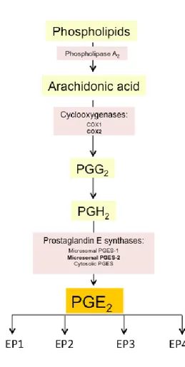
5. Prostaglandins ...32
5.1. Biosynthesis of Prostaglandin E2...34
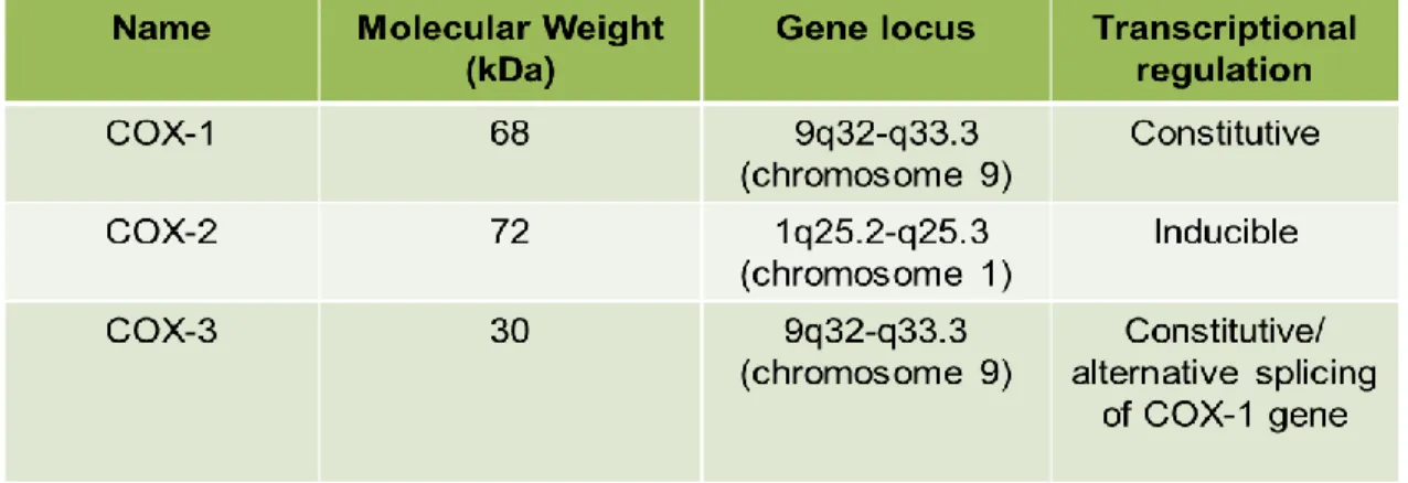
5.2. Cyclooxygenases: expression and regulation...34
5.2.1. Cyclooxygenase-1...37
5.2.2. Cyclooxygenase-2...37
5.2.3. Cyclooxygenase-3...39
5.3. Prostaglandin E synthases: expression and regulation...39
xi
5.3.3. Cytosolic prostaglandin E synthase...43
5.4. Cyclooxygenase-2/microsomal PGES-1/PGE2 pathway in Osteoarthritis...43
6. Reactive oxygen species ... ...45
6.1. Nitric oxide...46
6.1.1. Biosynthesis of nitric oxide ...46
6.1.2. Nitric oxide synthases: expression and regulation...48
a. Inducible nitric oxide synthase...48
b. Endothelial nitric oxide synthase...50
c. Neuronal nitric oxide synthase...51
6.1.3. Inducible NOS/NO pathway in Oteoarthritis...51
6.2. Hydrogen peroxide... ...54
7. Extracellular matrix proteinases... ...54
7.1. Matrix metalloproteinases...54 7.2. ADAMTS... ...56 8. Therapeutics of Osteoarthritis...57 8.1. Non-pharmacologic interventions...58 8.2. Pharmacologic interventions... ...59 8.2.1. Analgesics... ...59 8.2.2. NSAIDs...60 8.2.3. Intra-articular injections...61
8.2.4. Glucosamine and chondroitin sulfate...61
xii
1. Chromatin structure …... ...63
2. Epigenetic mechanisms…...65
2.1. Histone modifications: The histone code...66
2.2. DNA methylation... ...69
2.3. Micro RNAs...70
2.4. Chromatin remodeling... ...71
3. Histone methylation / demethylation...72
3.1. Histone lysine methylation... ...73
3.2. Histone arginine methylation...75
3.3. Histone methyltransferases...76
3.3.1.The lysine specific SET domain family...78
a. The SET1 family...78
b. The SUV39 family...80
c. The SET2 family...80
d. The EZH family...80
e. The SMYD family...81
f. The PRDM family...81
3.3.2.The non-SET HKMTs...81
3.3.3.The protein arginine methyltransferases...81
3.4. Histone demethylases... ...82
3.4.1.The lysine specific demetylase family...82
xiii
3.4.2.The Jumonji C family...85
a. The JHDM1 family...85
b. The JHDM2 family...85
c. The JHDM3 family...85
d. The JARID family...86
e. The PHF family...86
f. The UT family...86
4. Histone acetylation / deacetylation...87
4.1. Histone acetyltransferases...88
4.2. Histone deacetylasases... ...88
5. Epigenetic modifications in Osteoarthritis...91
5.1. Histone modifications in Osteoarthritis...91
5.2. DNA methylation in Osteoarthritis...92
5.3. Micro RNAs in Osteoarthritis...93
Thesis proposal
... ...96Chapter II- Articles
...98Article 1
... ...98Article 2
...139Chapter III- Discussion
...194Conclusion & Perspecives
... ...212xiv
Introduction
Figure 1: Schematic representation of the main constituents of a normal and osteoarthritic
knee………...…………2
Figure 2: The molecular composition of normal articular cartilage………...………9
Figure 3: The anatomy of articular cartilage and subchondral bone in normal knee………..…10
Figure 4: Comparison of the anatomy of articular cartilage and subchondral bone in normal and osteoarthritic knee ……….……17
Figure 5: Molecular and cellular mechanisms in osteoarthritis………..…19
Figure 6: Imbalance of cytokine production in osteoarthritic cartilage……….………23
Figure 7: Signaling pathway of interleukin-1in osteoarthritis………...………26
Figure 8: The prostaglandin biosynthetic cascade………..……..33
Figure 9: Pathway of PGE2 biosynthesis………...………35
Figure 10: The structure of cyclooxygenase-2 promoter………..…….……38
Figure 11: The structure of microsomal prostaglandin E synthase-1 promoter………...41
Figure 12: The biosynthesis of nitric oxide……….…….47
Figure 13: The structure of the inducible nitric oxide synthase promoter………49
Figure 14: Osteoarthritis treatment options……….………….58
Figure15: The chromatin structure………....64
Figure16: Histone modifications………..…..67
Figure 17: Histone lysine methylation………...………….74
xv
Figure 20: Histone lysine methylation demethylation in OA……….………….….205
Paper 1
Figure 1: Effect of interleukin-1 (IL-1) on histone H3K4 methylation at the inducible nitric
oxide synthase (iNOS) and cyclooxygenase 2 (COX-2) promoters………...…133
Figure 2: Effect of IL-1 on the recruitment of SET-1A and mixed-lineage leukemia 1 (MLL-1)
to the iNOS and COX-2 promoters ………134
Figure 3: Effect of 5’-deoxy-5’-(methylthio)adenosine (MTA) on IL-1–induced H3K4
methylation and COX-2 and iNOS protein expression ………..…………..………135
Figure 4: Effect of SET-1A silencing on IL-1–induced H3K4 methylation at the COX-2 and
iNOS promoters ………..……….……136
Figure 5: Effect of SET-1A silencing on IL-1–induced COX-2 and iNOS protein
expression……….……137
Figure 6: Expression of SET-1A protein in normal and osteoarthritic (OA) cartilage ……...……138
Paper 2
Figure 1: Effect of interleukin 1β on histone H3 lysine 9 methylation at the microsomal
prostaglandin E synthase 1 promoter ………....………185
Figure 2: Effect of interleukin 1β on the recruitment of lysine-specific demethylase 1 to the
xvi
Figure 4: Effect of lysine-specific demethylase 1 silencing on interleukin 1β–induced histone H3
lysine 9 demethylation at microsomal prostaglandin E synthase 1 promoter ………..………188
Figure 5: Effect of interleukin 1 on histone H3 lysine 9 methylation, lysine-specific demethylase
1 recruitment and flavin adenine dinucleotide levels in normal and osteoarthritis
chondrocytes………...189
Figure 6: Effect of interleukin 1β on histone H3 lysine 4 methylation at microsomal
prostaglandin E synthase 1 promoter………..…..…190
Figure 7: Expression of lysine-specific demethylase 1 protein in human normal and
xvii
Table I: The various pathophysiological effects of interleukin-1 in osteoarthritis………27
Table II: Characteristics of the cyclooxygenase synthases……….…...………..……36
Table III: Characteristics of the prostaglandin E synthases……….……39
Table IV: Characteristics of the nitric oxide synthases………..……45
Table V: The various physiological and pathological effects of PGE2 in osteoarthritic cartilage………48
Table VI: The various physiological and pathological effects of NO in osteoarthritic cartilage………...…….52
Table VII: Overview of different types and functions of identified histone modifications………68
Table VIII: Histone methyltransferases (HTMs): Specificity and transcriptional Effects…...……79
Table IX: Histone Demethylases (HDMs): Specificity and transcriptional Effects………....83
Table X: Histone acetyltransferases (HATs): Specificity and transcriptional Effects………89
xviii
AA: Arachidonic Acid
ADAMTS: A disintegrin and metalloproteinase with thrombospondin motif AIA: Antigen induced arthritis
AOL: Amine oxidase-like AP-1: Activating protein-1
ARE: Adenylate- and uridylate (AU)-rich elements ATP: Adenosine triphosphate
cAMP: Cyclic adenomonophosphate C/EBP: CCAAT/enhancer-binding protein CHD: Chromo-helicase/ATPase DNA binding
CIA: Collagen-induced-arthritis
cPGES: Cytosolic prostaglandine E synthase COX-2: Cyclooxygenase-2
CRE: Cyclic AMP response element
CREB: cAMP response element binding protein CREBBP: CREB-binding protein
Col-II: Type II Collagen
CoREST: Corepressor to the RE1 silencing transcriptionfactor DNA: Deoxyribonucleic acid
DNMTs: DNA methyltransferases ECM: Extracellular matrix
Egr-1: Early growth response gene-1 eNOS: Endothelial nitric oxide synthase EP: E prostanoid
ERK1/2: Extracellular signal-regulated kinase 1/2 FAD: Flavin adenine dinucleotide
GAG: Glycosaminoglycan GI: Gastrointestinal
GRE: Glucocorticoid-responsive elements H: Histone
xix
HDAC: Histone deacetylases
HDACi: Histone deacetylase inhibitors HDMs: Hsitone demethylases
HIF-1α: Hypoxia-inducible factor 1- alpha HMT: Histone methyltransferases H2O2: Hydrogen peroxide K: Lysine Kb: Kilobase kDa: Kilodalton KDMs: Lysine demethylases KMTs: Lysine methyltransferases KO: Knockout
ICE: IL-1-converting enzyme
IFN-: Interferon-gamma
IGF-1: Insulin growth factor INO80: Inositol requiring 80
iNOS: Inducible nitric oxide synthase IL: Interleukin
IL-1ß: Interlukin-1 beta IL-1R: Interlukin -1-receptor IL-1Ra : IL-1 Receptor antagonist
ISWIImitation switch
JNK: c-Jun N-terminal kinase
JHDM: JmjC domain-containing histone demethylase L-NIL: N-iminoethyl-L-lysine
L-NMMA: N-monomethyl-L-arginine
LIF: Leukemia inhibitory factor LOXs: Lipoxygenases
xx
MAPKs: Mitogen-activated protein kinases MBD : Methyl-CpG-binding domain
MGST1-L1: Microsomal glutathione transferase-1-like-1 MiRNAs: Micro RNAs
MMPs: Matrix metalloproteinases
mPGES-1: Microsomal prostaglandine E synthase type 1 m1: Mono-methylated
m2: Di-methylated m3: Tri-methylated
MTA: 5’-deoxy-5’-(methylthio)adenosine
NAB-1: NGF1-A-binding proteins-1 NaBu: Sodium butyrate
NFAT: Nuclear Factor of Activated T-cells NF-ĸB: Nuclear factor- kappa B
NO: Nitric oxide
nNOS: Neuronal nitric oxide synthase
NSAID: Non-steroidal anti-inflammatory drug NSD: Nuclear receptor SET domain
NuRD: Nucleosome remodelling and histone deacetylase
NURF: Nucleosome remodeling factor
OA: Osteoarthritis
PAD: Peptidyl arginine deiminases PARs: Protease-activated receptors
PGE2: Prostaglandin E2 PGI2: Prostaglandin I2 PGD2: Prostaglandin D2 PGF2: Prostaglandin F2 PGG2: Prostaglandin G2 PGs: Prostaglandins
xxi
PLA2: Phospholipase A2
PPARγ: Peroxisome proliferator-activated receptor gamma
PRDM: PRDI-BF1 and RIZ homology domain containing protein family PRMTs: Protein arginine methyltransferases
R: Arginine
RA: Rheumatoid Arthritis ROS: Reactive oxygen species SAM: S-adenosyl-L-methionine siRNA: Small interfering RNA SIRT: Sirtuin
Sox-9: Sex determining region- 9 Sp1: Specificity protein-1
STAT-1α: Signal transducer and activator of transcription-1 alpha SWI/SNF: SWItch/Sucrose NonFermentable
TACE: TNF-converting enzyme TCP: Tranylcypromine
TGF-ß:Transforming growth factor-ß
TLR: Toll-like receptor
TNF-: Tumor necrosis factor-alpha TNFR: Tumor necrosis factor receptor TSA: Trichostatine A
TXA2: Thromboxane A2
UTR: Untranslated region VA : Valproic acid
xxii
To my parents, my sister, my brother, To my grandma and grandpa, my aunties,
To all my family and friends.
Thank you for believing in me, thank you for your unfading love, thank you for your confidence, continuous encouragement, and sacrifice throughout my studies
xxiii
Acknowledgements
First and formost, I would like to express my sincere gratitude to my research director Dr Hassan Fahmi for his direction, assistance and guidance. My words cannot express the thanks I owe to him for his availability and expertise throughout this period of PhD studies. His unflinching encouragement kept me focused and motivated.
I am sincerely grateful to Dr Mohamed Benderdour, my co-director, for his valuable suggestions, help, and advices.
I would also like to thank Dr Johanne Martel-Pelletier and Jean-Pierre Pelletier, directors of the Osteoarthritis Research Unit-Notre dame Hospital, for their support and help.
My greatest regards and best thanks to the comittee members Dr Christopher Rose, Dr
Walid Mourad, Dr Fawzi Aoudjit, and Dr Noë
l
Raynal for accepting to review this work.I am deeply indebted to the research associate H. Afif, whose constant advices helped me to make necessary improvements during this work. Thanks are also due to all students of our lab.
Special thanks to all the research unit members for their help, kindness and friendship. Finally, I would like to show my appreciation to the Arthritis Society of Canada, the Canadian Institutes of Health Research (CIHR), the Fonds de la Recherche du Centre de Recherche du Centre Hospitalier de l’Université de Montréal (CHUM), and the CIHR Training on Mobility and Posture Deficiencies (MENTOR), for mentorship, funding, awards, and scholarships.
1
Part A-
1.
Osteoarthritis:
1.1 Definition of Osteoarthritis:
Osteoarthritis (OA) is a degenerative joint disease that affects a wide range of population (1). OA is considered as a disease of the entire joint involving all tissues. It is a composite of pathologic contribution from cartilage, synovial membrane, bone and adjacent soft tissues. The pathophysiological changes of OA include degradation and erosion of the articular cartilage, inflammation of the synovial membrane “synovitis”, subchondral bone remodeling, marginal osteophytosis, joint capsule fibrosis, tearing and fibrillation of intra-articular ligaments and menisci (2, 3). These changes are important for the onset and OA progression (Figure 1).
OA is a group of overlapping distinct diseases, which may have different etiologies, but with similar biologic, morphologic, and clinical outcomes. It is the result of both mechanical and biologic events. OA occurs when the equilibrium between the breakdown and the repair of joint tissues becomes unbalanced. This happens often when the mechanical loads applied exceed those that can be tolerated by the joint tissues causing joint pain, tenderness, limitation of movement, occasional effusion, and variable degrees of inflammation. Knees, hips, feet, and spine are the most frequently affected joints. Others such as finger and thumb joints may also be affected (4).
2
Figure 1: Schematic representation of the main constituents of a normal and osteoarthritic knee. OA is a disease of the whole joint with pathological changes occurring in all joint tissues. As shown, in the healthy (normal) cartilage, there is no degradation, no signs of synovial inflammation and no bone remodeling. However, in OA, cartilage is degenerated with lesions and fibrillation. This is accompanied by synovial inflammation, capsule fibrosis, and remodeling of bone leading to bony outgrowth and subchondral sclerosis (Adapted from 5).
3
1.2 Epidemiology of Osteoarthritis:
OA is the most common form of chronic arthritis. It is a highly prevalent disabling disease that has a large worldwide socioeconomic cost affecting approximately 15% of the population (6). In Canada, OA is one of the leading causes of disability and accounts for the majority of the disease burden for musculoskeletal disorders. Over 13% of Canadians are estimated to suffer from OA (7). In the United States, OA affects more than 10% of Americans (at least 27 million) (8, 9). People above the age of 65 years are more likely to develop OA. While only 7.6% of those 18-44 years of age and 29.8% of those 45-64 years of age, more than 50% of people older than 65 years
are diagnosed with OA(10).
OA is classified into two groups: primary and secondary OA. Primary OA, called also idiopathic OA, is the most common form of OA. It is classified as primary when there are clear
predisposing causes like age and heredity(11). Primary OA is a frustrating disease because both
the cause and cure are unknown. However, OA is classified as secondary when it is obviously associated with a defined pathology most likely developmental disorders, trauma and metabolic diseases (11, 12).
1.3 Symptoms of Osteoarthritis:
Symptoms of OA vary overtime between joint sites and individuals. Clinically, OA is described by joint pain, dysfunction, stiffness, deformity and joint space narrowing. It tends to follow periods of inactivity, such as sleep or sitting. Pain is typically accompanied by stiffness at the morning (11). As the disease advances, the pain may occur even when the joint is at rest.
OA Patients describe the most distressing aspect of living as fatigue, disability and reduced quality of life produced by chronic joint pain (13). Chronic pain in OA patients depends
4
primarily on the activation of sensory neurons that innervate the affected joint (14). In the joint and surrounding tissues, nociceptin, an endogenous peptide with opoid-like-activity, can be produced and delivered to its appropriate receptors, the so-called silent nociceptors which are located in peripheral tissues like capsule and ligaments (11, 15). In healthy joints, nerve fibers are quiescent. However, due to tissue injury or induction of inflammation, these receptors become active and start sending nociceptive information to the central nervous system. Moreover, joint nerves become sensitized to mechanical stimuli through the actions of eicosanoids, proteinase activated receptors and several others molecules. Patients with OA may also experience sensation of instability or buckling.
OA symptoms also includeloss of mobility and cracking noise with joint movement (16).
These signs are often associated with significant functional impairment and result in considerable impact on ability to perform activities of daily living (15, 17).
Radiography like X-rays and magnetic resonance imaging remain the best tools for OA diagnosis. Osteophyte formation and sclerosis are the most critical signs detected with these tests. Some patients, with no symptoms, have showed severe radiographic changes. However, other individuals experience significant joint pain with only minimal radiographic changes (13). This might be due to joint space width, which is too insensitive to determine structural alterations (14). More than 60 % of people above the age of 75 years old present radiologic signs. Further studies have confirmed discordance between radiographically diagnosed knee OA and symptoms like pain (14, 17).
5
1.4 Risk factors of Osteoarthritis:
The exact etiology of OA is unknown; however, a variety of risk factors have been described. They are classified into systemic and local risk factors. Systemic risk factors for OA include age, gender, sex, race, genetic predisposition, and obesity, whereas local risk factors include certain physical activities, tissue injuries, and developmental deformities (18, 19).
1.4.1. Systemic factors:
a. Age:
Aging is one of the most prominent risk factor for OA development. It is considered as a strong predictor of OA (6). Numerous studies have found that increased age promotes the initiation and progression of OA. Mitotic and synthetic cell activities decline with age, resulting in a reduced cartilage hydration (20).
Women are associated with a higher prevalence and severity of OA. Females are more likely to suffer from severe knee OA than males, especially following menopause. This has led to investigate the role of oestrogen in OA (6, 21).
b. Genetics:
Genetic factors are strong determinants in the pathogenesis of OA; they account for at least 40% of knee OA (6). Many genes have been shown to play crucial role in OA pathophysiological pathways. This includes genes involved in the regulation of inflammatory responses such as cytokines, other pro-inflammatory mediators, and genes involved in cartilage and bone metabolism (22). For instance, vitamin D receptors, insulin-like growth factor-1, and type II collagen (Col-II) genes have been demonstrated to be implicated in the susceptibility and the severity of OA and may represent therapeutic targets (6).
6
c. Obesity:
Obesity is another pivotal risk factor for OA incidence and progression (6, 23). It is one of the major OA modifiable risk factors (18, 19, 24). It has become a major focus since the identification of the white adipose tissue, which secretes adipokines. These biological active substances are highly produced in overweight people and may affect cartilage homeostasis. Leptin, the most abundantly produced adipokine, is released by adipose tissue. Leptin receptors are present in cartilage (25). Adipocyte cells share a common mesenchymal stem-cell precursor
with chondrocytes and osteoblasts (14, 26). Leptin regulates bone mass and mineralization via a
neuroendocrine pathway implicating the sympathetic nervous system (27). It has been shown to
increase levels of degradative enzymes like matrix metalloproteinases (MMPs), nitric oxide (NO) and pro-inflammatory cytokines (24, 28).
d. Diet:
Studies have emphasized the importance of early life nutrition (29). Low levels of vitamin D are associated with the pathological changes of OA. It has been reported that low levels of vitamin D may increase the incidence and the progression of knee and hip OA, predicting also loss of joint space and increased osteophyte growth in knee OA (30). In addition, low intake of vitamin C has been also shown to be associated with an increased risk of knee OA progression (6).
1.4.2. Local factors:
a. Occupation and physical activity:
Studies have found that people whose occupations require physical activity have twice the risk of developing knee OA than occupations that doesn’t. In addition, workers in some occupations like athletics, coal miners and farmers have also increased risks of knee OA (6).
7
b. Tissue injuries:
Tissue injury is the second major risk factor of OA (18, 19). Athletics like soccer and football players are exposed to a high risk of OA due to the high incidence of menisectomy and cruciate ligament injuries (11).
2.
Articulation components:
Joints provide support, stability, and protection. These are essential functions for normal and painless movement. The knee joint is a synovial joint that is composed of bones, cartilage, ligaments, tendons and joint capsule connecting the femur to the tibia. These bones, attached by ligaments, give strength and flexibility in the knee. The cartilage, the synovium, and the subchondral bone are the three basic elements that supply joint functions. Cartilage is a tissue that coats the ends of the bones; one of the few tissues in the body that does not have its own blood supply. Synovium is a membrane that surrounds the entire joint. It is filled with a lubricating liquid, the synovial fluid that supplies nutrients and oxygen to cartilage. The third element is the subchondral bone. The main role of subchondral bone is to provide structural support to the overlying articular cartilage (31) (Figure 1).
2.1. Articular Cartilage:
Cartilage is a specialized translucent connective tissue that covers the weight-bearing surfaces of articulating joints. There are three types of cartilage: the elastic cartilage, the fibro-cartilage, and the articular cartilage called also the hyaline cartilage. Articular cartilage is a hypocellular, aneural, and avascular tissue. This smooth lubricated tissue is derived mainly from
8
the synovial fluid, which consists of water and nutrients including electrolytes, small molecules and glucose (32).
The principal role of articular cartilage is reducing friction in the joint and absorbing the shock associated with locomotion (33). Articular cartilage affords a resistance to compressive forces. Thereby, it protects the underlying bones from mechanical damage during loading of the joint and allows for an efficient gliding motion during joint movement. This mechanical load is necessary for cartilage homeostasis. It induces fluid movement between the cartilage and the synovial fluid allowing the diffusion of molecules across cartilage and thus facilitating its nutrition (34). Thus, the main function of articular cartilage is the absorption and dissipation of mechanical load.
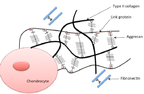
In its molecular composition, articular cartilage is composed of two main elements: the chondrocytes and the extracellular matrix (ECM) (Figure 2). Chondrocytes represent the unique cell type of cartilage that lies in the ECM. However, ECM is an extensive network of collagen
fibrils, proteoglycan molecules, and water (35). While water represents about 75% of the wet
weight, about 70% of the dry weight is collagen (9). Col-II is the principal type of collagen present in the articular cartilage.
9
Figure 2: The molecular composition of normal articular cartilage. In healthy articular cartilage, chondrocytes are surrounded by the extracellular matrix (ECM). The ECM is composed of several types of collagens (Type II collagen), collagen-binding proteins (link protein), large molecules of proteoglycan (aggrecan), small molecules of proteoglycan (fibronectin), and sulfate (S-S).
Articular cartilage consists of four zones: the superficial, middle, deep and calcified zone. The superficial zone is the thinnest zone of articular cartilage. It is composed of a highly structured network of uniform collagen fibers, proteoglycans, non-collagenous proteins and other ECM proteins. This layer maintains a high water content. The middle layer, called also the transitional zone, is composed of larger rounded chondrocytes. The collagen fibers are randomly oriented within this layer. Unlike the middle zone, the deep zone is constituted of collagen fibers that are arranged perpendicularly. The chondrocytes are grouped in columns. While the
10
concentration of water is low in this layer, the proteoglycan content is high. The calcified zone is the last layer of articular cartilage; it is composed of calcified cartilage and hypertrophic chondrocytes. This zone is characterized by the absence of proteoglycans (32). Of importance, the two last layers, the deep and calcified zones, are separated by a thick bundle of collagen named the tidemark. The tidemark is a thin line that marks the mineralization front between the calcified and the non-calcified articular cartilage (34). Small gaps that may exist in the tidemark allow the passage of nutrients through channels (32) (Figure 3).
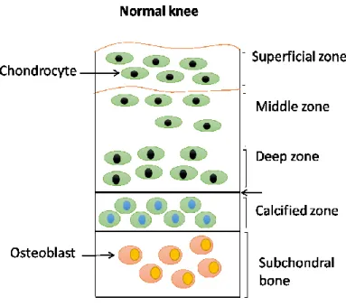
Figure 3: The anatomy of articular cartilage and subchondral bone in normal knee. Normal articular cartilage is divided into four zones: superficial zone, middle zone, deep zone, and calcified zone. Each zone is made of small number of chondrocytes embedded in collagen matrix. The calcified zone is separated from the deep zone by a tidemark (demarcation line). This calcified zone is located above the subchondral bone.
11 2.1.1. Chondrocytes:
Chondrocytes are the unique cellular component of articular cartilage (32). These cells
represent 1-5 % volume of the articular cartilage (35). They receive their nutrition by diffusion
through the matrix. The primary function of chondrocytes is to maintain cartilage homeostasis by the production of ECM components. They synthesize and degrade matrix components in response to environmental conditions like growth factors, cytokines, and biomechanical variations (9). Chondrocytes divide and produce new matrix in the peripheral zone. They produce collagens, proteoglycans, and non-collagenous proteins and organize all of these components in a highly ordered structure (32).
Number, size, and shape of chondrocytes vary depending on the layer of cartilage plate in
which they are located. For instance, in the superficial zone, chondrocytes show a flattened
ellipsoid form in parallel to the join surface. In this layer, they synthesize high concentration of collagen and low concentration of proteoglycans to provide the highest water content. However, in the transitional zone, chondrocytes are predominantly spheroid and the proteoglycan aggrecan concentration is higher. Chondrocytes in the superficial zone synthesize various relative amounts
of proteoglycans than do cells in the deeper zone. Unlike the previous layers, the calcified
cartilage zone contains small number of cells showing very low metabolic activities (35). Thus, most of the tissue contains water and inorganic salts such as sodium, calcium, and potassium chloride. The content of water and other molecules plays a crucial role in maintaining the resiliency of the tissue and contributing to the nutrition and lubrication system. These characteristics endow cartilage tissue with special properties like elasticity and ability to absorb and distribute loads.
12
Chondrocytes survive under hypoxic conditions (< 5% pO2) possessing a low metabolic
activity. Their metabolic activity differs in the various layers of cartilage. Chondrocytes maintain the balance between anabolism and catabolism mechanisms as well as a continual remodeling since there is replacement of matrix macromolecules lost through destruction. Together, the interaction between the chondrocytes and the ECM allows maintenance of the biological and mechanical properties of the articular cartilage.
2.1.2. Extracellular matrix:
The extracellular matrix is primarily made of tissue fluid and macromolecules like collagen, proteoglycans and non-collagenous proteins in specific distribution depending on the articulation. This texture provides tensile strength and resistance to compressive load.
More than 90% articular cartilage’s dry weight consists of two major components, Col-II and the large molecules of proteoglycan, aggrecan (36). Col-II is the most important type of the cartilage matrix. It is synthesized by chondrocytes. However, other collagen types such as VI, IX, XI, XII, and XIV that are contained in the ECM have also important structural and functional properties (37). While collagen forms a mesh to give support and flexibility to the joint, proteoglycans molecules are capable to ensure the high−fluid content in cartilage. Proteoglycan molecules are composed of glycosaminoglycan (GAG) subunits. They are bound to the protein core by means of sugar bonds. Due to link proteins, these chains are stabilized with a central hyaluronic acid (HA) chain (38) (Figure 2).
There are two major classes, large aggregating proteoglycan monomers, aggrecans, and small proteoglycans. Aggrecan is the main proteoglycan present in cartilage constituting 90% of the total cartilage proteoglycan mass. It is an elastic macromolecule that gives the tissue its ability to resist compression. The cartilage matrix contains also smaller proteoglycans like syndecans,
13
glypican, decorin, biglycan, versican, fibromodulin, lumican, and perlecan. These molecules are produced inside the chondrocytes and secreted in the matrix. They make up approximately 3% of the total proteoglycan mass (39, 40). Because of the hydrophilic nature of proteoglycans, the high water content of normal cartilage is maintained.
Transforming Growth Factor-β (TGF-) is another important protein produced in the matrix. It plays a critical role in a variety of physiological processes like cell proliferation, differentiation, and apoptosis. Chondrocytes secrete TGF-β in an inactive form, which is covalently bound to TGF-β binding proteins. TGF-β is activated from the growth plate by factors such as MMPs in order to bind to its receptor (41, 42).
TGF- has several regulatory mechanisms. It stimulates chondrocytes to induce aggrecan and Col-II production and also to intiate the first step of chondrogenesis. Of important, TGF- acts against inflammatory cytokines like IL-1, responsible for upregulation of MMPs like MMP-13. TGF- promotes cartilage ECM synthesis through counteracting the effects of catabolic cytokines (43).
The specific distribution and functions of collagens, proteoglycans, link proteins, hyluronic acids, and other components provide an integrated hydroelastic suspension system capable of resisting compression. Thus, the uniqueness of articular cartilage lies in its remarkable elasticity and ability to withstand enormous physical forces. Such extraordinary features of cartilage tissue are related to the collagen network and the high water content that is tightly held within the extracellular matrix.
14
2.2. Synovial membrane
The synovial membrane is a soft tissue made of layers that line the spaces of diarthrodial joints, tendons, and bursae. It is a tissue that secretes a glairy fluid named the synovial fluid. The synovial membrane plays an important role in maintaining normal joint physiology and function (44).
The synovial membrane covers all the intra-articular structures. It is composed of two layers: the synovial lining layer and the connective sublining layer (45). The synovial lining layer is made up of two kinds of cells: macrophage-like type A and fibroblastic type B cells. While type B cells synthesize and modify ECM and synovial fluid components, type A cells predominantly eliminate degradation products, including fluid and fine particulate materials from the joint space and from their ECM. In addition to these two different cells populations (type A and B), several additional studies have identified a third type of intermediate synovial lining cells which express CD68, a macrophage marker, indicating that these cells share both phenotypic properties of macrophages and fibroblastic cell types. According to that, it is thought that these three types of synovial lining cells originate from the same cell lineage and differentiate under the influence of local conditions (46).
The synovial fluid of joints functions as a biological lubricant and provides low friction
and low-wear properties to articulating cartilage surfaces in order to facilitate motion. These
lubricants, secreted by synovial cells in the synovium, are concentrated in the synovial space (47).
Moreover, hyaluronans are large polysaccharide molecules found naturally in the synovial fluid;
they help to create a viscous environment cushioning joints and preserving normal function. A
deficiency in this lubricating system may contribute to the erosion of articulating cartilage
15
2.3. Subchondral bone:
Bone is a vascularized tissue constituted of bone forming cells, osteoblasts, and osteocytes. Chemically, bone is made up of both organic and mineral components. While the organic component is primarily type I collagen, hydroxyapatite is the mineral component of bone. As bone matures, the size, crystallinity, and stoichiometry of the hydroxyapatite crystals change. These substitutions into the hydroxyapatite lattice are very important to bone strength and flexibility (49).
The subchondral bone is the epiphyseal bone located under the articular cartilage. It includes the subchondral bone plate and the underlying trabecular bone (4, 49). The subchondral bone provides structural support to the overlying articular cartilage. Several studies have demonstrated the potent role of abnormal subchondral bone cell metabolism in the initiation and progression of OA (11). In addition to the articular cartilage destruction, OA is characterized also by the increase of subchondral plate thickness and the formation of new bone at the joint margins, called osteophytes (50). It has been shown that subchondral bone changes may actually precede those of the synovial membrane and articular cartilage. The concept of crosstalk between subchondral bone tissue and articular cartilage that may be crucial for the initiation and/or progression of OA was highlighted (51).
16
3.
The pathophysiological mechanisms of Osteoarthritis:
In addition to the pivotal role of cartilage destruction as a hallmark in OA, the synovium and subchondral bone are implicated in OA development and progression. The cartilaginous changes are accompanied by synovial inflammation and pathological remodeling in the subchondral bone (15).
Under normal conditions, chondrocytes maintain a dynamic balance between synthesis and degradation of matrix components. In such non-stressed steady states, chondrocytes are quiescent
and there is very little low turnover of collagen network (34). However, In OA, a disruption of
matrix equilibrium leads to progressive degeneration of cartilage tissue with an increase in
matrix-degrading enzymes within the joint. A multitude of molecules drive cartilage breakdown and
disrupt cartilage homeostasis (14). These changes are accompanied by a tremendous loss of
proteoglycan from the upper zone followed by degradation of the collagen network (Figure 4). The metabolism of chondrocytes becomes unbalanced because of the excessive production of catabolic mediators with a down-regulation of anabolic mechanisms. Destruction of the ECM causes a gradual impairment of the articular cartilage accompanied with pain and physical disability (15). Further irregularities at the cartilage surface such as fibrillation are also features of cartilage damage in OA.
17
Figure 4: Comparison of the anatomy of articular cartilage and subchondral bone in normal and
osteoarthritic knee. Normal articular cartilage is divided into four zones: superficial zone, middle zone, deep zone, and calcified zone. Each zone is made of small number of chondrocytes embedded in collagen matrix. The calcified zone is separated from the deep zone by a tidemark (demarcation line). This calcified zone is located above the subchondral bone. In OA, Fissured articular cartilage induces vascularization of cartilage, which leads to exposure of subchondral bone to external surface. Microcracks that go through the cartilage and the subchondral bone contribute to reactivation and upward shifting of the tidemark.
The excessive catabolic activity which results in an imbalance of cartilage homeostasis and matrix breakdown is largely mediated by pro-inflammatory mediators including cytokines,
prostaglandins and other mediators. Chondrocytes produce mediators associated with
18
damage to the cartilage. Interleukin-1 (IL-1, MMPs, growth factors and free radicals, are key
contributors to cartilage destruction (14, 52) (Figure 5). The activation of these mediators causes
an aberrant expression of inflammation related genes including IL-1 converting enzyme (ICE/caspase-1), type IL-1 receptor (IL-1R), and tumor necrosis factor- (TNF-.
Synovial inflammation, or synovitis, may be either a primary event that initiate OA or a secondary mechanism that happens due to the accumulation of cartilage degradation products within the joint. Synovitis can result from both acute and chronic inflammatory state. It involves infiltration of mononuclear cells into the synovial membrane as well as production of
pro-inflammatory mediators like IL-1, TNF-, and chemokines. IL-1β and TNFα are able to excite
and sensitize nociceptors thereby inducing pain (14). Studies have showed that following acute
anterior cruciate ligament, high levels of inflammatory biomarkers can be detected in synovial fluids (53). As stated above, joint space is filled with synovial fluid that is abundantly composed of HA. In OA, the concentration and the molecular size of HA are diminished. These changes
result in less efficiency of lubrication. Furthermore, synovial mast cells are particularly implicated
19
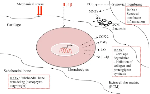
Figure 5: Molecular and cellular mechanisms in osteoarthritis. Mechanisms that drive cartilage destruction, synovial membrane inflammation as well as subchondral bone remodeling in osteoarthritis. Abbreviations: IL-1, interleukin-1; MMPs, matrix metalloproteinases; NO, nitric oxide; PGE2, prostaglandin E2;
20
The pathological structural changes that occur in the subchondral bone, both cortical and trabecular, are also one of the hallmarks of OA. The cortical subchondral plate becomes thick with
irregularities at the trabecular bone (54). The role of subchondral bone in OA biology has been an
interesting area of investigation. As the cartilage breaks down, changes occur in the underlying
bone. Changes in subchondral bone mineralization and bone volume have been detected in
samples with severe cartilage damage (55, 56). During the development of OA, subchondral bone undergoes adaptations like an increase in the subchondral plate thickness, sclerosis, reduced matrix mineralization, increased cancellous bone volume, osteophyte formation, and advancement of the tidemark associated with vascular invasion of the calcified cartilage (14, 57). However, it is still unknown whether these subchondral bone changes occur at the same time as changes in articular cartilage.
4.
Role of inflammation in Osteoarthritis:
The inflammatory response is a series of local cellular and vascular mechanisms triggered in response to injuries and damage that a tissue may face. Inflammation can be classified as either acute or chronic. Acute inflammation is the initial response of the body to harmful stimuli, whereas chronic inflammation is a prolonged response that leads to a progressive shift in the type of cells, present at the site of inflammation. Clinical manifestation of inflammation includes rubor (redness), tumor (swelling), dolor (pain), and fever (58). The inflammatory process is characterized by simultaneous destruction and healing of the tissue. A cascade of biochemical events propagates and matures the inflammatory response, involving the immune system, local vascular system, various cells, and different inflammatory mediators within the injured tissue.
21
Cytokines, prostaglandins (PGs) and reactive oxygen species (ROS) are the key players in the
inflammatory process (59, 60, 61).
OA is now well recognized as an inflammatory arthropathy. It is shown to be associated with signs and symptoms of inflammation. The involvement of an inflammatory response is marked by symptoms such as joint pain, swelling and stiffness. Numerous studies have shown that inflammatory mediators are highly implicated in OA (61).
Together, the articular cartilage, the synovial membrane, and the subchondral bone undergo alterations in the pathophysiology of OA. In fact, there is a coordinated release of cytokines and other inflammatory mediators from these three tissues. Such network makes them in a situation of interdependence, evidence that was supported by the magnetic resonance imaging techniques (11). Synoviocytes are considered as the principal cells mediating joint inflammation. This occurs through secretion of effector molecules that act on a variety of cells to modulate joint inflammation and promote matrix degradation. Cytokines and growth factors are the best example of these effector molecules that can be released. For instance, within the synovium, the presence of cytokine networks involves complex interactions between lymphocytes, synovial fibroblasts and macrophages. The secretion of IL-1or TNF-by monocytes/ macrophages followed by activation of resident tissue cells, such as fibroblasts, triggers the inflammatory cascade (45).
It is believed that synovial inflammation is a factor that contributes to dysregulation of chondrocytes function causing an imbalance between the catabolic and anabolic activities of chondrocytes. Interestingly, chondrocytes in OA cartilage express 1, ICE (caspase-1), and IL-1RI. IL-1is synthesized by chondrocytes at concentrations that are capable to induce the expression of MMPs, aggrecanases, and other catabolic genes. It colocalizes with TNF-, MMP-1, -3, -8, and -13, and Col-II cleavage epitopes in regions of matrix depletion in OA cartilage (62).
22
Cartilage breakdown products, resulting from mechanical or enzymatic destruction, can provoke, in turn, the release of collagenases and other hydrolytic enzymes from the synovial cells; thereby, leading to vascular hyperplasia in OA synovial membranes. This cascade sequentially results in the induction of synovial IL-1 and TNF-, which further the inflammatory outcome (37). Thus, high levels of cytokines and proteinases may exacerbate the inflammatory process.
4.1. Cytokines in Osteoarthritis:
Cytokines play a pivotal role during the inflammatory process in the pathophysiology of OA. They cause a loss of metabolic homeostasis through promoting the catabolic process. They might be produced either spontaneously or following stimulation of the joint tissue cells (15, 63).
Cytokines are classified with respect to their biological pro-inflammatory and anti-inflammatory effect. While IL-1, TNF-, interleukin-6 (IL-6), interleukin-15 (IL-15), interleukin-17 (IL-17), and interleukin-18 (IL-18) are categorized as pro-inflammatory mediators, interleukin-4 (IL-4), interleukin-10 (IL-10), and interleukin-13 (IL-13) are anti-inflammatory cytokines that modulate the inflammatory response (Figure 6).
4.1.1. The pro-inflammatory cytokines:
Pro-inflammatory cytokines have a crucial role in OA development and progression. They induce degradation of matrix molecules by enhancing the production and activation proteolytic enzymes like collagenases and aggrecanases.
In OA, cytokine expression is suggested to result from the mechanical insult. This is associated with subsequent MMP expression. For instance, IL-1 and TNF-, secreted by chondrocytes or other cells like synoviocytes, promotes the expression of matrix enzymes (32).
23
Either IL-1 or TNF-α, can recruit a unique set of receptor-associated proteins that transduce the stimulus into the cell upon ligand binding.
Figure 6: Imbalance of cytokine production in osteoarthritic cartilage. Chondrocytes are active players within the process of inflammation. The increased production of pro-inflammatory cytokines enhances the cartilage matrix turnover. Chondrocytes increase the catabolic activity through synthesizing most of the matrix degrading proteases and decrease the anabolic activity by down-regulating collagen and proteoglycan synthesis. Abbreviations: IL-1, interleukin-1; IL-4, interleukin-4; IL-6, interleukin-6; IL-10, interleukin-10; IL-13, interleukin-13; IL-17, interleukin-17; IL-18, interleukin-18; TNF-, tumor necrosis factor-; MMP, matrix metalloproteinase; ADAMTS, a disintegrin and metalloproteinase with thrombospondin motifs.
24
Although their receptors are different, IL-1 and TNF-α elicit series of shared phosphorylation events within the cells that facilitate transcriptional induction of MMPs as well as a number of distinct inflammatory and catabolic factors (64). These phosphorylation events are mediated by specific group of kinases, the mitogen activated protein kinases (MAPKs).
IL-1, TNF-, and IL-17 play important roles in OA progression. These cytokines increase cartilage destruction, synovial inflammation, and also bone resorption. However, there are other pro-inflammatory cytokines that have been shown to be expressed in OA tissues and have been considered as essential contributing factors. IL-6 has been proposed as an amplifier of the IL-1effects on the increased synthesis of MMPs (65), IL-8 for its chemotacic activity and ability of generating reactive oxygen metabolites (66), LIF that has diverse effects including the enhancement of IL-1expression in chondrocytes (67).
a. Interleukin-1:
Interleukin-1 expression and regulation:
IL-1 belongs to the IL-1 family; it is primarily produced as a cytosolic precursor protein pro-IL-1 (60, 69). The active form of IL-1 then results from an intracellular proteolysis accomplished by the ICE and finally released in the extracellular space (60, 70). IL-1 has two membrane receptors: interleukin-1 receptor-1 (IL-1R1) and interleukin-1 receptor-2 (IL-1R2). The activation of cells by IL-1 is mediated by its interaction with these receptors. These receptors may bind to a receptor antagonist named interleukin-1 receptor antagonist (IL-1Ra); thereby blocking their interaction with IL-1. In the joint, IL-1 is mainly produced by chondrocytes, osteoblasts, and cells of the synovial membrane (60, 71).
IL-1 induces its effect by activating several signaling pathways like nuclear factor-kappa B (NF-B), p38MAPK, and c-Jun N-terminal kinase (JNK) initiated once bound to its receptor
25
(72). Induction of the NF-κB pathway by IL-1 or TNFα results in phosphorylation of the IκB kinase. The subsequent degradation of this kinase unmasks the latent NF-κB, which translocates into the nucleus. This promotes the expression of many genes like cytokines, chemokines, MMPs and other inflammatory mediators (60, 73) (Figure 7).
Role of interleukin-1 in Osteoarthritis:
IL-1is the main inflammatory mediator implicated in numerous pathological features of OA. Patients with OA have elevated levels of IL-1 in the cartilage, the synovial membrane, the synovial fluid, and the subchondral bone. It has been reported that IL-1 induces the inflammatory response during the course of OA. Immunohistochemical studies revealed that IL-1is produced in the superficial zone of human OA cartilage in which the degenerative changes has been identified (60, 74).
Chondrocytes express not only IL-1 but also the receptor of this interleukin (IL-1RI). Higher levels of 1RI have been detected in OA patients (71). These destructive effects of IL-1in OA mediate elevation of cartilage catabolism, both by targeting MMPs for cartilage destruction, decreasing ECM synthesis, and leading to a down-regulation of anabolic activities of articular cartilage. Because of this high level of IL-1in OA cartilage, the correlation between the expression of IL-1and the severity of cartilage damage can be understood.
26
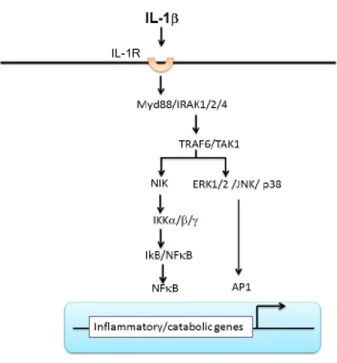
Figure 7: Signaling pathway of interleukin-1in osteoarthritis. Binding of IL-1 to IL-1R activates either NF-B (IKK complex) or AP1 (ERK/JNK/p38 pathways) transcription factors. IB-NF-B complex is inactive in the cytosol. After activation of IB-NF-B complex, free NF-B transfers into the nucleus and induces the expression of inflammatory, catabolic, and anti-anabolic genes. Abbreviations: IL-1, interleukin-1; IL-1R, interleukin-1 receptor; IL-6, interleukin-6; IL-8, interleukin-8; COX-2, cyclooxygenae-2; NF-kB, nuclear factor-kappa B; IB, inhibitor of B; ERK, extracellular signal-regulated kinases; JNK, c-Jun N-terminal kinases; AP1, activator protein 1.
27
As reported before, IL-1 has been shown to play a prominent role in cartilage degeneration. IL-1has potent bioactivities in repressing the expression of essential ECM components like Col-II and aggrecan, and inducing a spectrum of proteolytic enzymes like MMPs. It up-regulates the synthesis of MMPs such as MMP-1; -3; and -13 which have a catabolic effect on cartilage components as well as A disintegrin and metalloproteinase with thrombospondin motifs (ADAMTS), enzymes responsible for the proteolysis of aggrecans (40, 75, 76).
IL-1 induces its own secretion in cells of the joint in an autocrine way to stimulate the production of other cytokines such as TNF-, IL-6, and IL-8 (60, 77). It promotes the secretion of numerous enzymes and mediators implicated in the pathophysiology of OA like inducible nitric
oxide synthase (iNOS) producing NO, phospholipase A2 (PLA2), COX-2, and Prostaglandin E
synthase generating PGE2 (60, 78, 79) (Table I).
28 b. Tumor necrosis factor-:
Tumor necrosis factor- expression and regulation:
Tumor necrosis factor- (TNF-) belongs to the TNF superfamily. This cytokine is synthesized as a precursor protein in an inactive state and the proteolytic cleavage is done via a TNF-converting enzyme named TACE. TNF-α has two receptors: TNFR55 or TNF57 (80).
The mechanism of regulation of TNF- involves several signal transduction pathways including NF-B activation and MAPK pathway. Expression level of iNOS, COX-2 and
mPGES-1 as well as their produtcs NO and PGE2 has been shown to be increased in chondrocytes treated
with TNF-α (81).
Role of tumor necrosis factor- in Osteoarthritis:
TNF-α is implicated in maintaining the homeostasis of articular cartilage in combination with other cytokines and metabolic mediators like IL-1, insulin growth factor-1 (IGF-1) and transforming growth factor- (TGF-β). In the course of OA, TNF- has potent catabolic effects. Increased levels of TNF- were observed in cartilage, synovial membrane, synovial fluid and subchondral bone (60, 74). The level of TACE is also increased in OA (80). Like IL-1, TNF-is a potent inducer of matrix degradation and synovial membrane inflammation. It initiates a cascade of inflammatory response through the production of IL-1, IL-6 and IL-8 (82, 83). TNFR55 is the central receptor of TNF-α in articular cartilage during OA and its expression is increased in OA chondrocytes and synovial fibroblasts (83).
Inhibition of IL-1 and TNF-α block the amplification of the cleavage of Col-II and GAG in human OA cartilage (84). Anti-TNF-α treatments, with TNF-α antibodies, demonstrated a prolonged reduction of pain symptoms in OA (15).
29 c. Interleukin-:
Interleukin-17 expression and regulation:
Interleukin-17 (IL-17) is a pro-inflammatory cytokine secreted by T-cells. It belongs to the interleukin-17 family. The IL-17 family includes six ligands (IL-17A, IL-17B, IL-17C, IL-17D, 17E (25), and 17F), and five receptors (17RA, 17RB/ 25R, 17RC, IL-17RD/SEF and IL-17RE) (85). The evolving IL-17 family of ligands and receptors may play an important role in the homeostasis of tissues in health and disease beyond the immune system.
IL-17 was thought to be secreted only by T cells. However, it is now known to be produced by a variety of cells like macrophages, dendritic cells, natural killer T and lymphoid tissue inducer cells (85).
Role of interleukin-17 in Osteoarthritis:
IL-17 is another pro-inflammatory cytokine that has been shown to be involved in cartilage destruction. It has been demonstrated that IL-17 in combination with IL-1β enhances collagenase-3 in human OA chondrocytes through activator protein (AP)-1 (86). Furthermore, IL-17 can also increase the expression of NO in human OA chondrocytes (148 /faizeh). Studies on explants of
human OA knee menisci has revealed that NO and PGE2 production is increased by IL-17 (87).
4.1.2. The anti-inflammatory cytokines:
The anti-inflammatory cytokines are spontaneously elaborated by cartilage and synovial membrane. They are found in increased levels in OA patients. The anti-inflammatory and chondroprotective effects of these cytokines on cells of the articular cartilage and the synovium in OA have been well reported (60, 88, 89). The purpose of their production is to decrease the level of the pro-inflammatory cytokines, mainly IL-1 and TNF-; thereby, downregulating MMP production.
30
IL-4, IL-10, and IL-13 are the main anti-inflammatory cytokines implicated in OA. Increased levels of IL-4 have been observed in the synovial fluid and synovial cells (60, 90). IL-4 has a strong chondoprotective effect. Numerous studies have reported that IL-4 inhibits the degradation of proteoglycans in the articular cartilage, inhibits the secretion of MMPs, and reduces the variation in the production of proteoglycans in the course of OA (91). Chondrocytes treated with IL-4 showed a decreased synthesis of inflammatory cytokines like IL-1𝛽, TNF-𝛼, and IL-6 (60, 92, 93, 94). In addition, IL-4 has been found to decrease the secretion of other
inflammatory mediators like PGE2, COX-2, PLA2, and iNOS (92, 93, 95, 96).
IL-10 is another major anti-inflammatory cytokine that shows chondoprotective effects in OA. Chondrocytes express both IL-10 cytokine and its receptor IL-10R (97). It has been shown that IL-10 is implicated in stimulating the synthesis of Col-II and aggrecan. IL-10 can also inhibit the production of MMPs (98). Both IL-10 and IL-4 inhibit chondrocyte apoptosis (60, 99). Like
IL-4, IL-10 decreases the secretion of PGE2, COX-2, and PLA2.
Similar to IL-4 and IL-10, IL-13 has also chondroprotective effects. IL-13 showed inhibitory effects on the synthesis of pro-inflammatory IL-1𝛽, TNF𝛼, and MMP-3. While IL-10 has been shown to modulate TNF-production, IL-13 has been shown to inhibit the production of a wide range of pro-inflammatory cytokines and increase IL-1Ra production, a competitive inhibitor of IL-1R (89).
Thus, the effects of anti-inflammatory cytokines include increased proteoglycan synthesis, inhibited apoptosis of chondrocytes, decreased synthesis and secretion of MMPs, and decreased level of PGE2.