,)1,
3 T
Univers ity of Montreal
Regulation of lipocalin 2 (Lcn2) gene expression by iron stores, anemia and inflammation.
By Wenlei Jiang
Department of Biomedical Sciences faculty of Medicine
This thesis was submitted to the Faculty of Graduate Studies and Research in partial ffilfihlment of the requirements of the Degree of Master in Biomedical Sciences
December 2004 ©Wenlei Jiang, 2004
-LtQ‘A
Université
(HI
de Montréal
Direction des bibliothèques
AVIS
L’auteur a autorisé l’Université de Montréal
à reproduite et diffuser, en totalité ou en partie, pat quelque moyen que ce soit et sur quelque support que ce soit, et exclusivement à des fins non lucratives d’enseignement et de recherche, des copies de ce mémoire ou de cette thèse.L’auteur et les coauteurs le cas échéant conservent la propriété du droit d’auteur et des droits moraux qui protègent ce document. Ni la thèse ou le mémoire, ni des extraits substantiels de ce document, ne doivent être imprimés ou autrement reproduits sans l’autorisation de l’auteur.
Afin de se conformer à la Loi canadienne sur la protection des
renseignements personnels, quelques formulaires secondaires, coordonnées ou signatures intégrées au texte ont pu être enlevés de ce document. Bien que cela ait pu affecter la paginatïon, il n’y a aucun contenu manquant.
NOTICE
The author of this thesis or dissertation has granted a nonexclusive license allowing Université de Montréal to reproduce and publish the document, in part or in whole, and in any format, solely for noncommercial educational and research purposes.
The author and co-authors if applicable retaïn copyright ownership and moral rights in this document. Neither the whole thesis or dissertation, nor substantial extracts from it, may be printed or otherwise reproduced without the author’s permission.
In compliance with the Canadian Privacy Act some supporting forms, contact information or signatures may have been removed from the document. While this may affect the document page count, it does flot represent any loss of content from the document.
Univers ity of Montreal Faculty of Graduate Studies
This thesis entitled
“Regulation of lipocalin 2 (Lcn2) gene expression by iron stores, anemia and inflammation.”
Presented by Wenlei Jiang
Has been evaluated by a jury made up of the following people President ofjury: Dr. Marc Bilodeau
Director of research: Dr. Manuela Santos Member ofjtiry: Dr. Jianping Wu
December 2004 Thesis accepted on:
Summary
Lipocalin 2 (Lcn2) is a putative iron-trafficking protein. The aim of this study was to detenTiine whether Lcn2 plays a role in conditions associated with altered iron metabolism. Liver Lcn2 rnRNA levels were assessed in mouse models of dietary iron overload and iron deficiency. Two models of anemia were analyzed, including acute hernolysis induced by phenylhydrazine treatment and repeated phiebotomies. The Lcn2 response to CoC12-induced hypoxia and acute and chronic lipopolysaccharide (LPS)-CoC12-induced inflammation was also studied. Our resuits show that both types of anemia and hypoxia were associated with a dramatic increase in liver Ïipocaïin 2 gene expression and serum Lcn2 protein levels. Both acute and chronic inflammation up-regulate lipocalin 2 transcription and secretion. The up-regulation of Lcn2 gene expression by anemia or hypoxia suggests an important physiological role for Lcn2 in iron metabolism and is in une with its recently proposed role in the transferrin-independent iron delivery pathway. The increase of Lcn2 expression during acute and chronic inflammation is in agreement with the finding that lipocalin 2-mediated iron sequestration constitutes a host-defense mechanism of the iimate immune system.
Résumé
La lipocaline 2 (Lcn2) est une protéine active ayant pour fonction de favoriser la circulation du fer dans l’organisme. Le but de cette étude était de déterminer si la Lcn2 contribue ou non aux conditions associées à un métabolisme ayant un niveau de fer altéré. Les niveaux d’ARNm hépatique ont été déterminés dans des modèles de souris ayant soit une surcharge 011 une carence en fer. Deux modèles d’anémie ont été analysés, incluant l’hémolyse induite par la phénylhydrazine, ainsi que les phlébotomies répétées. La réponse de la Lcn2 à l’hypoxie induite par le CoCÏ2, ainsi que l’inflammation élevée et prolongée induite par les lipopolysaccharides (LPS), ont également été étudiées. Nos résultats démontrent que les deux types d’anémie et d’hypoxie sont associés à une augmentation drastique des niveaux d’expression génique de la Lcn2 dans le foie et des niveaux sériques de la protéine Lcn2. La relation entre l’expression du gène Lcn2, l’anémie et l’hypoxie suggère que la Lcn2 joue un rôle important dans le métabolisme du fer et confirme son rôle récemment proposé dans le métabolisme de transport du fer indépendant de la transferrine.
Contents
Surnrnary .
Contents y
List of figures viii
Listoftables x
List ofabbreviations xi
Acknowledgements xiv
Dedication xv
I. 1NTRODUCTION I
1. The significance ofiron for biological systems 2
1.1. Hemoproteins 2
1.2. Iron-sulfurproteins 3
1.3. Other iron-containing proteins 3
2. Iron toxicity 3
3. Disorder of iron metabolism in humans 4
3.1. Iron deficiency disorders 5
3.1.1. Irondeficiencyanemia 5
3.1.2. Iron-transport-deficiency anernia 5
3.1.3. Iron-utilization anemia 6
3.1.4. Iron reutilization anemia (ftinctional anemia) 6
3.2. Iron overload diseases 7
3.2.2. Type 1 HH (HFE-associated) .9
3.2.3. Non-HFE HH 10
3.2.4. Secondary iron overload (non-genetic) 12 3.2.5. Iron overload of undetermined origin 13 3.2.6. Neurodegenerative disorders related to iron rnetabolism 13
4. Cellular iron homeostasis 16
4.1. Iron uptake 16
4.1.1. Transferrin-dependent iron uptake 17
4.1.2. Iron uptake by reticuloendothelial (RE) macrophages 19 4.1.3. Iron uptake by intestinal absorptive cells 20 4.1.4. Transferrin-independent iron uptake 22
4.2. Iron storage 24
4.3. Iron export 25
4.4. Cellular iron homeostasis 27
5. Systernic iron homeostasis 29
5.1. Distribution of iron in aduits 29
5.2. Iron regulators 30
5.2.1. Ironregulators 30
5.2.2. Hcpcidin, a putative iron messenger 32
5.3. Animal models 33
6. Iron rnetabolism and immune system interactions 35
6.1. Structural similarities 35
6.1.2.NRAMP1 andDMTl .37
6.1.3.HFEandMflCclassI 38
6.2. Regulation of iron-metabolic proteins by inflammatory stimuli 39
6.3. Iron sequestration from bacteria 40
7. Recent findings inspiring our interest in studying Lcn2 41
II. OBJECTIVE AND SPECIFIC AIMS 43
1. Objective 44
2. Specific aims 44
III. MATERIALS AND METHODS 46
IV. RESULTS 54
V. DISCUSSION AND CONCLUSIONS 73
1. Discussion 74
2. Conclusions 79
VI. FUTURE PERSPECTIVES 80
List of figures
I. INTRODUCTION:
Figure 1. The transferrin (Tf) cycle 1$
Figure 2. Macrophage iron uptake and recycling 20
Figcire 3. The process of intestinal iron absorption 21
Figure 4. The composition offerritin 25
Figure 5. Iron export facilitated by Ceniloplasmin (Cp) and hephaestin (heph) 27
Figure 6. The IRE/IRP regulation system 29
Figure 7. Distribution of iron in adults 30
Figure 8. Protein alignment ofhuman defensins (defen B) and hurnan hepcidin 36 Figure 9. Protein alignment of human NRAMPI and hurnan DMT1 3$ Figure 10. Ribbon diagrams ofHFE and MHC class I (MHC I) proteins 39
IV. RESULTS:
Figure 1. Iron stores, Lcn2 and hepcidin mRNA levels in mice fed a carbonyl iron
supplemented diet 56
Figure 2. Iron stores, liver Lcn2 and hepcidin mRNA levels and senim Lcn2 plotein in
control and iron-deficiency anemic mice 59
Figure 3. Hematological indices in control, phlebotomized (PHL) and phenylhydrazine
Figure 4. Lipocalin 2 (Lcn2) and hepcidin (Hamp) levels in mice with acute
anernia 62
Figure 5. Lipocalin 2 (Lcn2) and hepciditi (Harnp) levels in hypoxic mice 63 Figure 6. Lipocalin 2 (Lcn2) and hepcidin (Hamp) levels in mice of LPS- and turpentine
(TP)- induced acute inflammation 65
Figure 7. Tissue expression pattem of Lcn2 in control and LPS-induced acute inflammatory
mice 67
Figure 8. Lipocalin 2 (Lcn2) levels in chronic inflarnrnatory mice 68 Figure 9. Iron concentrations, DMTI and fP1 levels, and iron deposition in the liver of
List of tables
I. INTRODUCTION:
Table L Classification ofhemosiderosis and bemochromatosis 8 Table 2. Useful rodent models for understanding iron metabolism and related
diseases 34
IV. RESULTS:
Table 1. Hernatological indices, serum iron (SI) and transferrin saturation (TS) in iron
deficient and iron-loaded mice 57
List of abbreviations
ACD: anernia of chronic disease, anemia of inflammation f32rn: beta-2-microglobulin
Cp: ceruloplasmin
DCT 1: divalent metal ion transporter 1 Dcytb: duodenal cytochrome b
DMT 1: divalent metal transporter 1 EPO: erythropoietin
Fe2: ferrous iron Fe3: fen-ic iron FP 1: feiioportin 1
GATA1: GATA binding protein 1 HAMP: hepcidin antimicrobial peptide I-lb: hernoglobin
HCT: hematocrit Heph: hepheastin
HFE: hemochromatosis (gene) HI-I: hereditary hemochromatosis HIF 1: hypoxia inducible factor 1 IFNy: interferon-y
IL-6: interleukin-6
IRE: iron responsive element Ireg 1: iron-rcgulated transporter 1 IRP: iron regulatory protein Lcn2: lipocalin 2
LIP: labile iron pool LPS: lipopolysaccharide
MCV: mean corpuscular volume
MHC: major histocompatibility complex MTP 1: metal transporter protein- 1 NF-KB: nuclear factor kappa B
NGAL: neutrophil gelatinase-associated lipocalin
NRAMP1: natural resistance-associated macrophage protein 1 NRAMP2: natural resistance-associated macrophage protein 2 PHL: phlebotomy
PHZ: phenylhydrazine RBC: red blood celi
ROS: reactive oxygen species SI: serumiron
Tf: transferrin
TfR1: transfenin receptor 1 TfR2: transferrin receptor 2
TIBC: total iron binding capacity TNFŒ: tumor necrosis factor (X TS transferrin saturation UTR: untranslated region
Acknowledgements
First and foremost, I would like to express my most sincere appreciation to my supervisor Dr. Manuela Santos, for her solid and skillftil guidance, as well as her encouragement throughout this dissertation. I feel honored to have been her student, and value greatly the wisdorn she has shared with me. Furthermore, I would like to acknowledge two of rny
colleagues, Hortence Markui and Marco Constante Pereira, both enthusiastic supporters of
my efforts. I also thank Christian Dallaire in assistance ofmy work. Finally I would like to honor my horneland, the great country of China, which gave me this precious opportunity for personal enrichment, and the opportunity to pursue my professional passion of inedical research.
Dedication
I dedicate rny work to my loving and caring parents, who have aiways been there to encourage and support me. You both taught me the importance of friendship, respect, and love, for all aspects of life. Your love and strength have sustained my energy to complete this work.
I. INTRODUCTION
Iron is an essential element for nearly ail living organisms. It is involved in a large number of metabolic processes, including oxygen transport, DNA synthesis and electron transport. However, iron concentrations in body tissues must be closely controlled since too much iron can lead to tissue injury due to the generation of free radicals, while too littie iron can lead to anemia. Thus, in light of the multiple biological functions and toxicity of iron, the existence of a variety of human diseases related to disturbed iron homeostasis is not surprising.
1. The significance of iron in biological systems
Trou is an essential element in the normal fiinction of ail cells. Many proteins are dependent on iron to perform their functions. These proteins illustrate the indispensable role of iron in biological systems. Proteins that contain iron can be classified into tbree categories: hemoproteins, iron-sulftir proteins and other iron-containing proteins.
1.1. Hemoproteins
Iron forms the core of heme, the main component of hemoproteins. They carry out three ftinctions: (1) oxygen transport and storage via hemoglobin and myoglobin; (2) electron transport and energy metabolism via cytochromes; and (3) antioxidant fiinctions through catalase and peroxidases, and beneficial pro-oxidant functions by myeloperoxidase. Oxygen transport through hemoglobin is one of the most important functions of iron. Through the ability of hemoglobin to acquire oxygen by contact and release it when
needed, hemoglobin in red blood celis (RBC) transports oxygen from the lungs to the rest ofthe body [1].
1.2. Iron-sulfiir proteins
Iron-sulfur proteins form a second class of iron-containing proteins, in which iron atoms are bound to sulfur, and they also perform a variety of physiological ftinctions. Rubredoxins and fenedoxins, for example, play roles in electron transport, and succinate dehydrogenase, nitrogenase and aconitase participate in redox and non-redox reactions [2].
1.3. Other iron-containing proteins
Other iron-containing proteins refer to a third class of non-heme, non-iron-suifrir iron containing proteins. These include lipoxygenases, which oxidize fafty acids into leukotrienes, transferrin, which transports iron in the blood, and ferritin, which stores iron in ceils.
The diverse range of functions performed by iron-containing proteins provides a sense of the indispensable role of iron in practically ail forrns of life.
2. Iron toxicity
In hurnan systems, iron is found in two oxidation states: Fe2 and Fe3. At physiological pH, Fe2 is soluble while Fe3 precipitates as oxyhydroxide polymers. The property of iron to readily participate in one-electron transfer reactions explains its potential to produce extremely toxic free radicals. Fe2 can mediate Fenton reactions to produce hydroxyl
radicals (0H) via the following reaction: Fe2 + H202 —* Fe3 + 0W + 0H. Fe3 can generate toxic radicals by catalyzing the Haber-Weiss reaction: 0[+ H202 —> OW+02+OW [3]. Hydroxyl radicals can cause damage to DNA, proteins, polyunsaturated fatty acids, mitochondrial DNA or lysosomes [4, 5].
The severity of iron-mediated toxicity is correlated with the concentration of low molecular weight iron in the labile iron pool within ceils. fron accumulation in tissues can increase the arnount of low molecular weight iron within the pool, thus catalyzing the production of more toxic free radicals than are present in healthy individuals, in whom iron is stored as a cofactor for different proteins to prevent its toxic effects.
The dismption of normal iron metabolism has been shown to lead to a variety of iron disorders, ranging from anemia to iron overload to neurodegenerative disorders. Thus, systernatic and cellular iron homeostasis must be properly maintained in the body in order to allow iron to be made available for physiological functions, and, at the same time, avoid its toxicity.
3. Disorders of iron metabolism in humans
The importance of maintaining iron homeostasis is clearly demonstrated by the existence of a group of diseases caused by dismrbed iron metabolism.
3.1. Iron deficiency dis orders
When iron is not available for ceils involved in RBC production, hemoglobin synthesis is reduced, resulting in a reduction in the rate of erythropoiesis as well as the production of a microcytic red blood celi population. In the early stage ofmicrocytic anemia, symptoms are often not apparent. In differential diagnosis, four types of microcytic anemia are considered: iron-deficiency, iron-transport deficiency, iron-utilization anernia, and iron reutilization anemia (anemia of chronic disease).
3.1.1. Iron deficiency anemia
Iron deficiency anemia has had many names: anemia of chronic blood loss, hypochromic microcytic anemia, chlorosis, hypochromic anemia of pregnancy, infancy, and childhood
[6].
The rnost common cause of anemia is blood loss from the gasterointestinal tract. Women may bleed from menstrual loss. Pregnant women may lose iron to the developing fetus. Increased iron requirement, such as in infants less than two years of age, and reduced iron absorption, as in the case of gastrectomy, may also cause iron deficiency [7].
3.1.2. Iron-transport deficiency anemia Atransferrinemia.
Iron-transport deficiency anemia is a rare disease caused by reduced levels oftransfeirin. In these individuals, iron caimot be transported from the intestine and liver to the bone
manow for erythropoiesis. Hemosiderosis of lymphoid tissue, particularly along the gastrointestinal tract, often occurs in this type ofanemia [8].
3.1.3. Iron-utilization anemias
Iron-utilization anemias include diseases such as hemoglobinopathies, primary thalassemia, and sideroblastic or myelodysplastic anemia [9]. Iron-utilization anemias are caused by the abnormal utilization of iron for hemoglobin synthesis, despite the normal presence of iron in the mitochondria of developing RBCs. Dyspoiesis increases the intramedullary death of RBCs, paradoxically leading to erythroid hyperpiasia with a relative or absolute reticulocytopenia. In feffokinetic studies, radiolabeled iron rapidly distributes from blood to the bone manow, but fails to reappear in developing RBCs at a normal rate. Ineffective erythropoiesis is thus the main characteristic ofthese anemias.
3.1.4. Iron-reutilization anemia
Anernia of chronic disease (ACD), also called inflammation anemia, is the second most common form of anemia. In ACD, bone marrow erythropoiesis does not respond appropriately to anemic conditions. The early stages of the disorder show symptoms of normocytic anemia whereas microcytic anemia is characteristic ofthe later stages [10].
ACD results from complications of many chronic disorders such as infection, inflainmatory disease, and cancer. Three pathophysiologic mechanisms are believed to be involved: (i) increased iron retention and storage within the reticuloendothelial system can induce hypoferremia, which limits iron availability to erythrons for hemoglobin (Hb) synthesis; (ii)
deficient erythropoiesis resulting from decreased erythropoietin (EPO) production or reduced bone marrow responses to EPO; and (iii) red blood celi suiwival times are reduced through mechanisms that are not entirely clear. In ail three mechanisms, inflarnmatory cytokines are involved, including interleukin-1 (IL-l) and turnor necrosis factorΠ(TNF-a).
3.2. Iron overload diseases
Chronic iron overload is characterized by an increase in tissue iron deposition either in a focal site or overail in tissues. The tenu liemosiderosis is cornmonly used for tissue iron overload. More severe states of iron ovcrload (greater than five grams of iron present in the body) are referred to as hemochromatosis. Hereditary hemochromatosis (HH), caused by mutations in the hereditary hemochromatosis gene (HFE), must be differentiated from other genetic iron overload, non-genetic iron overload, and increased iron storage of undetenuined etiology (Table 1).
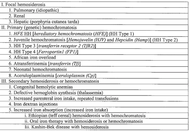
Table 1. Classification of hemosiderosis and hemochromatosis. 1. Focal hemosiderosis
1. Pulmonary (idiopathic) 2. Renal
3. Hepatic (porphyria cutanea tarda) II. Prirnary (genetic) hemochromatosis
1. HFE HH [herediatoryhernochrornatosis HFE)] (HH Type 1)
2. Juvenile hemochromatosis [Hernojuvelin (HJV) and Hepcidin (Harnp)J (HH Type 2) 3. HH Type 3 [transferrin receptor2 (‘T/R2)]
4. HH Type 4 [Ferroportin] (FF1)] 5. African iron overload
6. Atransferrinemia [transferrin (Tj)] 7. Neonatal hemochromatosis
8. Aceruloplasminemia [ceritioplasmin (Cp)J III. Secondary hemosiderosis or hemochromatosis
1. Congenital hemolytic anemias
2. Defective hemoglobin synthesis (thalassernia)
3. 1ncreased parenteral iron intake, repeated transfusions 4. Iron dextran injections
5. Jncreased iron absorption (increasediron intake)
i. Ethiopian (teffcereal) hemosiderosis with hemochromatosis ii. Oral iron therapy with hemosiderosis or hernochromatosis Iii. Kashin-Bek disease with hemosiderosis
3.2.1. Focal hemosiderosis
Focal hemosiderosis occurs mainly as a result of other diseases. Lungs andkidneys are the
most cornmonly affected sites [11, 12]. Pulmonary hemosiderosis is caused by recurrent pulmonary hemorrhage resulting in iron accumulation. Renal hemosiderosis is caused by various forms of hemolysis resulting in Hb deposits in the glomerulus. The renal parenchyrna is able to maintain its normal ftinction, but severe hemosiderinuria causes iron deficiency.
3.2.2. Type 1 HH tHFE-associated)
Type 1 hemochromatosis is an autosomai recessive iron disorder. A candidate gene for hemochromatosis (HFE) was found to be localizcd in the human chromosome 6p2l [13]. C282Y and H63D are two important mutations found in HFE [14]. The C282Y homozygote mutation has been found in 60—100% of Type 1 HH patients in the European population, and the H63D mutation is found in approximately 16% of these [13, 14]. HFE encodes a protein homologous to major histocompatibility compiex (MHC) ciass I molecules. Although the HFE protein retains many of the structural features of ciassicai class 1 molecules, its inward dispiacement ofthe cd domain helix prevents peptide binding. Given that the HFE protein interacts with transfenin receptor 1 (TfR1), a moiecule that plays a central foie in ceiiuiar iron uptake [15], HFE is thought to influence iron uptake in celis. The body obtains iron tbrough duodenai iron absorption. Duodenai iron absorption efficiency is reiated to the iron-sensing property of crypt celis, which receive signais about the body’s iron requirements, in part through the binding of transferrin to the HfE/TfRÏ complex. HFE mutations cause signaling of misinformation regarding iron requirements in the body. The consequence of this is excessive intestinal absorption of iron, which in tum ieads to a progressive iron ioading in the parenchymal ceils of important organs such as the liver, pancreas and heart. This can resuit in various iilnesses such as liver disease, diabetes meiiitus and cardiomyopathy [16]. Phlebotomy is the most efficient method for removing excess iron in type 1 HH patients.
3.2.3. Non-TIFF HH
Juvenile hemochromatosis (Type 2 HH)
Type 2 HH is a rare disorder originally identified in a gene heredity study of several Italian farnilies [17]. Juvenile hemochromatosis patients have accelerated iron loading rates as compared with Type 1, HFE-associated HH [17]. Due to the rapid iron loading in Type 2 HH, cardiomyopathics and endocrinopathies occur more cornmonly than liver dysftinction. Most individuals with juvenile hemochromatosis do flot survive beyond thirty years of age due to heart failure. Gene association studies in Type 2 HH have confirmed that the underlying Hemojuvelin (HJV) gene is located on human chromosome lp [1$]. HJV mutant patients have decreased hepcidin levels, which suggests that HJV may modulate hepcidin expression [19]. In addition, juvenile hemochromatosis was also found to be associated with mutations of Harnp, located on 19q13, which encodes hepcidin [20].
Type 3 HH
Type 3 HI-I is causcd by mutations in the transferrin receptor 2 (Tffl2) gene, and is an autosomal recessive disease. From fifteen Italian patients in five different families, four different mutations of TJR2 have been reported. Patients show a chronic increase in iron absorption resulting in high iron accumulation in the liver, heart and pancreas, suggesting a disniption in intestinal iron absorption due to the TfR2 mutation [21].
Type4HH
Type 4 HH, an autosomal dominant disease, is caused by ferroportin 1 (FF1) gene mutations [22, 23]. Iron concentrations increase in the liver, heart and pancreas in type 4 HH patients. Liver macrophages contain high amounts of iron and serum iron is relatively low. Two hypotheses regarding type 4 HH have been put forward at present. The loss-of ftmction hypothesis suggests that FF1 mutations impair iron recycling by macrophages, subsequently delivering abnormal signais to the intestine (mechanism as yet not understood), resulting in increased iron absorption and subsequent iron overioad [23]. The gain-of-function hypothesis suggests that FF1 mutations increase iron absorption in the duodenum [22]. In type 4 HH patients, since iron overioad is combined with anemia, phlebotomy treatment is not recommended.
African iron overioad
In the sub-Saharan region of Africa, approximately 10% of the population has a predisposition for iron overload, also referred to as Bantu siderosis [24]. A risk factor for African iron overioad is thought to be dietary iron intake from traditionally brewed beer. Since the prevaience of iron overioad in this region is high and flot ail beer drinkers are affected by iron overioad, it is difficuit to trace the inheritance pattem. Statisticai data, however, suggests that it is an autosomal dominant inherited disease, and the HFE gene has been exciuded as the causative gene for African iron overioad [25]. In African iron overioad patients, liver disease occurs more commonly than cardiomyopathy or diabetes. In addition, iron accumuiates in hepatic Kupffer ceiis and spleen in large amounts, suggesting a defect in iron recyciing by the reticuloendothelial system [26].
Atransfeninemia
Atransfeninemia is a very rare disease in which transferrin mutations cause impaired erythropoiesis, but iron accumulation is high in the liver due to an increase in non transfenmn bound iron (NTBI) [8, 27].
Neonatal hemochromatosis
Neonatal hemochromatosis is a rare, poorly characterized disease that occurs in newbom infants, with high iron accumulation in the liver, heart andlor pancreas [28]. Liver failure
mayoccur. It is unclear whether iron overload initiates the disease, or if other causes lead to iron overload [29]. Neonatal hemochromatosis is usually jointly present with other congenital diseases with similar liver manifestations, which suggests that it is a heterogenous disorder [30]. Familial cases have not been reported, and HFE gene mutations are not found in neonatal hemochromatosis. Liver transplantation is the only efficient therapy and has been found to prolong life in several cases [31].
3.2.4. Secondary iron overload (non-genetic)
Secondary iron overload includes transfusional iron overload and iron overload attributed to defective erytbropoiesis (congenital hemolytic anemias or hemoglobinopathies). In thalassemia major patients given transfusions without chelation, iron accumulation in the body is accelerated. Transfusional iron accumulation leads to progressive damage of the heart, liver and endocrine glands [32]. Iron overload-induced heart disease determines the
survival rate of the transfiised patients [33]. Liver disease is the second leading cause of death in these cases [34].
3.2.5. Iron overload ofundetermined origin
Some liver diseases are associated with elevated iron storage, for example, alcoholic liver disease, non-alcoholic steatohepatitis, and chronic hepatitis C [35]. The mechanisms are unknown. Although the patients do not have a genetic hemochromatosis background, iron unloading management may improve tiver function [36].
3.2.6. Neurodegenerative disorders related to iron metabolism
Neurodegenerative diseases related to iron metabolic disorders can be classified into two
categories based on the underlying iron defect: mitochondrial iron disturbance and brain iron disturbance. Mitochondrial iron disturbance is linked to friedrcich’s ataxia and X linked sideroblastic anernia with ataxia. Brain iron disturbance is related to neurofeiritinopathy, pantothenate kinase deficiency and acerulosplasminaemia.
Friedreich’s ataxia
Friedreich ataxia is an inherited lethal disease, and is characterized by spinocerebellar degeneration, cardiomyopathy and diabetes mellitus. In gene linkage studies, Friedreich’s ataxia was found to be associated with the gene encoding frataxin located on chromosome 9q [37]. Expansions of a trinucleotide repeat in an intron are usually found in patients, but point mutations havearc also been identified [37]. Affccted patients usually have defects in both alleles of the frataxin gene, ultimately leading to decreased frataxin levels. Frataxin is
a mitochondrial protein whose exact fiinction remains undetermined. Lack of frataxin homologues in yeast and mice leads to increased mitochondria toxicity, due to iron accumulation, elevated sensitivity to oxidative stress and depletion of iron-sulfiir proteins [38, 39].
X-Iinked sideroblastic anemia with ataxia
X-linked sideroblastic anemia with ataxia presents as mild microcytic anemia, bone manow sideroblastosis and nonprogressive cerebellar ataxia [40]. It is caused by an isoleucine to methionine substitution (1400M) in the ATP-binding cassette transporter 7 (ABC7) gene, locatcd on chromosome Xq. The exact pathogenesis of this disease is unknown. However, since Atmlp, the S. cerevisiae homologue of human ABC7, plays a role in iron export in yeast mitochondrion [41], iron accumulation within human mitochondria is thought to result from the loss-of-function mutation in A3C7.
X-linked sideroblastic anemia
X-linked sideroblastic anemia with ataxia is distinct from the non-neurodegenerative classical X-linked sideroblastic anemia, which is an X-linked recessive disease caused by mutations in the erythroid aminolevulinic acid synthase (e-ALAS) gene located on chromosome Xp [42]. The e-ALAS enzyme catalyzes the first step of heme synthesis. Dysftinctions in e-ALAS cause a defect in heme synthesis, leading to iron accunuilation in the mitochondria of erythroid precursors. Impaired erythropoiesis induces increased iron absorption from the intestine, resulting in iron overload. 11e anemia is usually nonuocytic; microcytic anemia is less common [43].
Neurofenitinopathy
Neurofenitinopathy is a late-onset disease of the basal ganglia, in which patients present with extrapyramidal features sirnilar to those found in Parkinson’s disease. Patients with neurofelTitinopathy usually have abnormal iron and fenitin aggregates in the brain. Mutations in the ferritin light chain gene leads to a structural change in the encoded protein. The resultant fenitin dysfiinction causes iron release from ferritin and iron accumulation in axons or synapses. This iron accumulation enhances oxidative stress in the neuron and resuits in neurodegeneration [44].
Pantothenate kinase-deficiency
Pantothenate kinase (PANK2)-deftciency is an example of a brain iron-overload disease involving a gene whose function is not related to iron metabolism. PANK2-deficiency usually develops within the first two decades of life, and clinically presents as dystonia, pigmentary retinopathy, optic atrophy, and high concentrations of iron in the globus pallidus [45]. PANK2 gene mutations cause a block in the coenzyme-A biosynthesis pathway. The subsequent increase in cysteine concentrations in the brain leads to iron accumulation due to the iron-chelating properties of cysteine. The increased free radical production and oxidative stress derived from iron accumulation result in neurodegeneration [46].
Acerulosplasminaemia
Aceniloplasminemia is a rare disease, in which decreased ceruloplasimin levels in plasma cause an inability for Fe2 to be oxidized to Fe3, which is critical for the binding of iron to
transfenin. In acerulosplasminaemia, mutations in the ceruloplasmin gene ieads to a decrease in iron released from neurons of the retina and the basal ganglia. Consequently, regional iron accumulation enhances neuronal oxidative stress, resulting in progressive neurodegeneration [47-49].
All the disorders mentioned in this section demonstrate the importance of maintaining iron horneostasis. They emphasize the importance of understanding the principles regulating iron rnetabolism at thc cellular and systemic levels. Within the past 10 years, advances in molecular biology techniques have permitted a dramatic increase in the information available on the various steps of iron metabolism: iron uptake, transport, storage, export and utilization.
4. Cellular iron homeostasis
Cellular iron homeostasis needs to be tightly controlled through iron uptake, storage and export. Thus, the molecular understanding of each step of iron metabolism helps elucidate the principles of normal iron homeostasis and the mechanisms of iron disorders.
4.1. Iron uptake
Iron uptake is necessary for ail cells. This section will detail identified pathways of cellular iron uptake: (j) classic transferrin-dependent iron uptake, which is used by almost all cells; (ii) ceil-type specific iron uptake in macrophages and enterocytes; and (iii) transferrin independent iron uptake, which piays a minor role.
4.1 .1. Transferrin-dependent iron uptake
Transferrin is the most important iron-transporting protein in the blood, and it binds iron with high affinity. In normal situations, ail circulating iron is bound to transfenmn. Two ftinctions are served by this transferrin-iron binding: (j) the prevention of free-iron derived toxicity through binding of fe3 in the blood; and (ii) the facilitation of iron transport in the plasma and iron uptake into ceils. Plasma transfenmn is mainly synthesized in and secreted from the liver into the blood, where it mediates the exchange of iron among the various sites of absorption, recycling, utilization, and storage.
The delivery of transferrin-bound iron into the cell is mediated by transferrin receptors located on the ceil surface. Receptor-mediated endocytosis is the classical pathway used by almost all mammalian cells in order to obtain iron [50-52]. Transferrins and thetransferrin receptor 1 (TfR1) are the components of this classical iron uptake and transferrin-recycling pathway (Figure 1). Cell surface transferrin receptors bind to the iron-transfenmn complex with high affinity under neutral pH conditions
[531.
Once the transferrin receptor-bound iron-transferrin complex has been endocytosed, it is intemalized into endosomes with a neutral pH. ATP-dependent proton pumps subsequently acidify the endosomal interior to pH 5.5 [54-5 7], which facilitates the dissociation of fenic iron (Fe3) from transferrin. The fenic iron released within the acidified endosomes is converted to ferrous iron and then transported by the divalent metal transporter 1 (DMTY) into the cytoplasm [58]. Transfenmns remain bound to their receptors after the release of iron in the acidified endosomes, and are recycled to the cell surface. In the neutral (pH 7.4) environment at the cell surface, the non-iron bound transfenmns lose affinity for their receptors, promoting theirdissociation. Transfenin is thus recycled back into circulation [51, 55]. The recycling process of transfenins enables one transfenin to be re-used up to one hundred times over the course ofits eight-day haif-life.
Transfenin receptor 2 (TfR2) and cubilin are ftvo alternative receptors for transfenin. TfR2 is bighly homologous to TfR1, yet it binds transferrin with a lower affinity [59]. The fact that TfR2 is only expressed in the liver and cannot rescue TfR1-mutant mice from early death suggests that TfR2 and TfR1 do not share the same roles [60, 61]. Cubilin is a novel potential transfenin receptor, which bas been shown to mediate endocytosis of the transferrin-iron complex, albeit only in the proximal tubules of the kidney and the yolk sac [62].
Extracellular space pH 7,4
Fe2-Tf Apo-Tf
%‘tHFE:H Proton pump
N
ç
en dosome Cytoplasm
Figure 1. The transferrin (If) cycle. Iron-loaded transferrin (fe2-Tf) binds to transfenmn receptors (TIR), which associate with 32m-HFE molecules on the ceil surface. Apo transfenin (Apo-Tf) and transferrin receptors can be re-used for further cycles of iron binding and iron uptake.
4.1.2. Iron uptake by reticuloendothelial (RE) macrophages
Macrophages can obtain iron in three ways: (i) transferrin receptor-mediated iron uptake; (ii) ceil surface receptor CD163-mediated hemoglobinlhaptoglobin complexes uptake [63]; and (iii) phagocytosis of senescent or damaged RBC (Figure 2). In the latter two pathways, hernoglobin is broken down by heme oxygenase 1. lion released from hemoglobin is subsequently transported from the phagosome into the cytoplasm by DM11. In the cytoplasrn, iron will either be stored in fenitin or transported back into the blood by ferroportin 1. Macrophages are the most important cells for iron recycling because they can obtain iron from RBCs and release digested iron back into the circulation for the development of erythron precurs ors.
Macrophage
Erythrocyte
Figure 2. Macrophage iron uptake and recycling. Macrophages can obtain iron by three different mechanisms: (i) TfR-mediated uptake of transferrin-iron complexes; (ii) CD 163-mediated acquisition of the haptoglobinlhemoglobin complex; and (iii) phagocytosis of senescent RBCs. In erythroid precursors, iron is taken up for heme biosynthesis through a TfR-mediated rnechanism. Nramp 1: natural resistance-associated macrophage protein 1, Hp-Hb: hemoglobinlhaptoglobin complexes.
4.1.3. Iron uptake by intestinal absorptive ceils
Dietary-derived iron is the normal source of exogenous iron. The intestine thus performs an important function in absorbing iron from exogenous sources. The developing enterocyte and mature enterocyte obtain iron through
two
different pathways. In the duodenum, the apical villus enterocyte matures and migrates from developing crypt ceils. The former can absorb exogenous iron, whereas the latter does not. Crypt ceils can take up iron onlythrough the transferrin receptor-mediated pathway. This section will focus on iron absorption by the apical absorptive celis of the duodenum. Low molecular weight iron and heme-iron are the two forms of iron that can be absorbed in this manner.
The process of transporting dietary-derived low molecular weight iron across the intestinal absorptive celis occurs in three steps (Figure 3): (i) uptake of iron through the apical membrane; (ii) distribution of iron in the ceil and transport of iron to the basolateral membrane; and (iii) the transfer of iron through the basolateral membrane into the blood [64, 65]. In the first step, dietary ferric iron (fe3) is reduced to fenous iron (Fe2) by the ferric reductase Dcytb, which is located at the apical membrane [66]. Fe21 is then transported into the celi cytoplasm by DMT1. Iron thus joins the intracellular labile iron pool (LIP) [67], from which iron either is directed to ferritin, where iron is stored, or is transported ftirther across the basolateral membrane into the blood by the iron exporter called fenoportin 1 [68].
ii1
Fe3DM11
won poo1L L_Fe2+
Fe2 Fpl Fe3Dcytb Feffitin TfR(ciypts) HFE (crypts) 2m (ciypts)
Figure 3. The process of intestinal iron absorption. Dietary iron is taken up through the apical membrane of the intestine into the cytoplasm. lion is then transferTed across the basolateral membrane into the blood. Only crypt ceils of the intestine obtain iron via the TfR-mediated pathway. Heph: hepheastin.
It is noteworthy that iron contained in heme derived from meat (containing hemoglobin, myoglobin and other heme proteins) is another important source of iron. Dietary heme iron accounts for approximately 30% of the total dietary iron in North American and Europe [69]. Heme-derived iron can be released by heme oxygenase 1 in the enterocyte, although a heme receptor at the apical membrane has not yet been identified.
An alternative ferric iron uptake mechanism has been identified for dietary iron absorption, refeiied to as the integrin-mobilferrin pathway (IMP). IMP transports only ferric iron (Fe3j, and no other metals, into enterocytes [70]. Proteins associated with this pathway includc mobilfenin, f33-integrin and flavin monooxygenase, whose macromolecular complex (520 kDa) is called parafenitin. The IMP complex is only found in enterocytes, and it is not known to what extent this pathway contributes to dietary iron absorption.
4.1.4. Transfenin-independent iron uptake
The existence of transfenin-independent iron uptake is suggested by two classes of iron disorders: transferrin shortage (atransfeninemia) and iron overload (hemochromatosis,
thalassemia). In hypotransferrinernia the reduction of transferrin levels leads to severe hypochromic microcytic anemia and massive iron loading in ail non-hematopoietic tissues, in particular in the liver and pancreas [71-73]. In hereditary hemochromatosis and transfusional thalassemia, two iron overload diseases, non-transferrin bound iron (NTBI) is found in the blood [74-77]. NTBI distributes iron into tissues different from those covered by transferrin-bound iron [74-77]. Furthermore, three pathways have been identified which support the existence of a transferrin-independent iron uptake, mediated by ferritin, L-type Ca2 channcls, and lipocalin 2.
Feiiitin-rnediated iron uptake is supported by several studies. In rats, when ferritin containing radiolabeled iron is injected into the circulation, iron is taken up and stored in
the liver [78]. In addition, the liver expresses ferritin receptors, and hepatocytes take up iron-containing ferritin via receptor-mediated endocytosis [79].
The L-type Ca2 channel provides a pathway for iron entry into cardiomyocytes. This pathway is organ specffic, because L-type Ca2 channels are specffic to cardiomyocytes. This pathway is important in iron overload conditions, as high concentrations of NTBI can utilize this pathway to enter the heart, leading to cardiac iron accumulation, and consequent cardiornyopathy [$0].
Lipocalin 2 is a newly identified putative iron transporter. Lipocalin 2 is the mouse homologue of the human neutrophil gelatinase-associated lipocalin (NGAL) gene. Structural analyses have shown that lipocalin 2 can bind siderophores (bacterial-derived
iron binding compounds) and iron [81]. Lipocalin 2-mediated iron transport has been shown to deliver amounts of iron sufficient for the regulation of iron-regulated gene expression in several celi unes [$2], including mouse embryonic kidney collecting duct celis, human Wilms tumor kidney ceils, dog kidney collecting duct ceils, rat embryonic kidney epithelial celis and mouse kidney stroma celis. Lipocalin 2-mediated iron transport can also provide iron for the development of the mesenchyme into epithelia during kidney development [$2].
4.2. Iron storage
When iron is present in the cytoplasm, ferrous iron is carried by a carrier molecule to the labile iron pool (LIP). This iron carrier molecule has not been identified, because of the very srnall amount of iron in the LIP. In the cytoplasm, iron diffuses to various intracellular cornpartments, such as the mitochondria for heme biosynthesis, or to ferritin for storage.
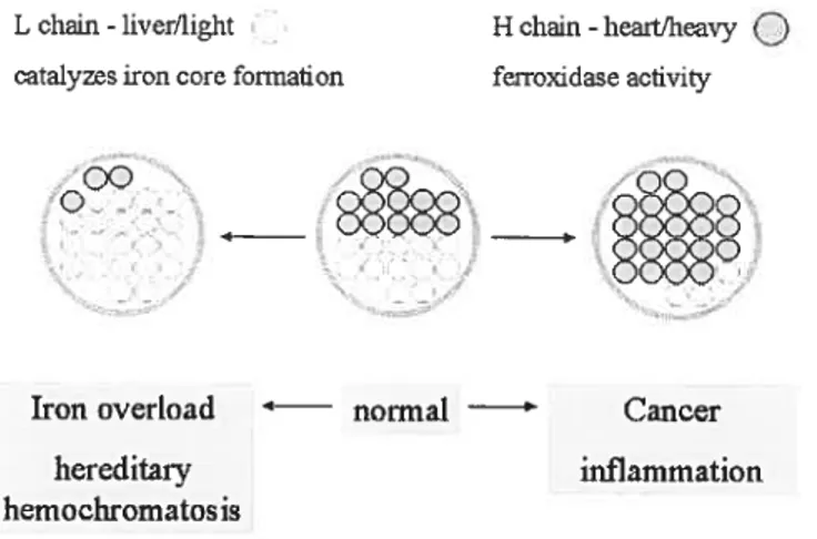
ferritin is a heteropolymer complex, composed of twenty-four subunits of H (heavy/heart) and L (light/liver) chains [$3]. The central cavity of ferritin can store up to 4500 iron ions [83]. The composition of ferritin varies with the ratio of H and L chains, depending on the organ in which it is found (Figure 4). ferritin found in the liver and spleen is rich in L chains, which are associated with high iron storage, whereas ferritin found in the heart is rich in H-chains, which are associated with high iron utilization [$3, 84]. ferritin thus has a variable iron storage capability, with a high H:L ratio when heme synthesis or cell proliferation increases [54, 85]. The degradation of ferritin and the associated release of
iron facilitate the mobilization of iron for cellular utilization, and is associated with lysosomes [86, 87].
L chain-liverllight ï H chain-heait/heavy
O
catalyzesfron core formation ferroxidase adivityoQ
__E98
.88*8’
88°
Iron overload — normal Cancer
hereditaiw inflammation
hemochromatosis
Figure 4. The composition of ferritin. Each ferritin molecule is composed of 24 chains, either L or H chains. The ratio of L and H chains in fenitin varies among organs. Furthermore, the L/H chain ratio in ferritin can be modified in response to iron overload or inflammation.
4.3. Iron export
Duodenal enterocytes, macrophages, hepatocytes and other types of celis have similar mechanisms for releasing iron in order to meet the iron needs of the whole body and also to maintain iron balance within celis. Ferroportin 1 (FP 1) is a ubiquitously expressed protein. Fenoportin 1 fimnctions as a cellular iron exporter, by transporting iron from the cytoplasm into the blood [88, 89]. However, ferroportin 1 needs the cooperation of a ferroxidase to perform this frmnction (Figure 5). Two types of copper ferroxidases are found in the body:
ceniloplasmin (Cp) and hephaestin, both of which oxidize Fe2 into Fe3, for Fe3 binding to transferrin in the blood.
Hephaestin is the homologue of the plasma protein cemloplasmin. Arnino acids involved in copper binding and disulfide bond formation are conserved between cemioplasmin and hephaestin. Unlike ceniloplasmin, hephaestin is a trans-membrane protein. Since ceniloplasmin circulates in the blood and hephaestin is expressed only in the intestinal villi, ceniloplasmin can facilitate iron export from non-intestinal cells while hephaestin mediates intestinal iron export. Sflidies in hephaestin knockout mice have shown that ceruloplasmin cannot replace the role of hephaestin in the process of enterocyte iron export [90, 91].
The cernioplasmin protein is a member of the multicopper oxidase family, which can utilize copper to catalyze biochemical reactions [92]. Since ceniloplasmin contains greater than 95% of the copper found in plasma, it was first thought to be involved in copper metabolism. However, cemioplasmin knockout mice exhibited normal copper metabolisrn during copper absorption, transportation, distribution, and excretion steps [93]. These evidence suggest that copper is only required for ceruloplasmin to carry out ferroxidase frmnctions, and that cemioplasmin does not play an essential role in copper metabolism.
O 1.0 o
1.0
FP1
hepatocytes
Ifintes final celis
Ifoffier celis
Figure 5. Iron export facilitated by ceniloplasmin (Cp) and hephaestin (heph). Ceruloplasmin and hephaestin are structurally similar, and both have ferroxidase activity, which can oxidize Fe2 into Fe3.
4.4. Cellular iron homeostasis
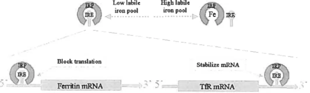
Cellular iron homeostasis is regulated by the iron responsive proteinliron responsive elernent (IRP/IRE) system in the ceil (Figure 6). IREs are found in the 3’- or 5’-untranslated inRNA regions of several critical proteins involved in iron rnetabolism, including ferroportin 1, DMT1, ferritin, and TfRY [94, 95). IRPs normally exist in the cytoplasrn as cytosolic aconitases. IRPs can bind to IREs to regulate the translation of proteins.
The affinity of JRPs to bind IREs is dependent upon the iron concentration in the labile iron pool (LIP). Under high iron concentrations, IRPs are fihled with a 4Fe-4$ cluster that prevents the binding of IREs. Under low iron conditions, the 4Fe-4S cluster dissociates from IRPs, which can then bind to IREs.
Through the binding of IRPs to IREs, the IRP/IRE system post-transcriptionally controls protein synthesis. The location of IREs on either the 5’- or 3’-untranslated region of mRNAs determines the consequences of IRP-IRE binding. For example, in ferritin, where 1 IRE is located in the 5’-untranslated region of the mRNA, the IRP/IRE binding blocks the translation pathway, thus decreasing ferritin synthesis. Conversely, in transferrin receptor 1, where IREs are locatcd to the 3’ -untranslated region of the mRNA, the IRP/IRE binding stabilizes the mRNA, and thus enhances TfRl synthesis.
Through the regulating activity of the IRP/IRE system, the up- or down-regulation of iron responsive genes enables the celi to maintain proper iron homeostasis. For example, when the iron concentration is high in the LIP, ferritin protein synthesis is up-regulated to store more iron, while TfRl is down-regulated, reducing the uptake of iron.
The IRP/IRE system can be influenced not only by iron concentrations, but also by nitric oxide (NO) levels, oxidants and hypoxia [96, 97]. These factors provide the ceil with a greater degree of control over iron levels via a complex seif-regulation mechanism.
Lowiabile Highlabilc won pool won pool
Block translation
Stabilize mRNA
PetrftinmRNA TIRrnRNA
Figure 6. The IRP/IRE regulation system. The affinity ofIRPs for IREs depends on the iron level in the labile iron pool. When the iron level is low, the binding of IRPs to IREs at the 5’-untranslated region blocks mRNA translation, while binding of IRPs to ifiEs at the 3’-untranslated region increases mRNA stability, and thus protein synthesis.
5. Systemic iron homeostasis 5.1. Distribution of iron in aduits
The average human has on average 4g of total body iron and approximately 1 -2mg of iron are lost daily from the epithelial surfaces of the gut and skin and, in females, through menstruations (figure 7). These daily losses are replenished through the transfer ofingested dietary iron to the portal circulation. Internai iron requirements, for the production of hemoglobin, are met by recycling iron from the destruction of RBCs by the reticuloendothelial (RE) system and subsequent release of iron into the plasma, where iron is transported while bound to transferrin (If) [9$]. Excess iron is stored in ferritin in the liver in a non-toxic form that can be mobiiized as required. Under normal conditions, liver iron stores are close to 1g, but growing chiidren and menstruating women have reduced liver iron stores due to their increased iron requirements [99]. Another smailer proportion of iron is located in muscle myoglobin and body enzymes.
Liveriron stores l000mg
__
‘N
Duodenumriasma
V loss ______________________ Myoglobin and - 1-2mg enzymes 30mgFigure 7. Distribution of iron in aduits. Iron is absorbed from the duodenum, transported into the plasma, recycled by macrophages, utilized by erythrocytes and enzymes, and stored in the liver. Iron regulatoiy factors can change the ftinction of these organs and celis, thus redistributing iron to establish a new iron homeostasis in the body.
5.2. Iron regulators
5.2.1. The four iron regulators
Systemic iron homeostasis is maintained through the control of iron absorption, iron stores and iron recycling to meet the iron demands of erythropoiesis and iron-dependent enzyme activity.
Four iron regulators can change iron mobilization and iron redistribution in the body: (j) The iron stores regulator refers to the amount of iron stored in the body [100]; (ii) the hypoxia regulator refers to oxygen insufficiency in the body; (iii) the erythropoetic
regulator refers to the need for the production of RBCs; and (iv) the inflammatory regulator refers to infection and inflammation stimuli.
In normal aduits, acute iron requirements are satisfied by the release of stored iron from the liver and recyclcd iron from macrophages. When the release of iron from body stores cannot meet the iron demand, as is the case in iron deficiencies, iron absorption from the intestine is accelerated. The erythropoetic regulator can dramatically increase iron absorption from the intestine. It can even override the effect of the stores regulator. For instance, in transferrin-mutant mice, hypotransfeninemia causes insufficient iron delivery into erythroid precursors. These mice develop severe anemia, which requires increased iron absorption. Paradoxically, the mouse also has iron overload in the body, which leads to a decrease in iron absorption. In this case, the erythropoetic regulator will dominate the stores regulator, and thus increased iron absorption from the intestine continues [101].
The activation of the hypoxia regulator can also increase iron absorption from the intestine, which produces an effect similar to that of the erythropoetic regulator. However, the interaction of the hypoxia regulator with the other regulators has not yet been ffilly investigated.
Infection and inflammation is another strong regulator of iron metabolism. The pathophysiologic response of iron metabolism to inflammation is to withhold iron from invading bacterial pathogens, through reducing iron release from the liver and the macrophages and by suppressing intestinal iron absorption, thus decreasing serum iron
levels. The inflammatory regulator can override the erythropoetic regulator. For example, in anemia of chronic diseases, inflammatory cytokines reduce bone marrow RBC production [102-104]. $uch anemia activates the erythropoetic regulator to increase iron absorption from the intestine. Paradoxically, at the same time, the inflammatory regulator produces a decrease in iron absorption. In this contradictory situation, the inflammatory regulator dorninates the erythropoetic regulator, ultimately resulting in a decreasc in intestinal iron absorption.
This hierarchy between iron regulators predicts the existence of messengers circulating in the blood needed to deliver signais among the sites of iron utilization, storage, absorption, and recycling, to conectly coordinate the efficiency of iron mobilization arnong these sites.
5.2.2. Hepcidin, a putative iron messenger
Hepcidin is a newly identified iron messenger. It is a small peptide secreted by the liver and excreted through the kidneys [105-107]. Hepcidin can respond to ail four known iron regulators. For example, hepcidin expression is increased by iron overload diets in rodents, which represents an iron store regulator [107]. It is also reduced in non-anemic hypoxia, a hypoxia regulator [108], and is eievated in response to anemia, an erythropoetic regulator [109]. Finally, hepcidin expression is increased in different types of inflammation in mice and humans, which represents an inflammatory regulator [10$-110].
Hepcidin functions as a regulator of iron absorption in the intestine and iron release from macrophages, as has been demonstrated in hepcidin-knockout mice and hepcidin-transgenic
mice. Hepcidin depletion in knockout mice is associated with increased body iron [111, 112]. lncreased hepcidin levels in transgenic mice resuits in iron deficiency [112]. Thus, the level of serum hepcidin provides a counterbalance to the level of body iron stores. This regulation may be explained by the recent finding that hepcidin regulates iron efflux via its binding to ferroportin 1 (FPY). This binding subsequently leads to FPY intemalization and degradation, resulting in a decrease in FP1 concentration at the ceil surface [113].
5.3. Animal models
Studying animal models takes advantage of the similarities between human and rodent iron rnetabolisrn regulation. $pecific gene dismptions in rodents, including targeted disruptions and spontaneous mutations, have provided crucial information about each gene involved. Studying the function of targeted genes in these rodents has extended our knowledge of the pathways of iron metabolism at the molecular level and the defects that occur in iron rnetabolism disorders. (Table 2)
Table 2: Useftil rodent models for understanding iron metabolism and related diseases.
Iron phenotype human disease
Mouse model Gene affected
mode ling Tncreased iron absorption, iron overload,
Hfe Hfe hepatocellular iron deposition, decreased Type I HH macrophage iron, clevated transferrin saturation
Increased iron absorption, iron overload,
J32nï 3-2 microglobulin hepatocellular iron deposition, decreased Type 1 HH macrophage iron, elevated transferrin saturation
Increased iron absorption, iron overload,
Usf2 Hepcidin hepatocellular iron deposition, decreased Type 2 HH macrophage iron, elevated transferrin saturation
Hamp (liver Decreased iron absorption severe iron Hepcidin
transgene) deflctency and anemia
Microcytic hypochromic anemia, tissue iron
Hpx Transferrin Atransferrinernia
deposition
TfR’ Transferrin Microcytic hypochromic erythrocytes, decreased receptor-1 iron stores, embiyonic lethality
TfR12245x/245x Transferrin Increased iron absorption, iron overload Type 3 HH rcceptor-2
Decreased iron absorption, systemic iron b (Belgrade rat) DMT 1 (GI 85R) deficiency, impaired iron uptake in duodenum
and in_erythroid_precursors Decreased iron absorption, systemic iron
mk DMTI(G185R) deficiency, impaired iron uptake in the —
duodenum and in_erythroid_precursors
Weh Hypochromicanemia, impaired iron transfer —
. Ferroportin 1
(Weissherbst) from yolk sac to embryo
-/- Iron accumulation in hepatocytes and
Cp Ceruloplasmin Aceruloplasminemia
macrophages
. Microcytic hypochromic anemia, impaired
sla Hephaestin —
intestinal iron transfer Elevated tissue and serum L-ferritin
Ftlf’ H-ferritin Embryonic lethality —
Frda (fleuron Mitochondrial iron deposits, neurodegeneration
Frataxin Friedreich ataxia
/heart knockout) and cardiomyopathy
Frda (muscle Mitochondrial iron deposits, cardiomyopathy
Frataxin Friedreich ataxia
knockout)
i Anemia, low serum iron levels, tissue iron
Hmoxl Hemeoxygenase 1 Hmoxl deficiency
6. Iron metabolism and inflammation
One of the most obvious connections between iron metabolism and inflammation is the necessity of iron for the devclopment of the immune system. Iron is reqtiired for the normal proliferation and maturation of lymphocytes. Iron deficiency causes a reduction in
peripheral T-cell populations and atrophy of the thymus [114, 115]. In addition, the relationship between iron and inflammation is generally recognized in certain baterial infections and inflammations. Since iron is an important nutrient for the proliferation of bacteria, the response of increased iron sequestration from bacteria becomes an important host defense rnechanism. This section will illustrate the connection between iron metabolisrn and the inflammation (anti-bacterial effect) in three aspects: structural similarity of some proteins; regulation of iron metabolism by inflammatory cytokines; and iron sequestration in bacterial infection.
6.1. Structural and functional similarities
The findings that there are structural similarities between sorne iron metabolism-related proteins and proteins that are particularly important for inflammation suggest that there may be a relationship between iron rnetabolism and inflammation. Three examples are dernonstrated here: (i) hepcidin and defensin; (ii) Natural resistance-associated macrophage protein 1 (Nramp]) and DMT1; and (iii) HFE and MHC class I.
6.1.1. Hepcidin and defensin
Hepcidin shares functional similanties with defensins. Antimicrobial peptides are a valuable component of innate immunity in many species [116]. In mammals, antimicrobial
peptides such as defensins [117] represent a substantial part of the immune system for resistance to cellular pathogens. The bactericidal spectrum of hepcidin is similar to that of hurnan defensin- 1, which can inhibit the proliferation of a range of pathogens including Gram-negative Neisseria cinerea and the yeast Saccharomvces cerevisiae [105]. In addition, hepcidin and defensin both appear in human blood [118], and the expression of both can be induced by lipopolysaccharide, matching their roles in host defense.
In addition to these functional sirnilarities, hepcidin’s structural features are sirnilar to that of antirnicrobial defensins in tenns of size and cysteine content [117, 119]. Hepcidin is a small peptide with an approximate molecular mass of 3 kDa; defensins range from 4-6 kDa in size. The hepcidin peptide sequence contains eight cysteine residues in four disulfide bonds; the human f3-defensins have three intramolecular cysteine-bonds linldng cystcines 1-5, 2-4, 3-6 [120] (Figure 8).
6.1.2. NRAMP1 ana DMTY
The natural resistance of inbred mice strains to infection by Salmonelta typhinturium, Mycobacteria, and Leishrnania donovaiti was documented nearly 30 years ago [121, 122]. A single locus (Bcg), mapped to mouse chromosome 1, has been shown to influence bacterial replication in macrophages [123]. Positional cloning of Bcg identified the gene for natural resistance-associated macrophage protein (NRAMF]) as being responsible for the resistance to infection by bacteria [122].
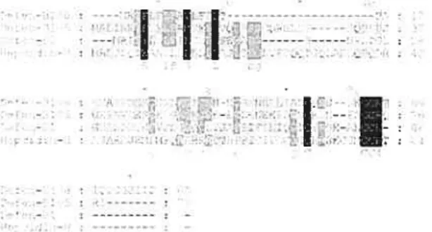
The mammalian NRAMP family is divided into two classes, NRAMF] and DMT1 (also known as NRAMP2). Although the cloning ofthe DMT1 gene was carried out afier that of NRAMF] and its identification was based solely on its homology to NRAMF] (f igure 9), the ftmction of DMT 1 was known before that of NRAMP]. A comparative study between NRAMF] and DMT1 was aptly used to elucidate the function of NRAMP]. Since DMT1 is a divalent cation transporter and can transport Fe2, Mn2, and Zn2 in several celi lines, sirnilar studies have been performed on NRAMF] that showed that NRAMP1 can also transport Fe2, Mn2, and Zn2, albeit with less efficiency than DMTY [58, 124, 125]. DMT1 and NRAMP1 are both located within endosomes and lysosomes [126, 127], which ftirther suggests a functional similarity, although some NRAMP1 expression can also be found within phagosomes [128, 129]. However, the ubiquitous expression of DMT1 in the body, in contrast to the exclusive expression of NRAIVIP 1 within macrophage cells, points to a difference of some ofthe functions ofthese two proteins [58, 130].
Major controversy exists conceming the direction in which NRAMP 1 delivers iron ions. Although DM11 has been demonstrated to transport Fe2 across the plasma membrane into the celi cytoplasm, the iron transport direction of NRAMP 1 remains unclear. Two schools of thought have emerged. One advances the hypothesis that NRAMP 1 functions to increase intraphagosomal Fe2 in order to provide a catalyst for the Fenton reaction, which generates the highly toxic hydroxyl radical for bactericidal activity [131]. The other hypothesizes that the function of NRAMP 1 is to remove the fe2 required by intraphagosomal bacteria for growth and the other divalent cations, such as Zn2 and Mn2, necessary for superoxide dismutase thus providing an effective antioxidant defense [132].
—
—
=
____-
-Lt!t-_
L____
_i
I I I
Figure 9. Protein alignment ofhuman NRÀIvIP1 and human DM11.
6.1.3. HFE and MHC class I
The HFE protein structurally belongs to the class I major histocornpatibility complex (MHC) antigen, an important protein family in the immune system (Figure 10). The HFE gene is located on chromosome 6p2l, close to the HLA class I gene cluster, and it encodes
tUE: r: uiip< 9 IF.
a MHC class I protein [133]. As with other MHC class I molecules, the extracellular portion of HFE is composed of ai, a2 and a3 domains, and the a3 domain binds f32-microglobulin [134]. The similarity between HFE and MHC class I molecules has raised questions regarding the ftmction ofHFE. MHC class I molecules normally participate in the cellular immune response by binding and presenting antigens to T-cells [135].
aI a2 OEl
t 132m
a3 •1 (32m I j
F1FE MMC I
Figure 10. Ribbon diagrams of HFE and MHC class I (MHC I) proteins. ai, a2, and a3 domain helixes of HFE and MHC I proteins are shown. (32m binds to both HFE and MHC I. Transferrin receptor (TfR)-binding site on HFE and T-cell receptor (TCR)-binding site on MHC I are indicated. (from reference: Lebron JA and Bjorkman PJ, 1999 [136])
6.2. Regulation of iron-metabolic proteins by inftammatory stimuli
Many iron-metabolic proteins can be regulated by vanous inflammatory stimuli. For example, hepcidin synthesis in the liver and its secretion in the serum are known to be increased by lipopolysaccharide (LPS) [107], turpentine [10$] and IL-6 [137] administration in mice [13$, 139]. Ferritin transcription, translation and secretion are also up-regulatcd by LPS [140, 141], turpentine [142] and IL-6 [143], as well as by TNFa and interleukin la (IL-la) [144-146] in different tissues and cell types. IFN-y has been shown
to up-reguiate DMT1 expression [147] and to down-regulate that of FP1 in the liver [14$]. Ail of these changes in iron metabolic proteins induced by inflammatory stimuli ultimateiy iead to hypoferrernia and iron accumulation in the liver.
6.3. Iron seguestration from bacteria
It has been shown that animais injected with iron, when infected with dangerous bacteria, dernonstrate an increased rate of mortality.
Bacteria are aiso iron-restricted species, and in muiticeiiuiar organisms colonized by bacteria, there is no freeiy availabie iron for bacterial nutrition [149]. There are two general rnechanisms by which bacteria obtain iron. The ftrst is characterized by direct contact between the bacteria and an exogenous iron source. In this mechanism, iron is taken up through bacterial ccii surface transporters. The second involves the synthesis and release of iron-chelating compounds from bacteria into the extracellular medium and capture of these compounds by their receptors. One known bacterial iron-chelating compound is caiied siderophore [150]. Siderophore chelates iron with a very high affinity in order to extract iron from most organic complexes.
In humans, severai iron-binding proteins, such as lactoferrin and lipocalin 2, are expressed in an iron-unsaturated form. This iron-unsaturated form enabies the sequestering of iron as a defence against invading bacteria.
7. Recent findings inspiring our interest in studying Lcn2
Neutrophil gelatinase-associated lipocalin (NGAL) was first identified as a 25-kDa glycoprotein associated with purified human neutrophil gelatinase [151]. Homologous proteins were also identified in the mouse [lipocalin2(Lcn2)/24p3/utcrocalin] and rat (F32-rnicroglobulin-related proteinlneu-related lipocalin) [152]. NGAL/Lcn2 has been shown to be highly expressed in the colonic epithelial areas of inflammation, and its expression was thought to be related to its anti-inflammatory ftinction [153]. The expression of Lcn2 in utems during the implantation and parturition stages of pregnancy suggests that it may function to induce apoptosis during involution [$2, 154, 155]. In addition, lipocalin 2 has also been shown to induce apoptosis of a wide variety of leukocytes [156]. Furthermore, microanay analyses performed in our laboratory two years ago obtained a spectnlm of genes, including Lcn2, that could be modulated by different iron regulators.
Two categories of recent findings, combined with the results from microarray analysis, inspired our interest in studying Lcn2.
The finding that NGAL/Lcn2 can function as an iron-binding molecule is supported by the dernonstration that it mediates a transfenmn-independent iron uptake pathway activated during kidney development [157]. fron delivered to differentiating epithelial cells by NGAL/Lcn2 is capable of regulating iron-dependent genes, such as ferritin and transfenmn receptor 1 (TfRÏ) [157], that are sensitive to the cellular iron status and are regulated by
iron-responsive elements (IREs) [158, 159]. This finding indicates that such ceils can utilize iron provided by NGAL/Lcn2. These observations, demonstrating that NGAL/Lcn2 mediates a transfenin-independent iron uptake pathway in vitro, raised thc question as to whether this pathway can become activated in response to increased iron demand in in vivo
rodent models.
It lias also been shown that NGAL/Lcn2 binds catecholate-type bacterial ferric siderophores [160], such as enterobactin. Siderophores have a high affinity for ferric iron and are able to acquire iron from mainmalian iron-binding proteins, including transfenin and lactoferrin [161]. Since NGAL/Lcn2 binds enterobactin with a higher affinity than the
E. cou enterobactin transporter, it effectively interferes with bacterial iron uptake in culture
conditions, and, in fact, acts as a bacteriostatic agent [160]. We have thus chosen to assess lipocalin 2 expression in two models of inflammation: systernic (LPS) and local (turpentine) inflammation, in evaluating its hypothesized bacteriostatic potential through an iron depletion strategy.
II. OBJECTIVE AND SPECIFIC AIMS 1. Objective
The purpose of this study is to investigate the role of Lcn2 in iron metabolism and inflammation by assessing its expression profile under conditions of altered iron storage, increased iron demand for hemoglobin synthesis, and induced iron redistribution during inflammation.
2. Specific aims
Aim 1: To study lipocalin 2 expression in response to altered iron metabolism. Iron absorption, storage, transport and utilization are regulated in response to altered iron metabolism. Many iron-metabolic proteins can be regulated to perform their needed ftinction and meet the body’s iron request. To define the role of Lcn2 in iron rnetabolism, Lcn2 levels are assessed in different iron storage conditions by using mouse models of iron overload and iron deficiency. The response of Lcn2 to an increased iron demand for enhanced hemoglobin synthesis is assessed in mouse models of anemia and hypoxia. Mouse rnodcls of anemia use PHZ (phenylhydrazine)-induced hemolytic anemia, iron deficient anemia, and phlebotomy-induced anemia. The mouse model of hypoxia uses CoC12-induced hypoxia. Lipocalin 2 mRNA in the liver and its secreted protein in the senim are analyzed.
Aim 2: To investigate lipocalin 2 regulation during inflammation. Since Lcn2 was found to bind bacteria-derived siderophore in vitro, the potential of Lcn2 to prevent bacteria growth in vivo is promising. Thus we are interested in assessing Lcn2 expression in mouse models
of bacterial component (LPS)-induced inflammations. Lcn2 transcription in the liver and its secretion in the serum are analyzed in inflammation. Since the LPS-induced Lcn2 tissue expression pattem may indicate the exact function of Lcn2 during inflammation, we are thus interested in assessing the tissue expression pattem of lipocalin 2 during acute inflammation. Lipocalin 2 mRNA levels are measured in the liver, spleen, duodenum, heart, kidney, thymus, brain, bone manow, and in isolated hepatocytes and liver mononuclear ceils. Lcn 2 expression in response to sterile inflammation is also assessed in a turpentine (TP)-induced acute inflammation mode!. During inflammation, iron redistribution and hypoferremia commonly occur; this is closely re!ated to the anemia of chronic disease (ACD). b examine the mechanism ofhypofenemia and the role ofLcn2 in the pathogenesis of ACD, Lcn2, DMT1, FP1 !eve!s are assessed in the liver of a mouse mode! of anemia of chronic inflammations. Iron accumulation in hepatocytes and macrophages are a!so analyzed to investigate their ro!es in the pathogenesis of hypoferremiaand ACD.