University of Montreal
Expression and regulation of microsomal prostaglandin E synthase-1 in human osteoarthritic cartilage and chondrocytes
By Xinfang Li
Department of Biomedical Sciences Faculty of Medicine
This thesis was submitted to the Faculty of Graduate studies and Research in partial fulfilment of the requirments of the degree of Master
in Biomedical Sciences. August 2005 udec CrDcc3ré\ ©Xinfang Li, 2005 f. 2ii1o
uJ
H
USB
n
q1
coq
n
o
Université
(III
de Montréal
Direction des bihiiothèques
AVIS
L’auteur a autorisé l’Université de Montréal à reproduite et diffuser, en totalité ou en partie, pat quelque moyen que ce soit et sur quelque support que ce soit, et exclusivement à des fins non lucratives d’enseignement et de recherche, des copies de ce mémoire ou de cette thèse.
L’auteur et les coauteurs le cas échéant conservent la propriété du droit d’auteur et des droits moraux qui protègent ce document. Ni la thèse ou le mémoire, ni des extraits substantiels de ce document, ne doivent être imprimés ou autrement reproduits sans l’autorisation de l’auteur.
Afin de se conformer à la Loi canadienne sur la protection des
renseignements personnels, quelques formulaires secondaires, coordonnées ou signatures intégrées au texte ont pu être enlevés de ce document. Bien que cela ait pu affecter la pagination, il n’y a aucun contenu manquant.
NOTICE
The author of this thesis or dissertation has granted a nonexciusive license allowing Université de Montréal to reproduce and publish the document, in part or in whole, and in any format, solely for noncommercial educational and research purposes.
The author and co-authors if applicable retain copyright ownership and moral
rights in this document. Neither the whole thesis or dissertation, flot
substantial extracts from it, may be printed or otherwise reproduced without the author’s permission.
In compliance with the Canadian Privacy Act some supporting forms, contact information or signatures may have been removed from the document. While this may affect the document page count, it does flot represent any loss of content from the document.
University of Montreal Faculty of graduate studies
This thesis entitled
Expression and regulation of microsomal prostaglandin E synthase-1 in human osteoarthritic cartilage and chondrocytes
Presented by Xinfang Li
Has been evaluated by a jury made up of the following people
President ofjury: Dr. Muhammad Zafarullah
Director of research: Dr. Hassan Fahmi
Member ofjury: Dr. Moharned Benderdour
Summary
Prostaglandin E2 (PGE2) is the major prostanoid synthesized in the joint and
play an important role in inflammation and pathogenesis of arthritis. High concentrations of PGE2 have been detected in serum and synovial ftuids from arthritic patients. In the PGE2 biosynthesis pathway, the synthesis of PGE2 from arachidonic acid (AA) requires 2 enzymes acting sequentially. Cyclooxygenases catalyze the conversion of AA to the intermediate prostanoid PGH2. Subsequently, PGES converts COX-derived PGH2 into PGE2. At least three distinct PGES isoforms have been identified, which are called microsomal PGES-l (rnPGES-l), mPGES-2, and cytosolic PGES. Among them, mPGES-l is induced by various inflamrnatory stimuli in some ceils and tissues and exhibits preferential functional coupling with COX-2.
Pro-inflammatory cytokines IL-113 and TNF-Πhave been shown to induce mPGES-l
expression in several tissues and ceil types. However, littie is known about the expression and regulation ofmPGES-1 in cartilage.
In order to better understand the regulation of PGE2 production in joint tissues, we analyzed mPGES-1 expression in nornal and OA cartilage. Furthemore, we explored the effects of different inflammatory agonists on the expression of
rnPGES-l in OA chondrocytes and tested the effeci of 15-PGJ2 on IL-1-induced
rnPGES-1 expression in OA chondrocytes. Our present study showed that levels of mPGES-1 mRNA and protein were markedly elevated in OA versus normal hurnan cartilage. Treatment ofchondrocyte with IL-1M induced the expression ofmPGES —l protein in a dose- and time-dependent manner. This appears to occur at the transcriptional level as IL-113 induced the expression of mPGES- 1 rnRNA and the activity of this gene prornoter. Furthermore, TNF-Œ and IL-17 also up-regulated the expression of mPGES-1 protein and displayed a synergistic effect with IL-113. The resuits obtained with 15-PGJ2 and PGE2 on mPGES protein expression of chondrocytes were also interesting. We showed that 1 5-PGJ2 inhibits IL-1 13-induced mPGES-1 protein expression, an effect that was reversed by exogenous PGE2.
II
To conclude, our study shows that rnPGES-1 expression is up-regulated in OA versus normal cartilage, proinflarnrnatory cytokines increased mPGES-1 expression and PPAR7 ligand 1 5d-PGJ2 repressed IL-1 3-induced mPGES- 1 expression in chondrocytes. These data suggest that mPGES- 1 may prove to be an interesting therapeutic target for controlling PGE2.
Key words:
Osteoarthritis, Microsornal prostaglandin E synthase-1, Cartilage, Chondrocytes, PGE2, PPARy, and 15d-PGJ2.
RÉSUMÉ
La prostaglandine E2 est une prostanoïde majeure synthétisée dans l’articulation. Celle-ci joue un rôle important dans l’inflammation et dans la pathogenèse de l’arthrite. De hautes concentrations de PGE2 ont été détectées dans le sérum et dans les liquides synoviaux des patients arthritiques. Dans la voie de la biosynthèse de la PGE2, la synthèse de la PGE2 à partir d’acide arachidonique (AA) nécessite l’action séquentielle de deux enzymes. La cyclooxygénase catalyse la conversion de l’AA en prostanoïde intermédiaire PGH2. Ensuite, la PGE synthase (PGES) convertit la PGH2 dérivée de la COX-2 en PGE7. Pas moins de trois isoformes distincts de PGES ont été identifiés PGES-l microsomal (rnPGES-1), mPGES-2 ainsi que PGES cytosolique. Parmi eux, rnPGES-l est induite par divers stimuli inflammatoires dans certaines cellules et certain tissus, et elle est préférentiellement couplée à COX-2.
Il a été démontré que les cytokines pro-inflammatoires IL-1 j3 et TNT-Œ
induisent l’expression de la rnPGES-1 dans plusieurs tissus et types de cellules. Toutefois, très peu est connu au sujet de l’expression et de la régulation de la mPGES
1 dans le cartilage.
Dans le but de mieux comprendre la régulation de la production de la PGE2 dans les tissus de l’articulation, nous avons analysé l’expression de la rnPGES-l dans le cartilage normal et celui ostéoarthritique (OA). De plus, nous avons exploré les effets que différents agonistes ont sur l’expression de la mPGES-1 dans les chondrocytes OA et nous avons testé l’effet de la 15-PGJ2 sur la mPGES-l induite par
IL-1W dans les chondrocytes OA. La présente étude démontre que les niveaux
d’ARNm et de protéines de mPGES-l sont élevés dans le cartilage OA comparativement au cartilage normal. Le traitement des chondrocytes avec IL-l
fI
induit l’expression de la protéine mPGES-1 de manière dose et temps dépendante. Ceci semble survenir à l’étape de transcription au cours de laquelle IL-ifI induit l’expression l’ARNm de mPGES-1 et l’activité de ce promoteur de gène. De plus,Iv
TNf-Œ et IL-17 augmente aussi l’expression de la protéine mPGES-1 et possède un effet synergique avec IL-113. Les résultats obtenus avec 15-PGJ2 et PGE2 sur l’expression de la protéine mPGES des chondrocytes sont également intéressants. Nous avons démontré que 1 5-PGJ2 inhibe l’expression de la protéine mPGES-1 induite par IL-l f3, un effet qui a été contré par la PGE2 exogène.
En conclusion, notre étude démontre que la mPGES-I est exprimé de manière plus significative dans le cartilage OA que dans le cartilage normal. De plus, les cytokines pro-inflammatoires augmentent l’expression de la mPGES-1, tandis que la 15d-PGJ2, un ligand de PPARg, diminue l’expression induite de la rnPGES-l par 1’IL-lb dans les chondrocytes. Ces résultats suggèrent que la mPGES-1 est une cible thérapeutique intéressante pour contrôler la production de la PGE2.
Mots clés
Ostéoarthnte, Prostaglandine microsomale E synthase- 1, Cartilage, Chondrocytes, PGE2, PPARy, 15d-PGJ2.
TABLE 0F CONTENTS
SUMMARY 1N ENGLISH I
SUMMARY IN FRENCH III
TABLE 0F CONTENTS V
LIST 0F FIGURES VIII
LIST 0F ABBREVIATIONS IX
ACKNOWLEDGEMENT XII
A. INTRODUCTION 1
I. Osteoarthritis 1
1.1. Definition and classification ofosteoarthritis 1
1.2. Epiderniology of osteoarthritis 1
1.2.1. Prevalence and incidence ofosteoarthritis 1
1.2.2. Risk factors for osteoarthritis 2
1.3. Articular cartilage 5
1.3.1. General structure of articular cartilage 5
1.3.2. Composition ofthe articular cartilage 8
1.4. Pathology of osteoarthritis 9
1.4.1. Catilage 9
1.4.2. Bone 10
1.4.3. Synovium 10
1.5. Molecular mechanisrn ofosteoarthritis 11
1.5.1. Destruction of articular cartilage in OA 11
1.5.2. Mechanism responsible for matrix destruction and disease
VI
II. Prostaglandîns biosynthesîs pathway .16
11.1. Biosynthesis ofeicosanoids 16
11.1.1 Eicosanoids 16
11.1 .2 Release of arachidonic acid and PLA2 enzymes E 6
11.2. Prostaglandins biosynthesis 16
11.3. Cyclooxygenases 20
11.3.1. Gene structures and expression ofCOX isoforms 20
11.3.2. Regulation ofCOX-2 expression 22
11.3.3. Biological function ofCOX-2 24
11.4. Prostaglandin E synthases 27
11.4.1 The MAPEG-supperfamily 27
11.4.2. PGES identification 27
11.4.3. rnPGES-1 gene stntcture and catalytic function 2$
11.4.4. Features ofmPGES-1 promoter 29
11.4.5. Expression and regulation ofmPGES-1 31 11.4.6. rnPGES-1 implication in physiology and pathology 33
11.4.7. cPGES 36
11.4.8. mPGES-2 37
III. PPARy and 15d-PGJ2 3$
111.1 PPARs 3$ 111.2. PPAR ligand 39 111.3. 15d-PGJ2 40 III.3.1.15d-PGJ2 synthesis 40 111.3.2. PGD synthase and PGD2 41 111.3.3. PPARy-dependent 1 5d-PGJ2 action 42 111.3.4. PPARy-independent 15d-PGJ2 action 43 111.4. PPAR-y 44
IV. Proinflammatory cytokines .45
IV.1. Inter1eukin-l 46
IV.2. Turnor necrosis factor-a 42
W.3. Interleukin-17 50
V. Research hypothesis 53
B. ARTICLE:
Expression and regulation ofmicrosornal prostaglandin E synthase-1 in human
osteoarthritic cartilage and chondrocytes 54
C. DISCUSSION 84
D. CONCLUSION 95
VIII
List of Figures
Introduction
Figure 1: Pathogenesis ofosteoarthritis with putative risk factors 4 Figure 2 : Diagrammatic representation of the general stnicture of hurnan articular cartilage from an aduit to show the zones, regions, and relationship with
subchondral bone 6
figure 3 : Sumrnary of some the metabolic pathway responsible foi- cartilage
degradation in OAjoints 15
figure 4 : General pathway for synthesis ofprostaglandins 18 figure 5 : Regulatory elements in the mouse mPGES-1 promoter. Egr-1 binds to
the proximal GC-box and triggers mPGES-1 transcription 30
Article
Figure I : Relative expression ofmPGES-1 and cPGES-1 in normaland OA human
cartilage 74
Figure 2 : Representative immunostaining of human normal and OA cartilage for
rnPGES-1 75
Figure 3 : Effect of IL-113 on mPGES-1 protein expression in OA chondrocytes...76 Figure 4: IL-113 induced rnPGES-1 expression at the transcriptional levet 77 Figure 5 :Effect of TNF-Πand IL-17 on mPGES-1 protein expression in OA
chondrocytes 78
Figure 6: PPARy ligands inhibited IL-1 13-induced mPGES- 1 protein expression in
OA chondrocytes 79
Figure 7 : PPARy ligand-inhibited IL-113-induced mPGES-1 expression is alleviated
List of abbreviations
AA: Arachidonic acid
ADAMTS: A disintegrin and metalloprotease with thrombospondin motifs AHR: Aryl hydrocarbon response element
AIA: Adjuvant-induced arthritis
ATF/CRE: Activating transcription factor/cyclic AMP response element CAlA: Collagen antibody-induced arthritis
cAMP: Adenosine 3’, 5’-cyclic monophosphate C/EBP: CAAT enhancer-binding protein
CNS: Central nervous system COX: Cyclooxygenase
CRE: cAMP responsive element
CREB: cAMP regulatory binding protein
CRTH2: Chemoattractant receptor-homologous molecule expressed on T helper (Th)2 Cells
DP: Prostaglandin D receptor 15d-PGJ2: 15-deoxy-delta-12-14- PGJ2 ECM: Extracellular matrix
EGF: Epidermal growth factor
EM$A: Electrophoretic mobility shifi assay FGF: fibroblast growth factor
Egr-l: Early growth response -1 EIA: Enzyme Immunoassay
ELISA: Enzyme-Linked Immunosorbent Assay EP: Eicosanoid receptor
fLAP: 5-lipoxygenase-activating protein FGF: fibroblast growth factor
GREs: Glucocorticoid response elements GSH: Reduced glutathione
GST: Glutathione-S-transferase HCG: Human chorionic gonadotrophin
X HODE: Hydroxy-octadecadienoic acid
Hsp9O: Heat shock protein 90
ICAM-1: Intracellular adhesion molecule-i
IGf: Insulin-like growth factor iNOS: Inducible nitric oxide synthase KO mice: Knock out mice
IL-1: Interleukin-1
IL-1 R: Interleukin- 1 receptor
LH: Luteinizing (luteinising) hormone LOX: Lipoxygenase
LPS: Lipopolysaccliaride LTA4: Leukotriene A4 LTC4: Leukotriene C4
MAPEG: Membrane-associated proteins in eicosanoid and glutathione
metabolism
MAPK: Mitogen-activated protein kinase MGST: Microsomal glutathione S-transferase
MGST 1-Li: Microsomal glutathione S-transferase -1 -like 1 MMP: Matrix metalloprotease
Mil -MMP: Membrane type 1 -MMP Nf-icB: Nuclear factorKB
NO: Nitric oxide
NSAID: Nonsteroidal anti-inflammatory drugs
OA Osteoarthritis
PDGF: Platelet-derived growth factor PGD2: Prostaglandin D2
PGDS: Prostaglandin D synthase
PGE2: Prostaglandin E2
PGES: Prostaglandin E synthase
PGF2a: Prostaglandin f
PGH2: Prostaglandin H2 PGI2: Prostaglandin 12 PGJ2: Prostaglandin J2 PGs: Prostaglandins PLA2: Phospholipase A2 PKA: Protein kinase A
PPARs: Peroxisome proliferator-activated receptors
PPARy: Peroxisome proliferative activated receptor, gamma PPRE: Peroxisome proliferator response element
RA: Rheumatoid arthritis
RT-PCR: Reverse transcription-polymerase chain reaction RXR: Retinoid X receptor
TGf: Transforming growth factor
TIMP- 1: Tissue inhibitor of metalloproteinases TNF-Œ: Tumor necrosis factor a
TPA: Tumor-promoting phorbol esters TXA2: Thromboxane A2
TZD: Thiazolidinediones
USf-1: Upstream transcription factor 1 WT mice: Wild type mice
XII
Acknowledgement
This thesis is finished under Dr. Hassan Fahrni’s supervision. Dr. Hassan Fahmi gave excellent suggestions during its proposai, experirnental design, experirnentai procedure and thesis writing. I wouÏd like to thank him for ail his heips.
I would like to thank Dr. Hassan Afif for his technical support during experirnental
procedure. I also would like to thank Saranette Cheng, Katherine fanajota for their nice discussions and heips. I also thank Arnal Bessissow for transiating my
surnmary into French.
I would like to thank rny family for support, love and understanding during my
A. INTRODUCTION
I. Osteoarthritis (OA)
1.1. Definition and classification of OA
The terni arthritis refers to many diseases, the most common of which is osteoarthritis (OA). OA is a group of overlapping distinct diseases, which may have different etiologies but with similar biologie, morphologic, and clinical outcornes. The disease processes not only affect the articular cartilage, but aiso invoive the entire joint, including the subchondral bone, ligaments, capsule, synovial membrane, and periarticular muscle. Ultirnately, the articular cartilage degenerates with fibrillation, fissures, ulceration, and full thickness loss of the joint surface. OA diseases are a result of both mechanical and biologie events that destabilize the normal coupling of degradation and synthesis of articular cartilage chondrocytes, extracellular matrix, and subchondrai bone (Brandt et al, 2003). When clinically evident, OA diseases are characterized by joint pain, tendemess, limitation of movement, crepitus, occasional effusion, and variable degrees of inflammation without systemic effects (Brandt et al, 2003).
OA is classified into two groups: primary (idiopathic) and secondary. Primary OA is most common form and has no known cause, aÏthough it often is reiated to aging and heredity. Primary OA is divided into two forms: localized and generalized. The locaiized form affects single joint site (hands, feet, knee, hip, spine). The generalized form involves three or more
j
oint groups. Secondary OA: An antecedent factor induces the disease, then the OA that follow is tenned secondary. The factors include trauma, congenital or developmental diseases, metabolic diseases, endocrine diseases and other bone and joint diseases etc (Brandt et ai, 2003).1.2. Epidemiology of OA
1.2.1. Prevalence and incidence ofOA
OA is an extremely common joint disorder in ail population. Its high prevalence, especialiy in the elderly, and the frequency of OA-related physical
2 disability make OA one of the leading causes of disability in the elderly. OA of the hip and knee represents two of the most significant causes of aduit pain and physical disability. It ranks fourth in health impact in women and eighth in men in the western world (Munay & Lopez, 1996). The physical and economic burden of OA is enormous, affecting up to 15% ofthe total population (>50% ofthe aging population over 60 years of age) (Poole et al, 2002).
OA has a higher prevalence, and more ofien exhibits a generalized distribution, in women than in men. Before the age of 50, men have a higher prevalence than women, but, aller the age of 50 women have a higher prevalence, and this sex difference in prevalence further increases with age (Felson et ai, 2000).
Overail, OA is the most common form of arthritis. It occurs frequently in knees, hands, hips, back, neck, spares wrists and ankies. The incidence and prevalence of this disease are higher in women than in men, especially aCter the age of 50. Many people have joint symptoms without X-ray change and vice versa.
1.2.2. Risk factors for OA
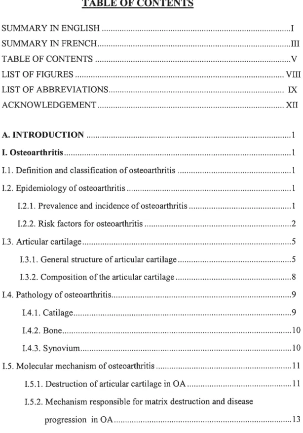
Risk factors for OA include systemic factors and local biomechanical factors (shown in Figure 1).
$ystemicfactors:
Age, Sex, and ethnicity: The most potent systemic vuinerabilities are increasing age and female gender. Disease incidence and prevalence increase dramatically with age. The framingham study found that 27% of those aged 63 to 70 had radiographic evidence of knee OA, increasing to 44% in the over $0 age group (Felson et ai, 1995). Racial factor is another systemic factor with those of Asian people having very low rates ofhip OA (Haq et ai, 2003).
Genetics: OA is a group of clinicaliy heterogeneous disorders. Many genes have been linked to OA. There is most concordance with chromosomes 2q, 4 and 16. Families have been found with rare autosomal dominant pattems of inheritance of OA. The defective genes are often coding for structural proteins of the extraceliular matrix (ECM) ofthe joint and collagen proteins (Haq et ai, 2003).
Hormonal status: Some women afler age 50 deveiop “menopausal arthritis” at the time of menopause. These gender and age reiated prevalence pattems are consistent with a role for post-menopausal hormone deficiency in increasing the risk of OA. Estrogen loss has been strongiy implicated as a risk factor. Epidemiologic studies provide evidence that estrogen replacement therapy is associates with a reduction in the risk of knee and hip OA (Nevitt et al, 1994).
Nutritional factors: Peopie in the lower vitamin C and vitamin D blood levels had a threefold risk of progression of knee OA (felson et ai, 1995). Vitamin C protects against damage by reactive oxygen species and it serves as a cofactor for enzymes contributing to type II collagen synthesis. Vitamin D sufficiency is necessary for active bone turnover which may be critical in OA (Brandt et ai, 2003).
Local biomechanicalfactors.
Obesity: This is the strongest modifiable risk factor. Three to six times the body weight is transferred across the knee joint during walking. Any increase in weight should be muitipiied by this factor to estimate the excess force across the knee joint when an overweight patient walks. Population-based studies show that overweight persons are at higher risk of OA than non-overweight control. In the framingham study, women who lost an average of 11 lbs decreased their risk for knee OA by 50% (Felson et ai, 1992).
Major joint injury: With a major joint injury, a person cari sustain permanent damage of many of the structures within a joint. This damage alters the biomechanics of the joint, increases stress across particular areas of the joint and oflen dramaticaiiy increases the risk of OA. The framingham study found men with a history of knee injuries had a relative risk of 3.5 for subsequent knee OA; for woman the relative risk xvas 2.2 (Feison, 1990). Prior joint surgery also is a risk factor in OA deveiopment.
Occupational and athietic activities: OA is common in those performing heavy physicai work, especiaily if this invoives knee bending, squatting, or kneeling. Dockers and miners have been found to have a higher prevaience of knee OA than those in sedentary jobs (Hunter et ai, 2002). There is a significant relationship between occupational kneeling and repetitive use of joints during work and OA. Competitive
4
athietes are at greater risk for later development of OA. Epidemiologic study has demonstrated that participation in certain competitive sports increase the risk for OA (Buckwalter et ai, 1997). Sports activities that appear to increase the risk for OA inciude those that demand high-intensity, adute, direct joint impact as a resuit of contact with other participants, piaying surfaces, or equipment (Buckwalter et ai, 1997). Repetitive joint impact and torsionai ioading also appear to be associated with joint degeneration.
:Systemic factoi LocalbïomechancaI
Age fadors
Sex Obes•iy
Ethnfc haracteistics Jointinjury
Joint dffGrmity Bone density SusceptibiIty Sports parUdpation Etrogen replacement to osteoarthritis Musde wekness
thetapy (in post menopausai w.ornen) Nutritiona factors ?)
Genetics Site and severity
of asteoarthrîtîs
Othtrsystemic factors
1.3. Articular cartilage
Cartilage is known as elastic cartilage, fibrocartilage or hyaline cartilage, depending on its different physical properties. Articular cartilage is a specialized avascular and neural connective tissue that provides covering for the osseous components of diarthrodial joints. It serves as a load-bearing material, absorbs impact, and is capable of sustaining shearing forces. The unique properties of this tissue are related to the composition and structure of its ECM, which is composed mainly of a high concentration of proteoglycans entangled in a dense network of collagen fibers and a large amount of water.
1.3.1 .General structure of articular cartilage
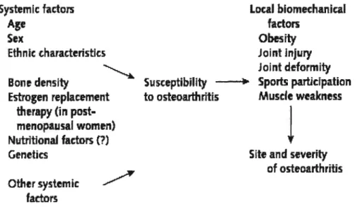
Articular cartilage is organized in a manner that reftects the tensile and compressive force and shear stresses acting on this tissue. This tissue is composed of an extensive ECM synthesized by chondrocytes. It contains different zones with respect to depth from the articular surface and has a regional organization around the chondrocytes. The cartilage is classified into four zones: superficial zone, mid- zone, deep zone and calcified zone (Poole et al, 2001). (shown in Figure 2)
6 SUPERFICIAL ZONE MIDDLE ZONE DEEP ZONE Aggrecan moet cancentrated and collagen content al its Iowest here CalcIIled
r
zone -Superficiel col) protein (also known as lubricin)Decorinand blglycan Pericellular region (decorin Type VI coHagen) Territorial reglon (more intact aggrecan) Interterrilorlal reglon fdegraded aggrecan) Tide mark Type X collagen Hyperirophic chondrocyte Subchondral bone
Figure 2. Diagrammatic representation of the general structure of human articular cartilage from an aduit to show the zones, regions, and relationship with
—-.-- ___4 -—i-- ..-‘--‘. -& ai .aJ Articular surface ‘4—
i.t_
2’
r
-1LL4_:--Subchondrai bone marrowIn the superficial zone the chondrocytes are flattened. The tissue in this region is maximally exposed to the shearing, compressive, and tensile forces of articulation. The collagen fibrils throughout the more superficiai matrix are much thinner and are frequentiy arranged parallel to each other and to the articular surface. Here, the small proteoglycan decorin is most concentrated, being associated with the collagen fibrils, whereas the large proteoglycan aggrecan is present in its lowest concentration. Beiow the superficiai zone is the mid-zone where celi density is iower. The mid-zone consists of rounded celis surrounded by an extensive ECM, rich in the proteoglycan aggrecan. In the deep zone, celi density is at its lowest but aggrecan content and fibril diameter are maximal, although collagen content is minimal. Ceils in this zone are oflen grouped in clusters and resemble the hypertrophic chondrocytes of the growth plate. Adjacent to the deep zone is the calcified zone. The calcified cartilage zone separates the hyaline cartilage from subchondral bone. It appears to serve as an anchor of the cartilage to the bone as collagen fibrils from the radial zone penetrate into the calcified cartilage. In this zone, the celi population is very scarce and chondrocytes are usually smaller.
The cartilage matrix consists of distinct regions sunounding the chondrocytes of articular cartilage. Ail chondrocytes are surrounded by a thin pericellular matrix up to 2im thick that contains few well-defined collagen fibrils, consists mainly of filamentous and fine fibrillar material. A territorial region sunounds this pericellular region that is present throughout the cartilage. In the deep zone, it is well-demarcated from the territorial region by differences in proteoglycan aggrecan structure and composition. This region is called the interterritorial region. It is the part of the matrix most remote from the chondrocytes. Degradation products of aggrecan probably are most concentrated here, produced as a result of incomplete proteolysis and retention of degradation products that retain binding for hyaluronan.
8
1.3.2. Composition ofthe articular cartilage
Condrocytes: Articular cartilage contains oniy one celi type, the chondrocyte. This occupies only approximately 2% of the total cartilage volume in human aduits (Stockweil et al, 1979). The remainder is occupied by an extensive ECM that is synthesized by these celis. This contrasts to fetal and young immature (0—2 years) cartilages where ceil volume is very much higher during growth. With increasing age, there is a progressive decrease in cell content and in matrix synthesis, the latter reaching its iowest point when the individuai is 20 to 30 years (Stockwell et ai, 1979). The chondrocytes are responsible for the metaboiism of ECM.
ECM of cartilage: The ECM is composed of 65 to 80% water. The water content of cartilage plays an important role in maintaining the resiliency of the tissue and contributing to the nutrition and lubrication system. Collagen (mainly type II) accounts for about 15 to 25% of the wet weight. Its concentration is usually progressively reduced with increasing depth from the articular surface. It forms a fiber network that provides the shape and form of the tissue. The proteoglycan content (mainly the very large molecule called aggrecan) accounts for up to 10% of the wet weight. Aggrecan content increases with depth. It is responsible for the compressive properties associated with ioad bearing. The remainder of the matrix is normally accounted for by other collagens including V, VI IX, XI and XIV, link protein and a number of matrix proteins (Koopman et al, 1997).
Collagens: The structural backbone of cartilage matrix is the collagen fibril. It is composed mainly of type II collagen. It also contains type IX collagen and type XI collagen, both within and on the surface of the fibril, and as well leucine-rich proteoglycans, including decorin, fibromodulin, and biglycan (Koopman et ai, 1997). Type II collagen that makes up the bulk of these fibers is specific for cartilage and is the primary collagen of articular cartilage. The fibrils that contain type II collage are composed of tropocollagen molecules (each of which contains a triple lieux of three identical Œ chains), with nonhelical amino- and carboxyl-terminal telopeptide domains. Tropocollagen molecules assemble to form fibrils and larger fibers, stabiÏized by covalent interfibrillar cross-links. The most important mechanical properties of collagen fibres are tensile stifffiess and strength.
Proteoglycan aggrecan: The major non-collagenous component in articular cartilage is proteoglycan aggrecan. It constitutes the second largest portion of solid phase in articular cartilage. This very large molecule consists of a central protein core of 2000 amino acids with several distinct domains and different functions. Its core protein contains three globular domains and two glycosaminoglycan-attachment domains. These domains play various roles to maintain cartilage structure and function. An N-terminal globular domain Gi contains a site for binding with hyaluronan and link protein to form huge aggregates. The link protein that has many structural features similar to the Gi domain of aggrecan stabilizes the complex. The G2 domain is homologous with the major part of the Gi domain. But it does flot interact with HA. The C-terminal, G3 domains contain sequences homologous to the epidermal growth factor, complement regulatory component and a lectin and may be involved in interaction with other ECM glycoproteins. Other domains with important functional properties are the chondroitin sulfate domains, CS1 and CS2. They carry a very large number of negatively charged glycosaminoglycan chains of chondroitin sulfate. An extended protein domain next to the chondroitin sulphate region has a rather specific repeat structure and carnes a number of keratan sulphate chains. Aggrecan provides the compressive stiffness of cartilage. This is achieved by hydration of the large numbers of chondroitin sulfate and keratan sulfate chains that occupy the core protein in the keratan sulfate and chondroitin sulfate nch regions between the G2 and G3 domains. Aggrecan creates a highly hydrated matrix, but the collagen fibrillar network limits hydration and swelling. Thus, aggrecan is only partially hydrated and exhibits a swelling pressure (Koopman et al, 1997). It is this property that endows cartilage with its compressive stiffness and ability to resist deformation and dissipate load.
1.4. Pathology of OA
1.4.1. Cartilage
The pathology of OA reflects both damage to the joint and reaction to the damage. The most striking gross changes are usually seen in the load-bearing areas of articular cartilage. In the earlier stages the cartilage is thicker than normal (Brandt et
10
aï, 2003). Excess mechanical stress induces edema, with stretching and thinning of the superficial layer. Cartilage edema makes the perichondral collagen fibers more susceptible to deformation damage, resulting in increased apoptosis and necrosis of the vulnerable chondrocytes (Hashimoto et al, 1998). The reduction of the chondrocyte population decreases the capacity of the tissue to secrete and maintain matrix proteoglycans, initiating a cycle that accelerates the susceptibility to injury (Brandt et al, 2003). With disease progression, the joint surface thins and the proteoglycan concentration diminishes, leading to softening of the cartilage. The integrity of the surface is lost and vertical clefis develop (fibrillation). With joint motion the fibrillated cartilage is lost, exposing underlying bone. Areas of fibrocartilaginous repair may appear (Brandt et aï, 2003). The chondrocytes replicate, forming clusters called clones. Later, the remaining cartilage becomes hypocellular.
1.4.2. Bone
While loss of articular cartilage represents the pathologic hallmark of OA, remodeling and hypertrophy of bone are also major features. At the beginning, activation of osteoclast-osteoblast system resuits in bone resorption and incremental bone formation. The remodeled bone matrix is more hydrated and less dense than bone more distant from the joint surface. Appositional bone growth occurs in the subchondral region, leading to the sclerosis; with decreased stress, bone resorption leads to osteoporosis (Brandt et aï, 2003). Later in the OA process, with extensive erosion of the cartilage surface, trabecular microfractures may contribute to the stiffening of subchondral bone. Bone cysts form beneath the surface and weaken the osseous support for the overlying cartilage. Growth of cartilage and bone at the joint margins leads to osteophytes, which alter the contours of the joint and may restrict movement.
1.4.3. Synovium
The synovium consists of a single, discontinuous, intimal layer comprised of macrophages (type A ceils) and fibroblasts (type B cells), embedded in connective
tissue containing thin collagen fibrils aligned parallel to the synovial surface. In early phases of OA, edema is within the synovium. As the edema fluid is resorbed, the matrix proteoglycan content increase. With the progression of OA, the synovial lining becomes more continuous as the intimai ceils proliferate and as macrophages migrate into the tissue. In OA effusions, proteolytic enzymes secreted by the synovium act to digest cartilage matrix that has been sheared mechanically from the joint surface (Brandt et ai, 2003).
1.5. Molecular mechanism of OA
1.5.1. Destruction of articular cartilage in OA
Cartilage loss is central to OA. The process of cartilage destruction in OA is basically an error in cartilage homeostasis. Normally, anabolic and catabolic pathways governing the synthesis and maintenance of ECM are in balance. While articular cartilage ECM protein turnover is quite modest under normal conditions, chondrocytes are able to synthesize and integrate into the ECM, those ECM proteins such as proteoglycans, collagen, fibronectin, integrins and other adhesive proteins which enable cartilage to maintain high tensile strength and low compressibility under load throughout the life-span of the individual. Chondrocytes function in response to cytokines and growth factor signais, and to direct physical stimuli, which interact in a complex manner. The end result is a change in the rate of synthesis versus that of enzymatic breakdown of the cartilage matrix, occurring both around the celis and at some distance. Both autocrine and paracrine actions have been demonstrated in chondrocyte and in synovial lining cells. In normal cartilage, there is strict regulation
of matrix turnover: a delicate balance between synthesis and degradation. In OA, this
balance is disturbed, with both degradation and synthesis usually enhanced. However, in OA, this equilibrium between anabolism and catabolism is weighted in favour of degradation.
Degradative proteinases, secreted by articular cartilage chondrocytes, such as
matnx metalloproteinases (MMPs) play a major role in the degradation in OA. These
12
coliagenase- I (MMP- 1), coiiagenase-2 (MMP-8), coliagenase-3 (MMP- 13) and MT 1-MMP (membrane type 1-1-MMP or collagenase 4 or 1-MMP-14) (Shlopov et ai, 1997). Most of these enzyme activities are increased in OA, whether by the mechanism of increased synthesis, increased activation ofproenzymes by other MMPs or plasmin, or decreased inhibitor activity. In nearly ail OA ceils, MMP-3, MMP-8 and MMP-13 were elevated. Many of these MMPs are stimuiated by exposure of the celis to inflammatory cytokines. To agonize the effects of MMPs, expression levels of inhibitors such as tissue inhibitor ofmetalloproteinases (TIMP)-1 are reduced in OA.
In OA, coilagenase is responsible for the breakdown of collagen type II scaffolding in cartiiage. Ail three coiiagenases, collagenase-1 (MMP-I), collagenase-2 (MMP-$), coilagenase-3 (MMP-13) cleaves type II collagen. Coilagenase-3 is the enzyme responsible for most of the collagen degradation (Billinghurst et ai, 1997), which plays the greatest part in the pathoiogy of OA degrading the resident collagen fibrils more remote from the ceil in the territorial and interterritiorial matrix. This coliagenase is also used to remodel matrix in the growth plate. CoÏlagenase-1 is believed to be more involved in the degradation of newly synthesized collagen. The activities of collagenases are clearly increased in both advanced and end stage OA and in the early deveiopment of focal OA lesions. In addition, stromelysin-1 (MMP-3) can cieave in the nonhelical telopeptide of type II and IX collagens (Wu et ai, 1991), leading to disntption of a collagen crosslink. This cleavage could result in a disrnpted fibril structure. Furthermore, type II collagen telopeptide can also be cleaved by MMPs 7, 9, 13 and 14. These findings indicated the presence in OA of a host of enzyme candidates capable of disrupting the collagen network. Disruption of this network will eventually lead to destabilization ofthe joint.
The large proteoglycan aggrecan, is cieaved by different MMPs and is also degraded by a special class of MMPs known as aggrecanases at distinct sites in the core protein. Two aggrecanases (aggrecanase-1 and aggrecanase-2) have been identified as part of the ADAMTS family. These proteinases selectively cleave particular peptide linkages in the G1-G2 interglobuiar domain and are largely responsibie for the turnover of aggrecan in articular cartilage of both normal and OA joints. There is evidence for the activities ofboth types ofproteinases in OA. Analysis
of the proteoglycan aggrecan have revealed that excessive cleavage occurs in OA
cartilage in the core protein, particularly in the intergiobular domain between the G1 and G2 domains (Poole, 1999).
Afier the initial cleavage of type II collagen by collagenases it is denatured and lost. Chondrocyte subsequently undergo further phenotypic change becoming hypertrophic and expressing and secreting type X collagen. This differentiation is normally seen in the growth plates as part of endochondral ossification (Poole et ai, 2001). Calcification of articular matrix also occurs in OA in association with these changes. Thus, chondrocyte differentiation in OA seems to be a response to extensive damage to the collage fubrillar network. Moreover, hypertrophic ceils eventualiy undergo apoptosis. This is commonly seen in OA cartilage.
1.5.2. Mechanism responsible for matrix destruction and disease progression in OA During matrix degradation, excessive catabolism of articular cartilage results in the release into synovial fluid of matrix breakdown products including chondroitin sulfate and keratan sulfate peptides, PG fragments, type II collagen peptides, fibronectin fragments, chondrocyte membranes, etc. ail of which are antigenic and elict an inflammatory response in the synovial membrane (Smith et al, 1997). The activated synovial macrophages in the membrane release cytokines (IL-1, TNf-Œ, etc), PGE2, proteinases and oxygen free radicals (superoxide, nitric oxide (NO)) into adjacent tissues and the synovial fluid. These mediators in tum can act on chondrocytes and synovial fibroblasts, modifiing their biosynthesis of PGs, collagen and hyaluronan as well as promoting release of catabolic mediators. Some of the matrix breakdown products are known to induce the expression and secretion of MMPs and prodegradative cytokines sucli as IL-1 and TNF-Œ. There is increased expression in OA chondrocyte of the IL-i and its receptors (Meichiorri et ai, 199$; Martel-Pelletier et ai, 1992). TNf-Œ is also upregulated in OA (Melchiorri et ai, 199$) and TNf-Œ receptors show increased expression when compared to normal cartilage (Webb et ai, 1997). These cytokines derived from chondrocytes and the synovial lining play a key role in cartilage degeneration in OA. Cytokines are responsible for
14
accelerating the destruction of cartilage ECM via their ability to up-regulate metalloproteinase gene expression. They also serve to suppress compensatory ECM protein biosynthesis by chondrocytes. Another pathway involving the induction of NO in cartilage by cytokines appears relevant to programmed ceil death (apoptosis) and OA pathology (Lotz, 1999).
While cytokines are clearly important in up-regulating MMP gene expression, other pathways relevant to the process include potent biological activity of fibronectin fragments. fibronectin fragments enhance levels of catabolic cytokines and also up regulate MMP expression, significantly enhance loss of proteoglycans from cartilage and transientÏy suppress proteoglycans synthesis (Malemud, 1999). Changes in ECM loading can also induce ECM cleavage as well as changing the synthesis of ECM macromolecules (Poole, 1999). The pathological changes in cartilage ECM in OA are likely to resuit in a disturbance ofthe normal balance between mechanical loading and direct cytokine/growth factor signalling changing gene expression. Figure 3 summarizes some metabolic pathways responsible for cartilage degradation in OA joint.
figure 3. Summary of some the metabolic pathway responsible for cartilage degradation in OA joint (Studer et ai, 2000).
16
II.
Prostagla ndins biosynthesis pathway II.1.Biosynthesis of eicosanoids:11.1.1. Eicosanoids
The term eicosanoid refers to any twenty-carbon (C20) fatty acid. Prostaglandins, thromboxanes, leukotriene and lipoxins are related compounds known as eicosanoids, which have a large variety of biological activities. Most eicosanoids are biosynthesized from C20 polyunsaturated fatty acids, primarily arachidonic acid (AA). AA is the most plentiful C20 polyunsaturated fatty acid in most mammals (Zubay, 198$).
11.1.2. Release ofAA and phospholipase A2 (PLA2) enzymes
AA is a 20-carbon unsaturated fatty acid distributed throughout the lipid bilayer of ail mammalians. It is derived directly from the diet or via modification of linoleic acid, and normally is stored in the ceil membranes, esterified in the sn-2 position of phospholipids (Irvine, 1982). Under normal conditions, the level of ftee AA is low, but upon stimulation, AA is released by the hydrolytic action of PLA2 enzymes (involving secretory, cytoplasmic or both types of PLA2. AA is metabolized tbrough oxygenation by three enzymatic pathways in mammals. Through cyclooxygenase (COX) pathway, AA is converted to prostaglandin H2 (PGH2). PGH2 is then converted to PGE2, PGF2, TXA2, PGD2, or PGL2. Through the 5-lipoxygenase (LOX) pathway, AA can be converted to a leukotriene A4 (LTA4). LTA4 can be further metabolized into various leukotrienes and monooxygenase pathway leads to a series of epoxy-and hydroxy-acid derivatives. Many PLA2 enzymes are active within the cell or in the close vicinity and have distinct, but interconnected roles in AA release. At least 19 PLA2 enzymes have been indentified in mammal, amongst which the cytosolic PLA2 (cPLA2), secretory PLA2 (5PLA2) and Ca2+-independent PLA2 (iPLA2) families have been implicated in eicosanoid production (Kudo, 2002).
11.2. Prostag]andins biosynthesis
Prostaglandins are small lipophilic molecules that are produced by a variety of cell types in response to both physiological and pathological stimuli. The general pathway for the biosynthesis of prostaglandins is illustrated in figure 4. The first step
in the pathway for the biosynthesis of prostaglandins involves intracellular release of AA from plasma membrane phospholipids via the action of PLA2. AA is then converted sequentially to PGG2 and PGH2 by the COX and peroxidase activities of a single enzyme, PGH synthase (also called COX). There are two forms of PGH synthase, a constitutive form (COX-1) and an inducible form (COX-2). Different terminal synthases then convert PGH2 to the 5 primary prostaglandins: thromboxane
A2 (TXA2), PGD2, PGE2, PGF2Œ, or prostacyclin (PGI2). PGD2 gives rise to the
important derivatives 9a, 1113 PGF2 and the J-series PGs including PGJ2, A12PGJ2 and 15d-PGJ2, the latter through a series of non-enzymatic steps. Additional active and inactive prostaglandins also denve from further isomerization of PGE2, PGF2a, PGJ2 and TXA2. The resulting products then exit the ceil via a carrier mediated process to activate G protein-linked prostanoid receptors or in some cases may interact with nuclear receptors.
Physiological actions of PGf2Œ, PGI2 TXA2 , PGD and PGE2 series
prostaglandins are mediated by binding to specific high afflnity G-protein coupled cell surface prostanoid receptors.There is one PGF receptor termed FP, one PGI receptor termed IF, one TXA receptor termed TP receptors, and two PGD receptor termed DP and CRTH2. PGE has 4 separate receptors, termed EP1-EP4, each encoded by a distinct gene. Depending on the receptor, the consequence of ligand binding to these receptors can be increased cyclic AMP, decreased cyclic AMP, or a phosphoinositide response (Narumiya, 1995). In addition to plasma membrane receptors, recent evidence shows that prostanoids also can bind and signal through nuclear hormone receptors, PPARs. Three distinct PPAR isoforms- PPARx, 13/6 and y- have been isolated and characterized. PPARy binds some AA metabolites, especially the PGD2 metabolites such as 15d-PGJ2. PPARct and 6 bind a stable analog of PGI2, carbaprostacyclin (cPGI).
Membrane Phospholipid 1 5d-PGJ2 Phospholipase A2
NSAIDs
TXA2
cox-1 COX-2 PGfPGF2
synthasePGD2
I
PGE2
DehydrationPGI2
The nature of the final active product depends on the celi type, the stimulus, and the presence of distinct PG synthase. TXA2 synthase (TXAS) is present in a platelets and macrophages, prostacyclin synthase (PGIS) is present in the utems, two type of PGD2 synthase (PGDS) are found in brain and mast ceils, and enzymes responsible for the isomerization of PGH2 to PGE2 are expressed in synovial fibroblasts (Stichtenoth et ai, 2001).
Prostaglandins play critical roles in numerous biological prosesses, including kidney development, reproduction, bone metabolism, inflammation, maintenance of gastrointestinal integrity, angiogenesis, modulation of immune responses, apoptosis and mitogenesis. In contrast to some homones, which are released from a specific site but have broad systemic effects in distant organs, prostaglandins are synthesized in broad range of tissue type and serve as autocrine or paracrine mediators to signal changes within the immediate environruent.
PGE2 produced is released from the celis and act on four types of the PGE receptors, EP1, EP2, EP3 and EP4, ail of which are coupled with the trimeric G-protein signalling. PGE2 plays crucial roles in various biological events, such as neuronal functions, female reproduction, vascular hypertension, tumorigenesis, fever, gastric mucosal protection, pain hypersensitivity, anti-allergic response and inflammation associated bone resoption.
Overproduction of PGE2 is oflen associated with various diseases. Elevated production of PGE2 plays an important role in the pathogenesis of arthritis. Several studies suggest that PGE2 is the major prostaglandin (PG) produced by articular joint celis and is involved in inflammation, apoptosis, angiogenesis, and tissue degradation that characterize arthritic diseases. The induction of cartilage degradation by PGE2 is due to the inhibition of collagen synthesis, induction of MMPs production and induction of chondrocyte apoptosis. PGE2 is largely produced in arthritic joint tissues and excessive production of PGE2 has been reported in serum and synovial fluids of rheumatoid arthritic and osteoarthritic patients. Treatment with neutralizing anti-PGE2 antibodies prevents acute and chronic inflammation in a rat adjuvant arthritis model (Portanova et ai, 1996). Mice lacking COX-2 or PGE2 receptors display reduced incidence and severity of collagen-induced arthritis. These animais showed reduced
20
inflammation and less cartilage and bone destruction (Myers et ai, 2000). The role of PGE2 in arthritis is also supported by effective suppression of pain and inflammatory responses in arthritis by nonsteroidal antiinftammatory drugs (NSAIDs) that reduce PGE2 biosynthesis (Crofford, 2002).
11.3. COXs
COX, also called PGH synthase, is a heme-containing enzyme that catalyzes the first two steps in the biosynthesis of the prostaglandins from the substrate AA. Two sequential enzymatic reactions are the bis-oxygenation of AA leading to production of PGG2 (COX reaction) and reduction of 15-hydroperoxid of PGG2 leading to formation of PGH2 (hydroperoxidase reaction). Three COX isoforms, COX-1, COX-2 and COX 3, are found in mammals. COX enzyme was first purified in 1976 and cloned in 1988 (Merlie et ai, 1988). In the early 1990s, COX was demonstrated to exist as two distinct isoforms. COX-1 is constitutively expressed as a “housekeeping” enzyme in most tissues. COX-2 is flot constitutively expressed in appreciable amounts by most normal tissues, but is rapidiy induced by proinflammatory cytokines, tumor promoters, oncogenes, and growth factors. COX-3 was recently identified and shown to exhibit the catalytic features of COX-1 and COX-2. COX-1 and COX-2 have very different expression profiles in several physiological processes. The COX isozymes are also involved in pathological processes. COX-1 is involved in thrombosis, while COX-2 mainly involved in inflammation, pain, fever, angiogenesis, cancer, Alzheimer’s disease and several forms of arthritis. COX-l and COX-2 are of particular interest because they also are the major targets ofNSADs including aspirin, ibuprofen, and the new COX-2 inhibitors.
11.3.1. Gene structures and expression ofCOX isoforms
COX-1 and COX-2 have a molecular weight of 71 KDa and are almost identical in length. The COX monomer consists of three structural domains: (a) An N-terminal epidermal growth factor (EGF)-like domain of 50 amino acids at the N terminus. The EGf-like domain may play a role in the integration of maturating COX into the lipid bilayer. (b) A membrane-binding domain of about 50 amino acids. The membrane-binding domain contains four short, consecutive and amphipathic a
helices. This creates a hydrophobic surface that would interact with the one face of the lipid bilayer, allowing COX enzymes to integrate into membranes through the monotopic mechanism (Picot et ai, 1994). (c) A large C-terminal giobular catalytic domain (about 460 amino acids) with a heme-bonding site. This domain is almost entireiy comprised of Œ-helical structure, shares a great deal of structural simiiarity to myeloperoxidase. The C-terminal PTEL and STEL sequences in COX-1 and COX-2, respectively, represent an ER retention signai. The major sequence differences between COXs isoforms occur in the membrane binding domain (Spencer et ai, 1999). A unique difference between COX-1 and 2 is 1$ amino acids inserted 6 residues in from the C terminus of COX-2 that are not present in COX- 1. Mature, processed COX-1 contains 576 amino acids; the mature form of COX-2 contains 587 amino acids. There is a 60%-65% sequence identity between COX-1 and 2 from same species and 85%-90% identity among individual isoforms from different species. However, the gene for COX-1 is approximateiy 22 kb in length with 11 exons and is transcribed as a 2.8 kb mRNA, whereas that for COX-2 is approximately 8.3 kb in length with 10 exons and is transcribed as 4.4 kb mRNA (Tanabe & Tolmai, 2002). The COX-1 and COX-2 genes map to human chromosomes 9q32-q33.3 and 1q25.2-q25.3, respectively (Tanabe & Tohnai, 2002)
COX-1 and COX-2 have significant sequence homoiogy and identical catalytic activity, but their expression pattem is markedly different. COX-1 has been found in neariy ail tissues under basal conditions and is thought to piay a ‘housekeeping’ role. Nevertheiess, COX-1 is preferentiaily expressed at high ievel in selected ceils and tissues, inciuding endothelium, monocytes, plateiets, renai coilecting tubuies, and seminal vesicles, indicating that it is deveiopmentaiiy reguiated (Smith et ai, 2000). In contrast to COX-1, ievels of COX-2 are typicaiiy low or absent in most tissues. However, COX-2 can be induced by severai physiologicai and proinflammatory stimuli, including IL-1, TNF-a, lipopolysaccharides (LPS), transforming growth factor
(TGF)-F3, epidermai growth factor (EGf), piatelet-derived growth factor (PDGF), and
fibroblast growth factor (FGF) and hormones in many ceil types like macrophages, monocytes, synoviocytes, chondrocytes, osteobiasts, leukocytes and endothelial celis (Dubois et ai, 1998). The induction of COX-2 is usualiy transient, with a retum to base
22
une within 24-48 hours (Williams et ai, 1999). COX-2 is expressed constitutively in the brain, kidney, during ovulation and blastocyst implantation.
11.3.2. Regulation of COX-2 expression
The COX-1 promoter region lacks a functional TATA or CAAT box ami is GC rich, which is consistent with a housekeeping gene. There are several putative transcriptional regulatory elements in the promoter region of the COX-1 gene, such as two Sp-i sites, two AP-2 sites, NF-IL-6 site and GATA. The two Spi sites contribute to constitutive expression of COX-1 (Xu et ai, 1997). To date these Spi sites are the only cis-acting elements documented to regulate transcription of COX- 1.
The promoter of the COX-2 gene contains a TATA box and various transcription elements, such as NF-IL-6, AP-2, Spi, NF-kB, CRE and E-box. $o far, only a limited class of elements are shown to be involved in the regulation of COX-2 transcription, ofien in synergy, such as E-box, ATF/CRE sequences, NF-1L6 CAAT enhancer binding sequence(C/EBP) and two NF-kB binding sites. The transcription factors that bind and activate COX-2 transcription involved C/EBP3 and C/EBPa for the NF-IL-6 elements, AP-1, ATF and CREB for the CRE elements, and USF-1 for the E-box. Dependence on Nf-kB signalling for COX-2 expression has been demonstrated by use of pharmacologie inhibitors offlcB kinase (Gallois et ai, 1998).
The signalling pathways that mediate COX-2 expression are tissue-specific and depend on the stimulus. A number of signalling pathways are likely to regulate transcription of COX-2. These include NF-kB and C/EBP, two common signalling pathways in inftammatory response, and three mitogen-activated protein kinase (MAPK) signalling cascades, ERK1/2, INKISAPK, and p38. Each of these signalling pathways has been shown to contribute or be solely required for increased expression of COX-2 in one or more cultured ceil systems.
The MAPK cascade is a very important signalling pathway for COX-2 expression and consists of three different subgroup of kinase (ERK: extracellular regulated kinase, INXISAPK: Jun N-terminal kinase/stress activated protein kinase, and p38: p38 mitogen-activated protein kinase). The ERKs are mainly activated by growth factors and oncogenes including v-src and v-ras. Cellular responses to mitogens are generally mediated by sequential activation of receptor tyrosine kinases, Src, Ras, and
one or more of the MAPK pathways. Expression of COX-2 following stimulation with serum and PDGF, or in response to v-Src or Ha-Rasexpression, requires activation of the ERK1/2 and JNKJSAPK pathways (Xie & Herschman, 1995; Sheng et ai, 1998). The COX-2 gene has been shown to be an important Ras target since oncogenic mutation in Ras and overexpression of COX-2 is found in many forms of human cancers, including breast cancer and colorectal carcinoma (Subbaramaiah et ai, 1996).
The JNXISAPK and p38 pathways are activated by inflammatory stimuli, including IL-1f3, TNF-a, and LPS, as well as the phorbol ester TPA and environmental stress, like oxidative stress. Proinflammatory cytokines including TNF-Πand IL-1
f3
have been shown to selectively activate INK ami p38 MAP kinase in cultured human articular chondrocytes (Geng et aI, 1996). Several studies demonstrated that the selective p38 MAPK inhibitors prevented IL-1 f3-induced COX-2 expression in human synovial fibrobÏasts (Faour et ai, 2001) and chondrocyte ceÏi une (Thomas et ai, 2002).The NF-icB pathway is a common mediator of inflammatoiy responses and plays an important role in COX-2 expression in several celi types, including rheumatoid synoviocytes (Crofford et aÏ, 1997). The COX-2 promoter contains two consensus sequences for the cis-acting regulatory sequences that are recognized by the NF-icB family of transcription factors. IL-1 f3 treatment of human synovial fibroblast induced binding of the
p65-p5O
heterodimer and the p50 homodimer to the COX-2 promoter, and pre-treatment of the ceils with NF-icB p65 antisense oligonucleotides prevented NF-icB binding and markedly decreased COX-2 expression (Crofford et ai, 1997).furthermore, transfection experiments with reporter plasmid demonstrated that
mutations in the NF-KB cis-regulatory sites attenuate transcriptional activation of the COX-2 promoter in response to TNF-Πstimulation (Yamamoto et ai, 1995). Nf-KB regulates COX-2 expression in response to the appropriate activators in specific celI types.
The C/EBPf3 and C/EBP transcription factors are commonly involved in the regulation of inflammatory responses. This family of transcription factors is activated by most of the inflammatory stimuli that induce COX-2 expression. An NF/IL-6 regulatory element is present in the COX-2 promoters ftom ail species examined and C/EBP proteins have been shown to bind to these promoter sequences. In human
24 synovial fibroblasts, TNf-a induced c/EBP binding to COX-2 promoter in addition to NF-icB (Alaaeddine et aI, 1999). The C/EBP transcription proteins appear flot to work independentiy but instead to cooperate with USF-1, NF-icB and c-jun to activate COX
2 transcription (Morris & Richards, 1996).
Dexamethasone is a common anti-inflammatory steroid, which binds to the gïucocorticoid receptor and activates transcription of a number of genes via glucocorticoid response elements (GREs). Dexamethasone is an efficient suppressor of inftammatory-induced COX-2 expression aithough the COX-2 promoter does flot contain GREs. The mechanism for glucocorticoid-mediated repression of COX-2 induction invoïves suppression of the AP-1 and NF-KB-dependent transcription (Scheinman et ai, 1995; Yang-Yen et ai, 1990) and destabiiization and degradation of COX-2 mRNA and protein (Newton et ai, 199$). The nuciear receptor PPARy lias been shown to down-reguiate the expression of COX-2. PPARy reguiate gene expression by binding their heterodimeric partner retinoid X receptor to specific PPAR-responsive elements (PPREs). The promoter region ofthe human COX-2 gene harbors a PPRE at —
3721 to —3707 bp (Meade et ai, 1999). 15d-PGJ2, a PPARy iigand, covalently binds to
1KB kinase, leading to inactivation of the NF-icB pathway and thereby to repression of
COX-2 transcription (Straus et ai, 2000). On the contrary, other studies have shown that PPARy augments COX-2 transcription by binding to the PPRE element in the COX-2 promoter (Meade et ai, 1999).
11.3.3. Biological function ofCOX-2
COX-2 and COX-1 have significant differences in tissue expression and regulation. Therefore they have different biologicai function. COX-2 is associated with inflammation and many malfunctions.
COX-2 lias some role in regulating brain function. High basal levels of COX-2 are found in the brain. In the centrai nervous system (CNS), COX-2 is up regulated by neural activity. COX-2 protein or mRNA was detected in neurones and in the nonneuronal celis of the CNS (Yamagata et ai, 1993). These suggested that COX-2 enzyme might be invoived in CNS function. Proinflammatory cytokine IL-1 Ç3 was shown to be the major inducer of COX-2 up-reguiation in the CNS. Intraspinal
administration of an interleukin-converting enzyme or COX-2 inhibitor decreased inflammation-induced central PGE2 levels and mechanicai hyperalgesia. These indicated that IL-1 13-mediated induction of COX-2 in the CNS contributed to inflammatory pain hypersensitivity (Samad et ai, 2001). Fever is thought to be the effect of PGE2 in CNS. COX-2-knockout mice suppressed both fever and PGE2 level in the CNS (Li et aI, 1999), implying that PGE2 involved in the febriie response may drive from COX-2.
Prostaglandins (PGs) are involved in normai renal function including control of renin release, control of tubular function and regulation of vascular tone. In COX-2 nuli mice, the kidneys fail to develop normaliy resulting in death, whereas COX- 1 nuil mice fail to produce a detectabie renal pathology (Morham et ai, 1995). Additionaily, the involvement of COX-2 in renai functions was also suggested by ciinical studies. Clinical studies showed that the COX-2 inhibitors, similar to other NSAIDs, cause qualitative changes in urinary prostaglandin excretion, glomerular filtration rate, and sodium retention. (Brater et ai, 2001). Thus, COX-2 may play a role in physiological renal functions.
Ovulation, the process by which oocytes are reieased from the preovulation follicle in the ovary, is accompanied by induction of prostaglandin synthesis as a consequence of the LII surge. This marked response ied to the first observation of COX-2 induction during a normal physiological event. The induction of COX-2 is necessary for the successful rupture of the follicie, probabiy mediating directiy the generation or activation of proteolytic enzymes necessary for this process (Isafriri, 1995). Afier fertilization, COX-2 appears to mediate the embryo-uterine interactions during implantation. COX-2 nuil mice show multiple failures in reproduction ftinction, inciuding ovulation, fertilization, implantation, and decidualization, underscoring the multiple roies of PGs during these processes (Lim et ai, 1997).
Inflammation and arthritis: Studies using animal modeis of inflammation artbritis have provided evidence that increased expression of COX-2 is responsible for increased prostaglandin production seen in inflamed joint tissues (Anderson et ai, 1996). COX-2 induction lias been observed in human OA-affected cartilage (Amin et ai, 1997). Other studies found that COX-2, not COX-1, expression was elevated in a disease-related pattem in synovial tissues from patients with rheumatoid arthritis (RA),
26
ankylosing spondylitis, psoriatic arthritis, and OA (Siegie et ai, 1998). The proinflammatory agents IL-1, TNF-a and LPS, as well as the growth factors TGf-j3, EGF, PDGF, and fGF, have ail been shown to induce COX-2 expression in primary culture ceils derived from human synovial tissue or catilage. On the other hand, the anti-inflammatory cytokines IL-4 and IL-13, as well as the immunosuppressive glucocorticoids, are shown to decrease COX-2 levels (Crofford, 1997).
COX-2 enzymes also play functional roles in tumorigenesis. High levels of constitutive expression of COX-2 have been found in various cancer ceils and tissues, and studies employing overexpression, antisense suppression, and specific inhibitors of COX-2 have demonstrated that COX-2 contributes to the progression of severai types of cancer. Both human and animal colorectal tumors express high ievels of COX-2 (Eberhart et ai, 1994). There is a reduction in the relative risk of colorectai cancer in individuals taking N$AIDs (Mamett, 1992). Suppression of tumorignesis in COX-2 (-I
)
mice has confirmed epidemiological studies, demonstrating that N$AIDs suppress the incidence of colon cancer (Oshima et ai, 1996). COX-2 may biunt the apoptotic response in tumor cells and may play a roie in the regulation of angiogenesis associated with neopiastic tumour celis (Tsujii et ai, 1998).The cytoprotective actions of prostaglandin preventing gastric ulceration are mediated by endogenous prostacyclin and PGE2, which reduce gastric acid production, stimulate gastric fluid secretion, increase secretion of viscous mucus and exert a direct vasodilator action on gastric mucosa. Classical NSAID use causes a variety ofproblems in the gastrointestinai tract, including irritation and ulceration of the stomach lining. The primary mechanism of NSAIDs in the treatment of inflammation is the inhibition of both COX-1 and COX-2. COX-2 mediates the inflammatory response. An NSAID that inhibits COX-2 seiectively should decrease inflammation but flot influence normal physiologic functions and thus should cause fewer gastrointestinal side effects. Selective COX-2 inhibitors are widely used. Rofecoxib and the novel COX-2 inhibitors etoricoxib and valdecoxib have a higher degree of COX-2 selectivity than traditional NSAIDs. However, rofecoxib induces thromboembolic adverse effects more frequently than classical NSAIDs. Caution is warranted regarding the use of these drugs (Evensen et al, 2005).
11.4. PGE Synthase (PGES) 11.4.1. The MAPEG-supperfamiiy
A widespread superfamily MAPEG (membrane-associated proteins in eicosanoid and glutathione metabolism) lias been defined according to enzymatic activities, sequence motifs, and structural properties. A total of 136 proteins belonging to the MAPEG superfamily were found in database and genome screenings. Ail MAPEG proteins have similar molecular masses of 16-1$ kDa and, except 5-lipoxygenase-activating protein (FLAP). Ail MAPEG proteins have similar three dimensionai and membrane-spanning topographie properties (Jakobsson et ai, 2000). Multiple sequence alignments of human MAPEG members reveal six strictly conseiwed amino acids. The family consists of six human proteins including fLAP, leukotriene C4 (LTC4) synthase, microsomal glutathione $-transferase 1 (MG$T1), MGST2, MGST3, and MGST1-like 1 (MGST1-L1 or PGES). The genes encoding these proteins ail reside on different chromosomes. In addition, several nonmammalian members have been identified, including those from plants (Arabidopsis thaliana, Oryza sativa, and Ricinus communis), fungi (Aspergillus nidulans), and bacteria (Synechocystis sp. [SynMGST], Escherichia cou, and Vibrio cholerae)( Jakobsson et ai, 2000).
On the basis of the multiple sequence alignments, MAPEG family can be subdivided into four subgroups. The first subfamily consists of the members FLAP, LTC4 synthase, and MGST2 and is important for leukotriene biosynthesis. The second subfamily consists ofMGST3 together with the members found in plants and flingi. The third subfamily is composed of the proteins identified in bacteria (E. cou and V. cholerae). The human MGST1 and MGST1-L1 proteins constitute a fourth subgroup possibly involved in cytoprotection. (Jakobsson et ai, 2000).
11.4.2. PGES identification
Metabolism of AA by COX yields only the unstable intermediary PGH2, which can be further metabolized into PGE2, PGD2, PGf2, PGI2, or TXA2. The enzyme responsible for the isomerization of PGH2 into PGE2 is PGES, the terminal enzyme responsible for PGE2 synthesis. PGE$ activity, in most cases glutathione (G$H) dependent, bas been detected both in microsomal and cytosolic fractions of various ceils, more than one form of PGES exist. At least four distinct PGES isoforms have
28
been identified, including cytosolic PGES (cPGES), GST.t, microsomai PGES-1 (mPGES-1), and mPGES-2.
Human mPGES-1 is a member of the MAPEG superfamily. It was initialiy discovered and identified as a homologue of MGST1 with 38% identity on the amino acid sequence level. The protein thus was referred to as MGST1-iike 1(MGST-L1). The same protein was also identified as a p53-induced gene and referred to as PIG12. In 1999, Jakobsson et al first reported mPGES-1 (Jakobsson et ai, 1999). The recombinant human microsomal GST-l-like 1 (MGST1-L1) bas an ability to catalyze the coversion of PGH2 to PGE2 with strict substrate specificity. Then this protein was cloned from several animal species and shown to represent a long-sought membrane bound form of PGES. This enzyme now is called mPGES-i. Soon after mPGES discovery, two cytosolic forms of PGES were purified and identified. One enzyme termed cytosolic PGES (cPGES). cPGES is a 23 kDa cytosolic protein and identical to p23, a heat shock protein 90 (Hsp9O)-associated protein (Tanioka et ai, 2000). The other is a member of the ,t form of the cytosoiic GST family. In 2002, the second form of membrane
associated PGES, termed mPGES-2 was identified.
11.4.3. mPGES-1 gene structure and catalytic function:
The primary structures of mPGES- 1 proteins of various animal species reveai a high degree of sequence homology ($0%). mPGES-1 aiso shows significant homology with other MAPEG superfamily proteins, including MGST-1, MGST-2, MGST-3, FLAP and LTCS, with the highest homology being found with MGST-1 (‘-40%) (Jakobsson et al, 1999).
The gene for human mPGES-1 maps to chromosome 9q34.3 is divided into three exons and two introns, and span 14.8 kb. Exon-intron junctions follow the GT AG mie except for the 5’-site of intron 2, which consists of GC instead of GT. The intron sizes are 4.2 kb and 8.8 kb respectively (Forsberg et ai, 2000). The gene structure of mPGES is similar to its ciosest relative MGST1 with regards to exonlintron borders and differs from other MAPEG members that contain additionai exons. The cDNA for human mPGES-1 encodes a protein composed of 152 amino acid residues (—16 kDa). Mutation of Argi 10, in mPGES-1, which is the residue strictly conserved in ail
MAPEG proteins, abrogates its catalytic activity, indicating an essentiai role of this residue (Murakami et ai, 2000). The cofactor GSH is absoiutely required for mPGES-1 enzymatic activity. GSH invoives in detoxification reactions with hydrogen peroxide and organic peroxides and aiso has a stabilizing effect on solubiiized mPGES-1.
mPGES-1 activity was inhibited by the COX-2 inhibitory NSAffl NS-398 and sulindac sulfide with 1C50 values of 20,80 .iM. rnPGES-1 was aiso inhibited by MK 866, an inhibitor ofFLAP and LTC4 with 1C50 values of 1-5 tM (Mancini et ai, 2001). MK-866 binds to the AÀ-binding region of fLAP, which is highiy conserved in LIC4 and mPGES-1 and couid possibly be involved in the binding ofeicosanoids (Mancini et al, 2001). furthermore, 15d-PGJ2 and some poiyunsaturated fatty acids were also reported to inhibit the activity ofmPGES-1 (Quraishi et ai, 2002). mPGES-1 activity is flot inhibited by ciassicai cytosolic GST inhibitor, in contrast to cPGES.
11.4.4. features ofmPGES-1 promoter
The human mPGES-1 promoter is GC rich and lacks a TATA box at a functionai site and contains numerous potentiai transcription factor binding sites, inciuding two GC-boxes, two tandem Barbie boxes and an AHR (aryl hydrocarbon response element). The putative promoter region of mPGES-1 was shown to be transcriptionaiiy active and was inducted by IL-1
f3
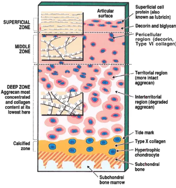
and down-reguiated by Phenobarbital (forsberg et ai, 2000).The mouse mPGES-1 promoter contains severai transcription factor binding sites (figure 5), including two tandem GC-boxes, C/EBPa and
-f3,
AP-1, and three GRE and two progesterone receptors (PR). The tandem GC box sequences in the mPGES promoter play a major role in reguiating its inducibie transcription. Egr-1 (eariy growth response factor-1), an inducibie zinc finger protein that recognizes the GC-rich consensus DNA sequence 5’-GCG(TIG) GGGCG-3’ binds to the proximal GC box in the mPGES promoter region and faciiitates inducibie transcription of the mPGES gene. Egr-1 gene is rapidly and transientiy induced by a variety of stimuli (TPA, cytokines, and LPS) or celiular stresses. Cytokine-induced mPGES-1 expression was reguiated by Egr-1 (Naraba et ai, 2002).30
The sequence of the mouse and human mPGES promoters (-1 to —640) is oniy 48% homologous, but, the homology between them around the tandem GC boxes (-70 to —124) is relativeiy high 78%. Thus, the tandem GC boxes are critical for transcriptional activation of both the human and the mouse mPGES gene (Naraba et ai, 2002).
EgT-1
‘4,
mPGES-1 gene
Figure 5. Regulatory eiements in the mouse mPGES-1 promoter. Egr-1 binds to the proximai GC-box and triggers mPGES-1 transcription