Université de Montréal
Angiopoietin like-2: a pro-inflammatory and pro-oxidative
protein that contributes to endothelial dysfunction
par Carol Yu
Département de Pharmacologie Faculté de Médicine
Thèse présentée à la Faculté de Médicine en vue de l’obtention du grade de Doctorat
en Pharmacologie
Août 2014 © Carol Yu, 2014
Université de Montréal
Faculté des études supérieures et postdoctorales
Cette thèse intitulée:
Angiopoietin like-2: a pro-inflammatory and pro-oxidative
protein that contributes to endothelial dysfunction
Présentée par: Carol Yu
a été évaluée par un jury composé des personnes suivantes:
Dre Hélène Girouard, président-rapporteur Dr Eric Thorin, directeur de recherche Dr Jean-Sébastien Joyal, membre du jury
Dr Ismail Laher, examinateur externe
ABSTRACT
Vascular aging is characterized by changes in the endothelium. Common cardiovascular risk factors, including obesity and hypertension, predispose the endothelium to increased oxidative stress, leading to endothelial dysfunction commonly characterized by diminished nitric oxide bioavailability. Although endothelial function can be a major determinant of cardiovascular risk prediction in patients, individual testing is still limited in clinical settings and thus there is increasing scientific interest in finding better biomarkers.
Angiopoietin like-2 (angptl2), a recently identified protein, is a pro-inflammatory and pro-oxidative protein involved in chronic pro-inflammatory disorders ranging from obesity to atherosclerosis. As inflammation and increased oxidative stress are established underlying mechanisms by which endothelial dysfunction occurs, this work focuses on the role of angptl2 in endothelial dysfunction, a topic that is largely unexplored. Specifically, this work aims to 1) determine the acute effects of angptl2 on endothelial function, 2) characterize endothelial function and contribution of different endothelium-derived relaxing factors in various vascular beds in a newly generated angptl2 knock-down (KD) mouse model, and 3) examine whether the lack of angptl2 expression protects against endothelial dysfunction induced by either a high-fat diet (HFD) or angiotensin II (angII) infusion in mice.
In the first study, we show that a recombinant angptl2 acutely evokes endothelial dysfunction in the femoral artery isolated from wild-type (WT) mice, likely due to increased production of reactive oxygen species. Also in the femoral artery, angptl2 KD mice display better endothelial function compared to WT, which may be a result of greater prostacyclin contribution to vasodilation. After a 3-month HFD, the main respective endothelium-derived relaxing factors in the femoral and mesenteric arteries are preserved in angptl2 KD mice only, which was associated with a better metabolic profile, such as lower total cholesterol-to-high-density lipoprotein and low-density-to-high-density lipoprotein ratios compared to WT mice. After a HFD, KD mice have less triglyceride accumulation in the liver and smaller adipocytes in their mesenteric and epididymal white adipose tissues compared to WT
mice, while inflammatory gene expressions in adipose tissues increase in WT mice only.
In the second study, we reveal that the lack of angptl2 in KD mice results in greater nitric oxide production compared to WT mice in their isolated cerebral arteries. Chronic infusion of pro-inflammatory and pro-oxidative angII results in cerebral endothelial dysfunction only in WT mice, which is acutely ameliorated with either N-acetylcysteine, apocynin, or indomethacin, suggesting increased reactive oxygen species, likely derived from the NADPH oxidases 1/2, and increased cyclooxygenase-derived endothelium-derived contracting factors. In contrast, apocynin reduces cerebral dilation in angII-treated KD mice, suggesting recruitment of a potential compensatory dilatory NADPH oxidase pathway.
These studies are the first to explore angptl2 contribution to endothelial dysfunction in different vascular beds, and strongly suggest that angptl2 can directly impair endothelial function by its pro-inflammatory and pro-oxidative properties. Translating this to the clinical setting, expression levels of angptl2 may be an indicator of endothelial function, and lowering angptl2 levels could become a potential therapeutic approach in the treatment of chronic inflammatory disorders including cardiovascular diseases.
Keywords: Angiopoietin like-2 (angptl2), endothelium-derived relaxing factors,
RÉSUMÉ
Le vieillissement vasculaire est caractérisé par une dysfonction de l’endothélium. De nombreux facteurs de risque cardiovasculaire tels que l’obésité et l’hypertension prédisposent l’endothélium à un stress oxydant élevé aboutissant à une dysfonction endothéliale, celle-ci étant communément accompagnée d’une diminution de la biodisponibilité du monoxyde d’azote. Bien que la fonction endothéliale soit un déterminant majeur de la prédiction du risque cardiovasculaire des patients, son évaluation individuelle reste très limitée. En conséquence, il existe un intérêt scientifique grandissant pour la recherche de meilleurs biomarqueurs.
L’Angiopoiétine like-2 (angptl2), une protéine identifiée récemment, joue un rôle pro-inflammatoire et pro-oxydant dans plusieurs désordres causés par une inflammation chronique allant de l’obésité à l’athérosclérose. L’inflammation et un stress oxydant accru ont été établis comme des mécanismes sous-jacents à l’apparition d’une dysfonction endothéliale, c’est pourquoi ce travail met l’accent sur le rôle de l’angptl2 dans la dysfonction endothéliale. Plus précisément, ce travail vise à: 1) déterminer les effets aigus de l’angptl2 sur la fonction endothéliale, 2) caractériser la fonction endothéliale et la contribution des différents facteurs relaxants dérivés de l'endothélium (EDRF) dans plusieurs lits vasculaires, et ce, dans un modèle de souris réprimant l’expression de l’angptl2 (knock-down, KD), et 3) examiner si l'absence d'expression angptl2 protège contre la dysfonction endothéliale induite par un régime riche en graisses (HFD) ou par perfusion d'angiotensine II (angII) chez la souris.
Dans la première étude, l’incubation aigue avec de l’angptl2 recombinante induit une dysfonction endothéliale dans les artères fémorales isolées de souris de type sauvage (WT), probablement en raison d’une production accrue d'espèces réactives oxygénées. Les artères fémorales de souris angptl2 KD présentent une meilleure fonction endothéliale en comparaison aux souris WT, vraisemblablement par une plus grande contribution de la prostacycline dans la vasodilatation. Après 3 mois d’une diète HFD, les principaux EDRF respectifs des artères fémorales et mésentériques sont conservés uniquement dans les souris angptl2 KD. Cette
préservation est associée à un meilleur profil métabolique, une moindre accumulation de triglycérides dans le foie et des adipocytes de plus petite taille. De plus, l’expression de gènes inflammatoires dans ces tissus adipeux n’est augmentée que chez les souris WT.
Dans la seconde étude, l’absence d’angptl2 résulte en une production accrue de monoxyde d’azote dans les artères cérébrales isolées par rapport à celles des souris WT. La perfusion chronique d’angII provoque, seulement chez les souris WT, une dysfonction endothéliale cérébrale probablement par le biais d’une augmentation de la production d’espèces réactives oxygénées, probablement dérivé des NADPH oxydase 1 et 2, ainsi que l'augmentation des facteurs constricteurs dérivés de l’endothélium issus de la cyclo-oxygénase. En revanche, l’apocynine réduit la dilatation cérébrale chez les souris KD traitées à l’angII, ce qui suggère le recrutement potentiel d’une voie de signalisation compensatoire impliquant les NADPH oxydases et qui aurait un effet vaso-dilatateur.
Ces études suggèrent fortement que l’angptl2 peut avoir un impact direct sur la fonction endothéliale par ses propriétés pro-inflammatoire et pro-oxydante. Dans une optique d’application à la pratique clinique, les niveaux sanguins d’angptl2 pourraient être un bon indicateur de la fonction endothéliale.
Mots-clés Angiopoietin like-2 (angptl2), facteurs relaxant dérivés de l’endothélium,
TABLE OF CONTENTS
ABSTRACT ... iii!
RÉSUMÉ ... v!
LIST OF TABLES ... xii!
LIST OF FIGURES ... xiii!
LIST OF ABBREVIATIONS ... xiv!
ACKNOWLEDGEMENTS ... xix!
1. Introduction ... 22!
Chapter 1: The Endothelium ... 24!
1.1. The endothelium – its function and dysfunction ... 24!
1.1.1. Endothelium-derived relaxing factors (EDRFs) ... 25!
1.1.1.1. Nitric oxide (NO) ... 26!
1.1.1.1.1. Regulation of eNOS activity and its downstream pathways ... 26!
1.1.1.1.2. Vascular protection and anti-oxidative effects of NO ... 28!
1.1.1.2. Prostaglandins (PGs) ... 29!
1.1.1.2.1. Other physiological roles of PGI2 ... 31!
1.1.1.3. Endothelium-derived hyperpolarizing factor (EDHF) ... 32!
1.1.1.3.1. Relevance of H2O2 in cerebral vasculature ... 33!
1.1.1.3.2. The role of eNOS in generating H2O2 ... 35!
1.1.1.4. Heterogeneity in EDRF contribution to vasodilation ... 37!
1.1.1.4.1. Interactions between different vasodilator pathways ... 39!
1.1.2. Endothelium-derived contracting factors (EDCFs) ... 40!
1.1.2.1. Prostanoids ... 40!
1.1.2.2. Endothelin-1 (ET-1) ... 41!
1.1.2.3. Angiotensin II (AngII) ... 42!
1.1.3. Endothelial dysfunction ... 44!
1.1.3.1. Alteration of the NO pathway contributing to endothelial dysfunction . 45! 1.1.3.2. Oxidative stress: reactive oxygen species (ROS) ... 46!
1.1.3.2.1.The uncoupled eNOS ... 47!
1.1.3.2.2. Mitochondrial ROS ... 48!
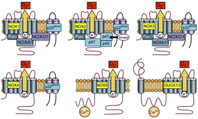
1.1.3.2.3. NADPH oxidases – focus on Nox1, Nox2, and Nox4 ... 49!
1.1.3.2.4. Generation of ROS by COX ... 52!
1.1.4. Adaptation of EDRFs to endothelial dysfunction ... 53!
1.1.5. Involvement of vasoconstrictors in endothelial dysfunction ... 54!
1.1.5.1. Role of ET-1 in endothelial dysfunction ... 54!
1.1.5.2. AngII and its signaling transduction pathways in endothelial dysfunction ... 54!
1.1.5.3. COX pathway alteration ... 56!
1.1.6. Pathologies associated with endothelial dysfunction ... 58!
1.1.6.1. Obesity, insulin resistance and endothelial dysfunction ... 58!
1.1.6.1.1. The adipocyte and its emerging importance in regulating endothelial function ... 60!
1.1.6.1.2. Dyslipidemia in obesity and endothelial dysfunction ... 61!
1.1.6.2. The renin-angiotensin system (RAS) and angII ... 61!
1.1.6.2.1. Hypertension and its implications ... 62!
1.1.6.2.2. AngII and its effects on the cerebrovasculature ... 64!
Chapter 2: Angiopoietin-like proteins ... 67!
1.2.2. History at a glance: identification and characterization of angiopoietin-like
proteins ... 69!
1.2.3. Physiological roles of angiopoietin-like proteins ... 72!
1.2.3.1. Angiopoietin-like proteins in angiogenesis ... 72!
1.2.3.2. Angiopoietin-like proteins in lipid metabolism ... 74!
1.2.3.3. Angiopoietin-like proteins in glucose metabolism ... 79!
1.2.4. Contribution of angptl proteins to cardiovascular risk factors ... 80!
1.2.4.1 Angiopoietin-like proteins in dyslipidemia ... 80!
1.2.4.2. Angiopoeitin-like proteins in obesity and insulin resistance ... 83!
1.2.5. Angiopoietin-like proteins in endothelial dysfunction and atherosclerosis ... 86!
Chapter 3: Angiopoietin-like-2 ... 89!
1.3.1. Angptl2 – a pro-inflammatory mediator ... 89!
1.3.2. Angptl2 – a pro-oxidative mediator ... 91!
1.3.3. Physiological roles of angptl2 ... 92!
1.3.3.1. Angptl2 in physiological angiogenesis ... 92!
1.3.3.2. Angptl2 in tissue repair and remodeling ... 93!
1.3.3.3. Angptl2 as a circadian gene ... 95!
1.3.3.4. Angptl2 in AT1R recycling ... 95!
1.3.4. Pathological roles of angptl2 ... 96!
1.3.4.1. Angptl2 in rheumatoid arthritis ... 96!
1.3.4.2. Angptl2 in cancer ... 97!
1.3.4.4. Angptl2 in vascular remodeling ... 103!
1.3.4.5. Angptl2 in inflammatory tissue disorders ... 104!
1.3.4.6. Angptl2 in abdominal aortic aneurysm (AAA) development ... 105!
1.3.4.7. Angptl2 in atherogenesis ... 106!
1.3.4.8. Angptl2 in endothelial dysfunction ... 107!
1.3.5. Reported molecular pathways associated with angptl2 ... 111!
1.3.6. Reported mechanisms regulating angptl2 transcription and expression . 112! 2. Research Overview ... 116!
2.1. Study #1: Does angptl2 knock-down protect against obesity-induced endothelial dysfunction? ... 117!
2.1.1. Background ... 117!
2.1.2. Hypothesis ... 117!
2.1.3. Specific aims ... 117!
2.2. Study #2: Does angptl2 knock-down protect against angII-induced endothelial dysfunction? ... 118! 2.2.1. Background ... 118! 2.2.2. Hypothesis ... 118! 2.2.3. Specific aims ... 118! 3. Articles ... 119! 3.1. Article 1 ... 119! 3.1.1. Contribution of co-authors ... 119! 3.2. Article 2 ... 140! 3.2.1. Contribution of co-authors ... 140!
4. Discussion ... 153!
4.1. Linking it all together: how does angptl2 mechanistically mediate endothelial dysfunction? ... 154!
4.2. Can angptl2 lower endothelial cell stress resistance? ... 157!
4.3. Could angptl2 also contribute to endothelial dysfunction via its role in AT1R recycling? ... 159!
4.4. Could protection against high-fat diet-induced metabolic dysfunction in angptl2 KD mice be a consequence of blunted angiogenesis? ... 160!
4.5. Is there a functional consequence of the cleaved angptl2 protein on adipocytes or ECs? ... 163!
4.6. Lowering levels of angptl2: is that the solution? But how? ... 163!
4.7. Limitations of the studies ... 166!
5. Conclusion and Perspectives ... 170!
LIST OF TABLES
LIST OF FIGURES
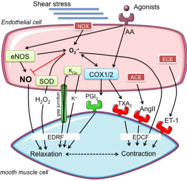
Figure 1. Proposed mechanisms of endothelium-derived relaxation and contraction
in the smooth muscle cell by various known EDRFs and EDCFs... 25
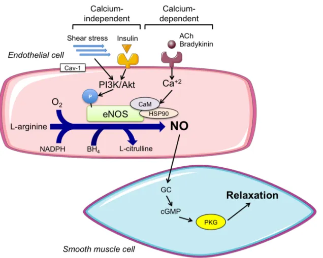
Figure 2. Calcium-dependent and –independent activation of eNOS to produce NO
for smooth muscle cell relaxation... 28
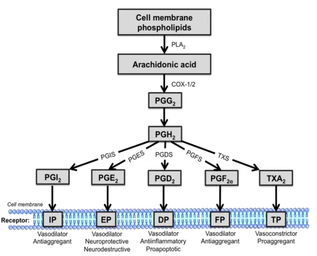
Figure 3. A schematic representation of AA metabolism... 30 Figure 4. Proposed mechanisms of H2O2 production in the cerebral arteries and its
downstream effects on the VSMC to cause vasorelaxation...35
Figure 5. EDRF heterogeneity in arteries of varying sizes...38 Figure 6. Activation of Nox isoforms...50 Figure 7. Proposed mechanisms by which angII stimulation of AT1R in the
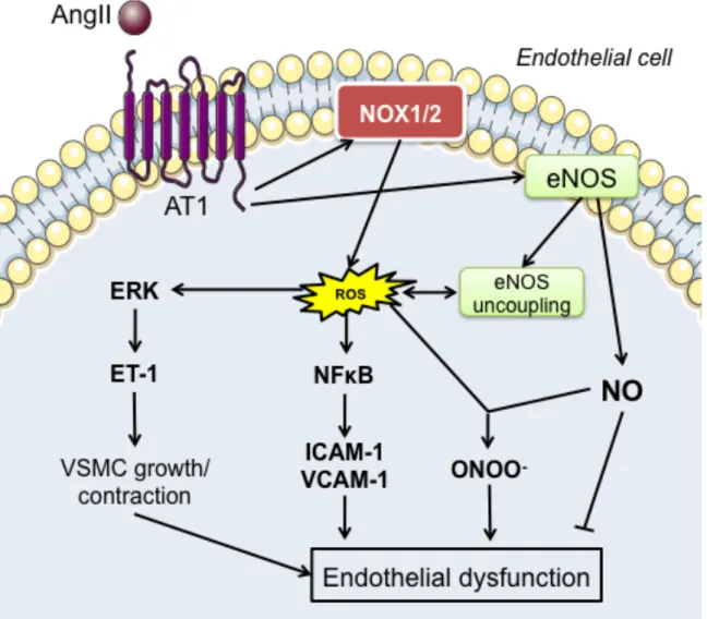
endothelium leads to endothelial dysfunction...56
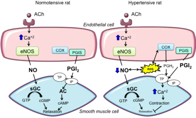
Figure 8. ACh-induced and endothelium-dependent effects in normotensive and
hypertensive rat aorta. ...63
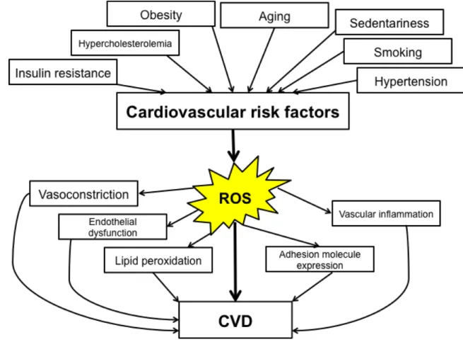
Figure 9. ROS is one of the links between cardiovascular risk factors and CVD....66 Figure 10. Schematic protein structure of an angptl protein...68 Figure 11. Alignment of the amino acid sequences and evolutionary relationships
between angptl2 and its relatives, human angptl1, angiopoietin-1 and -2,
demonstrating their homology...71
Figure 12. Summary of the different roles of angptls 3, 4, and 6 in maintaining
metabolic homeostasis...78
Figure 13. The role of angptl2 in physiological and pathological adipose tissue
remodeling...94
Figure 14. Proposed mechanism linking angptl2 and carcinogenesis...99 Figure 15. A proposed model of adipocyte-derived angptl2 contribution to
inflammation, insulin resistance, and vascular dysfunctions...109
Figure 16. Schematic representation of a selection of reported pathways that induce
angptl2 expression and the pathways that are in turn induced by angptl2...115
Figure 17. Exponentially growing scientific interests in angptl2 in physiology and
LIST OF ABBREVIATIONS
AA Arachidonic acid
AAA Abdominal aortic aneurysm
AC Adenylate cyclase
ACE Angiotensin converting enzyme
ACh Acetylcholine
Akt Protein kinase B
AngI Angiotensin I
AngII Angiotensin II
Angptl Angiopoietin like protein Angptl2 Angiopoietin like-2
ApoB Apolipoprotein B
ApoE Apolipoprotein E
ARAP1 Angiotensin II receptor-associated protein 1 ATF2 Activated transcription factor 2
AT1R Angiotensin II receptor type 1 AT2R Angiotensin II receptor type 2 BH4 Tetrahydrobiopterin
BMAL1 Brain and muscle aryl hydrocarbon receptor nuclear translocator-l like protein 1
BMI Body mass index
Ca2+ Calcium
CAD Coronary artery disease
CaM Calmodulin
cAMP Cyclic adenosine monophosphate
Cav-1 Caveolin-1
cGMP Cyclic guanosine monophosphate
ChREBP Carbohydrate responsive element binding protein CLOCK Circadian locomotor output cycles kaput
Cu/ZnSOD Copper- and zinc-containing superoxide dismutase CVD Cardiovascular disease
db/db Diabetic (leptin receptor-deficient) DETC Diethyldithiocarbamate
DP Prostaglandin D2 receptor
EC Endothelial cell
ECE Endothelin-1 converting enzyme ECM Extracellular matrix
EDCF Endothelium-derived contracting factor EDHF Endothelium-derived hyperpolarizing factor EDRF Endothelium-derived relaxing factor
EET Epoxyeicosatrienoic acid
ERK Extracellular signal regulated kinase ETC Electron transport chain
eNOS Endothelial nitric oxide synthase EP Prostaglandin E2 receptor ER Endoplasmic reticulum ET-1 Endothelin-1 ET-2 Endothelin-2 ET-3 Endothelin-3 ETA Endothelin receptor A ETB Endothelin receptor B FA Fatty acid
FAD Flavin adenine dinucleotide
FFA Free fatty acid
FMD Flow-mediated dilation FoxO1 Forkhead box protein O1 FP Prostaglandin F2α receptor
GC Guanylate cyclase
GLUT4 Glucose transporter type 4 GPCR G-protein coupled receptor
GPx Glutathion peroxidase GTP Guanosine tri-phosphate HDL High-density lipoprotein HFD High-fat diet H2O2 Hydrogen peroxide HO-1 Heme-oxygenase 1
HSC Hematopoietic stem cell HSP90 Heat shock protein 90
HUVEC Human umbilical vein endothelial cell ICAM Intracellular adhesion molecule
IFN γ Interferon γ
IL Interleukin
IκB Inhibitor of kappa B
iNOS Inducible nitric oxide synthase IP Prostaglandin I2/prostacyclin receptor
IRS-1 Insulin receptor substrate 1
JAK Janus kinase
K+ Potassium
KCa Calcium-activated potassium channel
KD Knock-down
Keap1 Kelch-like erythroid cell-derived protein with cap ‘n’ collar homology-associated protein 1
KO Knock-out
LCSFA Long chain saturated fatty acid LDL Low-density lipoprotein
LDLr Low-density lipoprotein receptor
LILRB2 Leukocyte immunoglobulin-like receptor B2 LNNA Nω-nitro-L-arginine
LPL Lipoprotein lipase
LXR Liver X receptor
MAPK Mitogen-activated protein kinase MCP-1 Monocyte chemoattractive protein 1
MMP Metalloproteinase
MnSOD Manganese superoxide dismutase
NAC N-acetylcysteine
NADPH Nicotinamide adenine dinucleotide phosphate NFκB Nuclear factor kappa B
NEFA Non-esterified fatty acid
NFAT Nuclear factor of activated T cells nNOS Neuronal nitric oxide synthase
NO Nitric oxide
NOS Nitric oxide synthase
NOX Nicotinamide adenine dinucleotide phosphate oxidase Nrf2 Nuclear-like factor 2
O2 Oxygen
O2-! Superoxide
ob/ob Obese (leptin-deficient) ONOO- Peroxynitrite PG Prostaglandin PGD2 Prostaglandin D2 PGE2 Prostaglandin E2 PGF2α Prostaglandin F2α PGG2 Prostaglandin G2 PGH2 Prostaglandin H2 PGD2S Prostaglandin D2 synthase
PGE2S Prostaglandin E2 synthase
PGF2αS Prostaglandin F2α synthase
PGG2S Prostaglandin G2 synthase
PGH2S Prostaglandin H2 synthase
PGI2 Prostaglandin I2/Prostacyclin
PIRB Paired immunoglobulin-like receptor
PKG Protein kinase G
PLA2 Phospholipase A2
PLC Phospholipase C
PPAR Peroxisome proliferator-activated receptor RAS Renin-angiotensin system
ROS Reactive oxygen species
RXR Retinoid X receptor
sGC Soluble guanylate cyclase
SOD Superoxide dismutase
STAT Signal transducers and activators of transcription
TG Triglyceride
TGF Tumour growth factor
TLL1 Tolloid-like 1
TLR Toll-like receptor
TNFα Tumour necrosis factor α TP Thromboxane A2 receptor
Trib3 Tribbles homolog 3
TXA2 Thromboxane A2
UCP-1 Uncoupling protein 1 UCP-2 Uncoupling protein 2 UCP-3 Uncoupling protein 3
VCAM Vascular cell adhesion molecule VEGF Vascular endothelial growth factor VLDL Very low-density lipoprotein VSMC Vascular smooth muscle cell
ACKNOWLEDGEMENTS
I have deliberately left this part of the thesis until the very end expecting myself to go through this section in no time. To my surprise, it is actually much more difficult than I imagined to express my gratitude towards each and everyone that has made this work possible. First and foremost, I would like to thank the members of my jury for this thesis. To Dr. Laher, Dr. Joyal, Dr. Girouard, and Dr. Lamontagne, thank you all for spending your time with this work and being on the jury panel, your comments are invaluable and I greatly appreciate them.
To Éric Thorin, my supervisor and mentor,
First of all, thank you for accepting me into your lab with open arms from the start. I remember I once joked that moving to Montreal was the best thing I have ever done, but in all honesty, it is one of the best decisions I have ever made. Not only has it been my honor working with you, it has been loads of fun. You are so down-to-earth, which makes work an enjoyable place to be at, and you always manage to have time for all your students. Of course it was not always easy and that “life is full of little challenges”, but you have proven time and again that things, inside or outside of the lab, do work out in the end. Thank you for being so patient, kind, generous, and overly optimistic with me throughout the past four years.
To Nathalie,
I cannot be more thankful for your teaching and help from the very beginning. It has been a great pleasure working with you. Thank you for the endless hours spent correcting this work and always giving me pointers for everything. I know you “suggest” them, but truly they are always so insightful. Although they are not always so peaceful, the discussions we have with Eric really open my mind. Working alongside the both of you, I was fortunate to be exposed to different kinds of thinking, and it has undoubtedly shaped me into a better thinker. And of course, thank you for all the delicious baked goods and food that help us last until the end of the day. Knowing that you are there, the lab is surely in good hands.
To Nada,
Thank you for your companionship throughout this time. This work would not have been completed as timely as it has without you. With you, there was never a moment of dullness, and you taught me what real multi-tasking is all about. Thank you for sharing the passion you have for your work, it is always inspiring working with you!
To Albert,
Where can I even start? Without that first-class training I had from you on the arteriograph, this work would not have been possible. It has been more than fun working with you. Thank you for always being so helpful, entertaining and witty.
To Xiaoyan,
Thank you for all your help with both of my projects. Without it, this would not have gone as smoothly as it did. You were always so patient and kind. Thank you for being so sweet to me. Working with you has been a great pleasure.
To Cécile,
It has been a great deal of awesomeness being with you both inside and outside of the lab. Thank you for all your help with all our little ones and with everything else, and always pushing me to keep going and improving.
To Adeline,
Merci for all the laughter we shared. It has been a blast working with you. You always manage to bring out my even more immature side and thanks to you, my French has “dramatically” improved, along with my exquisite taste in French cuisine.
To Franςois,
You are one of the most organized people I know, and I greatly appreciate your help with my transitions into the lab from the start (in fact, you were the first person I met from the lab!). Thank you for everything!
To Virginie,
Working with you has been a great pleasure. You were always so determined, hard-working and dedicated to your work, so thank you for being such a great lab-mate.
To Youri,
Your arrival to the lab made me the second loudest person. I know we joke around all the time, but in all seriousness, it has been an honor working next to you. You are dedicated and always pay attention to details. Thank you for being a cool bro.
To Nour,
Although it was only a short few months that we worked together, those times were great times! Thank you for being so much fun inside and outside of the lab!
To Bruce,
Even though you always just make fun of me, I would still like to thank you for all the insightful work-related and –unrelated West coast-style conversations we’ve had over the last few years, and the coffee too!
I would like to thank all of our collaborators for their help in this work. I would also like to acknowledge Anie, Yolanda, Sirirat, and Eric, who I did not have a chance to directly work with, your friendship means the world to me. Thank you for being there, through the good times and the bad, and the (super loud) laughter we share. And to Maya Mamarbachi, Louis Villeneuve, Natacha Duquette, and Robert Parent, thank you for all the technical help you supported me with. To my family and friends, thank you for all the local and long-distance moral support throughout this whole time. Last but not least, I extend my great thanks to my funding agencies, the Natural Sciences and Engineering Research Council of Canada (NSERC), and the Univeristé de Montréal, for the financial support in this achievement.
1. Introduction
“It has been said that one is as old as one’s arteries. In view of the supreme importance of endothelium in arterial function, I should like to modify... this statement by saying that one is as old as one’s endothelium.”
– Rudolf Altschul, 1954
Stated 6 decades ago, at a time when the endothelium was simply considered a passive cell-lining in the vasculature, this visionary statement clearly captures the relevance of endothelial health in the aging process. Indeed, endothelial cells (ECs) are known for their capabilities to secrete a wide spectrum of anti-atherosclerotic substances, as well as a balance of relaxing and contracting factors under physiological conditions (Sessa 1994; Moncada 1997; Feletou and Vanhoutte 2007; Feletou et al. 2011). However, under pathological conditions, where cardiovascular risk factors are present, persistent oxidative stress and endothelial dysfunction occur where ECs lose their protective role and become pro-atherosclerotic (Feletou and Vanhoutte 2006). Despite ample evidence demonstrating strong associations among cardiovascular risks, endothelial dysfunction and cardiovascular disease (CVD) (Feletou and Vanhoutte 2006), CVD remains one of the leading causes of death in Canada, and costs the Canadian economy more than $20.9 billion per year (Conference Board of Canada, 2010). The greatest and most established cardiovascular risk factor is metabolic syndrome (Katzmarzyk and Janssen 2004), which is characterized by obesity, often caused by sedentariness and excess energy intake, dyslipidemia, insulin resistance or glucose intolerance, a inflammatory and pro-thrombotic state, as well as hypertension (Grundy et al. 2004).
Over the past years, it has become strikingly clear that endothelial dysfunction can occur in early childhood and silently progress through age (Deanfield et al. 2007), and recent findings have shown potentials in lowering later cardiovascular events with lifetime risk management and reduction in risk factors starting at an earlier stage (Ulrich et al. 2000; Cohen et al. 2006). Nonetheless, studies that focus on the clinical impact of endothelial dysfunction concentrate heavily on patient cohorts presented with established CVD (Deanfield et al. 2007). Even though endothelial function testing in patients have
shown enormous benefits in understanding the development of endothelial dysfunction and ultimately CVD, it is still not yet suitable for individual screening (Deanfield et al. 2007). Undoubtedly, the endothelium is a dynamic organ and its regulation is vascular district-specific with varying results in different vascular beds (Shimokawa et al. 1996; Urakami-Harasawa et al. 1997). Thus, a greater and more comprehensive understanding of its regulation is clearly warranted.
In this introductory section, the physiology and pathophysiology of the endothelium will first be discussed, followed by an introduction to a relatively new family of proteins, the “angiopoietin like-proteins”, emphasizing on one in particular, angiopoietin like-2 (angptl2). How this protein is involved in regulating endothelial function, specifically in pathological settings, will be the focus of this work.
Chapter 1: The Endothelium
1.1. The endothelium – its function and dysfunction
The endothelium, consisting of a single layer of cells lining the interior surface of blood and lymphatic vessels, amount to roughly 1014 cells in the vasculature (Cines et al. 1998), and has been intensely studied since the discovery of nitric oxide (NO) as a vasodilating agent that is produced by ECs (Furchgott and Zawadzki 1980; Palmer et al. 1987). Indeed, vascular ECs play an extremely important role in maintaining cardiovascular homeostasis besides merely acting as a physical barrier between the lumen and vessel wall. It secretes a wide spectrum of mediators that can regulate cellular adhesion and permeability, vessel wall inflammation, smooth muscle cell proliferation, angiogenesis, and vascular tone (Sessa 1994; Moncada 1997). One of the most important features of the endothelium is its capacity to affect vascular tone by producing a balance of vasorelaxing and vasoconstricting factors (Luscher et al. 1989; Deanfield et al. 2007). Among them include NO (Furchgott and Zawadzki 1980), prostacyclin (PGI2) (Moncada
and Vane 1978), and endothelium-derived hyperpolarizing factor (EDHF) (Komori and Vanhoutte 1990; Garland et al. 1995), collectively called endothelium-derived-relaxing factors (EDRFs), which contribute to vasodilation, as well as endothelin-1 (ET-1) (Pernow et al. 2012), thromboxane (TXA2) (Feletou and Vanhoutte 2006), superoxide
anion (O2-!) (Katusic and Vanhoutte 1989), collectively called
endothelium-derived-contracting factors (EDCFs), which contribute to vasoconstriction, as depicted in Figure 1. Under the physiological state, endothelial nitric oxide synthase (eNOS)-generated NO mainly determines vascular tone (Sessa 1994; Moncada 1997), and is the major contributor of a quiescent state of the vascular wall by inhibition of inflammation and adhesion (Bath et al. 1991), cell proliferation (Yang et al. 1994), thrombosis (Ignarro 1989), and limits mitochondrial oxidative phosphorylation (Moncada and Erusalimsky 2002).
Figure 1. Proposed mechanisms of endothelium-derived relaxation and contraction in the
smooth muscle cell by various known EDRFs and EDCFs. Abbreviations: EDRF: endothelium-derived relaxing factor; EDCF: endothelium-derived contracting factor; AA: arachidonic acid; eNOS: endothelial nitric oxide synthase; O2-!: superoxide; SOD:
superoxide dismutase; KCa: calcium-activated potassium channels; K+: potassium ion;
NOX: nicotinamide adenine dinucleotide phosphate oxidase; COX: cyclo-oxygenase; PGI2: prostacyclin; TXA2: thromboxane A2; ACE: angiotensin converting enzyme;
AngII: angiotensin II; ECE: endothelin-1 converting enzyme; ET-1: endothelin-1.
1.1.1. Endothelium-derived relaxing factors (EDRFs)
With the knowledge that the endothelium serves as a regulator of vascular tone, research has begun to focus on its mechanisms. The endothelium responds to both physical (such as shear stress and flow (Davies 1995)) and chemical (such as
acetylcholine (ACh) and bradykinin (Groves et al. 1995; Drexler 1999)) stimuli. The following sections will highlight the most studied EDRFs and their respective mechanisms leading to vasodilation.
1.1.1.1. Nitric oxide (NO)
First discovered by Furchgott and Zawadzki and an EDRF (Furchgott and Zawadzki 1980), this endothelium-dependent relaxing factor was identified as NO (Palmer et al. 1987), and was first documented to derive from vascular ECs from the conversion of L-arginine (Palmer et al. 1988). The contribution of NO to vasodilation is well recognized, largely in conduit arteries such as the aorta, but also extends to all types of blood vessels (Forstermann et al. 1994). The generation of NO, whether basal or stimulated, is dependent on the endothelial, constitutively-expressed enzyme eNOS, a dimeric, bi-domain enzyme with a C-terminal reductase domain and N-terminal oxidase domain. eNOS is responsible for the conversion of L-arginine into NO through a two-step, 5-electron-oxidation process in the presence of cofactors including tetrahydrobiopterin (BH4) (Abu-Soud et al. 1994; Forstermann and Munzel 2006).
Subsequent to its generation, NO diffuses from the endothelium to the vascular smooth muscle cells (VSMC), where it activates guanylate cyclase (GC), leading to cyclic guanosine monophosphate (cGMP)-mediated vasodilation (Deanfield et al. 2007), as shown in Figure 2. NO has an extremely short half-life ranging from 3 to 5 seconds (Ignarro 1989), and is readily degraded by O2-! (Gryglewski et al. 1986) and oxidized
into nitrite and nitrate (Hibbs et al. 1988). Physiologically, a potent stimulator of NO generation is shear stress through a non-receptor-dependent mechanisms (Rubanyi et al. 1986; Corson et al. 1996), and pharmacologically, a number of agonists have been found able to stimulate NO generation including ACh and bradykinin, both of which bind to their respective receptor, which lead to subsequent downstream vasodilatory pathways (Doyle and Duling 1997).
1.1.1.1.1. Regulation of eNOS activity and its downstream pathways
There are 4 distinct isoforms of NOS – neuronal (nNOS), inducible (iNOS), endothelial (eNOS) (Forstermann et al. 1993), and red blood cell NOS (Jubelin andGierman 1996). As eNOS is the dominant NOS isoform expressed in the vasculature that produces NO under physiological conditions, it will be the primary focus of this chapter. The activity and expression of eNOS are tightly regulated both transcriptionally (Wang and Marsden 1995) and translationally (Sase and Michel 1997). Post-translationally and in its inactive form, eNOS is bound to inhibitory protein caveolin-1 and localized at caveolae in the cell membrane (Lisanti et al. 1994). Stimulation such as receptor-mediated rise in intracellular calcium has been shown to activate eNOS by disrupting the protein-protein interaction between eNOS and caveolin-1 (Brouet et al. 2001), thus increasing NO production.
There are two main branches of eNOS activation – calcium-dependent and calcium-independent activation, as shown in Figure 2. Classical agonists such as ACh act via G-protein-coupled receptors (GPCRs) to generate intracellular rise in calcium, which then binds to calmodulin and together bind to eNOS, resulting in its activation (Busse and Fleming 1995). In the other case, independently of calcium, stimuli such as shear stress (Ayajiki et al. 1996) and insulin (Zeng and Quon 1996) activate phosphatidylinositol 3-kinase (PI3K) and protein 3-kinase B (Akt), which then phosphorylate and activate eNOS (Harris et al. 2001). This PI3K/Akt signaling pathway downstream of insulin stimulation is, interestingly, shared with insulin-dependent glucose transporter type 4 (GLUT-4)-mediated glucose uptake (Hsueh and Law 1999), which will gain importance in settings of insulin resistance and associated dysfunctions.
Subsequently, the activated eNOS generates NO, as described in section 1.1.1.1., which diffuses into VSMC and activates GC by binding to its heme group at the iron (Lowenstein et al. 1994). Activation of GC leads to its synthesis of cGMP from GTP, which in turn activates cGMP-dependent protein kinase G (Carvajal et al. 2000), initiating a cascade of phosphorylation reactions resulting in physiological effects such as lowering intracellular calcium in VSMC and ultimately leading to vasorelaxation (Francis and Corbin 1994; Lohmann et al. 1997), as shown in Figure 2.
Figure 2. Calcium-dependent and –independent activation of eNOS to produce NO for
smooth muscle cell relaxation. Abbreviations: ACh: acetylcholine; O2: oxygen; NADPH:
nicotinamide adenine dinucleotide phosphate; Cav-1: caveolin-1; eNOS: endothelial nitric oxide synthase; NO: nitric oxide; PI3K: phosphatidylinositol 3-kinase; Akt: protein kinase B; Ca2+: calcium; CaM: calmodulin; HSP90: heat shock protein 90; BH4:
tetrahydrobiopterin; GC: guanylate cyclase; cGMP: cyclic guanosine monophospate: PKG: protein kinase G.
1.1.1.1.2. Vascular protection and anti-oxidative effects of NO
Besides its ability to act as a vasodilating agent, vascular NO possesses many other physiological properties. Importantly, NO has anti-platelet (Radomski and Moncada 1993), anti-adhesive (Bath et al. 1991), anti-proliferative (Yang et al. 1994) and anti-inflammatory (Bath et al. 1991) effects, and these properties become extremely important in settings of atherogenesis. For example, NO can potently inhibit aggregation of platelets and leukocytes onto the vessel wall. It does so by interfering with the
adhesive bonding between the leukocyte adhesion molecule and the endothelium surface, which is an early event of atherogenesis (Forstermann et al. 1994). In addition, it has been reported that NO could suppress DNA synthesis, mitogenesis, as well as proliferation of VSMC (Forstermann et al. 1994).
As mentioned, interactions between vascular signaling such as that of NO and oxidants such as superoxide are well documented. NO is best known for its ability to impair oxidation of free fatty acids (FFA), phosphatidylcholine and low-density lipoprotein (LDL) particles. NO induces endothelial ferritin formation (Recalcati et al. 1998), which then can reduce oxidative damage by preventing superoxide generations as ferritin binds free iron ions (Balla et al. 1992). Mechanistically, NO induces expression of heme oxygenase-1 (HO-1), which can then increase formation of bilirubin and carbon monoxide (Maines 1997), which in turn can scavenge superoxide and activate soluble GC (sGC), respectively (Stocker et al. 1987). NO has also been documented to induce extracellular superoxide dismutase both in vitro and in vivo in VSMC (Fukai et al. 2000). As a result, NO is able to decrease both superoxide and peroxynitrite levels in the vessel wall.
1.1.1.2. Prostaglandins (PGs)
Synthesized mainly from fatty acid (FA) arachidonic acid (AA) derived from the cell membrane released by phospholipase A2, PGs were discovered before NO was
identified as an endothelium-derived vasoactive substance (Moncada et al. 1976). AAs are then metabolized by different enzymes – prostaglandin H synthases (PGH synthases, or more commonly cyclo-oxygenases COX-1 or -2) (Vane et al. 1998), lipoxygenases, or cytochrome P450 (Morrow et al. 1990). COX-1 and -2 are the first and also rate-limiting enzymes to process PGs, and give rise to prostaglandin G2 (PGG2), which is reduced into
short-lived prostaglandin H2 (PGH2). The fate of PGH2, in turn, depends on the actions
of PG synthases – prostaglandin D2 synthase (PGDS), prostaglandin E2 synthase (PGES),
prostaglandin F2α synthase (PGFS), prostaglandin I2 synthase (PGIS) and thromboxane
synthase (TXS), which form the five main PGs – prostaglandin D2 (PGD2), prostaglandin
(TXA2), respectively. A schematic representation of AA metabolism is shown in Figure
3.
Figure 3. A schematic representation of AA metabolism. AA is first derived from cell
membrane phospholipids by PLA2, which is metabolized by COX-1 and COX-2 into
PGG2, and is then reduced into PGH2. Subsequently, PGH2 is further metabolized by
different PG synthases into distinct prostanoids. Abbreviation: PLA2: phospholipase A2;
COX-1/2: cyclo-oxygenase 1/2; PGG2: prostaglandin G2; PGH2: prostaglandin H2; PGI2:
prostaglandin I2; PGE2: prostaglandin E2; PGD2: prostaglandin D2; PGF2α: prostaglandin
F2α; TXA2: thromboxane A2; PGIS: prostaglandin I2 synthase; PGES: prostaglandin E2
synthase; PGDS: prostaglandin D2 synthase; PGFS: prostaglandin F2α synthase; TXS:
thromboxane A2 synthase; prostaglandin I2 receptor: IP; prostaglandin E2 receptor: EP;
prostaglandin D2 receptor: DP; prostaglandin F2α receptor: FP; thromboxane A2 receptor:
PGI2, derived from PGI2 synthase (Hara et al. 1994),is typically described as an
endothelium-derived vasodilator, as it can bind to and stimulate its receptor, the IP receptor, to activate adenylate cyclase and increase intracellular cAMP concentration and produce smooth muscle relaxation (Wise and Jones 1996). PGI2 synthase is highly
expressed in ECs (Tang and Vanhoutte 2008) and to a lesser degree also in VSMC (Wu and Liou 2005). Furthermore, vasodilation by PGI2 has been accompanied by
hyperpolarization of adjacent VSMC, which may involve potassium channels (Corriu et al. 2001). Of note, plasma concentrations of PGI2 in human has been observed to peak at
infancy and decrease throughout life (Kaapa et al. 1982). Hence, PGI2 is usually not the
main vasodilator in most vascular beds (Shimokawa et al. 1996); however, its vasodilatory role could become important in the face of decreased NO bioavailability, for instance, PGI2 may compensate for the loss of NO in hypertensive patients (Bulut et al.
2003).
1.1.1.2.1. Other physiological roles of PGI
2In most arterial beds, many believed that the synthesis of PGI2 and downstream
responses were not altered under pathological settings. A prime example was showcased by the intact PGI2 vasodilating system in the coronary resistance arteries of
atherosclerotic mice, despite damages in NO-dependent dilations (Godecke et al. 2002). However, this is not always the case, as shown in the abrogated PGI2-dependent
vasodilation in the aorta of aldosterone-treated normotensive and hypertensive rats (Blanco-Rivero et al. 2005). Importantly and in addition to its vasodilating features, PGI2
is characterized by its powerful anti-coagulant and anti-adhesive properties (Moncada and Vane 1978). This was highlighted by the unexpected results coming from colorectal adenoma patients presented with adverse cardiovascular effects of rofecoxib, trademarked as Vioxx (Merck and Co.) (Bresalier et al. 2005), ultimately leading to its withdrawal from the market. This could be partly explained by its inhibition of protective PGI2 synthesis, in spite of reduced TXA2 synthesis (Griffoni et al. 2007). Indeed, PGI2 is
the most potent endogenous inhibitor of platelet aggregation (Bunting et al. 1976; Moncada et al. 1976). It can also act in a paracrine or endocrine manner to affect functions of other cells types via specific GPCRs (Negishi et al. 1995) or nuclear
receptors such as the peroxisomal proliferator-activated receptors (PPARs) (Forman et al. 1997), as well as regulate physiological events such as angiogenesis (Spisni et al. 1992) and apoptosis (Li et al. 2004).
1.1.1.3. Endothelium-derived hyperpolarizing factor (EDHF)
The most recently discovered group of EDRF has been broadly termed endothelium-derived hyperpolarizing factor (EDHF), with its first evidence reported in 1988 (Chen et al. 1988; Feletou and Vanhoutte 1988). By definition, EDHF is a substance or an electrical signal that is released from the endothelium and is able to hyperpolarize VSMC resulting in relaxation (Fleming 2000; Feletou and Vanhoutte 2007). Its contribution to vasodilation is found in the greatest levels in small arteries such as the resistance arteries (Shimokawa et al. 1996; Urakami-Harasawa et al. 1997; Luksha et al. 2009). Ongoing debates have accumulated over the recent years surrounding topics on the variable nature of EDHFs and their variable mechanisms of actions. Initially, EDHF referred to any relaxing factor that is neither NO nor PGI2, but over the past
decade, a number of possible candidates have been uncovered: epoxyeicosatrienoic acids (Rubanyi and Vanhoutte 1987), hydrogen peroxide (H2O2) (Beny and von der Weid
1991; Matoba et al. 2000), K+ ions (Edwards et al. 1998), C-type natriuretic peptide (Chauhan et al. 2004), as well as contact-mediated mechanisms (Rummery and Hill 2004). Although the contribution by EDHF to vasodilation is limited in large conductance arteries under healthy conditions, it has been documented that EDHF can compensate for loss of NO bioavailability in disease states (Csanyi et al. 2012).
The basic mechanism of EDHF release and action can be separated into two stages. The first stage involves the pathways that take place in the endothelium: an increase in intracellular Ca2+, K+ efflux upon activation of Ca2+-dependent K+ channel, followed by hyperpolerization and generation of the EDHF that diffuses through myoendothelial gap junctions (McGuire et al. 2001; Busse et al. 2002). The second stage involves those taking place in the VSMC: EDHF activating K+ channels and leading to
endothelium-dependent hyperpolarization, followed by closure of voltage-gated Ca2+ channels and lastly, relaxation of the VSMC (Busse et al. 2002; McGuire et al. 2001).
1.1.1.3.1. Relevance of H
2O
2in cerebral vasculature
While in most vascular beds eNOS-derived NO predominantly contributes to vasodilation including flow-mediated dilation (FMD) (Davies 1995), both NO (Iadecola 1992; White et al. 1998) and EDHF (Fujii et al. 1991) have been found involved in this mechanism. In particular, one of the EDHFs, namely H2O2, has been demonstrated to
cause vasodilation in the cerebral vasculature from mice (Wei and Kontos 1990; Fraile et al. 1994; Wei et al. 1996; Iida and Katusic 2000; Drouin and Thorin 2009). Importantly, we previously demonstrated that H2O2-mediated cerebral vasodilation was inhibited by
an sGC inhibitor, suggesting that H2O2 shared a similar downstream vasodilatory
pathway with NO (Drouin et al. 2007). H2O2, although not a free radical itself, is the
product of various superoxide dismutases (SOD), including copper- and zinc-containing SOD (Cu/ZnSOD) (Morikawa et al. 2003). In contrast to highly reactive reactive oxygen species (ROS) such as O2-!, H2O2 is readily diffusible and is relatively stable. Most
importantly, it does not react with NO to produce peroxynitrite (Pacher et al. 2007). Consistent with data from human coronary (Liu et al. 2011) and rat skeletal (Sindler et al. 2009) arterioles, H2O2 contributes to vasodilation of cerebral arteries in vitro (Fraile et al.
1994; Iida and Katusic 2000), with strong evidence pointing towards potassium channels mediating its effects (Iida and Katusic 2000; Paravicini et al. 2004; Sobey et al. 1997), which leads to hyperpolarization and vasodilation (Sobey et al. 1997). Indeed, in rat cerebral arterioles, H2O2 caused vasodilation via activation of calcium-dependent
potassium channels (Sobey et al. 1997), and in cat cerebral arterioles, H2O2 activated
ATP-sensitive potassium channels (Wei et al. 1996). Exogenous H2O2 and ACh-induced
H2O2 production may also activate sGC in mouse cerebral arteries (Drouin et al. 2007),
resulting in elevated cGMP levels that may cause hyperpolarization and subsequently relaxation (Nelson et al. 1990). Interestingly, cerebral arterial dilation to H2O2 can also
stimulate AA release from VSMC via phospholipase A2 activation (Rao et al. 1995), and
this was confirmed by sensitivity of H2O2-dependent cerebral arteriolar dilation to
indomethacin (Iida and Katusic 2000), a non-specific COX inhibitor, in addition to H2O2
-mediated cAMP increase (Iida and Katusic 2000). Consistent with this, topical application of H2O2 onto piglet cerebral arterioles increased formation of
in the VSMC may cause relaxation by activating potassium channels to induce hyperpolarization and decrease calcium influx (Nelson et al. 1990). Collectively, these findings suggest that H2O2 may also cause dilation indirectly via the COX-mediated
vasodilatory pathway. Proposed mechanisms of H2O2-mediated vasodilation are
summarized in Figure 4. Moreover, vasodilatory effects of H2O2 have been documented
in studies using different vasoactive stimuli – AA (Kontos et al. 1984), bradykinin (Kontos et al. 1984; Yang et al. 1991; Sobey et al. 1997), ACh (Drouin et al. 2007), as well as flow (Drouin and Thorin 2009).
The sources of H2O2 have been primarily reported from the dismutation of O2-!,
and also directly from various enzymes, such as lipooxygenases, xanthine oxidase and NADPH oxidase (Haas et al. 1994; Cai 2005). Importantly, the recent provocative proposal that eNOS may be involved as a source of H2O2 in cerebral arteries (Drouin and
Thorin 2009; Drouin et al. 2007), associated with evidence showing eNOS-dependent H2O2-mediated dilation (Zembowicz et al. 1993; Bharadwaj and Prasad 1995; Cai et al.
2003) have shed light on the relationship between eNOS, the main generator of NO, and H2O2, in the cerebral vascular tone regulation.
Figure 4. Proposed mechanisms of H2O2 production in the cerebral arteries and its
downstream effects on the VSMC to cause vasorelaxation. Abbreviations: NOX: nicotinamide adenine dinucleotide phosphate oxidase; COX: cyclo-oxygenase; eNOS: endothelial nitric oxide synthase; ETC: electron transport chain; O2-!: superoxide; Cu/Zn
SOD: copper- and zinc-containing superoxide dismutase; H2O2: hydrogen peroxide; Gpx:
glutathione peroxidase; H2O: water; O2: oxygen; sGC: soluble guanylate cyclase; GTP:
guanosine tri-phosphate; cGMP: cyclic guanosine monophosphate; PLA2: phospholipase
A2; cAMP: cyclic adenosine monophosphate; KATP: ATP-sensitive potassium channel;
KCa: calcium-dependent potassium channel; CaV: voltage-gated calcium channel.
1.1.1.3.2. The role of eNOS in generating H
2O
2The enzyme eNOS, under physiological conditions, is known for its ability to produce NO in its coupled state (Forstermann and Munzel 2006). However, in conditions where oxidative stress is abundant or cofactors are limited, eNOS is uncoupled resulting in lower NO generation but greater superoxide production (Forstermann and Munzel 2006; Vasquez-Vivar et al. 1998). In the presence of enzymes capable of reducing
superoxide such as SOD, dismutation reaction of superoxide can take place to result in H2O2 generation (Cai 2005).
The involvement of eNOS in H2O2 generation was confirmed in pressurized
mouse cerebral arteries. In a seminal study by Drouin et al. using young and healthy C57Bl/6 mice, ACh-mediated cerebral vasodilation was abolished by eNOS inhibitor Nω-nitro-L-arginine (LNNA), catalase, SOD inhibitor diethyldithiocarbamate (DETC) but not by NO inhibitor pyruvate (Drouin et al. 2007). It was demonstrated that eNOS was, although uncoupled, physiologically producing H2O2 in these cerebral arteries in
response to ACh (Drouin et al. 2007). The “uncoupled” state of eNOS to produce H2O2
was proposed to be physiological as H2O2 generation was not detected in eNOS KO mice
and was not due to low BH4 levels (Drouin et al. 2007), conditions where pathological
eNOS uncoupling is commonly observed (Forstermann and Munzel 2006; Vasquez-Vivar et al. 1998). On the other hand, excess BH4 induced both NO and H2O2 production
(Drouin et al. 2007). In addition, eNOS-derived H2O2 activated sGC (Drouin et al. 2007),
suggesting a shared vasodilatory pathway with NO, as previously proposed by others (Iesaki et al. 1999). Most likely, SOD was involved to dismutate superoxide into H2O2 as
Also, cerebral arteriole ACh-mediated dilation impairment was previously reported in heterozygous manganese SOD (MnSOD)+/- mice (Faraci et al. 2006).
In addition to ACh, it was reported that FMD in mouse cerebral arteries also involved eNOS to produce H2O2 (Drouin and Thorin 2009). In this study where cerebral
arterioles isolated from young and healthy C57Bl/6 mice were used, FMD activated eNOS in an Akt-dependent manner that was associated with H2O2 production (Drouin
and Thorin 2009). Accordingly, the dilation driven by H2O2 was prevented by eNOS
inhibition and H2O2 scavengers, but not by NO scavengers (Drouin and Thorin 2009),
strongly implying eNOS involvement in FMD mediated by H2O2.
Although superoxide generated from eNOS has traditionally been viewed as a pathological state (Vasquez-Vivar et al. 1998; Yang et al. 2009), these recent studies, especially ones examining smaller cerebral arterioles (Drouin and Thorin 2009; Drouin et al. 2007), have provided examples that the unstable superoxide can be converted into H2O2 by SOD and that eNOS can potentially regulate vascular tone through H2O2
1.1.1.4. Heterogeneity in EDRF contribution to vasodilation
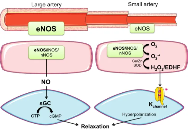
Relaxation of blood vessels dependent on the endothelium is the results of various agents such as ACh and bradykinin, or physical stimulus such as shear stress. Not only does the nature of the stimulus determine the specific EDRF responsible for the resulting signaling events, contribution by various EDRF to dilation of arteries also heavily relies on the specific arterial bed. As a general rule, larger conductance arteries usually utilize NO to a greater degree than other EDRFs, while smaller resistance arteries depend on EDHF more preferentially (Shimokawa et al. 1996; Urakami-Harasawa et al. 1997). For instance, the aorta, a conductance artery, mainly utilizes NO for dilation (Shimokawa et al. 1996) whereas smaller resistance arteries typically rely on EDHF (Shimokawa et al. 1996; Urakami-Harasawa et al. 1997), as illustrated by the key role of H2O2 in small
cerebral arteries (Drouin et al. 2007; Drouin and Thorin 2009). The contribution of PGI2,
on the other hand, usually does not play major roles regardless of vessel size (Shimokawa et al. 1996). This is summarized in Figure 5.
Figure 5. EDRF heterogeneity in arteries of varying sizes. NO is the main contributor to
vasodilation in larger conductance artery whereas its role decreases as artery size decreases, and EDHF gains important roles in the small artery. Abbreviations: eNOS: endothelial nitric oxide synthase; iNOS: inducible nitric oxide synthase; nNOS: neuronal nitric oxide synthase; NO: nitric oxide; sGC: soluble guanylate cyclase; GTP: guanosine tri-phosphate; cGMP: cyclic guanosine monophosphate; Cu/ZnSOD: copper- and zinc-containing superoxide dismutase; O2-!: superoxide; H2O2: hydrogen peroxide; EDHF:
endothelium-derived hyperpolarizing factor.
Adapted with the permission from the Rockefeller University Press: J Exp Med. Takaki A, Morikawa K, Tsutsui M, Murayama Y, Tekes E, Yamagishi H, Ohashi J, Yada T, Yanagihara N, Shimokawa H. Crucial role of nitric oxide synthases system in endothelium-dependent hyperpolarization in mice. J Exp Med. 205, 2053-2063. Copyright (2008).
However, the study of EDRF contribution to vasodilation is further complicated by their heterogeneity among different species and genders. Studies using different animal models have provided ample evidence demonstrating this rich diversity of vasodilatory pathways. For instance, FMD in porcine coronary arterioles exclusively depends on NO, demonstrated by complete abolishment of FMD by NOS inhibitors (Kuo
et al. 1991), while FMD is mainly mediated by PGI2 in cremaster muscle arterioles in rats
(Koller et al. 1995). In the guinea pig coronary arterioles, NO is the main vasodilator (Kelm and Schrader 1990; Kostic and Schrader 1992; Miura and Gutterman 1998), whereas in the same vascular bed in human, both NO and EDHF participate in vasodilation (Stork and Cocks 1994; Miura and Gutterman 1998; Miura et al. 1999), and in the rat, it is completely mediated by an EDHF (Fulton et al. 1995), whereas it is mediated by both NO and PGI2 in the rabbit (Lamontagne et al. 1992). Furthermore, an
evolution of vasodilators used in various stages of development has been observed in both animals and human. For example, in the vertebral artery, contribution of PGI2 to
ACh-mediated dilation is present at infancy but diminishes while NO takes over in adulthood (Charpie et al. 1994). As another example, endogenous H2O2 contributes to
skeletal arteriole vasodilation in juvenile rats but not in weaning rats (Samora et al. 2008). Taken together, the study of endothelium-dependent dilator mechanisms is complex – usually not only one vasodilator is involved, and different EDRFs can interact and/or compensate for each other. Hence, the endothelium is a dynamic organ with heterogeneous responses depending on different factors.
1.1.1.4.1. Interactions between different vasodilator pathways
In addition to the diversity of endothelium-dependent vasodilatory mechanisms, there are complex interactions between EDRFs. It has been proposed that COX and NOS enzymes influence each another, such that inhibition of either system could enhance the role of the other (Ichihara et al. 1998), while COX-2-derived vasodilators were observed to compensate for NO in renal arteries when NOS was acutely inhibited (Beierwaltes 2002). A more recent study reported a direct binding interaction between iNOS and COX-2 that facilitates NO S-nitrosylation and activation of COX-2 (Kim et al. 2005). Interactions between NO and cAMP, a downstream mediator in the COX pathway to mediate vasodilation, have also been reported (Zhang and Hintze 2001). Indeed, the study by Zhang and Hintze suggests that cAMP can activate protein kinase A, which in turn, through a PI3K-dependent pathway, phosphorylates eNOS by Akt (Zhang and Hintze 2001). Once again, there are complicated interactions among different vasodilatory pathways even within the same arterial bed in the same species.
In conclusion, the endothelium is capable of secreting different EDRFs, namely NO, PGI2, and EDHF in response to both mechanical and chemical stimuli in
context-dependent manners which is also context-dependent on vascular bed and vessel size, with complex interactions among them. With that, we will now focus on the other main group of endothelial mediators – the EDCFs.
1.1.2. Endothelium-derived contracting factors (EDCFs)
The endothelium, besides mediating relaxation, also plays a vital role to mediate vasocontraction to maintain vascular tone. Endothelium-dependent contractions are induced by both physical (such as pressure, stretch, and flow) and chemical (such as cytokines) stimuli (Luscher et al. 1992) and in response, the endothelium releases EDCFs. Endothelium-dependent contractions have been demonstrated in different vascular beds – aorta (Luscher and Vanhoutte 1986), carotid arteries (Traupe et al. 2002), cerebral arterioles (Mayhan 1992), femoral arteries (Shi et al. 2007) and many more. Particularly, in disease states such as hypertension, effects of EDCFs become more prominent as they may counteract actions of EDRFs to increase vascular tone. Many EDCFs have been identified and characterized, and will be briefly outlined below.
1.1.2.1. Prostanoids
Derived from COX-1 and -2 enzymes, prostanoids including endoperoxides (Auch-Schwelk et al. 1990), PGI2 (Rapoport and Williams 1996) and thromboxane A2
(Shirahase et al. 1988), have all been proposed to be EDCFs. Endoperoxides are immediate products of COX enzymes, which are then spontaneously or enzymatically converted into downstream prostanoids such as PGI2, prostaglandin D2 and E2 (Bos et al.
2004), but are also vasoconstrictors themselves (Ito et al. 1991).
PGI2, which was previously mentioned as one of the EDRFs, can also act as a
vasoconstrictor at high doses (Levy 1980; Williams et al. 1994) through activation of the TP receptors. This usually occurs in pathological states such as hypertension, where there are dysfunctions in the main IP receptor (Rapoport and Williams 1996).
Lastly, TXA2 is the major COX-derived vasoconstrictor resulting from the
and acts mainly on TP receptors (Figure 3), resulting in elevated calcium levels through calcium entry in VSMC, leading to contraction (Berridge and Irvine 1984; Shenker et al. 1991). The role of TXA2 becomes increasingly important both in aging (Drouin et al.
2011) and dyslipidemia (Gendron and Thorin 2007) in mouse cerebral arteries, as well as during atherosclerosis in mouse renal arteries (Gendron and Thorin 2007). In mouse cerebral arteries, increased TXA2 production was reported to limit eNOS activity (Drouin
et al. 2011), which was partly restored when the mice were given preventive polyphenol catechin treatment (Drouin et al. 2011). In mouse renal arteries, TXA2 production was
augmented in the presence of dyslipidemia, and was associated with a change in the redox environment (Gendron and Thorin 2007).
1.1.2.2. Endothelin-1 (ET-1)
Isolation of the 21-amino acid protein endothelin was first reported in 1988 (Yanagisawa et al. 1988) and include 3 main isoforms – ET-1, ET-2, and ET-3 (Inoue et al. 1989). ET-1, which is the predominant isoform expressed in the vasculature and released by ECs, potently vasoconstricts both in vitro and in vivo (Hickey et al. 1985; Yanagisawa et al. 1988; Miller et al. 1989), and will be the main focus herein. Through activation of two receptor subtypes – ETA (localized in VSMC only) and ETB (localized
in EC and VSMC), ET-1 exerts its vascular actions to result in phospholipase C (PLC) activation, increased cytosolic calcium and myosin kinase phosphorylation for smooth muscle contraction and ultimately vasoconstriction (Seo et al. 1994). In ECs, ET-1 activation of ETB would increase intracellular calcium leading to eNOS activation and
dilation (Tsukahara et al. 1994), whereas stimulation of ETB receptors in VSMC would
cause vasoconstriction (Haynes et al. 1995). Therefore, receptor localization and the balance between ETA and ETB receptors would ultimately determine the net effects of
ET-1. ET-1 can, in healthy human beings, cause increases in mean arterial blood pressure, reduction in heart rate, cardiac output and stroke volume (Weitzberg et al. 1993), suggesting involvement of this peptide to regulate vascular homeostasis.
1.1.2.3. Angiotensin II (AngII)
AngII is a peptide hormone well known to cause vasoconstriction, and in the long run, to cause increased blood pressure. AngII is considered the main final mediator of the renin-angiotensin system (RAS), where renin is a protease released by the juxtaglomerular cells of the kidney. Renin functions to cleave angiotensinogen, which is a glycoprotein released in the blood mainly from the liver, converting it into angiotensin I (angI) (Skeggs et al. 1954), which is then subsequently converted into angII (Skeggs et al. 1956b) by angiotensin converting enzyme (ACE) (Skeggs et al. 1956a). AngII then acts as the effector molecule by stimulating two possible receptor subtypes – the angII receptor type 1 (AT1R) (Murphy et al. 1991; Sasaki et al. 1991) and angII receptor type 2 (AT2R) (Chiu et al. 1989; Whitebread et al. 1989; Mukoyama et al. 1993). Besides merely acting as a vasoconstrictor, angII plays a pivotal role in the maintenance of cardiovascular homeostasis, while it is also implicated in many CVDs (Higuchi et al. 2007), which will be discussed later.
It is well established that the AT1R and AT2R subtypes, although sharing 34% sequence homology, are distinct in their expression patterns and functions (Lemarie and Schiffrin 2010). While the AT1R is ubiquitously expressed in the cardiovascular system, the AT2R is found highly expressed during fetal development and its expression is reduced rapidly after birth (Henrion et al. 2001). Furthermore, binding of angII to these receptors results in distinct signaling pathways and cellular responses (Lemarie and Schiffrin 2010). For instance, downstream signaling pathways of AT1 include vasoconstriction, superoxide production, cell proliferation and hypertrophy, whereas those of AT2 include vasodilation, growth inhibition and NO production (Lemarie and Schiffrin 2010). Importantly, pathological effects of angII have mainly been attributed to angII binding to the AT1R (Mehta and Griendling 2007; Lemarie and Schiffrin 2010).
The AT1R can be further categorized into the A and B subtype (Inagami et al. 1994), which share 96% homology as well as ligand binding and downstream signaling mechanisms. It is now clear that angII binding to AT1R leads to downstream effects that are also time-dependent. Within seconds, angII can activate PLC (Griendling et al. 1986), generating inositol phosphate and calcium mobilization (Brock et al. 1985), leading to