HAL Id: hal-02905794
https://hal.archives-ouvertes.fr/hal-02905794
Submitted on 2 Oct 2020HAL is a multi-disciplinary open access archive for the deposit and dissemination of sci-entific research documents, whether they are pub-lished or not. The documents may come from teaching and research institutions in France or abroad, or from public or private research centers.
L’archive ouverte pluridisciplinaire HAL, est destinée au dépôt et à la diffusion de documents scientifiques de niveau recherche, publiés ou non, émanant des établissements d’enseignement et de recherche français ou étrangers, des laboratoires publics ou privés.
Epigenetic regulation of the human mucin gene MUC4
in epithelial cancer cell lines involves both DNA
methylation and histone modifications mediated by
DNA methyltransferases and histone deacetylases
Audrey Vincent, Marie-Paule Ducourouble, Isabelle Seuningen
To cite this version:
Audrey Vincent, Marie-Paule Ducourouble, Isabelle Seuningen. Epigenetic regulation of the human mucin gene MUC4 in epithelial cancer cell lines involves both DNA methylation and histone modifi-cations mediated by DNA methyltransferases and histone deacetylases. FASEB Journal, Federation of American Society of Experimental Biology, 2008, 22 (8), pp.3035-3045. �10.1096/fj.07-103390�. �hal-02905794�
HAL Id: hal-02905794
https://hal.archives-ouvertes.fr/hal-02905794
Submitted on 2 Oct 2020HAL is a multi-disciplinary open access archive for the deposit and dissemination of sci-entific research documents, whether they are pub-lished or not. The documents may come from teaching and research institutions in France or abroad, or from public or private research centers.
L’archive ouverte pluridisciplinaire HAL, est destinée au dépôt et à la diffusion de documents scientifiques de niveau recherche, publiés ou non, émanant des établissements d’enseignement et de recherche français ou étrangers, des laboratoires publics ou privés.
Epigenetic regulation of the human mucin gene MUC4
in epithelial cancer cell lines involves both DNA
methylation and histone modifications mediated by
DNA methyltransferases and histone deacetylases
Audrey Vincent, Marie-Paule Ducourouble, Isabelle Seuningen
To cite this version:
Audrey Vincent, Marie-Paule Ducourouble, Isabelle Seuningen. Epigenetic regulation of the human mucin gene MUC4 in epithelial cancer cell lines involves both DNA methylation and histone modifi-cations mediated by DNA methyltransferases and histone deacetylases. FASEB Journal, Federation of American Society of Experimental Biology, 2008, �10.1096/fj.07-103390�. �hal-02905794�
The FASEB Journal
•
Research Communication
Epigenetic regulation of the human mucin gene MUC4
in epithelial cancer cell lines involves both DNA
methylation and histone modifications mediated by
DNA methyltransferases and histone deacetylases
Audrey Vincent, Marie-Paule Ducourouble, and Isabelle Van Seuningen1
Inserm U837, Jean-Pierre Aubert Research Centre, Place de Verdun, Lille, France
ABSTRACT The human gene MUC4 encodes a trans-membrane mucin, ligand of ErbB2, that is associated with pancreatic tumor progression. In the normal pan-creas, MUC4 is not expressed, whereas activation of its expression is observed in the early steps of pancreatic carcinogenesis. The molecular mechanisms responsible for MUC4 gene activation are however still unknown. The MUC4 5ⴕ-flanking region being GC-rich and includ-ing two CpG islands, we hypothesized that epigenetic regulation may be involved and undertook to decipher the molecular phenomenons implied. By treating can-cer cell lines with 5-aza-2ⴕ-deoxycytidine (5-aza) and trichostatin A (TSA), we were able to restore MUC4 expression in a cell-specific manner. We showed by bisulfite-treated genomic DNA sequencing and chroma-tin immunoprecipitation that methylation of five CpG sites and establishment of a repressive histone code at the 5ⴕ-untranslated region were associated with MUC4 silencing and impaired its activation by Sp1. Direct involvement of DNMT3A, DNMT3B, HDAC1, and HDAC3 was demonstrated by RNA interference and chromatin immunoprecipitation. Moreover, inhibition of histone deacetylation by TSA was associated with strong MUC4 repression in high-expressing cells. In conclusion, this work shows for the first time the importance of epigenetics in regulating MUC4 expres-sion and may represent a new strategy to inhibit its expression in epithelial tumors—Vincent, A., Ducour-ouble, M.-P., Van Seuningen, I. Epigenetic regulation of the human mucin gene MUC4 in epithelial cancer cell lines involves both DNA methylation and histone modifications mediated by DNA methyltransferases and histone deacetylases. FASEB J. 22, 000 – 000 (2008)
Key Words: acetylation
During tumor development, aberrant DNA hyper-methylation associated with histone deacetylation (1) is a common molecular mechanism that leads to the silencing of numerous genes implicated in cell differ-entiation, signaling, and DNA repair (2, 3). This pro-vides a survival advantage to neoplastic cells and influ-ences drug resistance and clinical outcome after
therapy (4). However, gene-specific hypomethylation occurs frequently during carcinogenesis and particu-larly in colonic and pancreatic cancers and correlates well with increased transcription levels (5). Detection of aberrant promoter methylation (or demethylation) may provide useful tools for early diagnosis and prog-nosis of cancers, including those of lung (6), pancreas (7), or colon (8). However, although common methyl-ation markers have been clearly established during the past few years (9), identification of panels of comple-mentary biomarkers that are cancer specific is still necessary to establish precise DNA methylation signa-tures that would be beneficial for patient screening. Hence, studying the methylation status of genes aber-rantly expressed in cancers may help in developing these early detection tools (10).
Pancreatic carcinogenesis is a stepwise process char-acterized by preneoplastic tumors called pancreatic intraepithelial neoplasia (PanIN) with different grades (PanIN1/2A/2B/3) that degenerate into carcinoma (11). Thus, targeting PanIN or genes expressed in PanIN represents a potential therapeutic approach to control pancreatic tumor progression. The human gene MUC4 encodes a transmembrane mucin charac-terized by a large growth factor-like extracellular do-main, ligand of ErbB2, that participates in ErbB2 signaling, cell proliferation, and tumor progression (12, 13). In pancreatic cancer, MUC4 has become an important molecular target (14), because it is not expressed in normal pancreas but is increasingly acti-vated during the carcinogenetic sequence as early as PanINs (15). Moreover, its overexpression in pancre-atic carcinomas is associated with a bad prognosis. For all these reasons, MUC4 has been recently proposed as a potent diagnostic (16) and prognostic (17) factor as well as a tumor marker (18) in pancreatic cancer. However, despite extensive data at the cellular level, the molecular mechanisms that control MUC4 de novo expression in pancreatic cancer are still unknown.
1Correspondence: Inserm, U837, Pl. de Verdun, 59045 Lille cedex, France. E-mail: isabelle.vanseuningen@inserm.fr
doi: 10.1096/fj.07-103390
1 0892-6638/08/0022-0001 © FASEB
AQ: 1
The characterization of the 5⬘-flanking region of
MUC4 in our laboratory showed that it is under the
control of two transcriptional units and that both the 5⬘-untranslated region (5⬘-UTR) and the proximal pro-moter are extremely GC-rich (up to 72%), whereas the distal promoter is not as GC-rich (up to 65%) (19). Moreover, the analysis of the genomic DNA sequence indicated the presence of numerous potential methyl-ated cytosine residues as well as two CpG islands. Considering the biological activities of the mucin MUC4 in tumor progression, its de novo expression in pancreatic cancer or overexpression in early stages of numerous epithelial cancers sustained during the car-cinogenetic sequence, and the structure of its 5 ⬘-flank-ing region, we hypothesized that epigenetic mecha-nisms may be involved and thus undertook to decipher the molecular mechanisms responsible for MUC4 epi-genetic regulation in epithelial cancer cells. In this work, we report the identification of key methylated CpG sites and the establishment of a repressive histone code at the 5⬘-UTR that are associated with the silenc-ing of MUC4 in epithelial cancer cell lines and discuss the possible implications in cancer therapy.
MATERIALS AND METHODS
Cell lines and cell culture
The pancreatic (PANC-1, CAPAN-1, and CAPAN-2) and gas-tric (KATO-III) epithelial cancer cell lines used in this study were cultured as described previously (19 –21). The inhibitor of methylation, 5-aza-2⬘-deoxycytidine (5-aza; 5 M, Sigma, Saint-Quentin Fallavier, France), and the inhibitor of histone deacetylase (HDAC), trichostatin A (TSA; 0.3 M, Sigma), were added to proliferating cells (PCs) or confluent cells (CCs) for 72 and 24 h, respectively, as described before (22). Cells were then lysed and processed for total RNA extraction or whole cellular extract preparation as described thereafter.
RNA extraction, reverse transcriptase-polymerase chain reaction (RT-PCR), and quantitative real-time
PCR (Q-PCR)
Total RNA was prepared using the QIAamp RNA blood and cell reaction kit (Qiagen, Courtaboeuf, France). Total RNA (1g) was used to prepare cDNA as described by Vincent et al. (22). RT-PCR was carried out on cDNA (5 l) using a specific pair of primers for MUC4 (19). The ribosomal RNA 28S subunit was used as the internal control (20). Single-stranded oligonucleotides were synthesized by MWG-Biotech (Ebersberg, Germany). PCR products (10l) were separated on a 1.5% agarose gel containing ethidium bromide run in 1⫻ Tris-borate-EDTA buffer. Multiplex Q-PCR was performed in triplicate using the AB 7500 Fast Real-Time PCR System and the Absolute Q-PCR Rox Mix (Applied Biosystems, Courtaboeuf, France). The ribosomal RNA Control Reagent (Applied Biosystems) was used as an internal amplification control. Primer sets for MUC4 were as follows: forward primer: 5⬘-TCAGCTGAGGCCTTGCCTT-3⬘, reverse primer: 5⬘-TCAGTCACCTTCCCTTTTCCA-3⬘, and probe:FAM 5⬘-TAAG-GCGCCATTGCTTTTGGGAGA-3⬘Tamra. Relative transcript lev-els were calculated using the 2⫺⌬⌬CTmethod. Untreated cells were used as reference samples.
Immunohistochemistry
PANC-1 cells were passed at 1.5⫻ 106cells/75 cm2flask and grown under standard conditions until they reached conflu-ency, at which time they were treated either for 48 h with 5-aza (5M) or 24 h with TSA (0.3 M). Cells were then trypsinized, centrifuged, and washed once with 1⫻ PBS. The cell pellet was fixed in 4% (w/v) formaldehyde and embed-ded in paraffin, and 3m sections were prepared. Immuno-histochemistry, including positive and negative (omitting primary antibody) controls, was performed using an auto-matic immunostainer (ES; Ventana Medical System, Illkirch, France), as described previously (23). The monoclonal anti-MUC4 antibody, which recognizes the tandem repeat region of the apomucin (24), was used at a 1/2000 dilution.
Whole cell extract preparation and Western blotting
Total cellular extracts from CAPAN-1 and KATO-III cells were prepared using standard procedures as described previously (25). Protein content was measured using the bicinchoninic acid method as described in the manufacturer’s instruction manual (Pierce, Perbio Sciences, Brebie`res, France). MUC4 protein (⬎900 kDa) and -actin (42 kDa) expression were respectively analyzed on 2% (w/v) SDS-agarose gels and 10% SDS-PAGE followed by transfer on nitrocellulose membrane as described in Piessen et al. (25). Prestained protein molec-ular weight standards were from Life Technologies (Cergy-Pontoise, France). The monoclonal anti-MUC4 antibody was used at a concentration of 1g/ml. The MUC4/-actin ratio was calculated after scanning protein bands with GelAnalyst-GelSmart software.
Sodium bisulfite-treated genomic DNA sequencing
Genomic DNA was extracted from the three cell lines used in this study with the Blood and Cell Culture DNA Maxi Kit (Qiagen). DNA content was quantified at 260 nm and stored at⫹4°C until use. Sodium bisulfite conversion was performed using 5g of genomic DNA from cell lines as described by Ghoshal et al. (26). The promoter sequences of interest were amplified by PCR using AmpliTaq Gold polymerase (Applied Biosystems) as described previously (26). Primer sequences covering the 5⬘-UTR, the proximal and distal promoters, as well as the two CpG islands were designed using the MethPrimer software (http://www.urogene.org/methprimer/;
Table 1). PCR products were cloned into pCR2.1 vector (Invitro-gen, Cergy Pontoise, France). At least five positive clones per site were selected for plasmid preparation using QIaprep 8 Mini-prep Kit (Qiagen) and sequenced on both strands on an infrared based ABI 3730 XL sequencer (GATC, Konstanz, Germany) using T7 and M13-RP universal primers. The percent-age of conversion of non-CpG cytosines was used as an index of overall bisulfite reaction efficiency. Clones with a conversion efficiency of⬎97% were included in the study.
Site-directed mutagenesis, in vitro methylation of MUC4 proximal promoter and 5ⴕ-UTR, and cell
transfection studies
QuickChange site directed mutagenesis kit (Stratagene, Am-sterdam, The Netherlands) was used to generate site-specific mutations. Oligonucleotides containing the desired muta-tions are shown in Table 2. Wild-type (WT) and mutated MUC4-pGL3 constructs (⫺461/⫺1 region) were digested with SacI-MluI restriction enzymes (Roche Diagnostics, Mey-lan, France), and the insert was gel-purified as described by
T1
Perrais et al. (21) before being methylated (10g) with 20 U of mSssI (CG) and mHpaII (CCGG) enzymes (New England Biolabs, Ozyme, St-Quentin en Yvelines, France) for 3 h at 37°C. The degree of methylation of the fragment was con-firmed by testing its resistance to HpaII digestion. The methylated fragments were then ligated into pGL3 basic vector. DNA concentration was measured at 260 nm before being used in transfection experiments in CAPAN-2 cells as described by Perrais et al. (19). Influence of methylation of WT and mutated MUC4 promoter (1g) on its transactiva-tion by Sp1 was studied by carrying out cotransfectransactiva-tions experiments in the presence of pCMV-Sp1 (0.5g) expres-sion vector as described in Van Seuningen et al. (27). Samples were tested in triplicate in at least three independent exper-iments.
Chromatin immunoprecipitation assay
Cells (1.0⫻106per antibody) were fixed in 1% (v/v) formal-dehyde, and chromatin was sonicated and immunoprecipi-tated as described in Vincent et al. (22) with either 5g of specific antibodies against histone H3 (antiacetylated lysine, methylated lysine 9, and trimethylated lysine 27; Upstate, Hampshire, UK) or with specific antibodies against chroma-tin modifier enzymes: DNMT1, DNMT3B, HDAC1, HDAC2, HDAC3 (Abcam, Paris, France), and DNMT3A (Imgenex, CliniSciences, Montrouge, France) or with normal rabbit IgGs (Upstate) at⫹4°C. Immunoprecipitated chromatin (50
ng) was then used as a template for PCR using the following primers: forward primer: 5 ⬘-TTTTGTCCTCTTCCCAG-GTTC-3⬘ and reverse primer: 5⬘-TGGCTGCGGCAAAAGTCC-3⬘, covering the ⫺153/⫹1 region of MUC4 proximal pro-moter and 5⬘-UTR. PCR was performed using AmpliTaq Gold polymerase as described in Piessen et al. (25) with an anneal-ing temperature of 55°C. PCR products (20l) were sepa-rated on a 2% agarose gel containing ethidium bromide run in 1⫻ TBE buffer.
Small interfering RNA (siRNA) assays
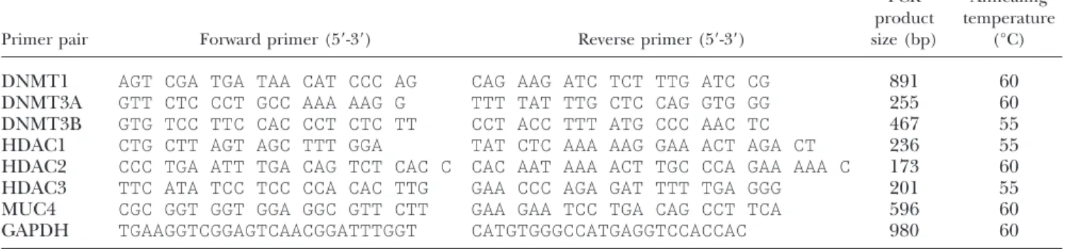
KATO-III cells were seeded the day before transfection in 24-well tissue culture plates at a density of 3⫻ 105cells/well in antibiotic free medium. Cells were transfected with either 100 nM of DNMT1, DNMT3A, DNMT3B, HDAC1, HDAC2, or HDAC3 ON-TARGETplus SMARTpool siRNA using 1l of DharmaFECT4 transfection reagent (Dharmacon, Brebie`res, France) as described in Piessen et al. (25). Controls included mock-transfected cells and cells transfected with siControl nontargeting siRNA or siControl GAPD siRNA. Total RNA was isolated 48 h after transfection, and RT-PCR was per-formed as described above. Primers used for amplification of the internal control GAPDH, and chromatin modifier en-zymes are described in Table 3. Samples were tested in triplicate in at least three independent experiments. The MUC4/GAPDH ratio was calculated as described above. TABLE 1. Sequences of the pairs of primers used for bisulfite sequencing studies
Primer pair Forward primer (5⬘-3⬘) Reverse primer (5⬘-3⬘)
Primer position (PCR product size)
Annealing temperature
(°C)
MUC4 (I) GGGATAGTGTGTGGTTTAAAAAGTT CCCTAATCAATAACCCAACATAAAA Distal prom. ⫺3600/⫺3380 (221 bp)
56
MUC4 (II) TTTAATTTTGGAAAATGGGTATATTG AACCAACCAAAATACAAAAAAAATC Distal prom. ⫺2801/⫺2595 (207 bp)
50
MUC4 (III) AGGTGTATTTTTATTTTATAGGTGAA ACAAATAACTAACCTCTTTCCCATAC CpG island II ⫺2400/⫺2211 (190 bp)
50
MUC4 (IV) TTTATTTAGAGTTGGAGGGATTGTT ACTCAAATTTCTACATTCCCAAAC Prox. prom. ⫺1595/⫺1370 (226 bp)
50
MUC4 (V) TTATGGGTTTGGGGTTTGTTAT AAACAAAAACAAAAATACACTATATACC CpG island I ⫺787/⫺532 (256 bp)
50
MUC4 (VI) GTTTAGGTTGATGAGAAGTAGAGTAA CAACAACTACAATATAAAAAACAAAC 5⬘UTR ⫺265/⫺47 (219 bp)
50
PCR product size and primer position refer to the first ATG (19).
TABLE 2. Sequences of the pairs of primers used for site-directed mutagenesis
Primer pair Sequence (5⬘-3⬘) Position
Sp1 WT CCCAGGTTCCCTGGCCCCTTCGGAGAAACGC ⫺129/⫺122 Mut#1 CCCAGGTTCCCTGGCATCTTCGGAGAAACGC
Sp1 WT CCTGGTGGGGTAGTGGGGTGGGGCTGAGGAGAGAAAAGGG ⫺205/⫺193 Mut#2 CCTGGTGGGGTAGTGATGTGGTGCTGAGGAGAGAAAAGGG
Mutated nucleotides are italicized and underlined. Antisense oligonucleotides were also synthe-sized and used for site-directed mutagenesis as described in Materials and Methods.
3 EPIGENETIC REGULATION OF MUC4 MUCIN GENE
Statistics
All values in this article are mean⫾ sd. When indicated, data were analyzed by Student’s t test using GraphPad Prism 4 software (GraphPad, San Diego, CA, USA) with differences P⬍ 0.05 considered significant.
RESULTS
Influence of DNA methylation and histone deacetylation on MUC4 expression in epithelial cancer cells
To study the role of methylation and histone acetyla-tion on MUC4 expression, one nonexpressing pancre-atic cancer cell line (PANC-1) and one high-expressing pancreatic cancer cell line (CAPAN-1) were treated with either the demethylating agent 5-aza (Fig. 1A) or the HDAC inhibitor TSA (Fig. 1B). As an intermediate
model, the low-expressing gastric cancer cell line (KATO-III) was used. Studies were conducted in PCs and CCs to evaluate epigenetic regulation of MUC4 in both cellular situations.
In nonexpressing pancreatic PANC-1 PCs and CCs, treatment with 5-aza and TSA induced MUC4 expres-sion at the mRNA level (Fig. 1A, B, lanes 2 and 4). Induction of MUC4 protein expression was also ob-served in a few cells by immunohistochemistry after either 5-aza or TSA treatment (Fig. 1C).
In low-expressing KATO-III PCs, 5-aza and TSA treat-ments strongly increased MUC4 expression both at the mRNA (4.07- and 3.03-fold increase by Q-PCR, respec-tively, Fig. 1A, B, lane 2) and protein (Fig. 1D) levels. In KATO-III CCs, DNA demethylation with 5-aza still induced a 3-fold activation (Q-PCR) of MUC4 mRNA expression and inhibition of histone deacetylation with TSA induced a 2.39-fold activation (Fig. 1A, B, lane 4 and Q-PCR).
TABLE 3. Sequences of the pairs of primers used for RT-PCR studies
Primer pair Forward primer (5⬘-3⬘) Reverse primer (5⬘-3⬘)
PCR product size (bp) Annealing temperature (°C)
DNMT1 AGT CGA TGA TAA CAT CCC AG CAG AAG ATC TCT TTG ATC CG 891 60 DNMT3A GTT CTC CCT GCC AAA AAG G TTT TAT TTG CTC CAG GTG GG 255 60 DNMT3B GTG TCC TTC CAC CCT CTC TT CCT ACC TTT ATG CCC AAC TC 467 55 HDAC1 CTG CTT AGT AGC TTT GGA TAT CTC AAA AAG GAA ACT AGA CT 236 55 HDAC2 CCC TGA ATT TGA CAG TCT CAC C CAC AAT AAA ACT TGC CCA GAA AAA C 173 60 HDAC3 TTC ATA TCC TCC CCA CAC TTG GAA CCC AGA GAT TTT TGA GGG 201 55 MUC4 CGC GGT GGT GGA GGC GTT CTT GAA GAA TCC TGA CAG CCT TCA 596 60 GAPDH TGAAGGTCGGAGTCAACGGATTTGGT CATGTGGGCCATGAGGTCCACCAC 980 60
Figure 1. Epigenetic regulation of MUC4 in epithelial cancer cell lines. A, B) RT-PCR was performed as de-scribed in Materials and Methods. The expected sizes for PCR products of MUC4 (lanes 1 to 4) and 28S (lanes 5 to 8) are 596 and 231 bp, respectively. PCR products (10l and 5l) were separated on a 1.5% agarose gel contain-ing ethidium bromide. Untreated (A: ⫺, lanes 1, 3, 5, and 7) or treated (⫹, lanes 2, 4, 6, and 8) cells with 5-aza (5 M, 72 h) or TSA (B: 0.3 M, 24 h). C) MUC4 immunohistochemical detection in untreated (left panel) and 5-aza- or TSA-treated (right panel) PANC-1 cells was performed as described in Materials and Methods. View⫻400. D) Influence of 5-aza and TSA treatment on MUC4 apomucin expression in KATO-III PCs and CAPAN-1 CCs. Untreated (⫺) and 5-aza- or TSA-treated cells (⫹). Total cellular extracts were prepared as described in Materials and Methods; 20g of proteins was loaded on a 2% (w/v) SDS-agarose gel (MUC4) or 10% SDS-PAGE (-actin) before being transferred onto nitrocellulose membrane and processed for MUC4 and -actin immunohistochemical detection. MUC4/-actin ratios are indicated, ratio corresponding to untreated cells was arbitrarily set to 1.
F1
C
O
L
O
R
In high-expressing pancreatic CAPAN-1 PCs and CCs, 5-aza treatment did not affect MUC4 mRNA level (Fig. 1A and as determined by Q-PCR), whereas TSA treatment induced a strong inhibition of MUC4 mRNA expression (3.22- and 1.87-fold decrease, respectively, Fig. 1B, lanes 2 and 4 and Q-PCR) correlated to a strong decrease of the apomucin level in the cells (Fig. 1D).
Mapping of methylated CpG sites within MUC4 promoters and 5ⴕ-UTR
In silico analysis of MUC4 5⬘-flanking region using the
MethPrimer software indicated the presence of two CpG islands at ⫺2370/⫺2236 (CpG island II) and ⫺738/⫺593 (CpG island I), both located in the prox-imal promoter (Fig. 2A). The 5⬘-flanking region has a very high GC content (up to 72%) almost exclusively concentrated within the proximal promoter and 5 ⬘-UTR. Bisulfite-treated DNA sequencing was used to map methylated cytosines within the distal and proxi-mal promoters and 5⬘-UTR of MUC4 in the three cell lines studied (Fig. 2A). Primer information is given in Table 1. Our results indicate that the two CpG islands are heavily methylated regardless of the level of MUC4 expression in cells. Most of the CpG sites studied are also heavily methylated in the three cell lines except for cytosines in the ⫺239/⫺73 region of the proximal promoter and 5⬘-UTR (Fig. 2A). Five of these CpG sites, all located in the 5⬘-UTR (at ⫺81, ⫺93, ⫺102, ⫺113, and ⫺121) were significantly more methylated in the nonexpressing cell line PANC-1 (92% methylation) compared with the expressing cell line CAPAN-1 (4% methylation). As expected, in low-expressing KATO-III cells, these CpG sites showed an intermediate methyl-ation profile (16% methylmethyl-ation). A more remote cyto-sine of the proximal promoter at ⫺1432 was also
significantly less methylated in CAPAN-1 cells com-pared with PANC-1. Thus, the pattern of methylation of these six CpG sites is well correlated to the mRNA level of MUC4 in the cells. Among them, the five CpG sites clustered in the 5⬘-UTR showed a methylation pattern strictly correlated to MUC4 expression. We thus focused the rest of the work on these five CpG sites. To identify among these five CpG sites which ones would be sensitive to 5-aza and thus demethylated after cell treatment with that drug, we submitted nonexpressing PANC-1 CCs to 5-aza treatment and compared their methylation profile to genomic DNA from untreated cells. Our results indicate that DNA demethylation by 5-aza was associated with demethylation (60%) of cy-tosines at ⫺81 and ⫺93 (Fig. 2B). This suggests that activation of MUC4 expression in 5-aza-treated PANC-1 cells is correlated to demethylation of these two CpG sites.
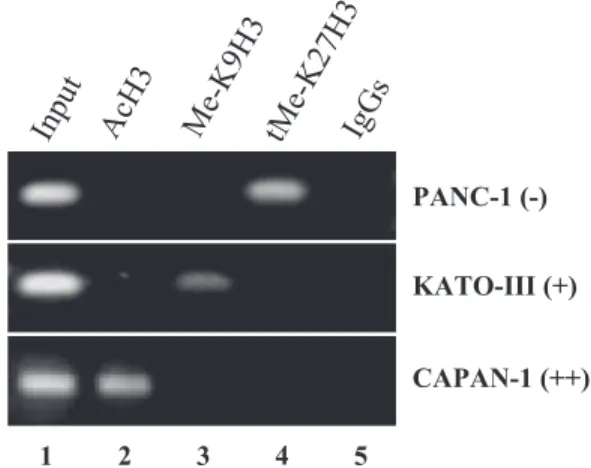
Study of histone H3 modifications at MUC4 5ⴕ-UTR
To establish histone H3 status in MUC4 5⬘-UTR, ChIP assays were carried out with chromatin from the three cell lines used in this study (Fig. 3). In nonexpressing PANC-1 cells, MUC4 5⬘-UTR DNA covering the ⫺153/⫹1 region was associated with deacetylated his-tone H3 (lane 2) and trimethylated K27H3 (lane 4), whereas K9H3 was not methylated (lane 3). In low-expressing KATO-III cells, histone H3 was also deacety-lated at MUC4 5⬘-UTR (lane 2), which was enriched in methylated K9H3 (lane 3). In high-expressing CA-PAN-1 cells, MUC4 expression was correlated to both acetylation and demethylation of histone H3 (lanes 2– 4). Thus, inhibition of MUC4 expression in these three epithelial cancer cell lines is associated with
Figure 2. Mapping of methylated cy-tosines within MUC4 distal and proximal promoters and 5⬘-UTR by bisulfite-treated DNA sequencing. A) Studies were performed in PANC-1 CC nonexpressing (⫺), KATO-III CC low-expressing (⫹), and CAPAN-1 CC (⫹⫹) MUC4 high-ex-pressing cells. Numbering refers to the first ATG (⫹1) of MUC4 gene (19); broken arrows indicate the transcription start sites situated at⫺199 and ⫺2603, determining the proximal and the distal promoters of MUC4 (19). Vertical thin bars indicate CpG sites. Horizontal thick bars indicate the positions of the two CpG islands. At least 5 individual clones were analyzed per CpG site (vertical bars) and per cell line. Black squares indicate methylated CpG sites; white squares indicate unmethylated CpG sites. Key CpG methylated sites are topped with a star. B) Mapping of methylated CpG sites of MUC4 5⬘-UTR in untreated (⫺, nonexpressing) and 5-aza-treated (⫹, MUC4-expressing) PANC-1 cells.
5 EPIGENETIC REGULATION OF MUC4 MUCIN GENE
F2
establishment of a repressive histone code, including histone H3 deacetylation and methylation.
DNMT3A, DNMT3B, HDAC1, and HDAC3 chromatin modifier enzymes regulate MUC4 silencing
To identify which DNA methyltransferases (DNMT1, DNMT3A, and DNMT3B) and class I HDACs (HDAC1, HDAC2, and HDAC3) regulate MUC4 endogenous expression, knockdown assays were performed with specific siRNAs in the KATO-III cell line (Fig. 4A). DNMT1 and HDAC1 knockdown led to a 1.8- and
2.9-fold increase of MUC4 expression in KATO-III cells (Fig. 4B). Knocking down of DNMT3A, DNMT3B, HDAC2, and HDAC3 also led to an increase of MUC4 mRNA expression that was significant (4.4-fold,
P⬍0.05; 4.6-fold, P⬍0.05; 3.7-fold, P⬍0.005; and
4.7-fold, P⬍0.05, respectively).
To show whether DNMTs and HDACs may directly modify the methylation profile and chromatin struc-ture at MUC4 5⬘-UTR, ChIP assays were then carried out with chromatin from the three cell lines used in this study (Fig. 4C). In nonexpressing PANC-1 cells, bind-ing of DNMT3A (lane 4), DNMT3B (lane 5), and HDAC1 (lane 6) to DNA covering the⫺153/⫹1 region of MUC4 5⬘-UTR was observed. In low-expressing KATO-III cells, we found binding of DNMT3B (lane 5) and a strong binding of HDAC3 (lane 8). On the contrary, in high-expressing CAPAN-1 cells, neither DNMTs nor HDACs were found to bind MUC4 5⬘-UTR. Thus, DNMT3A, DNMT3B, HDAC1, and HDAC3 are directly involved in MUC4 silencing by binding to its 5⬘-UTR in a cell-specific manner.
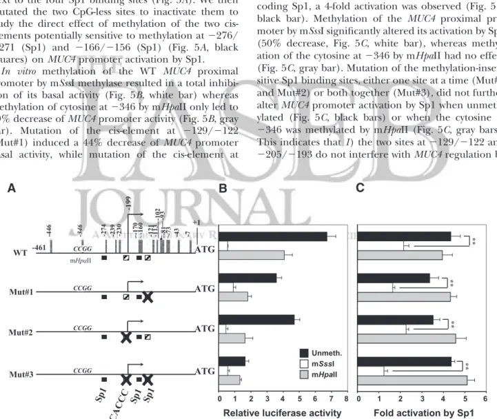
Effect of DNA methylation on MUC4 promoter basal activity and regulation by Sp1
Having shown that methylation was involved in MUC4 repression, we undertook to study whether promoter methylation could interfere with MUC4 promoter basal activity and regulation by the transcription factor Sp1, a known regulator of MUC4 transcription (19). These studies were performed in MUC4-expressing pancreatic CAPAN-2 cell line in which MUC4 transactivation by Sp1 was previously shown to be the strongest (19). In that part of the 5⬘-flanking region (⫺461/⫺1), which includes part of the proximal promoter and the 5
⬘-Figure 3.Histone modification status of MUC4 promoter in PANC-1, KATO-III, and CAPAN-1 cell lines. ChIP assays were performed as described in Materials and Methods. Three histone H3 modifications were analyzed: acetylated lysine (AcH3), monomethylated lysine 9 (Me-K9H3), and trimethyl-ated lysine 27 (tMe-K27H3). Input DNA was used as a positive amplification control; IgG indicates ChIP performed using rabbit IgGs as an isotypic antibody control.
Figure 4. Role of chromatin modifier enzymes (DNMT1, DNMT3A, DNMT3B, HDAC1, HDAC2, and HDAC3) on the endogenous expression of MUC4. A) siRNA knockdown of chromatin modifier enzymes in KATO-III cell line was carried out as described in Materials and Methods. Expression level for each DNMT and HDAC was evaluated by RT-PCR using specific primers (Table 3). Levels of expression of the different genes are shown for cells transfected with nontargeting siRNA (control, lanes 1 and 2) or with specific siRNA (lanes 3 and 4). B) MUC4 and GAPDH mRNA levels were assessed by RT-PCR. PCR products (10l MUC4, 6 l GAPDH) were analyzed on a 1.5% agarose gel. Diagram shows data expressed as MUC4/GAPDH ratio. Control corresponds to the mean value from mock cells and cells transfected with nontargeting siRNA. Transfections were carried out in triplicate in at least three independent experiments. *P⬍ 0.05; **P ⬍ 0.005. C) Binding of chromatin modifier enzymes to MUC4 5⬘-UTR was assessed by ChIP assays using specific antibodies against DNMT1, DNMT3A, DNMT3B, HDAC1, HDAC2, and HDAC3.
UTR, three Sp1 cis-elements and one Sp1-binding CACCC box have been previously identified (Fig. 5A, black and hatched squares; ref. 19). Among these four cis-elements, those situated at ⫺129/⫺122 (Sp1) and ⫺205/⫺193 (CACCC box) do not contain any CpG site (Fig. 5A, hatched squares) and thus are insensitive to DNA methylation.
In vitro methylation of the two other CpG-containing
Sp1 sites (⫺276/⫺271 and ⫺166/⫺156, black squares) was carried out with two methylases: mSssI, which methylates cytosine residues in CpG dinucleotides, and mHpaII, which methylates the second cytosine residue in the CCGG sequence. In the MUC4⫺461/⫺1 region, only one mHpaII-sensitive element was found at⫺346, next to the four Sp1 binding sites (Fig. 5A). We then mutated the two CpG-less sites to inactivate them to study the direct effect of methylation of the two cis-elements potentially sensitive to methylation at⫺276/ ⫺271 (Sp1) and ⫺166/⫺156 (Sp1) (Fig. 5A, black squares) on MUC4 promoter activation by Sp1.
In vitro methylation of the WT MUC4 proximal
promoter by mSssI methylase resulted in a total inhibi-tion of its basal activity (Fig. 5B, white bar) whereas methylation of cytosine at⫺346 by mHpaII only led to 40% decrease of MUC4 promoter activity (Fig. 5B, gray bar). Mutation of the cis-element at ⫺129/⫺122 (Mut#1) induced a 44% decrease of MUC4 promoter basal activity, while mutation of the cis-element at
⫺205/⫺193 (Mut#2) induced a 30% decrease and mutation of both sites (Mut#3) almost totally abolished
MUC4 promoter basal activity (78% decrease) (Fig. 5B,
black bars). As expected, methylation of the three mutants (Mut#1, Mut#2, and Mut#3) by mSssI resulted in a total inhibition of their basal activity (Fig. 5B, white bars). Methylation of the cytosine at⫺346 by mHpaII did not further alter transcriptional activity of the three mutants, as the decrease was similar to that observed with the WT construct when compared with respective unmethylated constructs (30 – 60% inhibition; Fig. 5B, compare gray bars to black bars).
When cotransfection was performed in the presence of WT MUC4 promoter and an expression vector en-coding Sp1, a 4-fold activation was observed (Fig. 5C, black bar). Methylation of the MUC4 proximal pro-moter by mSssI significantly altered its activation by Sp1 (50% decrease, Fig. 5C, white bar), whereas methyl-ation of the cytosine at⫺346 by mHpaII had no effect (Fig. 5C, gray bar). Mutation of the methylation-insen-sitive Sp1 binding sites, either one site at a time (Mut#1 and Mut#2) or both together (Mut#3), did not further alter MUC4 promoter activation by Sp1 when unmeth-ylated (Fig. 5C, black bars) or when the cytosine at ⫺346 was methylated by mHpaII (Fig. 5C, gray bars). This indicates that 1) the two sites at⫺129/⫺122 and ⫺205/⫺193 do not interfere with MUC4 regulation by
Figure 5.Influence of methylation on MUC4 promoter basal activity and regulation by Sp1. A) Schematic representation of the deletion mutant covering the⫺461/⫺1 region of MUC4 proximal promoter and 5⬘-UTR. Numbering refers to the first ATG (⫹1) of MUC4 gene; broken arrow indicates the transcription start site situated at ⫺199 (19). Black squares indicate CpG-containing Sp1 cis-elements; hatched squares are CpG-less Sp1 binding sites (19). Vertical bars indicate CpG sites. The mHpaII sensitive site is indicated (⫺346, CCGG). B) Luciferase diagram showing the effect of MUC4 promoter methylation by mSssI (CG) or mHpaII (CCGG) on its activity in pancreatic CAPAN-2 cell line. Unmethylated pGL3-MUC4 (⫺461/⫺1) (black bars) and corresponding mSssI- (white bars) or mHpaII-methylated (gray bars) promoter constructs. C) Study of the effect of promoter methylation on MUC4 transactivation by Sp1. Results are expressed as relative luciferase activity of the unmethylated pGL3-MUC4 constructs (black bars) or the corresponding methylated promoter construct with either mSssI (white bars) or mHpaII (gray bars) cotransfected with pCMV4-Sp1 expression vector. Transfections were carried out in triplicate in 3 independent experiments. **P⬍ 0.005.
7 EPIGENETIC REGULATION OF MUC4 MUCIN GENE
Sp1, and 2) methylation of the adjacent cytosine at ⫺346 does not alter activation by Sp1 either.
Promoter methylation by mSssI (Fig. 5C, white bars) did not further alter MUC4 activation by Sp1 when the binding site at⫺129/⫺122 (Mut#1) or at ⫺205/⫺193 (Mut#2) was mutated when compared with the corre-sponding unmethylated constructs (51 and 35% de-crease, respectively). On the contrary, activation by Sp1 was significantly altered with almost total inhibition when the two CpG-less binding sites were mutated (Mut#3, 72% decrease). This indicates that direct methylation of the two CpG-containing Sp1 binding sites at ⫺276/⫺271 and ⫺166/⫺156 (Fig. 5A, black squares) dramatically impairs MUC4 activation by Sp1.
DISCUSSION
In this study, we have investigated the role of epigenet-ics in the regulation of MUC4 mucin gene expression. Our results show that MUC4 is regulated by DNA demethylation and establishment of a derepressive histone code at its 5⬘-UTR involving DNMT3A, DNMT3B, HDAC1, and HDAC3. Moreover, the use of three cell lines with different levels of MUC4 expression allowed us to propose a model for MUC4 epigenetic regulation in epithelial cells (Fig. 6). We propose that in nonexpressing cells, 5⬘-UTR methylation and a re-pressive histone code cooperate to silence MUC4 mucin gene, whereas in low-expressing cells, progressive
de-methylation of the promoter but a repressive histone code allows low expression of MUC4; in high-expressing cells, selective hypomethylation and permissive chro-matin allow binding of factors such as Sp1 and full transcription of MUC4.
The use of demethylating agent 5-aza and HDAC inhibitor TSA in three cancer cell lines showing differ-ent levels of MUC4 expression indicated that DNA methylation and histone deacetylation play an impor-tant role in MUC4 silencing. In nonexpressing and low-expressing cells, treatment with the two drugs in-duced a strong increase in MUC4 expression. This result differs from the paradigm that, in the collabora-tion between DNA methylacollabora-tion and histone deacetyla-tion in silencing gene expression, DNA methyladeacetyla-tion is dominant (28), whereas TSA alone is unable to reacti-vate genes with densely methylated CpG islands (29). Our results clearly show that both epigenetic mecha-nisms of DNA methylation and histone deacetylation are involved in the silencing of MUC4, which is consis-tent with other studies reporting the influence of histone acetylation on the methylation status of genes (30).
Interestingly, we observed that inhibition of histone deacetylation by TSA specifically induced the decrease of both MUC4 gene and apomucin expression in high-expressing cells. We and others (22, 31, 32) have already shown that HDAC inhibitors are able to inhibit mucin gene expression in cancer cells. In a previous study, Ferguson et al. (33) showed that occupancy levels of the Sp1 transcription factor on the promoter of
Hmga2, a gene involved in the control of chromatin
structure, decreased significantly after TSA treatment. As Sp1 is an activator of MUC4 transcription (19), the TSA-mediated inhibition of Sp1 DNA binding may explain the decrease in expression observed in this study. However, TSA and other HDAC inhibitors, such as butyrate, have been shown to 1) influence cell maturation by repressing secretory markers such as the mucin gene MUC2 (32), 2) induce apoptosis and cell cycle arrest, and 3) enhance the response to chemo-therapeutic agents in pancreatic cancer cells (34). As MUC4 is implicated in cell proliferation, invasion, and metastasis in pancreatic cancer (13), the TSA-mediated inhibition of MUC4 we observed next to that of other genes (34) may thus represent the molecular basis for the antitumoral activity of this drug in pancreatic cancer. Moreover, TSA has also been shown to repress expression of the oncogenic ErbB2 (35). Because MUC4 is the ligand of ErbB2, it participates in ErbB2-mediated intracellular signaling, its localization, and trafficking (12, 36); therefore, the concerted inhibition of both MUC4 and ErbB2 by TSA could also contribute to the antitumor activities of this drug in epithelial cancers implying MUC4-ErbB2 complex formation, a mechanism known to promote tumor proliferation and progression (37). Finally, because MUC4 is often over-expressed in epithelial cancers and is considered as one of the most attractive targets for novel therapeutic strategies (14, 38), another application would be to
Figure 6. Model for epigenetic regulation of MUC4 expres-sion in epithelial cells based on mechanistic studies in epi-thelial cancer cell lines expressing different levels of MUC4 gene. Me, methylated cytosine; Ac, acetylated histone. AQ: 3
target MUC4 high-expressing cells by using pharmaco-logical drugs such as TSA.
The results obtained in pancreatic cancer cell lines (this study) are in accordance with a previous study in which Singh et al. (39) suggested that DNMT and HDAC activity may be involved in the down-regulation of MUC4 in prostate cancer cell lines. However, no mechanisms regarding direct silencing of MUC4 by methylation of CpG sites and/or histone modifications have been studied. It will be interesting in the future to assess whether the mechanisms identified in pancreatic cells are also active in prostate cancer cells.
In this study, we demonstrate that most of the CpG sites in both promoters and 5⬘-UTR are highly methyl-ated. Interestingly, the two CpG islands were hyper-methylated in the three cancer cell lines studied regard-less of the level of MUC4 expression. Hypermethylation of CpG islands is a common characteristic of cancer cells (28), and we recently showed that promoters of other mucin genes, encoding secreted mucins MUC5B and MUC6, also have hypermethylated CpG islands whether these genes are expressed or not in the cancer cells of interest (22).
Among the six differentially methylated CpG sites that were identified in this study, five CpG sites located in the 5⬘-UTR at ⫺81, ⫺93, ⫺102, ⫺113, and ⫺121 appear as key CpG sites to target in MUC4-expressing tumors because 1) their methylation pattern directly corresponds to the expression level of MUC4 in the cells and 2) in vitro methylation of the DNA region encompassing these CpG sites totally represses MUC4 promoter activity. Moreover, we demonstrated that 5-aza treatment, leading to activation of MUC4 expres-sion was associated with demethylation of two of these CpG sites. To our knowledge, this is the first time that such a mechanism involving direct regulation of MUC4 by methylation of a short region of the 5⬘-UTR has been reported.
We found differential profile of DNMT binding to
MUC4 5⬘-UTR in nonexpressing cells (DNMT3A and
DNMT3B directly bound to MUC4 5⬘-UTR in which CpG sites were highly methylated) compared with low-expressing cells in which only DNMT3B was found to bind MUC4 5⬘-UTR (Fig. 6). This result suggests that binding of DNMT3A is essential for the establishment of the hypermethylated state of MUC4 5⬘-UTR. Surpris-ingly, DNMT1, which is known to be responsible for maintaining DNA methylation through cell divisions and which is involved in repression of numerous genes including MUC2 and MUC5B (22), did not bind to
MUC4 5⬘-UTR, and knocking down DNMT1 by siRNA
only led to a slight increase of MUC4 mRNA expression, indicating that this enzyme does not play a major role in MUC4 silencing.
It is known that DNA methylation and histone deacetylation are often tightly linked and cooperate to repress gene transcription (28). Our studies of the chromatin status in MUC4 locus and especially histone modifications at the 5⬘-UTR covering the five CpG sites of interest indicated that complete or partial repression
of MUC4 expression is associated with histone H3 deacetylation and methylation. On the contrary, MUC4 expression is clearly associated with H3 acetylation and demethylation (Fig. 6).
The role of histone deacetylation was confirmed in our siRNA experiments in which we showed that the three class I HDACs were involved in MUC4 regulation. Interestingly, ChIP assays revealed that control of
MUC4 expression involved different HDACs whether it
was completely silenced or simply repressed. Indeed, in the nonexpressing cell line, our results showed HDAC1 binding to MUC4 5⬘-UTR, whereas in the low-express-ing cell line, we found bindlow-express-ing of HDAC3. It is known that class I HDACs may not be completely redundant and that HDAC3 may have some unique properties (40). In particular, they are associated with different multiprotein silencing complexes, HDAC1 and HDAC2 being associated with Mi2-NuRD and Sin3A complexes (41), while HDAC3 is associated with the nuclear receptor corepressor (N-CoR) complex (40). Here we show that complete or partial silencing of MUC4 in-volves different mechanisms and thus may probably result in differences in the established histone code. This hypothesis is supported by our ChIP assays show-ing that MUC4 5⬘-UTR is associated with methylation of K9H3 in the nonexpressing cell line, whereas it is associated with trimethylation of K27H3 in the low-expressing cell line.
The effect of DNA methylation on promoter activa-tion by Sp1, a transcripactiva-tion factor that binds GC-rich cis-elements, is rather controversial because some stud-ies (42, 43) have shown that Sp1 binding was inhibited by CpG methylation, while others showed that Sp1 was able to bind and activate transcription even when the binding site was CpG methylated (44). We have already shown that activation of MUC2 and MUC5B mucin genes by Sp1 was impaired by promoter methylation (21, 22). In this work, we found that methylation of two Sp1 cis-elements previously identified in the⫺461/⫺1 region (19) altered MUC4 activation by Sp1, whereas methylation of a neighboring mHpaII-sensitive element had no effect. These findings are different from a previous study in which methylation of adjacent bind-ing sites of p21Cip1promoter but not methylation of the central CpG dinucleotides affected its activation by Sp1 (43). Hence, the effect of methylation on Sp1 transac-tivating and binding activities is gene specific and may depend both on the strength of the Sp1 interaction with DNA and on the sequence of the Sp1 binding site (consensus or containing mismatch) present in the promoter. Other binding sites for transcription factors involved in MUC4 regulation and that could be influ-enced by methylation are present in the MUC4 proxi-mal promoter and 5⬘-UTR. Especially, two functional PEA3 cis-elements (45) could potentially be methylated (46) and consequently alter MUC4 regulation by PEA3. Moreover, it has been suggested that CpG methylation regulates the cell specificity of transforming growth factor (TGF)--responsive genes (47). TGF- was shown to be an activator of MUC4 expression in the 9 EPIGENETIC REGULATION OF MUC4 MUCIN GENE
CAPAN-2 pancreatic cancer cell line (20). The process implies binding of the Smad4 transcription factor to the MUC4 proximal promoter. Hence, methylation of
MUC4 5⬘-UTR could also be a limiting step in its
regulation by TGF-.
In conclusion, this work shows for the first time the important role of epigenetics in the regulation of
MUC4 mucin gene expression and indicates that both
DNA methylation and histone modifications coexist and are responsible for MUC4 silencing in epithelial cancer cell lines. MUC4 may thus be considered as a new complementary biomarker, cancer specific, use-ful to establish methylation signatures. Mechanisms at the DNA level have been deciphered, and we have identified five key CpG sites located in the 5⬘-UTR, which methylation correlates with MUC4 repression. These CpG sites may provide new biological tools to detect early reactivation of MUC4 expression in can-cers and easily screen epithelial tumors overexpress-ing MUC4. Histone deacetylation, mediated by HDAC1 or HDAC3, also plays an important role in silencing the gene in nonexpressing and low-express-ing cell lines. Finally, an important findlow-express-ing was that the HDAC inhibitor TSA was able to strongly repress MUC4 expression in a high-expressing cell line. This result thus provides a new way of repressing MUC4 expression, the ligand of ErbB2 oncogene, by using HDAC inhibitors. It will certainly have important implications at the therapeutic level, because these two proteins form a complex in cancer cells, promote tumor proliferation, and can be inhibited by HDAC inhibitors. Targeting them in tumors in which they are overexpressed with TSA or other HDAC inhibi-tors appears thus as a very promising therapeutic strategy.
We thank M. Cre´pin (Laboratoire de Biochimie, Hormo-nologie, Me´tabolisme-Nutrition, Oncologie, CHRU-Centre de Biologie-Pathologie, Lille, France) and D. Demeyer and B. He´mon for excellent technical help. We especially thank C. Brand (Inserm U545, Lille, France) for access to Q-PCR technical facility and technical help. A.V. is the recipient of a Conseil Re´gional Nord-Pas de Calais and the Institut National de la Sante´ et de la Recherche Me´dicale Ph.D. fellowship. This work was supported by a grant from l’Association pour la Recherche sur le Cancer (Isabelle Van Seuningen, grant 3872).
REFERENCES
1. Santos-Rosa, H., and Caldas, C. (2005) Chromatin modifier enzymes, the histone code and cancer. Eur. J. Cancer 41, 2381–2402
2. Esteller, M. (2005) Aberrant DNA methylation as a cancer-inducing mechanism. Annu. Rev. Pharmacol. Toxicol. 45, 9 – 656 3. Jones, P. A., and Baylin, S. B. (2002) The fundamental role of
epigenetic events in cancer. Nat. Rev. Genet. 3, 5– 428
4. Glasspool, R. M., Teodoridis, J. M., and Brown, R. (2006) Epigenetics as a mechanism driving polygenic clinical drug resistance. Br. J. Cancer 94, 87–1092
5. Wilson, A. S., Power, B. E., and Molloy, P. L. (2007) DNA hypomethylation and human diseases. Biochim. Biophys. Acta 1775,138 –162
6. Belinsky, S. A. (2004) Gene-promoter hypermethylation as a biomarker in lung cancer. Nat. Rev. Cancer 4, 707–717 7. Matsubayashi, H., Canto, M., Sato, N., Klein, A., Abe, T.,
Yamashita, K., Yeo, C. J., Kalloo, A., Hruban, R., and Goggins, M. (2006) DNA methylation alterations in the pancreatic juice of patients with suspected pancreatic disease. Cancer Res. 66, 1208 – 1217
8. Grady, W. M., Rajput, A., Lutterbaugh, J. D., and Markowitz, S. D. (2001) Detection of aberrantly methylated hMLH1 pro-moter DNA in the serum of patients with microsatellite unstable colon cancer. Cancer Res. 61, 900 –902
9. Laird, P. W. (2003) The power and the promise of DNA methylation markers. Nat. Rev. Cancer 3, 253–266
10. Shames, D. S., Minna, J. D., and Gazdar, A. F. (2007) Methods for detecting methylation in tumors: from bench to bedside.
Cancer Lett. 251, 187–198
11. Hruban, R. H., Wilentz, R. E., and Kern, S. E. (2000) Genetic progression in the pancreatic ducts. Am. J. Pathol. 156, 1821– 1825
12. Carraway, K. L., Ramsauer, V. P., Haq, B., and Carothers Carraway, C. A. (2003) Cell signaling through membrane mu-cins. Bioessays 25, 66 –71
13. Chaturvedi, P., Singh, A. P., Moniaux, N., Senapati, S., Chakraborty, S., Meza, J. L., and Batra, S. K. (2007) MUC4 mucin potentiates pancreatic tumor cell proliferation survival and invasive properties and interferes with its interaction to extracellular matrix proteins. Mol. Cancer Res. 5, 309 –320 14. Singh, A. P., Chaturvedi, P., and Batra, S. K. (2007) Emerging
roles of MUC4 in cancer: a novel target for diagnosis and therapy. Cancer Res. 67, 433– 436
15. Swartz, M. J., Batra, S. K., Varshney, G. C., Hollingsworth, M. A., Yeo, C. J., Cameron, J. L., Wilentz, R. E., Hruban, R. H., and Argani, P. (2002) MUC4 expression increases progressively in pancreatic intraepithelial neoplasia. Am. J. Clin. Pathol. 117, 791–796
16. Jhala, N., Jhala, D., Vickers, S. M., Eltoum, I., Batra, S. K., Manne, U., Eloubeidi, M., Jones, J. J., and Grizzle, W. E. (2006) Biomarkers in diagnosis of pancreatic carcinoma in fine-needle aspirates. Am. J. Clin. Pathol. 126, 572–579
17. Saitou, M., Goto, M., Horinouchi, M., Tamada, S., Nagata, K., Hamada, T., Osako, M., Takao, S., Batra, S. K., Aikou, T., Imai, K., and Yonezawa, S. (2005) MUC4 expression is a novel prognostic factor in patients with invasive ductal carcinoma of the pancreas. J. Clin. Pathol. 58, 845– 852
18. Iacobuzio-Donahue, C. A., Ashfaq, R., Maitra, A., Adsay, N. V., Shen-Ong, G. L., Berg, K., Hollingsworth, M. A., Cameron, J. L., Yeo, C. J., Kern, S. E., Goggins, M., and Hruban, R. H. (2003) Highly expressed genes in pancreatic ductal adenocarcinomas: a comprehensive characterization and comparison of the tran-scription profiles obtained from three major technologies.
Cancer Res. 63, 8614 – 862
19. Perrais, M., Pigny, P., Ducourouble, M. P., Petitprez, D., Por-chet, N., Aubert, J. P., and Van Seuningen, I. (2001) Character-ization of human mucin gene MUC4 promoter: importance of growth factors and proinflammatory cytokines for its regulation in pancreatic cancer cells. J. Biol. Chem. 276, 30923–30933 20. Jonckheere, N., Perrais, M., Mariette, C., Batra, S. K., Aubert,
J. P., Pigny, P., and Van Seuningen, I. (2004) A role for human MUC4 mucin gene, the ErbB2 ligand, as a target of TGF-beta in pancreatic carcinogenesis. Oncogene 23, 5729 –5738
21. Perrais, M., Pigny, P., Buisine, M. P., Porchet, N., Aubert, J. P., and Van Seuningen-Lempire, I. (2001) Aberrant expression of human mucin gene MUC5B in gastric carcinoma and cancer cells. Identification and regulation of a distal promoter. J. Biol.
Chem. 276, 15386 –15396
22. Vincent, A., Perrais, M., Desseyn, J. L., Aubert, J. P., Pigny, P., and Van Seuningen, I. (2007) Epigenetic regulation (DNA methylation, histone modifications) of the 11p15 mucin genes (MUC2, MUC5AC,. MUC5B,. MUC6) in epithelial cancer cells.
Oncogene 26, 6566 – 6576
23. Mariette, C., Perrais, M., Leteurtre, E., Jonckheere, N., Hemon, B., Pigny, P., Batra, S. K., Aubert, J. P., Triboulet, J. P., and Van Seuningen, I. (2004) Transcriptional regulation of hu-man mucin MUC4 by bile acids in oesophageal cancer cells is promoter-dependent and involves activation of the phospha-tidylinositol 3-kinase signalling pathway. Biochem. J. 377, 701–708
24. Moniaux, N., Varshney, G. C., Chauhan, S. C., Copin, M. C., Jain, M., Wittel, U. A., Andrianifahanana, M., Aubert, J. P., and Batra, S. K. (2004) Generation and characterization of anti-MUC4 monoclonal antibodies reactive with normal and cancer cells in humans. J. Histochem. Cytochem. 52, 253–261
25. Piessen, G., Jonckheere, N., Vincent, A., Hemon, B., Ducour-ouble, M. P., Copin, M. C., Mariette, C., and Van Seuningen, I. (2006) Regulation of the human mucin MUC4 by taurodeoxy-cholic and taurochenodeoxytaurodeoxy-cholic bile acids in oesophageal cancer cells is mediated by hepatocyte nuclear factor 1alpha.
Biochem. J. 402, 81–91
26. Ghoshal, K., Majumder, S., Li, Z., Dong, X., and Jacob, S. T. (2000) Suppression of metallothionein gene expression in a rat hepatoma because of promoter-specific DNA methylation.
J. Biol. Chem. 275, 539 –547
27. Van Seuningen, I., Perrais, M., Pigny, P., Porchet, N., and Aubert, J. P. (2000) Sequence of the 5⬘-flanking region and promoter activity of the human mucin gene MUC5B in different phenotypes of colon cancer cells. Biochem. J. 348, 675– 686 28. Herman, J. G., and Baylin, S. B. (2003) Gene silencing in cancer
in association with promoter hypermethylation. N. Engl. J. Med. 349,2042–2054
29. Cameron, E. E., Bachman, K. E., Myohanen, S., Herman, J. G., and Baylin, S. B. (1999) Synergy of demethylation and histone deacetylase inhibition in the re-expression of genes silenced in cancer. Nat. Genet. 21, 103–107
30. Cervoni, N., and Szyf, M. (2001) Demethylase activity is directed by histone acetylation. J. Biol. Chem. 276, 40778 – 40787 31. Gaudier, E., Jarry, A., Blottiere, H. M., de Coppet, P., Buisine,
M. P., Aubert, J. P., Laboisse, C., Cherbut, C., and Hoebler, C. (2004) Butyrate specifically modulates MUC gene expression in intestinal epithelial goblet cells deprived of glucose. Am. J.
Physiol. Gastrointest. Liver Physiol. 287, G1168 –G1174
32. Augenlicht, L., Shi, L., Mariadason, J., Laboisse, C., and Velcich, A. (2003) Repression of MUC2 gene expression by butyrate, a physiological regulator of intestinal cell maturation. Oncogene 22,4983– 4992
33. Ferguson, M., Henry, P. A., and Currie, R. A. (2003) Histone deacetylase inhibition is associated with transcriptional repres-sion of the Hmga2 gene. Nucleic Acids Res. 31, 3123–3133 34. Donadelli, M., Costanzo, C., Beghelli, S., Scupoli, M. T.,
Dan-drea, M., Bonora, A., Piacentini, P., Budillon, A., Caraglia, M., Scarpa, A., and Palmieri, M. (2007) Synergistic inhibition of pancreatic adenocarcinoma cell growth by trichostatin A and Gemcitabine. Biochim. Biophys. Acta 1773, 1095–1106
35. Scott, G. K., Marden, C., Xu, F., Kirk, L., and Benz, C. C. (2002) Transcriptional repression of ErbB2 by histone deacetylase inhibitors detected by a genomically integrated ErbB2 promoter-reporting cell screen. Mol. Cancer Ther. 1, 385–392
36. Carraway, C. A., and Carraway, K. L. (2007) Sequestration and segregation of receptor kinases in epithelial cells: implications for ErbB2 oncogenesis. Sci. STKE, 381, re3
37. Carraway, K.L. 3rd, Funes, M., Workman, H. C., and Sweeney, C. (2007) Contribution of membrane mucins to tumor progres-sion through modulation of cellular growth signaling pathways.
Curr. Top. Dev. Biol. 78, 1–22
38. Singh, M., and Maitra, A. (2007) Precursor lesions of pancreatic cancer: molecular pathology and clinical implications.
Pancre-atology 7, 9 –19
39. Singh, A. P., Chauhan, S. C., Bafna, S., Johansson, S. L., Smith, L. M., Moniaux, N., Lin, M. F., and Batra, S. K. (2006) Aberrant expression of transmembrane mucins, MUC1 and MUC4, in human prostate carcinomas. Prostate 66, 421– 429
40. Karagianni, P., and Wong, J. (2007) HDAC3: taking the SMRT-N-CoRrect road to repression. Oncogene 26, 5439 –5449 41. Sansom, O. J., Maddison, K., and Clarke, A. R. (2007)
Mecha-nisms of disease: methyl-binding domain proteins as potential therapeutic targets in Cancer Nat. Clin. Pract. Oncol. 4, 305–315 42. Clark, S. J., Harrison, J., and Molloy, P. L. (1997) Sp1 binding is
inhibited by (m)Cp(m)CpG methylation. Gene 195, 67–71 43. Zhu, W. G., Srinivasan, K., Dai, Z., Duan, W., Druhan, L. J.,
Ding, H., Yee, L., Villalona-Calero, M. A., Plass, C., and Otter-son, G. A. (2003) Methylation of adjacent CpG sites affects Sp1/Sp3 binding and activity in the p21(Cip1) promoter. Mol.
Cell. Biol. 23, 4056 – 4065
44. Holler, M., Westin, G., Jiricny, J., and Schaffner, W. (1988) Sp1 transcription factor binds DNA and activates transcription even when the binding site is CpG methylated. Genes Dev. 2, 1127– 1135
45. Gaston, K., and Fried, M. (1995) CpG methylation and the binding of YY1 and ETS proteins to the Surf-1/Surf-2 bidirec-tional promoter. Gene 157, 257–259
46. Fauquette, V., Perrais, M., Cerulis, S., Jonckheere, N., Ducour-ouble, M. P., Aubert, J. P., Pigny, P., and Van Seuningen, I. (2005) The antagonistic regulation of human MUC4 and ErbB-2 genes by the Ets protein PEA3 in pancreatic cancer cells: implications for the proliferation/differentiation balance in the cells. Biochem. J. 386, 35– 45
47. Yamane, K., Suzuki, H., Ihn, H., Kato, M., Yoshikawa, H., and Tamaki, K. (2005) Cell type-specific regulation of the TGF-beta-responsive alpha2(I) collagen gene by CpG methylation. J. Cell.
Physiol. 202, 822– 830
Received for publication December 5, 2007. Accepted for publication April 10, 2008.
11 EPIGENETIC REGULATION OF MUC4 MUCIN GENE
/tapraid4/z38⫺faseb/z38⫺faseb/z3800808/z386983d08a