Introduction
Fractures of the coronoid process are often an indirect sign of luxation or subluxation of the elbow [6, 7, 9]. Depending on the fragment size the indication for open reduction and internal fixation of coronoid process is controversial. The coronoid does play an important role in posterior stability [3], so overall large fracture fragments should be fixed to avoid unfavourable out-come [8, 11]. Heterotopic ossification is a recognized complication in complex fracture-dislocation of the el-bow. Operative treatment of the coronoid is technically demanding and requires additional dissection; this might play a role in increasing the risk of HO and elbow stiffness [12]. We present a minimal invasive surgical technique to fix a type III (Regan-Morrey) coronoid fracture associated with a dislocation of the elbow. As the surgical trauma to the elbow remains minimal the risk of heterotopic ossification should not be increased.
Case report and surgical technique
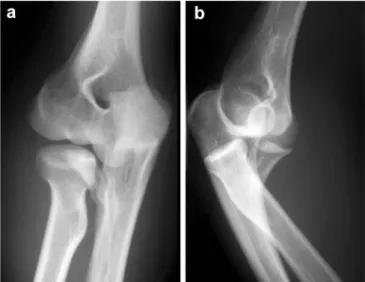
A 20-year-old man had fallen on his left elbow during a basketball match. He presented himself with a defor-mation of the left elbow. The elbow was swollen and fixed in 35° of flexion. Neurovascular testing of the left arm and hand were normal. Anteroposterior and lateral radiographs confirmed the diagnosis of posterior elbow dislocation associated to a type III coronoid fracture (Fig.1). X-ray films after reduction didn’t show other lesions.
The patient was operated upon 48 h after the acci-dent through a minimally invasive anterior and posterior approach: under general anesthesia the patient is placed in supine position and the left arm is positioned on a radiolucent arm board. The operation was performed without tourniquet to allow the surgeon to feel the anterior brachial vascular bundle. Under Fluoroscopic control a first 1.6-mm K-wire is introduced anteriorly from a 1-cm skin incision and Kryle protection and used R. Garofalo C. Bollmann C. Kombot B. Moretti O. Borens E. Mouhsine
Minimal invasive surgery for coronoid
fracture: technical note
Received: 31 March 2004 Accepted: 22 September 2004 Published online: 28 January 2005 Ó Springer-Verlag 2005
Abstract Operative treatment of coronoid fracture often requires a large dissection of soft tissue, resulting in elbow stiffness and functional limitation. The authors present a minimal invasive, safe technique, useful in the case of iso-lated coronoid fracture associated with elbow dislocation. This tech-nique does not require soft tissue dissection and allows an early unlimited resumption of sports activities.
Keywords Coronoid Æ Fracture Æ Elbow Æ Trauma
DOI 10.1007/s00167-004-0594-5 Knee Surg Sports Traumatol Arthrosc
(2005) 13: 608–611 S P O R T S M E D I C I N E
R. Garofalo Æ C. Bollmann Æ C. Kombot B. Moretti Æ O. Borens Æ E. Mouhsine (&) Department of Orthopaedics and
Traumatology, University Hospital, OTR-BH 14 CHUV, 1011 Lausanne, Switzerland
E-mail: [email protected] Tel.: +41-21-3142791
as a joystick to reduce the fracture, which was stabilized by two 1.6-mm K-wires, introduced anteriorly and with the same technique as the first wire. These two K-wires are introduced across the coronoid, the olecranon and the posterior skin. This technique permits the introduction of two cannulated 3.5 screws posteriorly as a definitive stabilisation (Fig.2).
The K-wires are then removed. At the end of surgery the elbow stability was assessed and only a posterolat-eral rotatory instability in full extension was noted.
Postoperatively the elbow is immobilized in a plaster cast with the elbow flexed at 30° for 10 days and then gravity-assisted active range of motion exercises are started. The postoperative course was uneventful.
Three months after surgery the fracture was united and the patient returned to his sportive activity without pain, and normal range of motion of the elbow and forearm rotation. One year after surgery the patient still had unlimited sports activities; the X-ray films showed an excellent fracture union and no signs of heterotopic ossification (Fig.3).
Discussion
Fractures of the coronoid often occurred in association with the elbow. Regan and Morrey [11] proposed a radiographic classification of the coronoid that have a prognostic value. They classified coronoid fractures based on the size of the fragment. Type I fracture is described as a small shear fracture and it indicates that a minimal dislocation of the elbow must have occurred. Type II fractures involve 50% of the coronoid and type III are shear fractures of the whole coronoid process (> 50%).
As the mechanism of these fractures are mostly shearing forces caused by posterior dislocation some authors [9, 11] believe that they are pathognomonic for elbow instability. Type I fractures are usually too small for an open reduction and internal fixation. This frac-ture can be managed by immobilization (maximum 3 weeks) and early range of motion. Rarely do these type I fractures require removal of intraarticular frag-ments [8].
Recently, Terada [14] suggested that even some small (‘‘fleck-fractures’’) type I or II coronoid fractures should be repaired in order to achieve sufficient elbow stability: they think that with this method even partial lesions on the anterior capsule can be healed by replacing these small fragments, thus improving elbow stability.
In type II lesions the most important criteria is instability; if the elbow is unstable after reduction in 50– 60° of flexion [10] the associated coronoid fracture should be fixed; the stability can also be assessed fluo-roscopically [13].
The importance of the radial head as secondary sta-bilizer of the elbow joint has been shown by several authors[5, 7] and the association of coronoid fracture with radial head fracture and elbow dislocation is rec-ognized as terrible triad injury [6]. Josefsson [7] at first had pointed out the importance of coronoid fractures associated with elbow-instability: he noted that redislo-cation of the elbow occurred in patients who underwent excision of radial head and had an associated coronoid fracture. In particular, type II fractures produce elbow instability near to full extension [11]; so, this kind of lesion when associated with an ipsilateral radial head fracture and elbow dislocation should be operated on [10].
The type III coronoid fractures should be treated surgically [8, 10]. This fracture is often part of a more
Fig. 1 a Anteroposterior and b lateral view of the fracture-dislocation
Fig. 2 Technique of anterior insertion of the 1.6-mm K-wires used as joystick for reduction, device for stabilisation and for screws guidance
complex elbow injury, such as posterolateral rotatory instability. So, the operative treatment is directed to-ward maintaining the congruity of the joint and conse-quently reduction of the coronoid; this allows a safe postoperative mobilisation of the elbow.
Closkey [3] showed in human cadaveric model an increased posterior elbow translation with type III cor-onoid fracture from 60° to 105° of flexion. When asso-ciated with radial head resection this lesion rendered the elbow grossly unstable [11].
Stiffness after fracture-dislocation of the elbow is a recognised problem [1,4,11].
Ka¨licke [8] found in 39 patients with fracture and dislocation of the elbow seven had developed hetero-topic ossification and needed reoperation (arthrolysis). All these seven patients presented coronoid fracture
associated to either olecranon or radial head fracture. Thirty patients of this series had prophylaxis with in-dometacin and nine patients had a postoperative radia-tion therapy. The reported frequency of heterotopic ossification is between 5% and 50% in case of complex fracture-dislocations of the elbow [2]. The additional dissection needed to repair the coronoid might play a role in increasing the risk of heterotopic ossification and elbow stiffness [12]. For this reason, we think that a minimal invasive approach to reduce this fracture might be better. Our method is easy and safe as there are no noble structures likely to be injured in this approach. On the other hand, it can be useful when the coronoid fracture is the only injury to be surgically repaired in case of an elbow trauma.
Fig. 3 a Anteroposterior and b lateral view of the fracture reduction and fixation
References
1. Breitfuss H, Muhr G, Neumann K, Neumann C, Rehn J (1991) Arthrolysis of post-traumatic stiff elbow. Which factors influence the end result. Unfall-chirurg 94(1):33–39
2. Broberg MA, Morrey BF (1987) Results of treatment of fracture-dislocations of the elbow. Clin Orthop 216:109–119 3. Closkey RF, Goode JR, Kirschenbaum
D, Cody RP (2000) The role of the coronoid process in elbow stability. A biomechanical analysis of axial loading. J Bone Joint Surg Am 82(A):1749–1753
4. Cobb TK, Morrey BF (1995) Use of distraction arthroplasty in unstable fracture dislocations of the elbow. Clin Orthop 312:201–210
5. Deutch SR, Jensen SL, Tyrdal S, Olsen BS, Sneppen O (2003) Elbow joint sta-bility following experimental osteoliga-mentous injury and reconstruction. J Shoulder Elbow Surg 12(5):466–471 6. Mezera K, Hotchkiss R (1996)
Frac-tures and dislocations of the elbow. In: Rockwood CAGD, Buchholz RW, Heckman JD (eds) Rockwood and Green’s fractures in adults, 5th edn. Lippincott-Raven, Philadelphia, pp 921–951
7. Josefsson PO, Gentz CF, Johnell O, Wendeberg B (1989) Dislocations of the elbow and intraarticular fractures. Clin Orthop 246:126–130
8. Kalicke T, Westhoff J, Wingenfeld C, Muhr G, Arens S (2003) Fracture dis-location of the elbow involving the coronoid process. Unfallchirurg 106(4):300–305
9. Linscheid RL, Wheeler DK (1965) El-bow dislocations. JAMA 194(11):1171– 1176
10. Morrey B, O’Driscoll SW (2002) Frac-tures of the coronoid and complex in stability of the elbow. In: Morrey B (ed) Master techniques in orthopaedic sur-gery—the elbow, 2nd edn. Lippincott Williams and Wilkins, Philadelphia, pp 127–138
11. Regan W, Morrey B (1989) Fractures of the coronoid process of the ulna. J Bone Joint Surg Am 71(9):1348–1354 12. Ring D, Jupiter JB (2002) Surgical
Exposure of Coronoid Fractures. Tech Shoulder Elbow Surg 3(1):48–56 13. Spencer EE, JC K (2003) A simple
technique for coronoid fixation. Tech Shoulder Elbow Surg 4(1):1–3
14. Terada N, Yamada H, Seki T, Urabe T, Takayama S (2000) The importance of reducing small fractures of the coronoid process in the treatment of unstable elbow dislocation. J Shoulder Elbow Surg 9(4):344–346