ORIGINAL ARTICLE
The impact of
18
F-FDG PET on the management of patients
with suspected large vessel vasculitis
Martin Fuchs&Matthias Briel&Thomas Daikeler&Ulrich A. Walker&Helmut Rasch& Scott Berg&Quinn K. T. Ng&Heike Raatz&David Jayne&Ina Kötter&
Daniel Blockmans&Maria C. Cid&Sergio Prieto-González&Peter Lamprecht& Carlo Salvarani&Zaharenia Karageorgaki&Richard Watts&Raashid Luqmani& Jan Müller-Brand&Alan Tyndall&Martin A. Walter
Received: 15 August 2011 / Accepted: 29 September 2011 / Published online: 10 November 2011 # Springer-Verlag 2011
Abstract
Purpose We aimed to assess the impact of18 F-fluorodeox-yglucose (FDG) positron emission tomography (PET) on the management of patients with suspected large vessel vasculitis.
Methods An international expert panel determined diagno-ses and clinical management in patients with suspected large vessel vasculitis, with and without the results of18 F-FDG PET, respectively. The accuracy of the clinical diagnosis and the resulting clinical management with and
without the 18F-FDG PET results were compared using logistic regression models.
Results The analysis included 30 patients referred to a tertiary care centre with large vessel vasculitis and 31 controls.18F-FDG PET had an overall sensitivity of 73.3% [95% confidence interval (CI) 54.1–87.7%], a specificity of 83.9% (95% CI 66.3–94.5%), a positive predictive value of 81.5% (95% CI 61.9–93.7%) and a negative predictive value of 76.5% (95% CI 58.8–89.3%). The diagnostic accuracy of 18F-FDG PET was higher in patients not
M. Fuchs
:
H. Rasch:
S. Berg:
Q. K. T. Ng:
J. Müller-Brand:
M. A. Walter (*)Institute of Nuclear Medicine, University Hospital, Petersgraben 4,
4031 Basel, Switzerland e-mail: [email protected] M. Briel
:
H. RaatzBasel Institute for Clinical Epidemiology and Biostatistics, University Hospital Basel,
Basel, Switzerland M. Briel
Department of Clinical Epidemiology and Biostatistics, McMaster University,
Hamilton, ON, Canada T. Daikeler
:
A. TyndallDepartment of Rheumatology, University Hospital Basel, Basel, Switzerland
D. Jayne
Vasculitis and Lupus Unit, Addenbrooke’s Hospital, Cambridge, UK
I. Kötter
Department of Internal Medicine II, University Hospital Tübingen, Tübingen, Germany
D. Blockmans
Department of General Internal Medicine, University Hospital Gasthuisberg, Leuven, Belgium
M. C. Cid
:
S. Prieto-GonzálezDepartment of Systemic Autoimmune Diseases, Hospital Clínic, University of Barcelona, IDIBAPS,
Villarroel 170, 08036-Barcelona, Spain P. Lamprecht
Department of Rheumatology,
University Hospital of Schleswig-Holstein, Campus Lübeck,
Lübeck, Germany C. Salvarani
Department of Rheumatology, Arcispedale S. Maria Nuova, Reggio Emilia, Italy Z. Karageorgaki
1st Department of Internal Medicine, Agios Dimitrios General Hospital, Thessaloniki, Greece
receiving immunosuppressive drugs (93.3 vs 64.5%, p= 0.006). Taken in context with other available diagnostic modalities, the addition of 18F-FDG PET increased the clinical diagnostic accuracy from 54.1 to 70.5% (p=0.04). The addition of 18F-FDG PET increased the number of indicated biopsies from 22 of 61 patients (36.1%) to 25 of 61 patients (41.0%) and changed the treatment recommen-dation in 8 of 30 patients (26.7%) not receiving immuno-suppressive medication and in 7 of 31 patients (22.6%) receiving immunosuppressive medication.
Conclusion 18F-FDG PET is a sensitive and specific imaging tool for large vessel vasculitis, especially when performed in patients not receiving immunosuppressive drugs. It increases the overall diagnostic accuracy and has an impact on the clinical management in a significant proportion of patients.
Keywords Positron emission tomography .
Fluorodeoxyglucose . Giant cell arteritis . Takayasu’s arteritis . Immunosuppressive drugs
Introduction
The diagnosis of large vessel vasculitis remains a challenge, especially in patients presenting with nonspecific symptoms and laboratory tests [1–3]. Standard diagnostic procedures include biopsy, angiography, ultrasound and magnetic resonance angiography. These procedures are either invasive, operator dependent or detect only morphological changes which mainly occur in later stages of the disease [4–9].
Positron emission tomography (PET) is an operator-independent, noninvasive metabolic imaging modality based on the regional distribution of the glucose analogue 18
F-fluorodeoxyglucose (FDG). Today,18F-FDG PET plays a major role in the management of oncology patients [10]. However, activated inflammatory cells also overexpress
glucose transporters and accumulate increased amounts of glucose and structurally related substances such as 18 F-FDG [11,12].
Remarkable images of patients with active vasculitis have been generated through 18F-FDG PET scans [13–15] and initial studies indicated that 18F-FDG PET might become useful for imaging giant cell arteritis [16] and Takayasu’s arteritis [17]. Large vessel involvement is found by 18F-FDG PET in about 83% of patients with giant cell arteritis [18]. Thereby, vascular FDG uptake is found especially in the subclavian arteries (74%), but also in the aorta (≥50%) and up to the femoral arteries (37%). The common 18F-FDG uptake pattern found in giant cell arteritis is linear and continuous and thoracic vessels are most frequently affected, followed by the abdominal vessels [19,20]. The common 18F-FDG uptake pattern in early phases of Takayasu’s arteritis is linear and continuous, while in late phases it becomes rather patchier than continuous [21]. Recent18F-FDG PET studies have shown sensitivities between 56 and 100% for detecting giant cell arteritis and sensitivities between 83 and 100% for detecting Takayasu’s arteritis [22–24]. Comparative studies employing18F-FDG PET and magnetic resonance imaging revealed comparable sensitivities of both methods, but18 F-FDG PET has been shown to identify significantly more affected vascular regions [19, 25]. Furthermore, 18F-FDG PET has shown a value for predicting the development of complications, e.g. thoracic aortic dilatation in patients with giant cell arteritis [26], and the ability to demonstrate response after successful initiation of immunosuppressive treatment [18]. Nevertheless, the diagnostic accuracy that 18
F-FDG PET adds to the diagnostic workup as well as its impact on the clinical management has not been systemat-ically evaluated yet.
This study therefore aimed to explore the diagnostic accuracy of18F-FDG PET on its own and as an add-on test to the conventional workup as well as its impact on the clinical management in patients with suspected large vessel vasculitis.
Materials and methods
A panel of international experts in the field of large vessel vasculitis determined diagnoses and clinical management in patients with suspected large vessel vasculitis, with and without the results of the18F-FDG PET scan, respectively. The accuracy of the clinical diagnosis and the resulting clinical management with and without the 18F-FDG PET results were compared. The study was approved by the local Ethics Committee (www.ekbb.ch, reference number 18/10). Because we used archived patient data the study was deemed exempt from requiring consent.
R. Watts
Norwich Medical School, University of East Anglia, Norwich Research Park,
Norwich NR4 7TJ, UK R. Luqmani
Department of Rheumatology, Nuffield Orthopaedic Centre, Oxford, UK
U. A. Walker
Department of Rheumatology of Basle University, Felix Platter Spital,
Basel, Switzerland R. Watts
Ipswich Hospital NHS Trust, Ipswich IP4 5pD, UK
Patients
We included patients with (1) suspected large vessel vasculitis who (2) were referred to our interdisciplinary tertiary referral centre of the Department of Rheumatology, University Hospital Basel, Switzerland and who (3) had received a whole-body PET at the Department of Nuclear Medicine, University Hospital Basel, (4) in the period between April 2002 and April 2010. We excluded patients in case of inconclusive or insufficient follow-up to establish or rule out large vessel vasculitis.
Reference standard
An expert panel of three experienced board certified rheumatologists (T.D., U.W. and A.T.) reviewed the complete patient history with the complete clinical follow-up including clinical symptoms, laboratory findings, ultra-sound, computed tomography, magnet resonance imaging, PET and/or biopsy. Diagnoses were established by consen-sus and were based on American College of Rheumatology (ACR) classification criteria from 1990 (n=24) or if not fulfilled by the combination of clinical symptoms, results of additional examinations, the exclusion of other diagnoses and the follow-up after treatment (n=6). Patients were judged by this panel as disease positive, negative or intermediate. Patients judged disease negative served as the control group, and patients judged with intermediate disease probability were excluded from the study (Fig.1). 18
F-FDG PET imaging
All scans were acquired after a 12-h fast with a dedicated full-ring PET camera system. Serum glucose levels were measured before18F-FDG application in all patients and were below 180 mg/dl (Glucometer Elite®, Bayer Diagnostics, UK).
Image acquisition was started 45 min after intravenous injection of 5 MBq/kg body weight of 18F-FDG with the patient in the supine position. Iterative reconstruction of the transaxial slices was performed using the ordered subset expectation maximization (OSEM) algorithm (2 iterations, 8 subsets).
All scans were assessed by a panel of a board certified nuclear medicine physician, a board certified radiologist and a physician board certified in nuclear medicine and radiology (M.A.W., S.B. and H.R.). The panel was blinded to the patients’ initial clinical, laboratory and imaging results as well as to the follow-up data. The images were judged as positive or negative for large vessel vasculitis as previously described [20]: The severity of large vessel18F-FDG uptake was visually graded using a 4-point scale: 0 = no uptake present, I = low-grade uptake (uptake present but lower than liver uptake), II = intermediate-grade uptake (similar to liver uptake) and III = high-grade uptake (uptake higher than liver uptake). Considering grade II or III uptake in the thoracic aorta and any visible uptake in other segments as pathological due to an active arterial inflammation has previously shown a high sensitivity for large vessel vasculitis [20].
Clinical scoring
All cases were scored by an international panel of nine board certified rheumatologists and internists (D.J., I.K., D.B., M.C. C., P.L., C.S., Z.K., R.W. and R.L.) twice: one time blinded to the18F-FDG PET results and one time with the addition of the 18F-FDG PET results, respectively. All cases were presented by a computer program, anonymized and in a random order. The case presentation included all clinical symptoms, laboratory findings, ultrasound, computed tomog-raphy and/or magnetic resonance imaging results and the medication at the time of the18F-FDG PET scan.
61 Patients with conclusive clinical follow up included
30 Patients included into analyses
67 Patients with conclusive18FDG PETdata
6 Patients without conclusive clinical follow up excluded
13 Patients without immunosuppressivedrugs
31 Controls included into analyses
17 Controls without immunosuppressive drugs 30 Patients with Large vessel Vasculitis included 31 Controls without Large vessel Vasculitis included
67 Patients scanned for suspected Large Vessel Vasculitis
17 Patients under immunosuppressivedrugs 14 Controls under immunosuppressive drugs
Each expert scored (1) the probability of large vessel vasculitis (in per cent) and based on the average score patients were judged as disease positive (probability 67– 100%), negative (0–33%) and intermediate (34–66%). Furthermore, each expert (2) recommended further diag-nostic workup from a panel of predetermined options including laboratory, ultrasound, computed tomography, magnetic resonance imaging and biopsy and (3) scored the disease extent and activity, each ranging from 1 (minimal) to 10 (maximal) or “indeterminable” and finally (4) the recommended drug management, e.g. in untreated patients to start or not to start glucocorticosteroids and/or other immunosuppressive drugs and in already treated patients to escalate or reduce the immunosuppression.
Study endpoints
The predefined study endpoints were (1) the diagnostic accuracy of 18F-FDG PET for large vessel vasculitis in patients with and without immunosuppressive drugs, (2) the accuracy of the clinical diagnosis with and without the addition of the18F-FDG PET results, (3) the intention to biopsy with and without the addition of the18F-FDG PET results and (4) the treatment recommendation with and without the addition of the18F-FDG PET results.
Statistical analyses
Sensitivity, specificity, positive predictive value, negative predictive value and diagnostic accuracy of the clinical scoring with and without 18F-FDG PET results were calculated using the expert panel assessment as the reference standard. Differences in proportions were tested with theχ2or Fisher’s exact test, as appropriate. Logistic regression models were used to examine the association of the pre-PET disease probability with the probability of18 F-FDG PET ruling out or confirming disease. Logistic regression was also used to examine the association of disease probability and the indication of biopsy.
Sensitivity analyses were performed to evaluate the effects described above in the subgroup of patients with giant cell arteritis.
All discrete variables are described by counts (percent-age) and continuous variables by their median (range), unless stated otherwise. Two-sided p values of <0.05 were considered to indicate statistical significance.
Results
Overall, 67 patients were considered for inclusion. Of these, large vessel vasculitis was confirmed in 30 (44.8%; 24 giant cell arteritis, 6 Takayasu’s arteritis) and ruled out
in 31 (46.3%). Six patients (8.9%) with inconclusive follow-up data were excluded from the study (Fig. 1). Of 61 patients included in this study, 28 (45.9%) received corticosteroids and 12 (19.7%) received other immunosup-pressive drugs (azathioprine, cyclophosphamide, cyclospor-ine or methotrexate). A total of 30 patients (49.2%) were PET scanned before and 31 patients (50.8%) were scanned after the start of steroid and/or other immunosuppressive drugs. The patients’ baseline characteristics are shown in Table1.
The diagnostic value of18F-FDG PET alone
Overall, 27 18F-FDG PET scans were positive for large vessel vasculitis (44.3%; 22 true-positives, 5 false-positives) and 34 were considered negative (55.7%; 26 true-negatives, 8 false-negatives). None of the scans were assessed as inconclusive. The resulting sensitivity was 73.3% [95% confidence interval (CI) 54.1–87.7%], the specificity was 83.9% (95% CI 66.3–94.5%), the positive predictive value was 81.5% (95% CI 61.9–93.7%) and the negative predictive value was 76.5% (95% CI 58.8–89.3%). The overall diagnostic accuracy of18F-FDG PET was 78.7%, i.e. 48 of 61 patients were labelled correctly (Table 2). In the subgroup of patients with giant cell arteritis the diagnostic accuracy was 81.8%. All eight false-negative 18F-FDG PET scans were performed under immunosuppressive drugs (Fig.2).
The diagnostic value of18F-FDG PET with and without immunosuppressive drugs
In 30 patients without immunosuppressive drugs, 13 scans were truly positive, 2 were falsely positive, 15 were truly negative and none were falsely negative. The resulting sensitivity was 99.6% (95% CI 69.8–100%), the specificity was 86.1% (95% CI 58.6–96.4%), the positive predictive value was 84.4% (95% CI 54.4–96.0%) and the negative predictive value was 99.6% (95% CI 73.2–100%). The diagnostic accuracy of 18F-FDG PET in patients without immunosuppressive drugs was 93.3% (28 of 30 patients were labelled correctly, Table2).
In the 31 patients on immunosuppressive drugs on the other hand, 9 scans were truly positive, 3 were falsely positive, 11 were truly negative and 8 were falsely negative. The resulting sensitivity was 52.9% (95% CI 27.8–77.0%), the specificity was 78.6% (95% CI 49.2–95.3%), the positive predictive value was 75.0% (95% CI 42.8–94.5%) and the negative predictive value was 57.9% (95% CI 33.5–79.7%). The diagnostic accuracy of 18F-FDG PET in patients on immunosuppressive drugs was 64.5% (20 of 31 patients were labelled correctly), significantly lower than in patients without immunosuppressive drugs (p=0.006).
The diagnostic value of18F-FDG PET in the clinical context
With all diagnostic workup data available except the18 F-FDG PET results, of 30 disease-positive patients 14 (46.7%) were identified correctly by the scoring board, 5 (16.7%) were falsely scored disease negative, while 11 (36.6%) were scored as intermediate probability. Of 31 disease-negative controls, 18 were correctly identified as disease negative (58.1%), 4 were falsely scored as disease positive (12.9%), while 9 patients (29.0%) were scored as intermediate probability. Without the18F-FDG PET results, 32 of 61 patients were diagnosed correctly (diagnostic accuracy 54.1%, Table2).
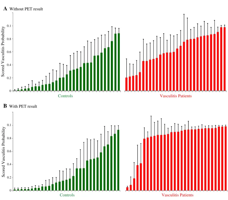
The detailed expert scoring results are presented in Fig.6 of theAppendix.
The addition of the 18F-FDG PET results changed the diagnosis in 18 patients (27.9%): 14 times from an intermediate probability or incorrect diagnosis to the correct diagnosis, 2 times from the correct diagnosis to an intermediate probability and 2 times from the correct
diagnosis to a wrong diagnosis. Overall, the addition of the 18F-FDG PET result reduced the number of patients scored undetermined from 20 to 10.
Of 30 disease-positive patients 25 (83.3%) were identi-fied correctly, 3 (10.0%) were falsely scored disease negative, while 2 (6.7%) were scored as intermediate probability. Of the 31 disease-negative controls 18 (58.1%) were correctly identified as disease negative, 5 (16.1%) were falsely scored as disease positive, while 8 (25.8%) were scored as undetermined. With the18F-FDG PET results, 43 of 61 patients were diagnosed correctly (diagnostic accuracy 70.5%), significantly more than without the 18F-FDG PET data (p=0.04). In the subgroup of patients with giant cell arteritis the addition of the 18 F-FDG PET results increased the diagnostic accuracy from 50.9 to 69.1% (p=0.05).
18
F-FDG PET had a higher additional diagnostic value in confirming than in ruling out large vessel vasculitis. In disease-positive patients, the addition of 18
F-FDG PET significantly changed the scored probability of disease from a median of 62.2% (range 20.6–97.4%) to Table 1 Baseline characteristics
aThe results were derived from
27 temporal artery biopsies and 2 aortic biopsies; biopsy-positive is defined as mononu-clear cell infiltration of the temporal artery with or without giant cells
Characteristic Finding Large vessel vasculitis
(n=30)
Controls (n=31)
Gender Female 22 18
Male 8 13
Age (years) Median 70.6 65.1
Range 17.6–81.4 24.3–86.9
Clinical findings Age at onset of symptoms≥50 years 22 28
New headache 14 12
Temporal artery abnormality 9 1
Elevated erythrocyte sedimentation rate
29 23
Age at onset of disease≤40 years 4 3
Claudication of an extremity 5 2
Decreased brachial artery pulse 3 0
Different systolic blood pressure in both arms
7 1
Bruit over the subclavian arteries or the aorta
6 2
Narrowing/occlusion of entire aorta 5 5
Biopsya Positive 12 0
Negative 9 8
Diagnosis Giant cell arteritis 24 0
Takayasu’s arteritis 6 0 Immunosuppressive treatment Total 23 19 Glucocorticosteroids 16 12 Methotrexate 7 3 Azathioprine 0 2 Cyclophosphamide 0 1 Cyclosporine 0 1
89.9% (5.3–98.1%, p=0.001, Fig.3). On the contrary, in the disease-negative controls the addition of the18F-FDG PET results did not significantly change the scored probability [25.6% (0.8–88.3%) to 16.7% (1.8–93%), p= 0.65, Fig.3]. T able 2 Diagnostic performance of 18 F-FDG PET and of clinical workup with and without 18F-FDG PET results T rue-positive False- positive T rue-negative False- negative Sensitivity Specificity Positive predictive value Negative predictive value Accuracy 18 F-FDG PET Overall 22 5 2 6 8 73.3 83.9 81.5 76.5 78.7 W ithout immunosuppression 13 2 1 5 0 99.6 86.1 84.4 99.6 93.3 W ith immunosuppression 9 3 1 1 8 52.9 78.6 75.0 57.9 64.5 Clinical diagnosis W ithout 18 F-FDG PET a 14 4 1 8 5 46.7 58.1 51.9 52.9 54.1 W ith 18 F-FDG PET b 25 5 1 8 3 83.3 58.1 65.8 78.3 70.5 Sensitivities, specificities, positive predictive values, negative predictive values and accuracy are expressed in % a Overall, 20 patients were scored as undetermined b Overall, 10 patients were scored as undetermined
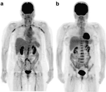
Fig. 2 18F-FDG PET scans of two patients with giant cell arteritis. The scan in a 68-year-old woman without immunosuppressive drugs (a) shows pathologically elevated 18F-FDG uptake in the aorta and its major
branches. These findings changed the treatment recommendation from ‘no immunosuppressive drugs’ to ‘start immunosuppressive drugs’. On the contrary, the scan in a 52-year-old woman under 40 mg per day prednisone (b) shows no elevated18F-FDG uptake in the large vessels, with, however, no change in the recommended clinical management
pre PET post PET pre PET post PET
Controls Vasculitis Patients
p=0.001 Scored Vasculitis Probability p=0.65 1.0 0.8 0.6 0.4 0.2 0
Fig. 3 Diagnostic value of18F-FDG PET in the clinical context. In disease-positive patients (p=0.001), but not in the controls (p=0.65) the addition of 18F-FDG PET significantly changed the scored probability of disease
The impact of18F-FDG PET on the biopsy recommendation
With all the diagnostic workup data available except the 18
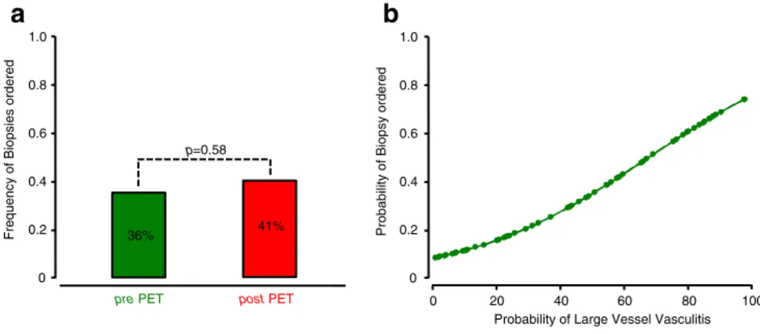
F-FDG PET results, biopsy was scored to be indicated in 22 patients (36.1%). The addition of the 18F-FDG PET results changed the biopsy indication from‘yes’ to ‘no’ in four patients and from‘no’ to ‘yes’ in seven patients. As a result, the addition of the 18F-FDG PET results did not significantly change the number of patients with indicated biopsy to 25 (41.0%, Fig.4a). Logistic regression revealed a positive correlation of higher biopsy rates when higher probabilities of large vessel vasculitis were scored (Fig.4b). The impact of18F-FDG PET on treatment recommendation The impact of 18F-FDG PET on the medical management was assessed in patients receiving and not receiving immunosuppressive medication.
Of all 61 patients, 30 (49.2%) had not received any immunosuppressive medication. With all diagnostic workup data available except the18F-FDG PET results, glucocorti-costeroids were considered to be indicated in 11 of the 30 patients, while no change in management was recommended in 19 patients. The addition of the 18F-FDG PET result
changed the indicated management in eight patients (26.7%, Fig. 5a). Four patients were changed from no drugs to corticosteroids, one patient was changed from no drugs to corticosteroids with additional immunosuppressives, while three patients were changed from corticosteroids to cortico-steroids with additional immunosuppressives.
Thirty-one patients (50.8%) were receiving
immuno-suppressive medication. Without the 18F-FDG PET
results, a dose increase was scored to be indicated in 3, a decrease to be indicated in 7, while no change in management was indicated in 21 patients. The addition of the 18F-FDG PET results changed the management in seven patients (22.6%, Fig.5b). Two patients were shifted from‘no change’ to ‘dose increase’ and five patients were shifted from ‘no change’ to ‘dose decrease’. In total,18 F-FDG PET changed the treatment recommendation in 15 patients. In 8 of these 15 patients (53.3%) the 18F-FDG PET had also changed the overall diagnosis.
Discussion
The present results indicate that18F-FDG PET is a sensitive and specific imaging tool that increases the overall diagnostic accuracy for large vessel vasculitis in the context
pre PET post PET 1.0 0.8 0.6 0.4 0.2 0 0 20 40 60 80 100 1.0 0.8 0.6 0.4 0.2 0
Probability of Biopsy ordered
a
b
Probability of Large Vessel Vasculitis
Frequency of Biopsies ordered 36% 41%
p=0.58
Fig. 4 18F-FDG PET and biopsy frequency. The addition of the18F-FDG PET results did not significantly change biopsy rates (a). Logistic regression revealed a positive correlation of higher biopsy rates with higher probabilities of large vessel vasculitis (b) Start Corticosteroids & Immunosuppressives n=0 Increase Dose n=3 Decrease Dose n=7
+3 after PET No changes
a
Patients not on Immunosuppressive Drugs (n=30)b
Patients on Immunosuppressive Drugs (n=31)No Change in Medication n=19 No Change in Medication n=21 Start Corticosteroids n=11 After PET Before PET
Fig. 5 18F-FDG PET and clini-cal management. The addition of the18F-FDG PET result
changed the indicated manage-ment in 26.7% of patients naïve to immunosuppressive drugs (a) and in 22.6% of patients under immunosuppressive drugs (b)
of the available diagnostic results. Thereby,18F-FDG PET has an impact on the medical management in a significant proportion of patients.
The sensitivities and specificities found are compara-ble to previously published results [22]. The underlying principle of PET imaging in inflammatory processes is the increased accumulation of glucose and structurally related substances such as18F-FDG into inflammatory cells [11, 12]. Alternatively, magnet resonance imaging, which is also used for imaging large vessel vasculitis, mainly analyses the thickness of the vascular wall [27]. We have previously shown that the accuracy of 18F-FDG PET increases in the state of active inflammation [20]. In line with these results, previous studies have indicated a lower diagnostic value of 18F-FDG PET in patients with suspected large vessel vasculitis under immunosuppres-sive drugs [28, 29]. The present study systematically evaluated the diagnostic accuracy of 18F-FDG PET with and without immunosuppressive medication and found a significantly reduced diagnostic value in patients on immunosuppressive drugs. Consequently, 18F-FDG PET should, whenever feasible, be performed in an interval without immunosuppressive drugs.
Until now it has remained elusive whether adding 18
F-FDG PET results to the usual diagnostic workup for large vessel vasculitis would increase diagnostic accuracy. Here, the present study showed that18F-FDG PET allows the overall accuracy of the diagnostic workup to be significantly increased. Furthermore, the present results showed that18F-FDG PET principally had a higher value in confirming than ruling out disease. The increase of diagnostic accuracy by 18F-FDG PET is relevant to the diagnostic workup of each individual patient as well as to the current efforts to establish diagnostic and classification criteria for systematic vasculitis [2]. 18F-FDG PET changed the indicated medical management in both groups, patients with and without immunosuppressive drugs, respectively. Thereby, with changes in management in about one of four patients, the extent of the influence of 18
F-FDG PET was similar in both groups. These results demonstrate that 18F-FDG PET increases diagnostic accuracy and changes medical management in a signifi-cant proportion of patients.
One of the aims of noninvasive PET imaging is to provide a whole-body status and to replace local invasive procedures such as biopsies. In this study, the addition of 18F-FDG PET did not decrease the frequency of recommended biopsies despite the increased diagnostic value. We found a positive correlation between the probability of larger vessel vasculitis and the probability of recommendation for biopsy. Higher pre-test probabili-ties increased the rate of recommended biopsies. These results indicate that, given the current state of evidence,
18
F-FDG PET is unlikely to replace biopsy procedures in the near future.
This study has strengths and limitations. First, large vessel vasculitis is a rare condition and accordingly the present cohort is relatively small. Nevertheless, the study was sufficiently powered to obtain significant results. Secondly, the present study shows that 18F-FDG PET increases the diagnostic accuracy of experts in the field of large vessel vasculitis that scored the disease probability on a scale from 0 to 100%, analogous to the visual analogue scale [30]. The effect on the diagnostic accuracy in less experienced physicians remains elusive. Third, the reference standard may be influenced by results of 18 F-FDG PET potentially leading to incorporation bias since the reference panel based its decisions on the complete clinical follow-up of patients including all diagnostic test results. Fourth, the study cohort comprises patients with giant cell arteritis and Takayasu’s arteritis. Accordingly, sensitivity analyses were performed that showed similar results in the cohort of giant cell arteritis patients. In accordance with previous studies [31], we were not able to detect enhanced uptake in temporal vessels. This limita-tion is mainly due to the high FDG uptake in the brain and the small diameter of the temporal arteries. Next genera-tion PET scanners, however, might be able to reliably measure FDG uptake in temporal vessels. Finally, the study showed that 18F-FDG PET has an impact on the medical management in a significant proportion of patients. In a next step further studies are warranted to evaluate the potential improvement in the clinical outcome by the additional use of18F-FDG PET.
Previous studies had already assessed the diagnostic
performance of 18F-FDG PET in patients with and
without immunosuppressive medication. The present study is the first to systematically analyse the impact of 18
F-FDG PET on biopsy and treatment recommendations. The results demonstrate the additional diagnostic value of 18
F-FDG PET in patients with suspected large vessel vasculitis and they define the conditions under which18 F-FDG PET is anticipated to be most helpful. Whenever feasible, 18F-FDG PET should be performed when the patient is not receiving immunosuppressive drugs. 18 F-FDG PET increases the overall diagnostic accuracy and has an impact on the medical management in a significant proportion of patients.
Acknowledgements The authors declare that they have no conflict of interest. Martin A. Walter gratefully acknowledges the support of the Swiss National Science Foundation. Matthias Briel and Heike Raatz are supported by santésuisse and the Gottfried and Julia Bangerter-Rhyner Foundation. Sergio Prieto-González and Maria C. Cid are supported by Ministerio de Ciencia e Innovacion (SAF 08/ 04328 and SAF 11/30073).
Appendix
The detailed expert scoring results are presented in Fig.6.
References
1. Salvarani C, Cantini F, Hunder GG. Polymyalgia rheumatica and giant-cell arteritis. Lancet 2008;372:234–45.
2. Basu N, Watts R, Bajema I, Baslund B, Bley T, Boers M, et al. EULAR points to consider in the development of classification and diagnostic criteria in systemic vasculitis. Ann Rheum Dis 2010;69:1744–50.
3. Mukhtyar C, Guillevin L, Cid MC, Dasgupta B, de Groot K, Gross W, et al. EULAR recommendations for the management of large vessel vasculitis. Ann Rheum Dis 2009;68:318–23.
4. Salvarani C, Silingardi M, Ghirarduzzi A, Lo Scocco G, Macchioni P, Bajocchi G, et al. Is duplex ultrasonography useful for the diagnosis of giant-cell arteritis? Ann Intern Med 2002;137:232–8.
5. Blockmans D. Utility of imaging studies in assessment of vascular inflammation. Cleve Clin J Med 2002;69 Suppl 2:SII95–9. 6. Tso E, Flamm SD, White RD, Schvartzman PR, Mascha E,
Hoffman GS. Takayasu arteritis: utility and limitations of magnetic resonance imaging in diagnosis and treatment. Arthritis Rheum 2002;46:1634–42.
7. Kissin EY, Merkel PA. Diagnostic imaging in Takayasu arteritis. Curr Opin Rheumatol 2004;16:31–7.
0 0.2 0.4 0.6 0.8 0.1 0 0.2 0.4 0.6 0.8 0.1
A
Without PET resultScored Vasculitis Probability
Scored Vasculitis Probability
B
With PET resultControls Vasculitis Patients
Controls Vasculitis Patients
Fig. 6 Waterfall diagram of the expert scoring results in controls and vasculitis patients without (a) and with the18F-FDG PET results (b). The inter-observer agreement was higher with the18F-FDG PET results (mean standard deviation 13.4) than without (mean standard deviation 19.5)
8. Seo P, Stone JH. Large-vessel vasculitis. Arthritis Rheum 2004;51:128–39.
9. Aschwanden M, Kesten F, Stern M, Thalhammer C, Walker UA, Tyndall A, et al. Vascular involvement in patients with giant cell arteritis determined by duplex sonography of 2x11 arterial regions. Ann Rheum Dis 2010;69:1356–9.
10. Weber WA, Grosu AL, Czernin J. Technology insight: advances in molecular imaging and an appraisal of PET/CT scanning. Nat Clin Pract Oncol 2008;5:160–70.
11. Ishimori T, Saga T, Mamede M, Kobayashi H, Higashi T, Nakamoto Y, et al. Increased (18)F-FDG uptake in a model of inflammation: concanavalin A-mediated lymphocyte activation. J Nucl Med 2002;43:658–63.
12. Jones HA, Cadwallader KA, White JF, Uddin M, Peters AM, Chilvers ER. Dissociation between respiratory burst activity and deoxyglucose uptake in human neutrophil granulocytes: implica-tions for interpretation of (18)F-FDG PET images. J Nucl Med 2002;43:652–7.
13. Turlakow A, Yeung HW, Pui J, Macapinlac H, Liebovitz E, Rusch V, et al. Fludeoxyglucose positron emission tomogra-phy in the diagnosis of giant cell arteritis. Arch Intern Med 2001;161:1003–7.
14. Brodmann M, Lipp RW, Aigner R, Pilger E. Positron emission tomography reveals extended thoracic and abdominal peri-aortitis. Vasc Med 2003;8:127–8.
15. Hara M, Goodman PC, Leder RA. FDG-PET finding in early-phase Takayasu arteritis. J Comput Assist Tomogr 1999;23:16–8. 16. Blockmans D, Maes A, Stroobants S, Nuyts J, Bormans G, Knockaert D, et al. New arguments for a vasculitic nature of polymyalgia rheumatica using positron emission tomography. Rheumatology (Oxford) 1999;38:444–7.
17. Meller J, Grabbe E, Becker W, Vosshenrich R. Value of F-18 FDG hybrid camera PET and MRI in early takayasu aortitis. Eur Radiol 2003;13:400–5.
18. Blockmans D, de Ceuninck L, Vanderschueren S, Knockaert D, Mortelmans L, Bobbaers H. Repetitive 18F-fluorodeoxyglucose positron emission tomography in giant cell arteritis: a prospective study of 35 patients. Arthritis Rheum 2006;55:131–7.
19. Meller J, Strutz F, Siefker U, Scheel A, Sahlmann CO, Lehmann K, et al. Early diagnosis and follow-up of aortitis with [(18)F]FDG PET and MRI. Eur J Nucl Med Mol Imaging 2003;30:730–6.
20. Walter MA, Melzer RA, Schindler C, Müller-Brand J, Tyndall A, Nitzsche EU. The value of [18F]FDG-PET in the diagnosis of large-vessel vasculitis and the assessment of activity and extent of disease. Eur J Nucl Med Mol Imaging 2005;32:674–81. 21. Webb M, Chambers A, AL-Nahhas A, Mason JC, Maudlin L, Rahman
L, et al. The role of 18F-FDG PET in characterising disease activity in Takayasu arteritis. Eur J Nucl Med Mol Imaging 2004;31:627–34. 22. Blockmans D, Bley T, Schmidt W. Imaging for large-vessel
vasculitis. Curr Opin Rheumatol 2009;21:19–28.
23. Henes JC, Müller M, Krieger J, Balletshofer B, Pfannenberg AC, Kanz L, et al. [18F] FDG-PET/CT as a new and sensitive imaging method for the diagnosis of large vessel vasculitis. Clin Exp Rheumatol 2008;26:S47–52.
24. Walter MA. [(18)F]fluorodeoxyglucose PET in large vessel vasculitis. Radiol Clin North Am 2007;45:735–44. viii. 25. Scheel AK, Meller J, Vosshenrich R, Kohlhoff E, Siefker U,
Müller GA, et al. Diagnosis and follow up of aortitis in the elderly. Ann Rheum Dis 2004;63:1507–10.
26. Blockmans D, Coudyzer W, Vanderschueren S, Stroobants S, Loeckx D, Heye S, et al. Relationship between fluorodeoxyglu-cose uptake in the large vessels and late aortic diameter in giant cell arteritis. Rheumatology (Oxford) 2008;47:1179–84. 27. Spira D, Kötter I, Ernemann U, Balletshofer B, Pfannenberg CA,
Fenchel M, et al. Imaging of primary and secondary inflammatory diseases involving large and medium-sized vessels and their potential mimics: a multitechnique approach. AJR Am J Roentgenol 2010;194:848–56.
28. Bleeker-Rovers CP, Bredie SJ, van der Meer JW, Corstens FH, Oyen WJ. Fluorine 18 fluorodeoxyglucose positron emission tomography in the diagnosis and follow-up of three patients with vasculitis. Am J Med 2004;116:50–3.
29. Iwabu M, Yamamoto Y, Dobashi H, Kameda T, Kittaka K, Nishiyama Y. F-18 FDG PET findings of Takayasu arteritis before and after immunosuppressive therapy. Clin Nucl Med 2008;33:872–3. 30. Miller MD, Ferris DG. Measurement of subjective phenomena in
primary care research: the Visual Analogue Scale. Fam Pract Res J 1993;13:15–24.
31. Brodmann M, Lipp RW, Passath A, Seinost G, Pabst E, Pilger E. The role of 2-18F-fluoro-2-deoxy-D-glucose positron emission tomography in the diagnosis of giant cell arteritis of the temporal arteries. Rheumatology (Oxford) 2004;43:241–2.