Deletion of hypothetical wall teichoic acid ligases in
Staphylococcus aureus activates the cell wall stress response
Vanina Dengler1, Patricia Stutzmann Meier1, Ronald Heusser1, Peter Kupferschmied1,
Judit Fazekas1, Sarah Friebe1, Sibylle Burger Staufer1, Paul A. Majcherczyk2, Philippe Moreillon2, Brigitte Berger-Ba¨chi1& Nadine McCallum1,3
1Institute of Medical Microbiology, University of Zurich, Zu¨rich, Switzerland;2Institute of Fundamental Microbiology, University of Lausanne,
Lausanne, Switzerland; and3Sydney Emerging Infectious Diseases and Biosecurity Institute (SEIB), The University of Sydney, Sydney, NSW,
Australia
Correspondence: Vanina Dengler, Institute of Medical Microbiology, University of Zu¨rich, Gloriastrasse 32, CH-8006 Zu¨rich,
Switzerland. Tel.: +41 44 634 26 94; fax: +41 44 634 49 06; e-mail: [email protected]
Received 19 December 2011; revised 25 April 2012; accepted 21 May 2012.
Final version published online 18 June 2012.
DOI: 10.1111/j.1574-6968.2012.02603.x
Editor: Andre Klier
Keywords
cell wall stress stimulon; LytR-CpsA-Psr; VraSR; wall teichoic acids; ligase; Staphylococcus aureus.
Abstract
The Staphylococcus aureus cell wall stress stimulon (CWSS) is activated by cell envelope-targeting antibiotics or depletion of essential cell wall biosynthesis enzymes. The functionally uncharacterized S. aureus LytR-CpsA-Psr (LCP) pro-teins, MsrR, SA0908 and SA2103, all belong to the CWSS. Although not essen-tial, deletion of all three LCP proteins severely impairs cell division. We show here that VraSR-dependent CWSS expression was up to 250-fold higher in sin-gle, double and triple LCP mutants than in wild type S. aureus in the absence of external stress. The LCP triple mutant was virtually depleted of wall teichoic acids (WTA), which could be restored to different degrees by any of the single LCP proteins. Subinhibitory concentrations of tunicamycin, which inhibits the first WTA synthesis enzyme TarO (TagO), could partially complement the severe growth defect of the LCP triple mutant. Both of the latter findings support a role for S. aureus LCP proteins in late WTA synthesis, as in Bacillus subtilis where LCP proteins were recently proposed to transfer WTA from lipid carriers to the cell wall peptidoglycan. Intrinsic activation of the CWSS upon LCP deletion and the fact that LCP proteins were essential for WTA-loading of the cell wall, high-light their important role(s) in S. aureus cell envelope biogenesis.
Introduction
Staphylococcus aureus mounts a general cell wall stress response in the presence of cell wall damaging agents, involving the upregulation of up to 50 genes collectively known as the cell wall stress stimulon (CWSS; Kuroda et al., 2003; Utaida et al., 2003; Jordan et al., 2008). Induction of CWSS genes is controlled by the VraSR two-component system (Belcheva & Golemi-Kotra, 2008), which is homologous to the cell wall stress-responsive sensor-transducer systems LiaFSR of Bacillus subtilis (Mascher et al., 2004), LiaFSR of Streptococcus mutans (Suntharalingam et al., 2009) and CesRS of Lactococcus lactis (Martinez et al., 2007). The sensor kinase VraS senses an unknown signal triggered by cell envelope disturbance and phosphorylates VraR, which then binds as a dimer to promoter-specific elements and facilitates
transcript induction (Martinez et al., 2007; Belcheva & Golemi-Kotra, 2008; Eldholm et al., 2010; Belcheva et al., 2012). There is a wide variation in the fold-induction levels of different CWSS genes, which is probably linked to the specificity of VraR-binding, although the exact VraR-binding consensus and the influence of specific nucleotide differences on expression and induction of different CWSS genes has not been thoroughly analysed (Martinez et al., 2007; Belcheva & Golemi-Kotra, 2008; Belcheva et al., 2012).
The magnitude of CWSS induction strongly depends on the class and concentration of cell wall antibiotics (Dengler et al., 2011). Disruption of wall teichoic acid (WTA) synthesis by targocil, which inhibits the WTA transporter TarG (TagG), was also shown to activate the CWSS (Campbell et al., 2012). WTA are anionic glyco-polymers that are attached to the peptidoglycan of
MICR
Gram-positive bacteria via a phosphodiester linkage, and they can constitute up to 60% of the total cell wall bio-mass. WTA of B. subtilis are composed of poly(glycerol phosphate) and poly(ribitol phosphate), whereas S. aureus contains mainly poly(ribitol phosphate) WTA. The bio-synthesis of WTA is catalysed by tag (teichoic acid gly-cerol) or tar (teichoic acid ribitol) genes in B. subtilis and S. aureus, respectively (reviewed in Swoboda et al., 2010). Besides the induction by cell wall active antibiotics, VraSR signal transduction is also triggered by internal disruption of cell wall synthesis caused by the depletion of essential cell wall biosynthesis enzymes such as MurA, MurZ, MurB (Blake et al., 2009), MurF (Sobral et al., 2007), PBP2 (Gardete et al., 2006) or depletion of enzymes involved in mevalonate biosynthesis, the direct precursor for undecaprenyl phosphate lipid carrier syn-thesis (Balibar et al., 2009). Induction of the CWSS enhances intrinsic resistance/tolerance to almost all cell wall damaging agents, regardless of their target or mode of action (Dengler et al., 2011; McCallum et al., 2011). Members of the CWSS directly linked to peptidoglycan synthesis, such as PBP2, FmtA, MurZ and SgtB, are thought to contribute to the stress response by stimulat-ing cell wall synthesis (Cui et al., 2009; Kato et al., 2010; Mehta et al., 2012). It is predicted that CWSS genes with unknown or poorly characterized functions are also likely to contribute to the stress response by directly or indi-rectly influencing cell wall synthesis.
All three S. aureus LytR-CpsA-Psr (LCP) genes, msrR, sa0908 and sa2103, belong to the CWSS (Utaida et al., 2003; McAleese et al., 2006; Over et al., 2011). LCP pro-teins are unique to bacteria with Gram-positive cell walls (Hu¨bscher et al., 2008; Kawai et al., 2011) and typically contain a short intracellular N-terminal region, a trans-membrane domain and a large extracelluar region con-taining the LCP domain (Hu¨bscher et al., 2008; Kawai et al., 2011). Deletion of LCP proteins in S. aureus alters cell surface properties and decreases virulence. Phenotypes of LCP deletion mutants include defective cell separation, increased TritonX-100-induced autolysis, increased beta-lactam susceptibility, and the cell wall WTA content was reduced in an msrR deletion mutant (Hu¨bscher et al., 2009). Phenotypes become more pronounced in double mutants, and growth is severely impaired in the LCP tri-ple mutant, which contains large amorphous cells with multiple septa (Over et al., 2011).
Recently, the LCP proteins of B. subtilis, TagT (YwtF), TagU (LytR) and TagV (YvhJ) were found to be essential for the formation of a WTA-loaded cell wall. Kawai et al. (2011) claim that LCP proteins catalyse the final, previ-ously uncharacterised, step in WTA synthesis, the linkage of WTA to peptidoglycan. WTA are not essential for the cell, but deletion of the first two synthesis steps, catalysed
by TarA (TagA) or TarO (TagO), leads to impaired cell division, colonization and infection in vivo (Weidenmaier et al., 2004; Weidenmaier & Peschel, 2008; D’Elia et al., 2009). However, the late-acting enzymes from TarB (TagB) onwards are conditionally essential; mutants are only viable when one of the first two steps of WTA synthesis is inhibited (Swoboda et al., 2010). Blocking the flux of WTA precursors into the WTA pathway prevents the deleterious sequestration of the universal undeca-prenyl phosphate lipid carrier that is also essential for peptidoglycan synthesis, and it prevents the accumulation of potentially toxic intermediates. LCP proteins in B. subtilis are also conditionally essential, and the LCP triple mutant is only viable when tagO (tarO) is deleted (Kawai et al., 2011). Whether LCP proteins fulfil the same function in S. aureus has not yet been verified.
In this study, reporter gene fusions were used to ana-lyse CWSS expression levels in LCP mutants and to iden-tify promoter regions essential for CWSS induction of LCP genes. The effect of LCP deletion on the WTA con-tent was determined and partial complementation of the LCP triple mutant by TarO (TagO) inhibition demon-strated, suggesting that LCP proteins play an important role in the WTA decoration of S. aureus peptidoglycan.
Materials and methods
Bacterial strains and growth conditions
The strains and plasmids used in this study are listed in Table 1. Bacteria were grown at 37°C in Luria Bertani (LB) broth (Difco Laboratories), shaking at 180 r.p.m. with a 1 : 5 culture to air ratio or on LB agar plates. Optical density (OD) measurements were taken at 600 nm. Media were supplemented with the following antibiotics when appropriate: 10lg mL 1 tetracycline (Sigma), 10lg mL 1 chloramphenicol (Sigma), 100 lg mL 1ampicillin (Sigma) or 200 ng mL 1 anhydrotetracy-cline (Vetranal).
Construction ofDVraR mutants
The pKOR1 system developed by Bae & Schneewind (2006) was used to inactivate VraR in the different LCP mutant strains, by inserting an XhoI site and two stop codons in-frame into the beginning of the vraR coding sequence, truncating VraR after the 2nd amino acid, as previously described (McCallum et al., 2011).
Northern blots
Northern blots were performed as previously described (McCallum et al., 2007). To compare relative expression
Table 1. Strains, plasmids and primers
Strain/plasmid/primer name Relevant genotype and/or phenotype (strain name) or primer sequence Source or reference S. aureus
RN4220 Restriction-de cient derivative of NCTC 8325-4 Kreiswirth et al. (1983) MSSA1112 Clinical isolate, bla, McsPenr Hu¨bscher et al. (2009)
DmsrR MSSA1112,DmsrR::ermB; McsEmr(JH100) Hu¨bscher et al. (2009)
Dsa0908 MSSA1112, marker-less sa0908 deletion mutant (RH53) Over et al. (2011) Dsa2103 MSSA1112, marker-less sa2103 deletion mutant (PS47) Over et al. (2011) Dsa0908/msrR MSSA1112,Dsa0908/msrR double-mutant (RH72) Over et al. (2011) Dsa2103/msrR MSSA1112,Dsa2103/msrR double-mutant (PS60) Over et al. (2011) Dsa2103/sa0908 MSSA1112,Dsa2103/sa0908 double-mutant (PS109) Over et al. (2011) Dsa2103/sa0908/msrR MSSA1112,Dsa2103/sa0908/msrR triple-mutant (PS111) Over et al. (2011) DVraR MSSA1112, truncated VraR after the 2nd amino acid (=DVraR) (PS199) This study DVraR/msrR MSSA1112,DVraR/msrR double-mutant (RH194) This study DVraR/sa0908 MSSA1112,DVraR/sa0908 double-mutant (PS202) This study DVraR/sa2103 MSSA1112,DVraR/sa2103 double-mutant (RH191) This study DVraR/sa0908/msrR MSSA1112,DVraR/sa0908/msrR triple-mutant (NM776) This study DVraR/sa2103/msrR MSSA1112,DVraR/sa2103/msrR triple-mutant (RH193) This study DVraR/sa2103/sa0908 MSSA1112,DVraR/sa2103/sa0908 triple-mutant (RH216) This study
SA113 Restriction-de cient derivative of NCTC 8325 (ATCC 35556) Iordanescu & Surdeanu (1976) SA113DtarO SA113,DtarO::ermB; Emr Weidenmaier et al. (2004)
E. coli
DH5a F u80d/acZDM15 recA1 Invitrogen Plasmids
pKOR1 S. aureus-E. coli shuttle vector, ori pAMa1, ori ColE1, E. coli Amr, S. aureus Cmr
Bae & Schneewind (2006)
pKOR1-VraR::stop pKOR1 construct containing mutant vraR insert with XhoI site and two inframe stop codons inserted between the 2nd and 3rd vraR codons.
McCallum et al. (2011)
pGC2 E. coli–S. aureus shuttle plasmid, ori ColE1-ori pC194 bla cat; E. coli Amr, S. aureus Cmr
Skinner et al. (1988)
pmsrR pGC2 containing 1.3-kb fragment comprising the msrR ORF and upstream flanking sequence
Hu¨bscher et al. (2009)
psa0908 pGC2 containing 1.9-kb fragment comprising the sa0908 ORF and upstream flanking sequence
Over et al. (2011)
psa2103 pGC2 containing 2.1-kb fragment comprising the sa2103 ORF and upstream flanking sequence
(Over et al. (2011)
pBUS1 S. aureus – E. coli shuttle vector, tetL; Tcr Rossi et al. (2003)
psas016p-luc+ pBUS1 containing the sas016 promoter-luciferase reporter gene fusion McCallum et al. (2011)
pvrap-luc+ pBUS1 containing the vraSR operon promoter-luciferase reporter gene fusion This study
pmsrRp-luc+ pBUS1 containing the msrR promoter-luciferase reporter gene fusion Over et al. (2011)
psa0908p-luc+ pBUS1 containing the sa0908 promoter-luciferase reporter gene fusion Dengler et al. (2011)
psa2103p-luc+ pBUS1 containing the sa2103 promoter-luciferase reporter gene fusion Over et al. (2011)
psas016D6bpp-luc+ pBUS1 containing the sas016 promoter with 6-bp deletion fused
to the luciferase gene (Fig. 2)
This study
psas016D6Bbpp-luc+ pBUS1 containing the sas016 promoter with 6-bp deletion variant B
fused to the luciferase gene (Fig. 2)
This study
pmsrRD12bpp-luc+ pBUS1 containing the msrR promoter with 12-bp deletion fused to
the luciferase gene (Fig. 2)
This study
pmsrRD18bpp-luc+ pBUS1 containing the msrR promoter with 18-bp deletion fused to
the luciferase gene (Fig. 2)
This study
psa0908D6bpp-luc+ pBUS1 containing the sa0908 promoter with 6-bp deletion fused to
the luciferase gene (Fig. 2)
This study
psa2103D6bpp-luc+ pBUS1 containing the sa2103 promoter with 6-bp deletion fused to
the luciferase gene (Fig. 2)
This study
Primers
vra.lucF AATTTGGTACCGCACATGTACTTAATTACTT This study vra.lucR ATTAACCATGGCTATCACCTTTTATAATAAGT This study
levels of sas016 in wild type and mutant strains, overnight cultures were diluted to OD 0.05 in prewarmed LB broth and cultures grown to OD 1.5, except for the LCP triple mutant that was sampled at OD 0.5 because of its severe growth defect. Uninduced culture samples were collected, and the remainder of the culture was induced with oxa-cillin (10lg mL 1) for 30 min before induced samples were collected. Total RNA was extracted as described by Cheung et al. (1994). RNA samples (9lg) were separated in a 1.5% agarose-20 mM guanidine thiocyanate gel in 19 TBE buffer (Goda & Minton, 1995). The sas016 digoxigenin (DIG)-labelled probe was amplified using the PCR DIG Probe synthesis kit (Roche) as previously described (Dengler et al., 2011).
Primer extension
The transcriptional start site of sas016 was determined by primer extension, as previously described (McCallum et al., 2007), using primer SAS016.PErev (Table 1) and 20lg of RNA harvested from a culture of S. aureus COL that had been grown to OD 0.5 and induced with 10lg mL 1of teicoplanin for 30 min.
Luciferase reporter gene fusions
The promoter region of the vraSR operon was PCR amplified from S. aureus strain COL using primer pair vra.lucF and vra.lucR (Table 1). The PCR product was
digested with Asp718 and NcoI and ligated directly upstream of the promoterless luciferase (luc+) gene in the vector pSP-luc+ (Promega). Fragments containing the resulting promoter-luc+ translational fusions were then excised with Asp718 and EcoRI and cloned into the Esc-herichia coli – S. aureus shuttle vector pBUS1 (Table 1). The fusion plasmids pvrap-luc+ and psas016p-luc+ (McCallum et al., 2011) were then electroporated into S. aureus RN4220, re-isolated and electroporated into S. aureus SA113, SA113DtarO, MSSA1112 and all LCP and VraR/LCP mutants.
Predicted VraR-binding sites of luciferase fusion con-structs were disrupted by amplifying each promoter as two fragments, using primers listed in Table 1. Comple-mentary fragments were digested and ligated together, to create recombinant promoters in which 6–18-bp regions were replaced by restriction sites. Promoters were then fused to the luciferase gene as described above, and the resulting plasmids were electroporated into RN4220.
Luciferase assays
To measure luciferase activities, cultures were grown from overnight cultures inoculated to an OD 0.05 in pre-warmed LB broth containing tetracycline. One-millilitre culture samples were harvested by centrifugation, and the pellets frozen at 20°C.
To determine relative light units (RLU), pellets were thawed briefly and resuspended in PBS (pH 7.4) to an
Table 1. Continued
Strain/plasmid/primer name Relevant genotype and/or phenotype (strain name) or primer sequence Source or reference sas016.lucF AATTAGGTACCTGGATCACGGTGCATACAAC McCallum et al. (2011) sas016.lucR AATTACCATGGCCTATATTACCTCCTTTGCT McCallum et al. (2011) sas016-D6bp.F AAATTAAGCTTGTTGATGTCACACATAAAAAT This study
sas016-D6bp.R AAATTAAGCTTTATCAACTTTTTATCAGAC AT This study sas016-D6bpB.F AAATTAAGCTTTTCTATGTCTGATAAAAAGTT This study sas016-D6bpB.R AAATTAAGCTTATTTACTAAGACTATTTATGT This study JR13 (msrR.lucF) GGGTACCTGAGCTAAAGTTAAGTCGCC Rossi et al. (2003) JR14 (msrR.lucR) TATCCATGGTTACCTACCTTATATCTTC Rossi et al. (2003) msrR-D12bp.F AATTTAAGCTTTTATTAAGAAATCACTTGCTT This study msrR-D18bp.F AATTTAAGCTTAGAAATCACTTGCTTTTTGAA This study msrR-D12bp/D18bp.R AATTAAAGCTTTCTAATGAAAGGATGTCAAA This study
sa0908.lucF AATTAGGTACCATAATAGTACACACGCATGT Dengler et al. (2011) sa0908.lucR TTAATCCATGGTTGATGCTCCTATATTAAATT Dengler et al. (2011) sa0908-D6bp.R AATTTAAGCTTTTCCTTGTAATTTGAATGTTT This study
sa0908-D6bp.F AATTTAAGCTTCATAACATTTGTATTTTTTAC This study lucF.sa2103 GGGGTACCAAAATGACGACTTTAGATGGTAAG Over et al. (2011) lucR.sa2103 CATGCCATGGCAATCCCACCACTCCTTTACTATTCC Over et al. (2011) sa2103-D6bp.F AATTAGAATTCAAGTATAGTAAAAAAATTAT This study sa2103-D6bp.R AATTAGAATTCACGTATAACTATTTTTTATC This study SAS016.PErev CTTCATGGTGATACTGTCGATA This study
Am, ampicillin; Cm, chloramphenicol; Em, erythromycin; Mc, methicillin; Pen, penicillin; Tc, tetracycline; r, resistant; s, susceptible. Restriction sites are underlined.
OD of either 10 or 1, depending on induction levels. Aliquots of the cell suspensions were then mixed with equal aliquots of Luciferase Assay System substrate (Pro-mega), and luminescence was measured for 15 s after a delay of 3 s on a Turner Designs TD-20/20 luminometer (Promega) as previously described (Dengler et al., 2011).
Bacitracin gradient plates and Etests
Qualitative differences in resistance levels for bacitracin (from Bacillus licheniformis, Sigma) were compared using antibiotic gradient plates as previously described (Hu¨bscher et al., 2009). LB medium was supplemented with ZnCl2 (25lg mL 1), and plates were incubated at 37°C for 48 h. Bacitracin minimum inhibitory concen-trations (MIC) were detected by Etest (Bio-Me´rieux) on Mu¨ller-Hinton plates swabbed with an inoculum of 0.5 McFarland and incubated at 37°C for 24 h.
Growth under subinhibitory concentrations of tunicamycin
Overnight cultures were diluted to OD 0.05 in LB media containing 0.05lg mL 1 tunicamycin (AG Scientifics). OD measurements were taken hourly for 8 h.
Preparation and quantification of WTA
Cell walls and WTA were prepared as previously described (Majcherczyk et al., 2003). The amount of WTA was indirectly quantified by determination of the cell wall phosphorus content (Ames & Dubin, 1960). Experiments were performed two to four times with three technical replicates per sample.
Results and discussion
Deletion of LCP proteins leads to increased VraSR-dependent basal expression of the CWSS LCP proteins are essential for optimal cell separation (Over et al., 2011). The severe cell division defects of double and triple LCP mutants resemble those resulting from the depletion of essential peptidoglycan biosynthesis enzymes or inhibition of WTA synthesis, which both trig-ger VraSR signal transduction and induction of the CWSS (Gardete et al., 2006; Sobral et al., 2007; Balibar et al., 2009; Blake et al., 2009; Campbell et al., 2012). The most sensitive indicator of staphylococcal CWSS activation is the sas016 gene, as demonstrated previously in Northern blot, promoter-luciferase fusion and microarray studies; however, its function is still unknown (McAleese et al., 2006; Dengler et al., 2011). We therefore determined the
basal CWSS transcription levels of single, double and tri-ple LCP mutants and compared them to those of the par-ent strain MSSA1112 using a probe against the CWSS gene sas016. Northern blots showed that sas016 transcrip-tion was detectably higher in single LCP mutants than in the wild type, with highest levels of transcription in the Dsa0908 mutant (Fig. 1a). Transcript levels were further increased in double LCP mutants, Dsa0908/msrR, Dsa2103/msrR and Dsa2103/sa0908, and were extremely high in the LCP triple mutant (Fig. 1a).
To compare and quantify CWSS expression at different growth stages, a promoter-luciferase reporter construct containing the sas016 promoter (psas016p-luc+) was used as previously described (McCallum et al., 2011). Figure 1b shows the luciferase activity levels measured in relative light units (RLU) in the wild type and LCP mutant strains at the time points indicated. The right graph shows the corresponding OD values of the cultures at each sampling point. To confirm patterns of CWSS upregulation, expression of the autoregulatory vra pro-moter from the vraSR operon was also measured, using the promoter-luciferase fusion pvrap-luc+ (Supporting information, Fig. S1). Both constructs, psas016p-luc+ and pvrap-luc+ displayed very similar luciferase activity profiles, with expression from the vraSR operon promoter being consistently lower than that from the sas016 promoter, reflecting differences in promoter activity that were observed in previous transcriptional analyses of the CWSS (McAleese et al., 2006). In all strains tested, the activity increased during exponential growth and decreased again as cells entered stationary phase, with maximum luciferase activity levels reached in late exponential growth, at around 4.5 h.
Luciferase activity profiles corresponded closely to the results from Northern blots (Fig. 1a). Expression was reproducibly higher in LCP single mutants than in the parent MSSA1112, with up to twofold increases in Dsa2103 and DmsrR mutants and a larger, up to sixfold increase, in Dsa0908. The luciferase expression from the sas016 promoter increased further in the double LCP mutants with the highest expression levels seen in Dsa2103/sa0908 and comparable levels in Dsa0908/msrR and Dsa2103/msrR. The most dramatic increase was apparent in the triple mutant, where expression levels were up to 250-fold higher than in the wild type, similar to levels reached after antibiotic stress (Fig. 1e). Activity peaked slightly later in some mutants, possibly reflecting minor differences in growth dynamics.
To verify that increased CWSS expression was VraSR dependent, a VraR mutation was introduced into the wild type strain MSSA1112 and all single and double mutants. The VraR mutation could not be introduced into the triple mutant, probably due to its cell separation
(a)
(b)
(c)
(d) (e)
Fig. 1. CWSS expression in LCP and VraR/LCP mutant strains. (a) Northern blot analysis showing the expression of the CWSS gene sas016 in LCP mutants. (b and c) Luciferase activities measured from reporter construct psas016p-luc+ in LCP mutants (b) and in VraR/LCP mutants (c).
Values shown indicate the RLU measured in each of the strains at the different growth stages indicated. Left, single LCP or VraR/LCP mutants; middle row, LCP or VraR/LCP double and triple mutants; right, corresponding OD values of the cultures at each sampling point for all strains. Samples were taken at 1.5-h intervals for up to 7.5 h. The RLU scales of the different graphs were adjusted to appropriate ranges for visualizing strain-dependent differences. Average values and standard deviations from three independent experiments are shown. (d) Complementation of theDsa0908 mutant strain by introducing sa0908 in trans. RLUs were measured from strains containing the reporter construct psas016p-luc+
that were harvested between OD 0.6 and 0.8. Values shown represent the averages and standard deviations from three independent experiments. (e) Luciferase activities of LCP and VraR/LCP mutants with and without oxacillin (10lg mL 1) induction. Cultures were grown to OD 1.5–1.8 before being split into two prewarmed flasks, one culture was induced with oxacillin and the other left uninduced, and cultures were grown for a further 30 min before samples were harvested. RLU values are shown on a logarithmic scale and represent the averages and standard deviations from three independent experiments. Untreated LCP mutants are shown in white, treated LCP mutants in black, untreated VraR/LCP mutants in grey/white hatched and treated VraR/LCP mutants in grey.
defects and temperature sensitivity (Over et al., 2011). Expression of the CWSS was measured over growth in the VraR/LCP mutants using psas016p-luc+. In all DVraR mutants, CWSS expression levels dropped clearly below wild type values (Fig. 1c). The minor differences in expression between all VraR/LCP mutants and MSSA1112DVraR, indicates that the increased basal CWSS expression levels in LCP mutants were VraSR dependent.
Complementation of Dsa0908, the single mutant with the strongest effect on CWSS expression, by re-introduction of sa0908 in trans, reduced luciferase activity back to wild type levels (Fig. 1d), demonstrating that differences in CWSS activity were directly linked to the LCP mutations.
LCP mutants are still responsive to cell wall stress
As the CWSS was already inherently activated to varying degrees in the absence of external stress in growing LCP mutants, we tested their potential to react to an external cell wall stress. Luciferase activity from psas016p-luc+ was measured in exponentially growing LCP and VraR/LCP mutants exposed to oxacillin for 30 min (Fig. 1e). Basal transcription levels were again increased in uninduced LCP mutants. Expression was still strongly induced by oxacillin stress in the single and double LCP mutants. Expression in the untreated LCP triple mutant appeared to already be close to the maximum level, as it only increased approximately twofold upon oxacillin stress (Fig. 1e).
Identification of promoter regions involved in CWSS induction
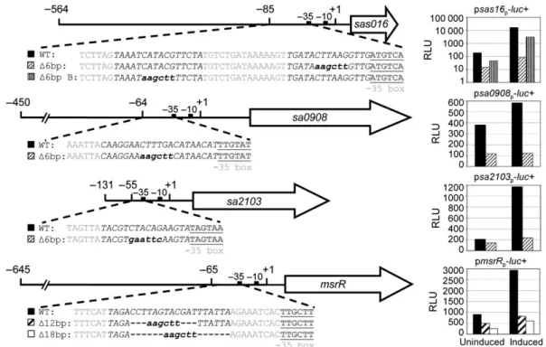
Two VraR-binding sites have been identified in the pro-moter of the vraSR operon with a tail to tail tandem repeat motif ACT(X)nAGT (X= A, C, T or G; n = 1–3; Belcheva & Golemi-Kotra, 2008; Belcheva et al., 2012). They are involved in the fine tuning of the VraR-dependent expression of the CWSS and have different affinities for VraR or phosphorylated VraR (Belcheva & Golemi-Kotra, 2008; Belcheva et al., 2012). VraR-binding sites in other CWSS promoters have so far only been studied in silico. A 16-bp palindromic sequence TCAGHCTnnAGDCTGA (H= A, T, C; D = A, T, G), deduced from the VraR homologue CesR in L. lactis (Martinez et al., 2007) and partially overlapping the motif identified by Belcheva et al., is present in the promoters of 26 VraSR-dependent genes of the S. aureus N315 genome (Martinez et al., 2007). As we found the induction levels of the three LCP genes and of the highly induced CWSS gene sas016 to
vary over a wide range, we analysed their specific VraR-binding motifs. The transcriptional start sites of msrR, sa0908 and sa2103 are known (Rossi et al., 2003; Over et al., 2011), and the transcriptional start site of sas016 was determined by primer extension to be 29-nt upstream of the ATG (data not shown). Potential VraR-binding sites were predicted in all four promoters investi-gated in this study, based on previously published motifs (Martinez et al., 2007; Belcheva & Golemi-Kotra, 2008; Belcheva et al., 2012). These sequences were then dis-rupted and/or deleted in the promoter regions of lucifer-ase reporter gene constructs (Fig. 2). Disruption of the predicted motifs decreased basal expression levels and lar-gely abolished induction by oxacillin (Fig. 2). In all four promoters, the regions essential for induction were located close to the 35 boxes. The promoter of sas016 contained a second region that was found to be essential for full induction. The presence of two VraR-binding sites could contribute to the extremely high induction levels of sas016. Alignment of the nucleotide sequences from the VraR-binding regions identified here revealed no obvious consensus sequence. The high-affinity VraR-binding region in the vraSR operon promoter (Belcheva et al., 2012) and the tcaA promoter region required for induc-tion (McCallum et al., 2007) were both also in close proximity to their respective 35 box. The msrR pro-moter region needed for induction corresponded to the CesR-like motif identified in silico by Martinez et al. (2007; Fig. 2); however, deletion of the suggested CesR-binding region for sa0908 did not affect transcription (data not shown). For the promoters of sas016 and sa2103, no CesR-like binding sites were previously pre-dicted (Martinez et al., 2007); however, the VraR-binding sites identified here both contained potential CesR-like sequences. To create a reliable VraR-binding consensus for S. aureus CWSS gene induction, detailed promoter analysis of more VraSR-dependent genes is required. The trend, however, seems to involve sequences with a close proximity to the 35 box of the CWSS gene promoter.
Bacitracin hypersensitivity of the LCP triple mutant
Bacitracin inhibits the recycling of the universal undeca-prenyl phosphate lipid carrier by preventing dephosphor-ylation of the undecaprenyl pyrophosphate (Stone & Strominger, 1971; Qi et al., 2008). Kawai et al. (2011) recently suggested that LCP proteins transfer WTAs and other anionic polymers from the lipid carrier to the cell wall peptidoglycan in B. subtilis. Comparative growth of LCP mutants on bacitracin gradient plates showed that the LCP triple mutant was highly susceptible (Fig. 3a). The bacitracin MIC of the triple mutant was 4lg mL 1
compared to 32 lg mL 1for wild type and all LCP single and double mutants. The hyper susceptibility of the LCP triple mutant to bacitracin could therefore be due to an additional shortage of the lipid carriers caused by the lack of the putative WTA ligase function of LCP proteins.
Deletion of all three LCP proteins in S. aureus depletes WTA content
In line with the proposed function of LCP proteins, pre-vious studies showed a decrease in the WTA content of LCP mutants in different species (Hu¨bscher et al., 2008; Kawai et al., 2011). Therefore, we analysed the WTA con-tent of LCP single mutants and the triple mutant in S. aureus, via detection of the cell wall phosphorus con-tent (Ames & Dubin, 1960). The previously described decrease in the WTA content of the DmrsR mutant (Hu¨bscher et al., 2009) could be confirmed here, and the WTA contents of the Dsa0908 and Dsa2103 mutants were decreased to 62% and 95% of the wild type level, respec-tively (Fig. 3b). An almost complete depletion of WTA was observed in the triple LCP mutant, with cell wall phosphorus content down to 2% of the wild type.
Re-introduction of single LCP genes into the triple mutant restored WTA levels to 94%, 81% and 69% of wild type levels for sa2103, msrR and sa0908, respectively. The capacity of all LCP proteins to restore the WTA con-tent to a certain degree confirmed a partial functional redundancy that has been shown for other phenotypes such as growth defects, beta-lactam resistance, biofilm formation and self-agglutination (Over et al., 2011). The very low WTA content of the LCP triple mutant con-firmed that LCP proteins in S. aureus have an essential function in WTA loading of the cell wall.
TarO (TagO) inhibition can partially complement the growth defect of the S. aureus LCP triple mutant
The three LCP genes in B. subitlis are conditionally essen-tial, meaning that an LCP triple mutant in B. subtilis is only viable when tagO (tarO) is also deleted, thereby pre-venting the flux of precursors into the WTA synthesis pathway (Kawai et al. 2011). The effect of TarO (TagO) inhibition on the LCP triple mutant was tested to detect a possible connection between LCP proteins with WTA
Fig. 2. Analysis of predicted VraR-binding sites in the sas016, msrR, sa0908 and sa2103 promoters. Nucleotide sequences of promoter regions and introduced promoter mutations are shown, together with their corresponding luciferase activities when fused to the luciferase gene and introduced into Staphylococcus aureus strain RN4220. RLU of the promoter constructs were measured with and without 30 min of induction with 10lg mL 1 oxacillin. Cultures were grown to OD 0.5–0.8 before splitting into two prewarmed flasks comprising the uninduced and oxacillin-induced samples. Predicted VraR-binding regions are shown in black italic capitals; 35 boxes in bold grey underlined capitals; restriction sites in bold italic lowercase letters; deleted regions are indicated by a dashed line. Representative results from three independent experiments are shown.
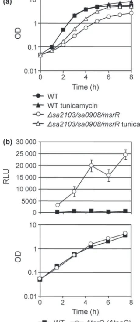
synthesis or assembly in S. aureus, as found for B. subtilis (Kawai et al., 2011). Subinhibitory concentrations of tu-nicamycin, which are known to inhibit TarO (TagO; Campbell et al., 2011), could partially complement the growth defect of the LCP triple mutant (Fig. 4a). The minimal doubling time of the triple mutant decreased from 49± 2 to 34 ± 2 min upon tunicamycin treatment. Inhibition of TarO in the wild type did not significantly affect the minimal doubling time of 25± 0.6 min but reduced the maximal OD reached after 8 h of growth from 8.2 to 5.5. This result supports an involvement of LCP proteins in a late step of WTA synthesis in S. aureus.
As LCP proteins in B. subtilis are essential, it could be that the staphylococcal LCP triple mutant is only viable because of compensatory mutations, which remains to be verified. However, it is also possible that the functions of LCP proteins in S. aureus are not identical to those in B. subtilis, because differences have been found in the WTA synthesis pathways of these closely related bacteria (Brown et al., 2010). Also, in contrast to S. aureus, WTA-deficient strains in B. subtilis have significantly decreased growth rates and lost their rod shape, indicating potential
(a)
(b)
Fig. 3. Bacitracin susceptibility and phosphorus content of the cell wall (WTA content) of LCP mutants. (a) Bacitracin gradient plates of LCP mutants. MICs of bacitracin were detected by Etest (BioMe´rieux): wild type MSSA1112 and LCP single and double mutants all had MICs of 32lg mL 1, LCP triple mutant had an MIC of 4lg mL 1.
(b) Relative levels of WTA contents are shown as percentages of wild type MSSA1112 content, determined indirectly by detecting the phosphorus content of the cell wall for LCP single mutants, the LCP triple mutant and complemented triple mutants. The experiment was performed two to four times with three technical replicates per sample. The average of the absolute values for the wild type was 0.81± 0.04 lmol phosphorus per mg cell wall.
(a)
(b)
Fig. 4. Growth of the LCP triple mutant under subinhibitory concentrations of tunicamycin and CWSS expression in a tarO (tagO) mutant strain. (a) Growth of the LCP triple mutant and wild type MSSA1112 with and without tunicamycin (0.05lg mL 1). Average
values and standard deviations from three independent experiments are shown. (b) Luciferase activity, in RLU, at different growth stages in the wild type strain SA113 and the SA113DtarO mutant strain, measured from reporter construct psas016p-luc+. Upper graph shows
luciferase measurements and lower graph, the corresponding OD values of the cultures at each sampling point for all strains. Average values and standard deviations from three independent experiments are shown.
differences in the roles of WTA ligases in B. subtilis and S. aureus cell division (Weidenmaier et al., 2004; D’Elia et al., 2006).
Deletion of tarO (tagO) induces the CWSS Measurement of CWSS expression in an S. aureus SA113DtarO (DtagO) mutant (Weidenmaier et al., 2004), with the reporter plasmid psas016p-luc+, revealed that inhibition of the first step of WTA synthesis induces the CWSS (Fig. 4b). This result is in conflict to the observa-tions by Campbell et al., (2011) who showed that inhibi-tion of TarO (TagO) by subinhibitory concentrainhibi-tions of tunicamycin does not induce the CWSS. They suggested that CWSS induction is triggered by the sequestration of the lipid carrier rather than WTA deficiency (Campbell et al., 2011, 2012). However, our analysis of the tarO (tagO) mutant indicates that further research is required to reveal the actual trigger of CWSS induction.
Conclusions
Deletion of LCP proteins increased basal expression levels of CWSS genes via the VraSR two-component system. The LCP triple mutant showed very high basal expression of the CWSS, close to levels triggered by antibiotic stress. The LCP double and single mutants, however, still responded to cell wall stress by further upregulating the CWSS.
Promoter regions required for VraR-dependent induc-tion of the LCP genes and sas016 shared low overall nucleotide similarity, but all contained fragments of the predicted CesR-like binding consensus or the VraR-binding motif of the vraSR operon and all were in close proximity to the 35 box of the gene’s promoter.
Hyper susceptibility of the triple mutant to bacitracin, the virtual absence of WTA and partial restoration of WTA levels by complementation with each of the single LCP proteins, as well partial complementation of its growth defect by TarO (TagO) inhibition, support the hypothesis that S. aureus LCP proteins have WTA ligase functions, as suggested by Kawai and colleagues for B. subtilis (Kawai et al., 2011).
An enzymatic analysis of all three LCP proteins will be required to confirm their specific WTA ligase functions, substrates and products.
Acknowledgements
We thank C. Weidenmaier for providing the tarO mutant strain. The research leading to these results has received funding from the European Union Seventh Framework Programme (FP7/2007-2013) under grant agreement no.
241446 (project ANTIRESDEV). This study was further supported by the Swiss National Science Foundation grant 31-117707 to B.B.-B.
References
Ames BN & Dubin DT (1960) The role of polyamines in the neutralization of bacteriophage deoxyribonucleic acid. J Biol Chem 235: 769–775.
Bae T & Schneewind O (2006) Allelic replacement in Staphylococcus aureus with inducible counter-selection. Plasmid 55: 58–63.
Balibar CJ, Shen X & Tao J (2009) The mevalonate pathway of Staphylococcus aureus. J Bacteriol 191: 851–861.
Belcheva A & Golemi-Kotra D (2008) A close-up view of the VraSR two-component system. A mediator of Staphylococcus aureus response to cell wall damage. J Biol Chem 283: 12354–12364.
Belcheva A, Verma V, Korenevsky A, Fridman M, Kumar K & Golemi-Kotra D (2012) The role of DNA sequence and sigma A factor in transcription of the vraSR operon. J Bacteriol 194: 61–71.
Blake KL, O’Neill AJ, Mengin-Lecreulx D, Henderson PJ, Bostock JM, Dunsmore CJ, Simmons KJ, Fishwick CW, Leeds JA & Chopra I (2009) The nature of Staphylococcus aureus MurA and MurZ and approaches for detection of peptidoglycan biosynthesis inhibitors. Mol Microbiol 72: 335–343.
Brown S, Meredith T, Swoboda J & Walker S (2010) Staphylococcus aureus and Bacillus subtilis W23 make polyribitol wall teichoic acids using different enzymatic pathways. Chem Biol 17: 1101–1110.
Campbell J, Singh AK, Santa Maria JP Jr, Kim Y, Brown S, Swoboda JG, Mylonakis E, Wilkinson BJ & Walker S (2011) Synthetic lethal compound combinations reveal a
fundamental connection between wall teichoic acid and peptidoglycan biosyntheses in Staphylococcus aureus. ACS Chem Biol 6: 106–116.
Campbell J, Singh AK, Swoboda JG, Gilmore MS, Wilkinson BJ & Walker S (2012) An antibiotic that inhibits a late step in wall teichoic acid biosynthesis induces the cell wall stress stimulon in Staphylococcus aureus. Antimicrob Agents Chemother 56: 1810–1820.
Cheung AL, Eberhardt KJ & Fischetti VA (1994) A method to isolate RNA from Gram-positive bacteria and mycobacteria. Anal Biochem 222: 511–514.
Cui L, Neoh HM, Shoji M & Hiramatsu K (2009) Contribution of vraSR and graSR point mutations to vancomycin resistance in vancomycin-intermediate Staphylococcus aureus. Antimicrob Agents Chemother 53: 1231–1234.
D’Elia MA, Millar KE, Beveridge TJ & Brown ED (2006) Wall teichoic acid polymers are dispensable for cell viability in Bacillus subtilis. J Bacteriol 188: 8313–8316.
D’Elia MA, Henderson JA, Beveridge TJ, Heinrichs DE & Brown ED (2009) The N-acetylmannosamine transferase
catalyzes the first committed step of teichoic acid assembly in Bacillus subtilis and Staphylococcus aureus. J Bacteriol 191: 4030–4034.
Dengler V, Stutzmann Meier P, Heusser R, Berger-Ba¨chi B & McCallum N (2011) Induction kinetics of the Staphylococcus aureus cell wall stress stimulon in response to different cell wall active antibiotics. BMC Microbiol 11: 16.
Eldholm V, Johnsborg O, Straume D, Ohnstad HS, Berg KH, Hermoso JA & Havarstein LS (2010) Pneumococcal CbpD is a murein hydrolase that requires a dual cell envelope binding specificity to kill target cells during fratricide. Mol Microbiol 76: 905–917.
Gardete S, Wu SW, Gill S & Tomasz A (2006) Role of VraSR in antibiotic resistance and antibiotic-induced stress response in Staphylococcus aureus. Antimicrob Agents Chemother 50: 3424–3434.
Goda SK & Minton NP (1995) A simple procedure for gel electrophoresis and northern blotting of RNA. Nucleic Acids Res 23: 3357–3358.
Hu¨bscher J, Lu¨thy L, Berger-Ba¨chi B & Stutzmann Meier P (2008) Phylogenetic distribution and membrane topology of the LytR-CpsA-Psr protein family. BMC Genomics 9: 617. Hu¨bscher J, McCallum N, Sifri CD, Majcherczyk PA, Entenza
JM, Heusser R, Berger-Ba¨chi B & Stutzmann Meier P (2009) MsrR contributes to cell surface characteristics and virulence in Staphylococcus aureus. FEMS Microbiol Lett 295: 251–260. Iordanescu S & Surdeanu M (1976) Two restriction and
modification systems in Staphylococcus aureus NCTC8325. J Gen Microbiol 96: 277–281.
Jordan S, Hutchings MI & Mascher T (2008) Cell envelope stress response in Gram-positive bacteria. FEMS Microbiol Rev 32: 107–146.
Kato Y, Suzuki T, Ida T & Maebashi K (2010) Genetic changes associated with glycopeptide resistance in Staphylococcus aureus: predominance of amino acid substitutions in YvqF/ VraSR. J Antimicrob Chemother65: 37–45.
Kawai Y, Marles-Wright J, Cleverley RM et al. (2011) A widespread family of bacterial cell wall assembly proteins. EMBO J 30: 4931–4941.
Kreiswirth BN, Lo¨fdahl S, Betely MJ, O’Reilly M, Schlievert PM, Bergdoll MS & Novick RP (1983) The toxic shock syndrome exotoxin structural gene is not detectably transmitted by a prophage. Nature 305: 709–712. Kuroda M, Kuroda H, Oshima T, Takeuchi F, Mori H &
Hiramatsu K (2003) Two-component system VraSR positively modulates the regulation of cell-wall biosynthesis pathway in Staphylococcus aureus. Mol Microbiol 49: 807–821. Majcherczyk PA, Rubli E, Heumann D, Glauser MP &
Moreillon P (2003) Teichoic acids are not required for Streptococcus pneumoniae and Staphylococcus aureus cell walls to trigger the release of tumor necrosis factor by peripheral blood monocytes. Infect Immun 71: 3707–3713. Martinez B, Zomer AL, Rodriguez A, Kok J & Kuipers OP
(2007) Cell envelope stress induced by the bacteriocin Lcn972 is sensed by the Lactococcal two-component system CesSR. Mol Microbiol 64: 473–486.
Mascher T, Zimmer SL, Smith TA & Helmann JD (2004) Antibiotic-inducible promoter regulated by the cell envelope stress-sensing two-component system LiaRS of Bacillus subtilis. Antimicrob Agents Chemother 48: 2888–2896.
McAleese F, Wu SW, Sieradzki K, Dunman P, Murphy E, Projan S & Tomasz A (2006) Overexpression of genes of the cell wall stimulon in clinical isolates of Staphylococcus aureus exhibiting vancomycin-intermediate – S. aureus-type resistance to vancomycin. J Bacteriol 188: 1120–1133.
McCallum N, Brassinga AK, Sifri CD & Berger-Ba¨chi B (2007) Functional characterization of TcaA: minimal requirement for teicoplanin susceptibility and role in Caenorhabditis elegans virulence. Antimicrob Agents Chemother 51: 3836–3843.
McCallum N, Stutzmann Meier PS, Heusser R & Berger-Ba¨chi B (2011) Mutational analyses of open reading frames within the vraSR operon and their roles in the cell wall stress response of Staphylococcus aureus. Antimicrob Agents Chemother 55: 1391–1402.
Mehta S, Cuirolo AX, Plata KB, Riosa S, Silverman JA, Rubio A, Rosato RR & Rosato AE (2012) VraSR two-component regulatory system contributes to mprF-mediated decreased susceptibility to daptomycin in-vivo-selected MRSA clinical strains. Antimicrob Agents Chemother 56: 92–102.
Over B, Heusser R, McCallum N, Schulthess B, Kupferschmied P, Gaiani JM, Sifri CD, Berger-Ba¨chi B & Stutzmann Meier P (2011) LytR-CpsA-Psr proteins in Staphylococcus aureus display partial functional redundancy and the deletion of all three severely impairs septum placement and cell separation. FEMS Microbiol Lett 320: 142–151.
Qi ZD, Lin Y, Zhou B, Ren XD, Pang DW & Liu Y (2008) Characterization of the mechanism of the Staphylococcus aureus cell envelope by bacitracin and bacitracin-metal ions. J Membr Biol 225: 27–37.
Rossi J, Bischoff M, Wada A & Berger-Ba¨chi B (2003) MsrR, a putative cell envelope-associated element involved in Staphylococcus aureus sarA attenuation. Antimicrob Agents Chemother 47: 2558–2564.
Skinner S, Inglis B, Matthews PR & Stewart PR (1988) Mercury and tetracycline resistance genes and flanking repeats associated with methicillin resistance on the chromosome of Staphylococcus aureus. Mol Microbiol 2: 289–292.
Sobral RG, Jones AE, Des Etages SG, Dougherty TJ, Peitzsch RM, Gaasterland T, Ludovice AM, de Lencastre H & Tomasz A (2007) Extensive and genome-wide changes in the transcription profile of Staphylococcus aureus induced by modulating the transcription of the cell wall synthesis gene murF. J Bacteriol 189: 2376–2391.
Stone KJ & Strominger JL (1971) Mechanism of action of bacitracin: complexation with metal ion and C 55-isoprenyl pyrophosphate. P Natl Acad Sci USA 68: 3223–3227. Suntharalingam P, Senadheera MD, Mair RW, Levesque CM &
envelope stress response in Streptococcus mutans. J Bacteriol 191: 2973–2984.
Swoboda JG, Campbell J, Meredith TC & Walker S (2010) Wall teichoic acid function, biosynthesis, and inhibition. ChemBioChem 11: 35–45.
Utaida S, Dunman PM, Macapagal D, Murphy E, Projan SJ, Singh VK, Jayaswal RK & Wilkinson BJ (2003) Genome-wide transcriptional profiling of the response of
Staphylococcus aureus to cell-wall-active antibiotics reveals a cell wall stress stimulon. Microbiology 149: 2719–2732. Weidenmaier C & Peschel A (2008) Teichoic acids and related
cell-wall glycopolymers in Gram-positive physiology and host interactions. Nat Rev Microbiol 6: 276–287. Weidenmaier C, Kokai-Kun JF, Kristian SA, Chanturiya T,
Kalbacher H, Gross M, Nicholson G, Neumeister B, Mond JJ & Peschel A (2004) Role of teichoic acids in
Staphylococcus aureus nasal colonization, a major risk factor in nosocomial infections. Nat Med 10: 243–245.
Supporting Information
Additional Supporting Information may be found in the online version of this article:
Fig. S1. CWSS expression in LCP mutant strains mea-sured with pvrap-luc+.
Please note: Wiley-Blackwell is not responsible for the content or functionality of any supporting materials sup-plied by the authors. Any queries (other than missing material) should be directed to the corresponding author for the article.