OPEN
SLY regulates genes involved in chromatin remodeling
and interacts with TBL1XR1 during sperm
differentiation
Charlotte Moretti
1,2,3,8, Maria-Elisabetta Serrentino
1,2,3,8, Côme Ialy-Radio
1,2,3, Marion Delessard
1,2,3, Tatiana A Soboleva
4,
Frederic Tores
5, Marjorie Leduc
6, Patrick Nitschké
5, Joel R Drevet
7, David J Tremethick
4, Daniel Vaiman
1,2,3, Ayhan Kocer
7and
Julie Cocquet*
,1,2,3Sperm differentiation requires unique transcriptional regulation and chromatin remodeling after meiosis to ensure proper
compaction and protection of the paternal genome. Abnormal sperm chromatin remodeling can induce sperm DNA damage,
embryo lethality and male infertility, yet, little is known about the factors which regulate this process. Deficiency in
Sly, a mouse Y
chromosome-encoded gene expressed only in postmeiotic male germ cells, has been shown to result in the deregulation of
hundreds of sex chromosome-encoded genes associated with multiple sperm differentiation defects and subsequent male
infertility. The underlying mechanism remained, to date, unknown. Here, we show that SLY binds to the promoter of sex
chromosome-encoded and autosomal genes highly expressed postmeiotically and involved in chromatin regulation. Specifically,
we demonstrate that
Sly knockdown directly induces the deregulation of sex chromosome-encoded H2A variants and of the H3K79
methyltransferase DOT1L. The modifications prompted by loss of
Sly alter the postmeiotic chromatin structure and ultimately
result in abnormal sperm chromatin remodeling with negative consequences on the sperm genome integrity. Altogether our
results show that SLY is a regulator of sperm chromatin remodeling. Finally we identified that SMRT/N-CoR repressor complex is
involved in gene regulation during sperm differentiation since members of this complex, in particular TBL1XR1, interact with SLY in
postmeiotic male germ cells.
Cell Death and Differentiation (2017) 24, 1029
–1044; doi:10.1038/cdd.2017.32; published online 5 May 2017
The postmeiotic phase of spermatogenesis is a fascinating
process in terms of transcriptional regulation and chromatin
re-organization. Indeed, after meiosis, during which the
genetic material is recombined and then partitioned in haploid
cells, round spermatids experience a differentiation program
characterized by profound morphological changes:
elonga-tion, nucleus condensation and acquisition of new structures
such as the acrosome and the flagellum. In many organisms
including mammals, this process involves transcriptional
regulation by master genes, and expression of thousands of
genes in round and early elongating spermatids, before the
spermatid chromatin is compacted and transcription is
progressively shut down.
1–6Chromatin compaction is achieved by a transition from a
nucleosome-based organization to a unique
genome-packaging structure based on non-histone proteins, called
protamines. The replacement of histones by protamines starts
with incorporation of spermatid-enriched histone variants and
post translational modifications of histone residues, the most
predominant of which is histone H4 hyperacetylation. These
steps are thought to open the chromatin to facilitate the action
of topoisomerases and the removal of histones; then,
transition proteins are incorporated and finally replaced by
protamines (for review, see
7). In mice, haploinsufficiency of
genes coding for protamines (i.e., Prm1 or Prm2) is sufficient
to result in male infertility and leads to sperm DNA damage
and embryo lethality;
8,9protamines are therefore essential to
mammalian fertility with a function in compaction, as well as
protection of the paternal genome until after fertilization. Little
is known about the factors and molecular mechanisms which
regulate chromatin remodeling during sperm differentiation.
Studies of mouse genetic models have identified a few nuclear
factors, histone variants and chromatin remodelers required
for histone-to-protamine transition, such as BRDT
(Bromodo-main testis specific) protein,
10histone H2B variant TH2B,
11and the chromodomain helicase DNA-binding protein 5.
12We and others have also shown that the mouse Y
chromosome long arm (MSYq) encodes genetic information
required for normal chromatin compaction during sperm
differentiation: males with deletions of MSYq have severe
sperm differentiation defects and produce deformed
sperma-tozoa with poorly compacted chromatin, which are unable to
1
Department of Development, Reproduction and Cancer, INSERM, U1016, Institut Cochin, Paris, France;2CNRS, UMR8104, Paris, France;3Université Paris Descartes, Sorbonne Paris Cité, Faculté de Médecine, Paris, France;4The John Curtin School of Medical Research, The Australian National University, PO Box 334, Canberra, ACT 2601, Australia;5Bioinformatics facility, Université Paris Descartes, Sorbonne Paris Cité, Institut Imagine, Paris, France;63P5 proteomic facility, Institut Cochin, Université Paris Descartes, Sorbonne Paris Cité, Paris, France and7Genetic Reproduction and Development, CNRS UMR6293– INSERM U1103 – Clermont Université, 63178 Aubière Cedex, France
*Corresponding author: J Cocquet, Development, Reproduction, Cancer, Institut Cochin– Inserm u1016 – Université, 24 rue du Faubourg St Jacques, PARIS 75014, France. Tel: +33144412319; Fax: +33144412302; E-mail: julie.cocquet@inserm.fr
8
These authors contributed equally to this work.
Received 20.9.16; revised 25.1.17; accepted 09.2.17; Edited by P Salomoni; published online 05.5.2017
Cell Death and Differentiation (2017) 24, 1029–1044 Official journal of the Cell Death Differentiation Association www.nature.com/cdd
fertilize oocytes in vivo and in vitro.
13Sly, a multicopy gene of
MSYq only expressed in postmeiotic cells,
14,15largely
con-tributes to these phenotypes since males with Sly specifically
knocked down (Sly-KD males) also present abnormal sperm
differentiation, including abnormal chromatin compaction and
increased sperm DNA damage.
15,16It has been shown that
Sly knockdown leads to the upregulation of ~ 100 sex
chromosome-encoded genes in round spermatids. At the
protein level, SLY lacks any conserved domain except for a
COR1 region identified in SYCP3, a protein involved in the
meiotic synaptonemal complex.
14,17Therefore, the
mechan-ism by which SLY controls gene expression and the origin of
the sperm differentiation defects observed in its absence
remain unclear.
In the present study, we investigated the molecular function
of SLY by performing chromatin immunoprecipitation followed
by high-throughput sequencing (ChIP-Seq) and by
co-immunoprecipitation followed by mass spectrometry. We
discovered that SLY associates with the transcriptional start
sites of thousands of genes expressed postmeiotically, many
of which are involved in gene regulation or chromatin
remodeling. We focused on SLY-target genes relevant to the
chromatin defects observed in Sly-deficient sperm, and found
that SLY controls the expression of genes coding for
spermatid-specific histone variants and for chromatin
regula-tors such as the H3K79 methyltransferase, DOT1L. Overall,
we show that Sly deficiency leads to changes in the chromatin
composition just prior to histone removal, which impact on
histone-to-protamine exchanges and, ultimately, on sperm
atozoa chromatin content and function, as well as on their
genome integrity. Finally, we found that SLY is part of the
SMRT/N-CoR complex and interacts with TBL1XR1.
Altogether our data identify for the first time the molecular
role of SLY and link the sperm chromatin compaction
pheno-type observed in Sly-deficient males to molecular pathways
important for chromatin remodeling during sperm
differentia-tion, in particular, the regulation of H3K79 methylation.
Results
SLY marks the sperm differentiation genetic program. To
investigate the molecular mechanism by which SLY controls
postmeiotic gene expression, we performed ChIP-Seq
analyses on purified round spermatids from wild-type (WT)
males with anti-SLY antibody. We found that SLY protein
preferentially binds to the start of genes, in the 1 kb region
surrounding the transcription start site (TSS) and, overall,
occupies the TSS of ~ 16% of mouse genes (6,381 genes
with SLY at TSS, 7,280 genes with SLY at
± 1 kb of TSS)
(Figure 1a, Supplementary Figure 1A and Table 1). No
particular bias towards the sex chromosomes was observed
(Figure 1b and Supplementary Figure 1B). ChIP followed by
real-time PCR (ChIP-qPCR) confirmed the ChIP-Seq results
(Figure 1c). Strikingly, comparison with published RNAseq
data
6,18showed a strong correlation between SLY-genomic
targets and genes expressed in round spermatids (~89%
versus 41% of all mouse genes, χ
2, Po0.0001; Figures 1d
and e), and more specifically with a high expression level
(91% of SLY-associated genes are among the 50% most
expressed genes,
χ
2, Po0.0001; 36% among the 10% most
expressed genes,
χ
2, Po0.0001). Interestingly, SLY was
found to bind to the TSS of master genes of spermatid
transcriptional regulation such as Crem, Creb1, Crebbp,
Kif17, Taf7l, Terf2, Tbpl1, Papolb, Piwil1 or Brd4.
1,2,19SLY
presence appears as an excellent marker of genes essential
Figure 1 SLY marks the sperm differentiation program and co-localizes with active epigenetic marks. (a) Annotation of SLY-enriched genomic regions (right) compared to whole genome (left), using Cis-regulatory Element Annotation System (CEAS). (b) Graphic representation of the percentage of genes found occupied by SLY protein by ChIP-Seq on each chromosome. (c) Validation of SLY-target genes by ChIP-qPCR on round spermatids using antibody against SLY. The TSS of Slx, Rbmx, Rhox11, Actrt1, Ctag2 and Hist1h3a were found enriched in SLY by ChIP-Seq (present study) and were previously found deregulated in Sly-KD versus WT samples by micro-array and/or RT-qPCR analyses.15The TSS of H2afb3, Jmjd1c and Dot1l were found enriched in SLY by ChIP-Seq (present study) and were found deregulated in Sly-KD versus WTround spermatids by
RT-qPCR (present study, cf. Figure 2b). Sex chromosome-encoded genes are represented in black and autosomal genes, in gray. The Y-axis represents the mean enrichment (% IP/input)± S.E.M. normalized to a negative control region (NC) located at ~ 170 kb from a TSS. All regions shown were found significantly enriched in SLY compared to the negative control region (t-test, Po0.05; n = 3–6 samples). (d) Graphic representation of SLY ChIP-Seq profile showing the average enrichment of SLYaround the TSS of genes expressed (in red) and not expressed (in black) in round spermatids. (e) Graphic representation of the percentage of genes that are expressed (in red) and not expressed (in black) in round spermatids, among all mouse genes (mm10 genome version) or among SLY-associated genes. SLY is significantly enriched at the start (i.e., TSS± 1 kb) of genes expressed in round spermatids (χ2, P
o0.0001). (f) Representation of SLY ChIP-Seq and input profiles at Sly gene locus. (g) Graphic representation of the comparison between SLY ChIP-Seq data set and ChIP-Seq data sets from chromatin marks (Kcr, H3K4me3, H3K9ac, H4ac, H3K27ac, H3K27me3, H3K9me3) and from BRD4, in round spermatids. In red is represented the percentage of SLY-enriched genomic regions which overlaps with that of the chromatin mark-/factor-enriched genomic regions; in black is represented the percentage of the genome covered by the chromatin mark/factor. A star indicates a significant enrichment at SLY-covered regions compared to global genomic coverage (χ2, Po0.03). (h) Table recapitulating the results of gene ontology analyses of SLY ChIP-Seq genes using EnrichR. See also Supplementary Figure 1C
Table 1 SLY ChIP-Seq details
SLY ChIP DNA Input DNA Total number of reads 37 495 530 26 337 788 Total number of alignments 23 699 247 25 636 677 Final number of tags (no duplicate
reads) 9 754 313 208 29 1 54 Normalized 9 754 313 754 313 MACS 1.4.2 peak calling Paired peaks 3631 Predicted fragment length 147 Final MACS peaks 13 664 Negative peaks 99 False discovery rate 0.72%
Annotation SLY-enriched genomic regions 13 664 Percentage of intervals within
10Kb of NCBI genes (mm10)
67.8% Percentage of Intervals within
Promoter Region (−7500/ +2500 bp of NCBI Gene Start)
46.8%
Percentage of NCBI genes (mm10) with SLY at TSS
16% Percentage of NCBI genes
(mm10) with SLY at± 1kb of TSS
19%
SLY controls sperm chromatin re-organization C Moretti et al
to the spermatid differentiation program. This was confirmed
by gene ontology analyses in which one of the most
significant hits was
‘spermatids’ (Supplementary Figure 1C).
Of note, Sly promoter itself is occupied by SLY protein
(Figure 1f).
Next, we compared SLY ChIP-Seq with published ChIP-Seq
of chromatin marks performed in round spermatids.
19–22SLY
profile was found to be very similar to that of active marks
(Figure 1g,Supplementary Figure 2A), i.e., chromatin marks
associated with the promoter of expressed genes,
19,21,23,24such as H3K4me3 (trimethylation of histone H3 lysine 4), Kcr
(histone lysine crotonylation), H3K9ac (acetylation of histone
H3 lysine 9), H3K27ac (acetylation of histone H3 lysine 27)
and H4ac (acetylation of histone H4). Interestingly, the overlap
of SLY with active marks is not restricted to gene TSS and
proximal promoters. Indeed, approximately half of
SLY-genomic targets are located at gene TSS and proximal
promoters (Table 1) while ~ 2/3rd of SLY-genomic targets
correlate with Kcr and H3K9ac (Figure 1g). This suggests that
SLY overlaps with active marks not only at gene TSS/proximal
promoter but also at distal promoters and enhancer regions,
since active chromatin marks are known to be enriched in
these regions. SLY also correlates with BRD4 genomic
localization, which has been shown to be enriched at
spermatogenesis-specific genes.
19On the other hand, it
differed significantly from chromatin marks associated with
transcriptional repression such as the repressive marks
H3K27me3 (trimethylation of histone H3 lysine 27) and
H3K9me3 (trimethylation of histone H3 lysine 9) (Figure 1g,
Supplementary Figure 2A). Benayoun et al. have described
that broad H3K4me3 regions correlate with a specific cell
identity.
25Here, of the broadest H3K4me3 domains of round
spermatids, 74% intersect with SLY domains, confirming that
SLY is a good marker of sperm differentiation program/
spermatid identity (Figure 1g).
SLY controls the expression of genes involved in
transcriptional regulation, chromatin remodeling and
the ubiquitin pathway. Gene ontology analyses for
mole-cular and biological functions of the list of 7280 SLY-bound
genes identified by ChIP-Seq revealed a clear enrichment for
genes encoding nuclear proteins involved in the regulation of
gene expression, chromatin binding, ubiquitin ligase activity,
stress, genomic/chromosomal instability and DNA repair
pathway (Figure 1h, Supplementary Figure 1C).
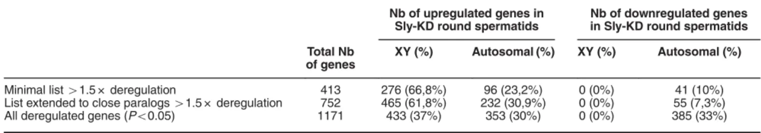
To compare SLY ChIP-Seq gene list with the list of genes
deregulated in Sly-deficient round spermatids (Sly-KD), we first
re-analyzed our previously published microarray data
15using
the most recent version of the mouse genome (GRCm38/
mm10) since it is more complete in term of sequence length and
annotation, especially of the Y chromosome, than the previous
version GRCm37/mm9 (i.e., in mm9 version, only 15% of the Y
chromosome was assembled). Over 400 genes were found
deregulated more than 1.5-fold (Po0.05), a majority of which
are encoded by the sex chromosomes, in agreement with our
previous observations.
15Since many of those genes are
present in multiple copies, we included closely related
paralogous genes and obtained a total of 752 deregulated
genes, again with a strong bias towards X- and Y- encoded
upregulated genes (Table 2). Comparison with SLY-associated
genes (i.e., the 7280 SLY ChIP-Seq genes) showed a higher
proportion of upregulated genes versus downregulated genes
(χ
2, P = 0.005) (Table 2, Supplementary Figure 2B). When
including all 1171 significantly deregulated genes (no threshold,
Po0.05), a higher proportion of autosomal upregulated and
downregulated genes was found and, this time, there were
more downregulated than upregulated genes among the genes
enriched in SLY at their TSS (χ
2, P = 0.012) (Table 2,
Supplementary Figure 2B).
Next, we investigated why some autosomal genes had SLYat
their TSS and yet were not found deregulated in Sly-KD round
spermatids by microarray. Since real-time PCR is a more
sensitive
technique
than
expression
microarrays,
26we
re-examined by quantitative real-time PCR (RT-qPCR) the
expression level of those autosomal genes, focusing on those
with the highest SLY enrichment at their gene start (10% of
genes with highest SLY peak, Supplementary Figure 3). In this
gene list are several members of the Speer gene cluster,
multicopy genes of chromosome 14 with yet unknown functions
and, interestingly, genes encoding proteins with a known role in
chromatin regulation, such as Dot1l which encodes an H3K79
methyltransferase (Figure 2a, Supplementary Figure 3). By
RT-qPCR we found several of those autosomal genes significantly
deregulated (up or downregulated) in Sly-KD spermatids
compared to WT spermatids (Figure 2b). With the same
approach, we identified additional sex chromosome-encoded
genes significantly upregulated in Sly-KD spermatids such as
Spin2d, Gmcl1l, Ube2a, Kdm5c and genes encoding
spermatid-specific histone variants, such as H2afb3, H2al1
(aka 1700012L04Rik) or H1fnt (Figures 2b and c).
7,27–30Their
closely related paralog H2afb1 encoded by an autosome, is not
regulated by SLY (Figures 2b and c). Since Sly knockdown
leads to increased transcription of H2afb3, we checked whether
it affects H2 A.B3 incorporation in the spermatid chromatin by
ChIP-qPCR and found that H2A.B3 level is higher at the TSS of
Table 2 Microarray analyses of Sly-KD versus WT round spermatids
Nb of upregulated genes in Sly-KD round spermatids
Nb of downregulated genes in Sly-KD round spermatids Total Nb
of genes
XY (%) Autosomal (%) XY (%) Autosomal (%)
Minimal list41.5 × deregulation 413 276 (66,8%) 96 (23,2%) 0 (0%) 41 (10%) List extended to close paralogs41.5 × deregulation 752 465 (61,8%) 232 (30,9%) 0 (0%) 55 (7,3%) All deregulated genes (Po0.05) 1171 433 (37%) 353 (30%) 0 (0%) 385 (33%)
expressed genes in Sly-KD compared to WT round spermatids
(Figure 2d).
Overall our data show that, when considering genes
expressed in spermatids, all sex chromosome-encoded genes
are upregulated in Sly-KD spermatids while autosomal genes
are either upregulated, downregulated or unchanged.
Sly-dependent deregulation of the H3K79 histone
methyl-transferase DOT1L impairs H3K79 methylation prior to
histone-to-protamine exchange. One of the genes with
highest enrichment of SLY at its start is Dot1l (Figure 2a)
which encodes the only known H3K79 histone
methyltrans-ferase. It is presumed to be important for chromatin
remodeling during sperm differentiation, because high levels
of H3K79 methylation precede histone removal during
spermatid elongation.
31,32Besides, analyses of published
RNASeq and microarray data show that Dot1l is particularly
expressed after meiosis in mice (Figure 3a) and humans
(Figure 3b). Immunofluorescence experiments on WT mouse
testicular samples show that DOT1L protein is enriched at the
sex chromatin in round spermatids and appears as nuclear
punctuated signals in step 9–11 elongating spermatids
(Figure 3c, Supplementary Figure 4). High H3K79
dimethyla-tion (H3K79me2) levels were also observed in round
spermatids and at the onset of spermatid elongation (step
10–12 elongating spermatids, Supplementary Figure 6) just
prior to histone-to-protamine exchange, as described by
Dottermusch-Heidel et al.
31,32Since Dot1l is downregulated
Figure 2 SLY controls the expression of genes involved in chromatin regulation during postmeiotic sperm differentiation. (a) Representation of SLY ChIP-Seq, its input, H3K4me3 Seq, Kcr Seq, H3K27me3 Seq and H3K9me3 seq at the TSS of Dot1l, one of the genes found to have the highest enrichment of SLY by ChIP-Seq. (b) Transcript level of autosomal genes found to be highly enriched in SLY protein at their TSS (10% of genes with highest SLY peak, Supplementary Figure 3) and of XY-encoded genes, measured by RT-qPCR in Sly-KD and WT round spermatids. The graph represents the geometric mean± S.E.M (after normalization with β-actin, n = 3–6 samples per genotype). For all except two genes (labeled with‘ns’ for non-significant), a significant difference between Sly-KD and WT samples was found with a P-valueo0.05 (t-test). (c) Representation of SLY ChIP-Seq, its input, H3K4me3 ChIP-Seq, Kcr ChIP-Seq, H3K27me3 ChIP-Seq and H3K9me3 ChIP-seq. SLY co-localizes with active epigenetic marks H3K4me3 and Kcr at the TSS of H2afb3 (located in an intron of Pdch11x) but not of its autosomal-encoded homolog, H2afb1. (d) ChIP-qPCR experiments on WT and Sly-KD round spermatids using antibody against H2A.B3. The TSS of expressed genes was found enriched in H2A.B3 in Sly-KD compared to WT round spermatids (n= 3 samples per genotype). The Y-axis represents the mean enrichment (%IP/input) normalized to the corresponding WT value. A significant increase in H2A.B3 level at the TSS was found in Sly-KD compared to WT samples when considering all tested genes (t-test, Po0.05)
SLY controls sperm chromatin re-organization C Moretti et al
in Sly-KD spermatids (Figure 2b, Supplementary Figure 5),
we looked at H3K79me2 expression: by
immunofluores-cence, there was a notable decrease in H3K79me2 levels in
step 10–12 elongating spermatids from Sly-KD males
compared to WT elongating spermatids (Figures 4a, b and
Supplementary Figure 6). We next purified elongating/
condensing spermatid fractions from WT and Sly-KD testes
by elutriation.
33Despite an elevated intra-genotype variability
we did not observe major differences in the proportion of
elongating/condensing spermatids between WT and Sly-KD
fractions; those fractions can therefore be compared. We
quantified H3K79me2 levels by ChIP-qPCR and western
blot and confirmed that H3K79me2 is reduced in Sly-KD
compared
to
WT
elongating/condensing
spermatids
(Figure 4c, Supplementary Figure 7A and C). Finally, we
observed by immunofluorescence that H3K79me2 persists in
spermatozoa (Figure 4d and Supplementary Figure 8).
Quantification of H3K79me2 revealed that the decrease
observed in Sly-KD elongating/condensing spermatids
per-sists in spermatozoa (when normalized to histone H3 level,
see below, Supplementary Figure 7B and D).
Sly deficiency leads to reduced histone H4 acetylation
prior to histone-to-protamine exchange. Extensive
acet-ylation of histone H4 (acH4) is a hallmark of chromatin
remodeling during spermatid differentiation and is detected at
the same postmeiotic stages than H3K79 dimethylation, just
prior to nucleosome eviction.
31,32It has recently been
demonstrated that DOT1L-mediated H3K79me2 facilitates
acH4.
34We therefore tested whether acH4 was impacted by
Figure 3 DOT1L is highly expressed in postmeiotic germ cells. (a) Schematic of Dot1l expression as found by RNA-Seq on Sertoli cells (SE) and mouse purified germ cells fractions (SG: spermatogonia; SC: spermatocytes; rST: round spermatids; eST: elongating spermatids) (analysis of GSE3500518). (b) Schematic diagram of DOT1L expression in humans as found by micro-array analyses of testicular biopsies with different germ cell contents (SG, SC, rST, eST) (analysis of Affymetrix HG-U133_Plus_2 probe no. 231297_at61). (c) Immunofluorescence detection of DOT1L protein (red) in germ cells at 4 stages of postmeiotic differentiation (from left to right: round spermatid, earlyelongating spermatid, late elongating spermatid, spermatozoa). DAPI (blue) was used to stain nuclei. H3K9me3 (in green) marks the chromocenter (i.e., the constitutive pericentromeric heterochromatin) and the X or Y chromosome (i.e., the postmeiotic sex chromatin, PMSC, indicated by an arrow).71Scale bar indicates 10μm. See also Supplementary Figure 4
Sly deficiency and found reduced level of acH4 in Sly-KD
versus WT step 10–12 elongating spermatids by
immuno-fluorescence (Figures 5a, b and Supplementary Figure 9). This
was confirmed by ChIP-qPCR analyses in all tested sites
(Figure 5c). Together, these data show that Sly deficiency
affects chromatin remodeling during spermatid differentiation.
Defects in postmeiotic chromatin remodeling lead to a
higher proportion of residual histones and increased
DNA oxidation in
Sly-deficient spermatozoa.
Approxi-mately 1–5% of histones remain in WT mouse
sperma-tozoa.
35To determine whether abnormal chromatin marks in
elongating spermatids have consequences on chromatin
content in spermatozoa, we next compared the quantity of
remaining histones in sperm and observed a ~ 2.5-fold
increase in histone H3 and a ~ 2.3-fold increase in TH2B in
Sly-KD compared to WT spermatozoa (Figures 6a and b).
Using antibody against protamine 2 (Hup2B) we also
detected a small (~20%) but significant decrease in the
quantity of protamine 2 in Sly-KD compared to WT sperm
(Figure 6c). No significant difference was observed for
protamine 1 (data not shown).
Finally, we tested whether abnormal chromatin content
could alter spermatozoa genome integrity by measuring the
proportion of spermatozoa showing oxidation of their DNA
(measurement of oxidized deoxy-guanosine, 8-oxo-dG). We
found an average of ~ 34% of WT spermatozoa with 8-oxo-dG
staining, as described in other studies,
36and a significant
increase to ~ 53% of 8-oxo-dG positive spermatozoa in Sly-KD
epididymis (Figure 6d).
SLY interacts with TBL1XR1 and other members of the
SMRT/N-Cor complex. To understand how SLY controls
gene expression during sperm differentiation, we searched
for its protein partners by co-immunoprecipitation followed by
mass spectrometry. We used FLAG antibody to
immunopre-cipitate SLY and its partners on two types of materials: (i)
testicular cells from a transgenic mouse model expressing
SLY protein fused to a FLAG-tag (Supplementary Figure 10)
and (ii) a spermatogonia cell line (GC1) transfected with a
FLAG-SLY construct. WT testes and GC1 cells transfected
with an empty vector were used as negative controls.
Immunoprecipitated
proteins
were
analyzed
by
liquid
chromatography coupled to tandem mass spectrometry
Figure 4 DOT1L-mediated H3K79 dimethylation is affected by Sly knockdown. (a) Immunofluorescence detection of H3K79 dimethylation (H3K79me2) in stage X testicular sections from WTand Sly-KD mice. Scale bar indicates 20μm. Pictures were taken using the same image capture parameters (see also Supplementary Figure 6). (b) Schematic showing H3K79me2 level quantified by immunofluorescence in Sly-KD and WT step 10–12 elongating spermatids. The graph represents mean ± S.E.M. The difference between the two genotypes is statistically significant (t-test on six samples per genotype, P= 0.0002). (c) ChIP-qPCR experiments on elongating/condensing spermatids from WTand Sly-KD mice, using antibody against H3K79me2. The Y-axis represents the mean enrichment (% IP/input)± S.E.M. normalized to a negative control region (NC) located at ~ 170 kb from a TSS, and to the corresponding WT value. A significant decrease of H3K79me2 level at gene TSS was found in Sly-KD compared to WT samples when considering all tested genes (t-test, Po0.05, n = 3–4 samples per genotype). (d) Immunofluorescence detection of H3K79me2 (red) in WTepididymal spermatozoa. Hoechst (blue) was used to stain nuclei. Scale bar indicates 5μm
SLY controls sperm chromatin re-organization C Moretti et al
(LC-MS/MS). Two complementary approaches (MASCOT
and label-free quantification (LFQ) Maxquant analysis) were
used to analyze LC-MS/MS data. They showed that TBL1XR1
and several other members of the SMRT/N-CoR repressive
complex (TBL1X, NCOR1 and HDAC3) were specifically
immunoprecipitated with SLY (Table 3). Interaction of SLY
and TBL1XR1 was confirmed in WT and FLAG-SLY transgenic
testes by western blot following immunoprecipitation using
anti-SLY, anti-FLAG or anti-TBL1XR1 antibody (Figure 7).
Discussion
In the present paper, we investigated the molecular role of SLY
and the consequences of its absence on chromatin structure;
based on acquired data we identified novel regulators of
postmeiotic gene expression and of chromatin remodeling
during sperm differentiation.
SLY and the regulation of postmeiotic gene expression.
First, we found that SLY protein is present at the TSS of
thousands of genes relevant to postmeiotic cell identity, with
a known role during postmeiotic differentiation and/or
significantly upregulated postmeiotically. We also showed
that SLY overlaps with active chromatin marks, such as
H3K4me3 or Kcr. Comprehensive transcriptome analyses of
Sly-deficient postmeiotic germ cells demonstrated that Sly
deficiency does not switch on genes normally silent in
postmeiotic cells but rather modulates the expression level
of
41000 genes expressed postmeiotically. Sly deficiency
chiefly induces upregulation of XY genes (37% of all
deregulated genes) with no XY gene found downregulated,
while hundreds of autosomal genes are either upregulated or
downregulated (respectively 30% and 33% of all deregulated
genes). The consequences of Sly deficiency on postmeiotic
gene expression are therefore different for XY genes
compared to autosomal genes, suggesting distinct regulatory
mechanisms, probably due to a different chromatin
environ-ment. Indeed, postmeiotically, the XY chromatin significantly
differs from that of autosomes as a consequence of the
meiotic silencing of sex chromosomes.
37,38How does SLY control gene expression? Despite the fact
that SLY is related to SYCP3, a protein of the meiotic
synaptonemal complex which has been proven to bind
double-stranded DNA,
14,17,39the mechanism by which SLY
regulates genes remained elusive. Indeed, SLY-SYCP3
conservation is relatively low (28% of identity) and SLY,
contrary to SYCP3, is very acidic (with an isoelectric point
~ 4.8); this most likely precludes direct DNA binding and rather
Figure 5 Sly deficiency impairs histone H4 hyperacetylation during postmeiotic sperm differentiation. (a) Immunofluorescence detection of histone H4 acetylation (acH4) in stage XII testicular sections from WT and Sly-KD mice. Scale bar indicates 20μm. Pictures were taken using the same image capture parameters (see also Supplementary Figure 9). (b) Schematic showing acH4 level quantified by immunofluorescence in Sly-KD and WT step 10–12 elongating spermatids. The difference between the two genotypes is statistically significant (t-test on 5–6 samples per genotype, P = 0.00012). (c) ChIP-qPCR experiments on elongating/condensing spermatids from WTand Sly-KD mice, using antibody against acH4. The Y-axis represents the mean enrichment (% IP/input)± S.E.M. normalized to the corresponding WT value. A significant decrease of acH4 level was found in Sly-KD compared to WT samples when considering all tested genes (t-test, Po0.01, n = 3–4 samples per genotype)
suggests that SLY is part of a protein complex able to recruit
regulators of gene expression, including proteins with
DNA-binding domains. In that respect, our findings that SLY
interacts with TBLX1R1 and other members of the
SMRT/N-CoR complex provide an interesting model. This complex,
expressed in many tissues, contains five proteins (NCOR1,
HDAC3, GPS2, TBL1XR1 and TBL1X) and has been shown to
interact with nuclear hormone receptors, transcription factors
or chromatin modifying enzymes.
40–43The recruitment/
release of SMRT/N-CoR complex on repressed gene
promo-ters is a dynamic process
44,45on which SLY could act to
control gene expression. Further studies will be required to
Figure 6 Sly deficiency leads to abnormal chromatin content and increased DNA oxidation in spermatozoa. (a) Western blot quantification of histone H3 (using anti-pan-H3) in protein extracts from WT and Sly-KD spermatozoa. Antibody against TUBULIN was used to normalize the signal. The scatter plot on the right represents histone H3 level normalized with TUBULIN (mean value± S.D.). The difference between the two genotypes is statistically significant (t-test on 8–9 samples per genotype, P = 0.0047). (b) Western blot detection and quantification of TH2B in protein extracts from WT and Sly-KD spermatozoa. Antibody against TUBULIN was used to normalize the signal. The right panel is a scatter plot showing quantification of TH2B (normalized to TUBULIN level) in Sly-KD and WT spermatozoa. The graph represents the mean level± S.E.M. The difference between the two genotypes is statistically significant (t-test, n= 3–5 samples per genotype, P = 0.009). (c) Western blot quantification of protamine 2 (using anti-Hup2B) in protein extracts from WT and Sly-KD spermatozoa. Antibody against TUBULIN was used to normalize the signal. The scatter plot on the right represents Protamine 2 level normalized with TUBULIN (mean value± S.D.). The difference between the two genotypes is statistically significant (t-test on 5–6 samples per genotype, P = 0.0199). The variability was elevated among Sly-KD samples and when excluding one outsider value, the P-value dropped to 0.0003. (d) Representative pictures obtained using antibody against 8-oxo-dG in WT and Sly-KD epididymal spermatozoa. WT picture shows two 8-oxo-dG negative spermatozoa while Sly-KD picture shows two 8-oxo-dG positive spermatozoa. Pictures were taken using the same image capture parameters. Scale bar indicates 5μm. The scatter plot on the right represents the percentage of oxidized spermatozoa (mean ± S.D.) measured using antibody against 8-oxo-dG. Ctl+ indicates values obtained when spermatozoa were pre-incubated with H202 (positive control). Mann–Whitney nonparametric test showed significant difference between WTand Sly-KD samples (5–7 samples per genotype, P = 0.01) and between WTand Ctl+ (P = 0.025)
SLY controls sperm chromatin re-organization C Moretti et al
characterize the role of SMRT/N-Cor complex in the context of
sperm differentiation.
By gene ontology analyses, we showed that many of
SLY-targets are involved in gene regulation and chromatin
modification. Focusing on those genes, we found that SLY
regulates XY-encoded H2A variants and DOT1L, a promising
candidate for the chromatin remodeling defects observed in
Sly-KD spermatids (see below).
The sex chromosomes encode several H2A variants, all of
which are particularly, if not specifically, expressed in
spermatids.
7,27–29We found that all of them are upregulated
in Sly-KD round spermatids and that H2A.B3, known to be
enriched at the start of active genes,
27is more incorporated in
Sly-KD than in WT spermatid chromatin. Several other genes
encoding histones are also SLY-targets (both enriched in SLY
at their TSS and deregulated when Sly is knocked down), such
as Hist1h3 and Hist1h4 clusters,
15H1fnt and so on. Their
deregulation could also contribute to the gene deregulation
and abnormal chromatin remodeling observed in Sly-deficient
spermatids.
It is quite intriguing that SLY controls the expression of
essential and evolutionary conserved genes but is itself not
conserved throughout evolution.
15,46,47Indeed, Sly has been
shown to be involved, together with its X-linked homolog Slx, in
an intragenomic conflict in which an unbalanced number of Sly
versus Slx gene copies leads to transmission distortion.
33The
fact that SLY regulates chromatin components fits with the
observation that transmission distorters are often involved in
gene/chromatin regulation processes.
48SLY and chromatin remodeling during sperm differentiation.
We particularly investigated Dot1l and the consequence of its
downregulation, as it is an interesting candidate gene
involved in spermatid chromatin remodeling and
histone-to-protamine replacement. DOT1L is the principal H3K79
methyltransferase identified to date and is ubiquitously
expressed and conserved throughout evolution.
49,50During
spermatogenesis, it has been shown to be expressed in
spermatocytes
51and spermatids;
31,32here, we show that
Dot1l is actually expressed at a higher level in spermatids
compared to spermatocytes or spermatogonia. In elongating
spermatids, high H3K79me2 coincides with histone H4
hyperacetylation, just prior to histone removal.
31,32Using
our mouse model, we showed that postmeiotic
downregula-tion of Dot1l induced by Sly deficiency leads to reducdownregula-tion in
H3K79me2 and in acH4 levels in elongating spermatids. At
Table 3 Mass spectrometry results from MASCOT and LFQ Maxquant analysis
Mascot Data IP:FLAG on whole testis
IP:FLAG on FLAGSLY testis IP:FLAG on WT testis
Score Nber peptides % Sqce covery EmPAI Score Nber peptides % Sqce covery EmPAI
HDAC3 0 0 0 0 0 0 0 0
TBL1XR1 34 1 4.5 0.09 0 0 0 0
NCOR1 33 2 0.9 0.04 0 0 0 0
SLY 276 11 23.9 1.56 0 0 0 0
TBL1X 0 0 0 0 0 0 0 0
Mascot Data IP:FLAG on transiently transfected cells
IP:FLAG on FLAGSLY cells IP:FLAG on empty-pcDNA3.1 cells
Score Nber peptides % Sqce covery EmPAI Score Nber peptides % Sqce covery EmPAI
HDAC3 86 1 3 0.1 0 0 0 0
TBL1XR1 143 4 11 0.8 0 0 0 0
NCOR1 0 0 0 0 0 0 0 0
SLY 566 16 51 8.56 0 0 0 0
TBL1X 273 7 0.8 24 0 0 0 0
MaxQuant Data IP:FLAG on whole testis IP:FLAG on transiently transfected cells
IP:FLAG on FLAGSLY testis IP:FLAG on WT testis IP:FLAG on FLAGSLY cells IP:FLAG on empty- pcDNA3.1 cells
LFQ LFQ LFQ LFQ HDAC3 0 0 141 110 0 TBL1XR1 0 0 45 558 0 NCOR1 92 305 0 0 0 SLY 6 544 900 0 5 026 100 0 TBL1X 0 0 754 720 0
Abbreviations: EmPAI, Exponentially Modified Protein Abundance Index; LFQ, label-free quantification
Top panel: MASCOT analysis of the co-immunoprecipitation assay performed on whole testis and of the co-immunoprecipitation assay performed on GC1 cells. The protein score is the sum of the highest MS/MS ions score for each distinct peptide sequence. The highest the protein score, the more it is represented in the sample. However, for an identical mole of protein, a larger protein can give more peptides than a smaller one. EmPAI value provides a complementary information as it depends on the number of detected peptides compared to the total number of detectable peptides. The higher the EmPAI, the more abundant the protein.
Bottom panel: Maxquant LFQ analyses of the co-immunoprecipitation assay performed on whole testis and of the co-immunoprecipitation assay performed on GC1 cells. Maxquant analysis is more accurate than EmPAI because it reflects the total LC-MS intensity of peptides for the proteins
the end of the differentiation process, Sly-KD spermatozoa
display a moderate but significant alteration in spermatozoa
chromatin content, with more residual histones, and less
protamination (~20% reduction). We propose this is a
consequence of the abnormal spermatid chromatin
composi-tion resulting from the deregulacomposi-tion of genes essential to
spermatid chromatin remodeling, in particular of DOT1L
(Figure 8). It has recently been shown that DOT1L-mediated
H3K79me2 facilitates histone H4 acetylation in the context of
MLL leukemia,
34and that BRD4-mediated acetylation
pro-motes chromatin decompaction and nucleosome eviction.
52We propose that, in the context of sperm differentiation,
DOT1L and H3K79me2 play a critical role in H4 acetylation
which itself is required for an open chromatin state enabling
nucleosome removal prior to protamine incorporation.
Finally, we show that abnormal chromatin content and
compaction of Sly-KD spermatozoa is associated with
increased susceptibility to oxidative stress. This could also
explain the increase in sperm DNA breaks that was previously
observed.
16A modest reduction in protamine levels (~33%) in
mice haploinsufficient for Prm1 or Prm2 gene results in
reduced sperm compaction, increased DNA damage and
embryo lethality.
9Experiments in which Sly-KD sperm were
directly injected into the oocytes (i.e., intracytoplasmic sperm
injection, ICSI) have shown that Sly deficiency does not
dramatically impair the early post-fertilization events and does
not lead to gross paternal chromosome breaks in the
zygotes.
16But, in light of the high incidence of sperm with
oxidized DNA in Sly-KD males that we reported here, ICSI with
Sly-KD sperm may produce offspring with increased
muta-tional load. If not properly repaired, DNA lesions such as
oxidized deoxy-guanosine (8-oxo-dG) can indeed lead to
mutations. Despite the existence of DNA repair strategies in
the mammalian zygote, studies have shown deleterious
consequences on genome integrity and embryo development
when incidence of DNA lesions is too high.
13,36,53Oxidative
damage to sperm DNA resulting from age, environmental or
lifestyle factors (such as smoking), has been shown to be
associated with increased incidence of diseases (such as
cancers, neurological disorders, etc.) in the progeny.
54Oxidative stress associated with impaired chromatin
remodel-ing in case of male infertility could similarly have negative
consequences on the health of children conceived using ICSI
to bypass the father’s infertility. Further studies using relevant
models will be needed to address this question.
Material and Methods
Seq analyses. SLY Seq was performed by Active Motif ChIP-Sequencing service, as follow: ~ 10 million of FACS-sorted enriched fractions of round spermatids (with a purity490%) were fixed with 1% formaldehyde for 15 min then quenched with 0.125 M glycine. Chromatin was isolated by adding lysis buffer,
Figure 7 SLY protein interacts with TBL1XR1 protein. (a) SLY and TBL1XR1 antibody detection of WT and Sly-deficient (Sly-KD) whole-testicular extracts (INPUT) and of corresponding extracts immunoprecipitated with SLY antibody (IP:SLY). SLY immunoprecipitation performed on Sly-KD testicular extracts and immunoprecipitation with purified rabbit IgG on WT extracts (IP:IgG) were used as negative controls. (b) SLY and TBL1XR1 antibody detection of whole-testicular extracts from WT and FLAG-SLY mice (INPUT) and of corresponding extracts immunoprecipitated with FLAG antibody (IP:FLAG). FLAG immunoprecipitation performed on WT testicular extracts was used as a negative control. (c) SLY and TBL1XR1 antibody detection of WT whole-testicular extracts (INPUT) and immunoprecipitated with TBL1XR1 antibody (IP:TBL1XR1). TBL1XR1 immunoprecipitation performed on Sly-KD testicular extracts and immunoprecipitation with purified rabbit IgG on WT extracts (IP:IgG) were used as negative controls. Arrows indicate the specific band (at expected sizes of 38kDa for SLY and 64kDa for TBL1XR1) and stars, non-specific bands (which disappear in negative controls)
SLY controls sperm chromatin re-organization C Moretti et al
followed by disruption with a Dounce homogenizer. Lysates were sonicated and the DNA sheared to an average length of 300–500 bp. Genomic DNA (input) was prepared by treating aliquots of chromatin with RNase, proteinase K and heat for de-crosslinking, followed by ethanol precipitation. Pellets were resuspended and the resulting DNA was quantified on a NanoDrop spectrophotometer. Extrapolation to the original chromatin volume allowed quantitation of the total chromatin yield. An aliquot of chromatin (30 μg) was precleared with protein A agarose beads (Life technologies, Carlsbad, CA, USA). Genomic DNA regions of interest were isolated using 12μg of anti-SLY antibody.14Complexes were washed, eluted from the beads with SDS buffer, and subjected to RNase and proteinase K treatment. Crosslinks were reversed by incubation overnight at 65 °C, and ChIP DNA was purified by phenol–chloroform extraction and ethanol precipitation. Illumina sequencing libraries were prepared from the ChIP and input DNAs using the Apollo 324 system (WaferGen, Fremont, CA, USA). After a final PCR amplification step, the resulting DNA libraries were quantified and sequenced on HiSeq 2500 (37 and 26 million of reads were obtained for ChIP and input DNAs, respectively).
Bioinformatics. SLY ChIP and input sequences (50-nt reads, single end) were aligned to the mouse genome (GRCm38/mm10) using BWA algorithm,55 and filtered by mapping quality using Samtools -q0.56 For reads with multiple good
alignments, one alignment was reported at random. Alignments were extended in silico at their 3′-ends to a length of 200 bp, which is the average genomic fragment length in the size-selected library, and assigned to 32-nt bins along the genome. The resulting histograms (genomic‘signal maps’) were stored in BAR and bigWig files. Data sets have been submitted to SRA http://www.ncbi.nlm.nih.gov/sra with accession number SRP055115. Sly peak locations were determined using the
MACS algorithm (v1.4.2)57with a cutoff of P= 1e − 7. Annotation of SLY-enriched genomic regions (Figure 1a and Supplementary Figure 1A) was performed using Cis-regulatory Element Annotation System available at http://liulab.dfci.harvard.edu/ CEAS/index.html. P-values for the significance of the relative enrichment with respect to the background were calculated using one-sided binomial test. BED files containing SLY ChIP-Seq regions (WT and Sly-KD) were intersected with Ensembl80 gene coordinates (gene start, or gene start ± 1 kb). Graphical representation of enrichment around the start of genes (expressed and not expressed in round spermatids) was done using Profiler and ComputeMatrix tools58 available at the Galaxy website https://mississippi.snv.jussieu.fr/. Round spermatids ChIP-Seq data sets for H3K4me3, H3K27me, Kcr and H3K9me3 were obtained from GSE42629, GSE32663 and GSE56526.19–21All data sets were re-analyzed using the last version of the mouse genome (GRCm38/mm10) as described in Moretti et al.38Overlap comparison was achieved by intersecting the intervals of two
ChIP-Seq data sets. Graphic representations of ChIP-Seq data were performed using IGV (Integrative Genomics Viewer, https://www.broadinstitute.org/igv/). Gene ontology analyses were performed using Genomatix (https://www.genomatix.de/), GSEA (http://www.broadinstitute.org/gsea/)59and EnrichR (http://amp.pharm.mssm. edu/Enrichr/).60
Microarray and RNA-Seq analyses. From Illumina microarray WG6 v2 of Sly-KD versus WT round spermatids,15all genes41.5 × fold deregulated (i.e.,
Log2 ratioo − 0.58 or 40.58) with a P-value ⩽ 0.05 were converted to ENSEMBL ID using Biomart (http://www.ensembl.org/biomart) and the last version of the mouse genome (GRCm38/mm10). Close paralogs (470% identity) were added to 41.5 × extended to close paralog list (752 genes in total). All genes significantly
Figure 8 Model presenting the mechanism by which SLY controls gene expression and chromatin remodeling during sperm differentiation. In WT round spermatids (left panel), SLY (in blue) interacts with the SMRT/N-CoR complex (which comprises TBL1XR1, TBL1X, NCOR1 and HDAC3) and is located at the start of genes involved in gene regulation, chromatin regulation and the ubiquitin pathway. In particular, SLY directly controls the expression of X-chromosome-encoded genes coding for H2.A variants (such as H2A.B3) and of the H3K79 methyltransferase DOT1L. In elongating spermatids, there is a wave of H3K79 dimethylation (orange circles) and of histone H4 acetylation (green circles); those modifications are expected to be a prerequisite to the efficient removal of nucleosomes (light pink oval) and replacement by protamines (purple oval), a process which is required to achieve optimal compaction of the spermatozoa nucleus. When SLY is knocked down (right panel), X-encoded H2.A variants are upregulated and more incorporated in the spermatid chromatin, while DOT1L is downregulated. DOT1L downregulation leads to a decrease in dimethylated H3K79 and acetylated histone H4 in elongating spermatids. Alterations in the spermatid chromatin structure affect the replacement of nucleosomes by protamines and lead to a higher proportion of nucleosomes and a decreased proportion of protamines. As a result, Sly-deficient spermatozoa are abnormally shaped, less compact and present a higher susceptibility to DNA damage than WT spermatozoa
deregulated with a P-value⩽ 0.05 were converted to ENSEMBL ID using Biomart and mm10 version of the mouse genome. This gave a list of 1171 deregulated genes. Gene expression data from human testicular biopsies were obtained from published microarray data set (ArrayExpress: E-TABM-234).61 Gene expression
data of mouse purified germ cells were obtained from RNASeq data sets GSE35005 and GSE437176,18and analyzed by GenoSplice (http://www.genosplice. com) using the following parameters: reads were aligned onto the mouse genome (Ensembl 75, mm10) using STAR v.2.3.0, with an exon–exon junction database built using annotations from Ensembl version 75. For each gene present in ENSEMBL, reads aligning on constitutive regions (that are not prone to alternative splicing) were counted. Based on these read counts, normalization and differential gene expression were performed using DESeq (v1.12.0 on R v3.0.0).
Mice. All animals used in the present study were on490% C57BL/6 background and processed at adult age (between 2- and 6-month old males). Sly-KD mice were obtained as described in Cocquet et al.15 FLAG-SLY1 transgenic mice were
obtained by pronuclear micro-injection of a linearized construct containing Sly1 open reading frame (i.e., Sly long and main isoform, see Riel et al.16fused with Flag sequence, under the control of the spermatid-specific promoter SP-10 (aka ACRV1).62,63Fertilized eggs from CBA/Ca x C57BL/10 mating were microinjected
with the construct, using standard protocols. Transgenic founders carrying the SP10-FLAG-SLY1 construct were identified by PCR using the following primers (35 cycles, annealing temperature 60 °C): Flag-F primer: 5′-GGA CTA CAA GGAC GA CGA TGA CAA-3′ and Flag-R primer: 5′- GCA GCC TGC ACC TGA GGA GT-3′ (711 bp).
Several founders were obtained and crossed with XYRIII males on a random-bred MF1 albino (National Institute for Medical Research colony) background. One line (called FLAG-SLY) was established from a male founder which transmitted the transgene with a ~ similar expression of FLAG-SLY1 compared to endogenous SLY1 protein. The line was maintained by further backcrossing transgenic males and females to B6/N mice and generate XYRIII males with (tsgic) and without (neg sib) the transgene. RT-qPCR and Western blot experiments showed a ~ 2-fold increase in Sly transcript and protein level in FLAG-SLY compared to WT testes. By immuno-fluorescence on FLAG-SLY and WT testicular tubules it was observed that FLAG-SLY transgene was only expressed in round spermatids (Supplementary Figure 10).
For all experiments, WT controls were of same age and background (i.e., non-transgenic siblings from the same mating). Animal procedures were subjected to local ethical review (Comite d’Ethique pour l’Experimentation Animale, Universite Paris Descartes; registration number CEEA34.JC.114.12).
Germ cell purification by FACS. Testicular cells were isolated from one adult male per experiment following a protocol adapted from64 with some
modifications previously described in Comptour et al.65Cell purity was assessed for
each collected fraction by microscope observation following DAPI (4,6-diamidino-2-phenylindole) staining (VECTASHIELD Mounting Medium with DAPI, Vectorlab, Burlingame, CA, USA) of cells spread onto glass slides and fixed with 4% buffered paraformaldehyde. Round spermatids and elongating spermatids were collected with a purity490%.
Germ cell purification by elutriation. Enriched fractions of round spermatids and elongating/condensing spermatids were obtained using two to three mice per experiment (i.e., 4–6 testes) by centrifugal elutriation as described previously.15 Cell purity was assessed for each collected fraction as described
above. Only fractions with490% purity were used for ChiP-qPCR analyses. It has previously been shown that Sly-KD and WT testicular tubules contain the same proportion of each type of round spermatids,15elutriated Sly-KD and WT round spermatid fractions can therefore be compared.
Collection of epididymal spermatozoa. Cauda epididymides were dissected out from 2- to 6-month-old males, freed of connective tissues and fat, and transferred to a small petri dish containing 1 ml of M2 medium (Sigma-Aldrich, Saint-Louis, MO, USA). Using small pipette tips, spermatozoa cells were very gently squeezed out of cauda epididymides, to limit contamination with somatic cells. Following 5 min of incubation at 37 °C, sperm suspensions were collected and counted using a Malassez hemocytometer. Sperm cell purity was assessed for each sample by microscope observation following DAPI (4,6-diamidino-2-phenylindole) staining (VECTASHIELD Mounting Medium with DAPI, Vectorlab) of sperm cells spread onto glass slides and fixed with 4% buffered paraformaldehyde. All samples used in our analyses contained⩾ 99% of spermatozoa. Samples were
aliquoted to five millions of spermatozoa per tube then centrifuged (600 g for 5 min) and washed once in 1 × PBS (Life technologies). Pellets were flash-frozen in liquid nitrogen.
SLY ChIP prior to qPCR. About 5 × 10⁶ elutriated round spermatids
(collected from two to three mice per sample) were crosslinked for 10 min at room temperature in 1 × PBS containing 1% PFA. Reaction was stopped by adding 125 mM Glycine and incubating for 5 min at room temperature. Cells were washed twice with ice-cold PBS and centrifuged at 500 g, 4 °C for 10 min. Cells were resuspended in 600μl of Lysis buffer (50 mM Tris pH 7.4, 300 mM NaCl, 0.1% NP-40, 0.1% DOC, 1 mM DTT) with Complete Protease Inhibitor Cocktail Tablets EDTA-free (Roche, Basle, Switzerland), 1 mM PMSF 1 mM and 10 mM Aprotinin added extemporaneously, and incubated on ice for 30 min. The suspension was then sonicated using PICO-Diagenode to obtain fragments of approximate size of 500 bp (as verified by 2100 Bioanalyzer, Agilent, Santa Clara, CA, USA). The lysate was centrifuged at 10 000 g at 4 °C for 5 min. Twenty microliter of supernatant were taken as input and the remaining supernatant was incubated on a rotating wheel at 4 °C overnight with Protein G Dynabeads (Life Technologies) which were previously coupled with antibody against SLY,14as recommended by the manufacturer. The
following day beads were washed subsequently in Lysis buffer, wash buffer no. 2 (50 mM Hepes pH7.4, 0.5M NaCl, 5 mM EDTA, 1% Triton, 0.1% NaDeoxycholate), wash buffer no. 3 (10 mM Tris-HCl pH8, 0.25 M LiCl, 0,5% NP-40, 0.5% NaDeoxycholate, 5 mM EDTA ph8) and in TE buffer. Supernatant was completely removed and beads were resuspended in Elution buffer containing 1% SDS. Input was defrosted on ice and diluted in Elution buffer. Samples were incubated for 15 min at 65 °C with gentle mix every 2 min. IP and input were reverse crosslinked overnight at 65 °C. Samples were then treated with Proteinase K and incubated for 1 h at 37 °C. IP and input DNAs were purified using NucleoSpin Kit (MachereyNagel, Hoerdt, France) and eluted in 50μl of distilled water. Experiments were repeated on independent pools of elutriated round spermatids (each representing 2–3 mice) three to six times. T-test was used for statistical analyses. H2A.B3 ChIP prior to qPCR. Mononucleosomes were prepared from elutriated round spermatids (490% purity) as previously described in Montellier et al.11with some modifications. Briefly, about 5 × 10⁶round spermatids purified by elutriation were lysed by incubation with 300μl of lysis buffer (KCl 60 mM, NaCl 15 mM, Tris HCl pH 7.4 15 mM, Saccharose 0.34 M, EDTA 2 mM, EGTA 0.5 mM, Spermidine 0.65 mM, DTT 1 mM, Triton 0.03%, Glycerol 1%, Complete Protease Inhibitor Cocktail Tablets Roche EDTA-free, PMSF 1 mM, Aprotinin 10 mM) for 20 min on ice, followed by centrifugation at 500 g at 4 °C for 10 min. The pellet was gently resuspended in 200μl of wash buffer (KCl 60 mM, NaCl 15 mM, Tris HCl pH 7.4 15 mM, Saccharose 0.34 M, spermidine 0,65 mM, DTT 1 mM, Complete Protease Inhibitor Cocktail Tablets Roche EDTA-free, PMSF 1 mM, aprotinin 10 mM) and centrifuged again. Nuclei were resuspended in 200μl of MNase buffer (Tris HCl pH7.5 10 mM, KCl 10 mM, CaCl2 2 mM) and 5U of Micrococcal Nuclease (ThermoFisher, Waltham, MA, USA) were added. Samples were immediately incubated in a waterbath at 37 °C for 10 min. MNase reaction was stopped by adding EDTA to 5 mM final concentration. The nucleosome fraction was isolated by centrifugation at 10 000 g, 4 °C, for 5 min. ChIP were performed by adding LSDB 250 (glycerol 20%, Hepes 50 mM, MgCl2 3 mM, KCl 250 mM, Complete Protease Inhibitor Cocktail Tablets Roche, PMSF 1 mM, Aprotinin 10 mM) to the nucleosome fraction to a final volume of 500μl. Twenty microliter of this suspension were kept as input and immediately frozen at− 20 °C and the remaining volume was incubated on a rotating wheel at 4 °C overnight with the beads (Dynabeads protein G, Life technologies) previously coupled with 5μl of anti-H2A.B3).27 Samples were
processed as described above. Experiments were repeated twice on independent pools of elutriated round spermatids (each representing 2–3 mice) and gave similar results.
ChIP of chromatin marks on elongating/condensing spermatids. Elongating/condensing spermatids were collected by centrifugal elutriation. Aliquots of about 1 × 10⁷ elongating/condensing spermatids were used for chromatin preparation as described previously by Montellier et al.11with some modifications.
Briefly, aliquots were suspended in 75μl of lysis Buffer (Tris pH 7.4 50 mM, NaCl 300 mM, NP-40 0.1%, DOC 0.1%, DTT 1 mM, Complete Protease Inhibitor Cocktail Tablets EDTA-free Roche, PMSF 1 mM, aprotinin 10 mM, sodium butyrate 5 mM) for 15 min on ice with gentle shaking every 3 min. Spermatids were centrifuged at 10 000 g at 4 °C for 10 min. Supernatant was kept on ice and the pellet was resuspended in 100μl of Lysis buffer and sonicated on PICO-Diagenode for 4 min
SLY controls sperm chromatin re-organization C Moretti et al
(30 sec ON–30 sec OFF) to allow the suspension of larger chromatin fragment. Spermatids were centrifuged again at 10 000 g at 4 °C for 10 min and supernatant was pooled with the first supernatant for MNase digestion. Seventy-five microliter of MNase Buffer (Tris, pH 7.5 10 mM, KCl 10 mM, and CaCl2 1 mM) were added and MNase digestion was performed by adding 10 U of MNase. Digestion was performed for exactly 10 min in a waterbath at 37 °C. Digestion was stopped by adding 5 mM EDTA. Five microliter were kept to check MNase digestion on Agilent’s 2100 Bioanalyzer. The remaining volume was diluted with Lysis buffer and 20μl were kept as input. The remaining volume was incubated on a rotating wheel, 4 °C overnight with magnetic beads (Dynabeads Protein G, Life technologies) previously coupled with the appropriate antibody (5μl of anti-H3K79me2 (ab-3594 from Abcam, Cambridge, UK), or 9μl of anti-H4panAcetyl antibody (06-866 from Millipore, Billerica, MA, USA)). IP and input were further processed as described above. Experiments were repeated three to four times on independent pools of elutriated elongating/condensing spermatids (each representing 2–3 mice). T-test was used for statistical analyses.
Real-time quantitative PCR. Real-time PCR was performed using Roche LightCycler 480 and SYBRgreen Mastermix (Roche). ChIP-qPCR were performed on purified round spermatids or on elongating/condensing spermatids obtained by elutriation. Each sample represents a pool of 2–3 animals. Primers were designed to amplify regions across the TSS of indicated genes, except for NC which represents a negative control region (used to normalize) located 170 kb away from any TSS. The sequences and qPCR condition of Akap4 and Actrt1 ChIP primers can be found in Soboleva et al.27for other primers see Supplementary Figure 11.
RT-qPCR were performed on RNA extracted from WT and Sly-KD elutriated round spermatids and reversed-transcribed as described in Cocquet et al.15using primers listed in Supplementary Figure 11. The sequences and qPCR condition of Jmjd1c primers can be found in Kuroki et al.,66of
β-actin primers in Cocquet et al.,15of Dot1l
H9LAN primers in Dottermusch-Heidel et al.,32of H2afb3 primers in Soboleva et al.,27
of H2al1, H2al2y and H2afb1 (aka H2al2) primers in Ellis et al.67For the quantification of Flag-Sly transgene expression, RT-qPCR was performed on whole testes as described in Cocquet et al.15using Sly global primers (which amplify Sly1 and other
Sly isoforms), Sly1 primers and Acrv1 primers.15,16Student's t-test was used for all
qPCR statistical analyses.
Immunofluorescence. Immunofluorescence on sections and on surface-spread testicular cells were performed as previously described.15,65 Antibody
against DOT1L (ab-64077 from Abcam), H3K79me2 (ab-3594 from Abcam) or AcH4 (06-866 from Millipore) were diluted 1/100 to 1/200. Pictures were taken with an Olympus BX63 microscope. Quantification was performed on pictures obtained from six samples per genotype using ImageJ 1.48v (http://imagej.nih.gov/ij/). T-tests were performed with GraphPad Prism 5.02.
Quantification of DNA oxidation. Detection of 8-hydroxy-2 ′-deoxy-guanosine (8-oxo-dG) was carried out on spermatozoa from cauda epididymis. Spermatozoa were resuspended in a decondensing buffer consisting of 2 mM DTT and 0.5% Triton X-100 in PBS 1 × and incubated for 30 min at room temperature. After centrifugation at 800 g for 5 min at room temperature, spermatozoa were washed in 1 × PBS and smeared on a glass plate (250 000 cells/plate). The 8-oxo-dG immunostaining were performed as previously described by Noblanc et al.68 using a monoclonal antibody against 8-oxo-dG (15A3, Novus biological, Interchim, France). To evaluate the percentage of spermatozoa with 8-oxo-dG staining, 300 spermatozoa per sample were scored, and 5–7 samples per genotype were analyzed. A positive control consisting in pre-treating spermatozoa with H202 was included in the analysis (n= 3). Mann–Whitney nonparametric tests were performed using GraphPad Prism 5.02.
Transfection and protein extraction of GC1 cells. The open reading frames of Sly116was cloned in-frame with an N-terminal FLAG tag under the control
of the CMV promoter of the pCDNA3.1 vector (Invitrogen, Carlsbad, CA, USA). GC1-spg cells were transfected using Lipofectamine 2000 (Invitrogen) according to the manufacturer’s instructions with pCDNA3.1-CMV-FLAG-SLY vector (i.e., FLAG SLY) and pCDNA3.1 empty vector. Cells were collected for protein extraction 24 h after transfection and pelleted by centrifugation for 5 min at 1000 g. Proteins were extracted using ice-cold lysis buffer (150 mM NaCl, 20 mM Tris/HCl pH 8.0, 5 mM EDTA, 0.5% Igepal CA-630 (Sigma-aldrich), 1 mM phenylmethanesulfonyl fluoride, 1 mM sodium orthovanadate and 1 × Complete, Mini, EDTA-free Protease Inhibitor Cocktail) at 4 °C for 30 min on a rotating platform. After centrifugation at 13 000 g at
4 °C for 5 min, the supernatant was collected and immediately used for the in vitro immunoprecipitation assay and mass spectrometry analysis.
Protein extraction from testes. Nuclear and cytosolic fractions were obtained from adult testes as described in Cocquet et al.33Protein extraction prior to immunoprecipitation was performed as follow: flash-frozen testes were ground and resuspended in 1: 9 w/v ice-cold extraction buffer (150 mM NaCl, 20 mM Tris/ HCl pH 7.5, 5 mM EDTA, 0.5% Igepal CA-630 (Sigma-aldrich), 1 × protease inhibitor cocktail (Sigma-Aldrich), 1 mM phenylmethanesulfonyl fluoride and 1 mM sodium orthovanade). After homogenization and incubation at 4 °C for 30 min, tissue lysates were centrifuged at 13 000 g at 4 °C for 10 min. The supernatant was collected and immediately used for the immunoprecipitation assays.
Co-immunoprecipitation. For immunoprecipitation assays in GC1 cells, 60μl of anti-FLAG M2 magnetic beads (Sigma-aldrich) were washed three times in 1 × TBS (150 mM NaCl, 50 mM Tris, pH 7.4) and incubated with 600μl of protein extract from transiently transfected cells for 2 h at 4 °C. The magnetic beads were then washed two times in 500μl of TBS with 0.5% Tween-20 and then incubated for a long wash for 5 min at room temperature in TBS only. For non-denaturing elution, beads were resuspended in 150μl elution buffer (0.1 M glycine/HCl, pH 3.0), and incubated at room temperature for 5 min. The pH was neutralized using triethylammonium bicarbonate buffer (pH 8.4).
For immunoprecipitation assays in whole-testicular extracts, 40μl of Bio-Adembeads PAG (Ademtech, Pessac, France) or were washed two times in 80μl of PBS 0.65% Tween 20 and resuspended with 4μg of SLY antibody, anti-TBLX1R1 antibody (ab 24550, Abcam) and purified rabbit IgG (02-6102, Invitrogen) for 30 min at room temperature to allow binding of the antibody. Beads were washed two times in PBS 0.65% Tween 20, incubated with 200μl of 20 mM of dimethylpimelimidate to covalently coupled the antibodies to the beads for 30 min at room temperature and the reaction was stopped by resuspended the beads with 40μl of 50 mM Tris pH 7.5 for 15 min. Bio-Adembeads PAG beads and 40 μl of anti-FLAG M2 magnetic beads (Sigma-aldrich) were incubated with 400μl of protein extract for 2 h at 4 °C, and washed two times in PBS 0.65% Tween 20 and then incubated for a long wash for 5 min in PBS only. For non-denaturing elution, Bio-Adembeads PAG beads were resuspended in 30μl elution buffer and anti-FLAG M2 magnetic beads were resuspended in 100μl elution buffer, and incubated at room temperature for 5 min. The pH was neutralized using triethylammonium bicarbonate buffer (pH 8.4).
Alternatively, Dynabeads Protein G (Life technologies) were used as instructed by the manufacturer and resuspended in 800μl binding buffer with anti-SLY antibody or purified rabbit IgG (Life technologies), and incubated overnight at 4 °C to allow binding of the antibody. Beads were washed twice 0.2 M sodium borate (pH 9.0), incubated in 30 mM dimethylpimelimidate for 30 min at room temperature to covalently couple the antibodies to the beads, washed three times in 0.2 M ethanolamine (pH 8.0), and then washed twice in 1 ml binding buffer. Antibody-coupled beads were incubated with whole-testis extracts for 1 h, and washed three times in extraction buffer and once in 1 × TBS with 0.05% Triton X-100 for 5 min. Elution was performed using 0.1 M glycine/HCl, pH 3.0. The pH was neutralized using triethylammonium bicarbonate buffer (pH 8.4).
Western blot. Protein extraction and western blot experiments on whole testes and elutriated spermatids were performed as described in Comptour et al.65In brief, 15μl of each immunoprecipitated sample and a volume of input sample corresponding to 10% of IP sample were denatured using 4 × NuPAGE LDS sample buffer (Life Technologies) with 10%β – mercaptoethanol, boiled for 10 min at 95 °C and loaded. Extraction of sperm proteins was as follow: five millions of spermatozoa (purity⩾ 99%) were resuspended in 200 μl of 4 × NuPAGE LDS sample buffer (Life Technologies) and boiled for 10 min at 95 °C. Ten microliter of sample were loaded per lane. Antibody against SLY14was diluted 1/3000, antibody against SLX/SLXL1,69 1/6000, H3K79me2 (ab-3594 from Abcam), anti-TBLX1R1 (ab 24550 from Abcam), anti-Tubulin antibody (T-9026 from Sigma-Aldrich), anti-panH3 antibody (05-928 from Abcam), anti-FLAG (M5 from Sigma-Aldrich) and anti-Hup2B antibody (Briar Patch Biosciences, Grass Valley, CA, USA), 1/1000.
Sample digestion and LC-MS/MS. Co-immunoprecipitated samples were subjected to a bottom-up analysis at the 3P5 university platform. After, cysteines reduction with dithiothreitol and alkylation with chloroacetamide, proteins were