Aberrant Ras/MAPK signaling in skeletal development
by Simona Nedelcu B.S. BiologyCalifornia Institute of Technology, 2007
Submitted to the Department of Biology in Partial Fulfillment of the Requirements for the Degree of
DOCTOR OF PHILOSOPHY
at the
MASSACHUSETTS INSTITUTE OF TECHNOLOGY
September 2013
© 2013 Massachusetts Institute of Technology. All rights reserved
Signature of Author__________________________________________________________________________ Department of Biology
July 30th, 2013
Certified by___________________________________________________________________________________
Jacqueline A. Lees Professor of Biology Thesis Supervisor Accepted by___________________________________________________________________________________ Stephen P. Bell Professor of Biology Chairman, Graduate Committee
Aberrant Ras/MAPK signaling in skeletal development
ABSTRACT
The Mitogen-‐activated protein kinase(MAPK) signaling pathway has been studied intensively in the context of neoplastic transformation. Other studies have focused on the roles of this pathway during development and have modeled syndromes, such as Noonan Syndrome (NS), that are caused by aberrant germline Ras/MAPK signaling. One hallmark of these developmental syndromes is a defect in the skeletal development of the patients. However, the in vivo role of MAPK
signaling during bone development is still controversial, with some studies supporting a positive role of the MAPK pathway during this process, and others arguing for a suppressive role. To analyze in depth the bone defects caused by aberrant K-‐ras/MAPK signaling during development and to understand how bone mass is regulated via K-‐ras/MAPK control of osteoblast differentiation we have generated two novel mouse models. Specifically, to investigate how hyperactive K-‐ ras affects skeletal development when expressed in a spatial and temporal specific manner we activated K-‐rasG12D at various stages during limb development, either in
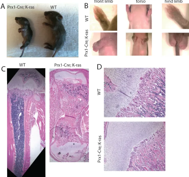
all mesenchymal lineages (Prx1-Cre;K-rasG12D mutants), or specifically in the
osteoblasts (Osx1-Cre;K-rasG12D). The Prx1-Cre;K-rasG12D mutant mice display greatly
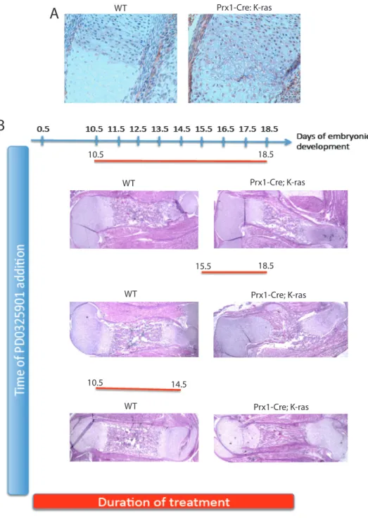
shortened and thickened bones that mirror the defects found in patients with NS. We determined that this bone defect appears during embryogenesis around E14.5 and is distinguished by impairment in formation of the bone collar. Furthermore, we describe a treatment strategy that rescues the skeletal phenotype, and identify a narrow developmental time window, where in utero treatment with MEK inhibitor is sufficient to completely rescue the bone phenotype. The Osx1-Cre;K-rasG12D
mutant mice allowed us to activate expression of hyperactive K-‐ras in the osteoblast population at different times during the development of the organism. Using this model, we defined two time windows when expression of active K-‐ras results in opposing bone phenotypes in the mutant mice. In the first time window
(approximately between E11.5 and E14.5) induction of active K-‐rasG12D in
osteoblasts precursors impairs terminal differentiation and leads to profound bone loss. In the second time window, activation of K-‐rasG12D at birth acts to promote
osteoblast differentiation and consequently long bone mineralization. Taken together, our data has revealed a critical involvement of K-‐ras/MAPK signaling in osteoblast differentiation during skeletal development, in embryogenesis and after birth. They also show how the MAPK pathway can be modulated chemically to rescue the skeletal defects seen in a mouse model for NS. These findings yield insight into diseases of the bone, including both developmental syndromes caused by aberrant MAPK activation, such as NS, and diseases characterized by an
imbalance in the bone mass, such as osteoporosis.
Thesis Supervisor: Jacqueline A. Lees Title: Professor of Biology
SIMONA NEDELCU
Koch Institute of Integrative Cancer Research, MIT
Dr. Jacqueline Lees Laboratory
56 Linnaean St, Cambridge, MA 02138
tescu@mit.edu, 857-‐499-‐0050 EDUCATION
Massachusetts Institute of Technology, Cambridge, MA 2008 – Present • PhD candidate in Biology, Department of Biology,
California Institute of Technology, Pasadena, CA 2004 – 2007 • BS with Honors, Biology
RESEARCH EXPERIENCE
Professor Jacqueline A. Lees, MIT, Cambridge, MA June 09 – Aug 13 • PhD Candidate
• Thesis Title: Aberrant Ras/MAPK signaling in skeletal development
Professor Robert H. Horvitz, MIT, Cambridge, MA July 07 – Aug 08 • Research Technician
• Project: Identification of genes that control aging in C. elegans
Professor Paul H. Patterson, Caltech, Pasadena, CA Jan 06 – June 07 • Undergraduate Research Fellow
• Project: IKKγ and mutant huntingtin regulation of Pleiotrophin expression in neuronal models of Huntington's Disease
Professor Henry A. Lester, Caltech, Pasadena, CA March 05 – Dec 05 • Undergraduate Research Fellow
• Project: Electrophysiology and fluorescence studies of the muscle nicotinic receptors
PUBLICATIONS
Hilgendorf KI, Leshchiner ES, Nedelcu S, Maynard MA, Calo E, Ianari A, Walensky LD, Lees JA. “The retinoblastoma protein induces apoptosis directly at the
mitochondria.” Genes Dev. 2013 May 1; 27(9): 1003-‐15.
Berdichevsky A, Nedelcu S, Boulias K, Bishop N, Guarente L, and Horvitz RH. “3-‐ Ketoacyl thiolase delays aging of C. elegans and is required for longevity mediated by sir-‐2.1” PNAS 2010 Nov 2; 107(44): 18927-‐32
Calo E, Quintero JA, Danielian PS, Nedelcu S, Berman SD and Lees JA. “Rb regulates fate choice and lineage commitment in vivo”. Nature 2010 Aug 26; 466(7310): 1110-‐ 4.
Khoshnan A, Ko J, Tescu S*, Brundin P, Patterson PH. “IKKalpha and IKKbeta regulation of DNA damage-‐induced cleavage of huntingtin”, PLoS One. 2009 Jun 2; 4(6): e5768.
*Publications before July 2009 appear under my maiden name: Simona Tescu
PRESENTATIONS
Nedelcu S, Lees JA. Activation of MAPK signaling pathway disrupts normal bone development in mice. Colrain Meeting, Colrain, MA (2012) Oral Presentation
Nedelcu S, Lees JA. Stage-‐Specific Effects of Activating Oncogenic K-‐ras on
Mesenchymal Development and Tumorigenesis. Mechanisms and Models of Cancer, Cold Spring Harbor (2012) Poster Presentation
Nedelcu S, Lees JA. Consequences of activating oncogenic K-‐ras during bone development. Koch Institute Focus Seminar Series, Cambridge, MA (2011) Oral Presentation
Nedelcu S, Lees JA. Effects of Activating Oncogenic K-‐ras During Skeletal Formation and Mesenchymal Lineage Specification. Koch Institute Retreat, MA (2011) Poster Presentation
Nedelcu S, Stanciu M, Calo E, Lees JA. Understanding the Mechanisms of
Osteosarcoma Formation and Metastasis. Ludwig Retreat, MIT Endicott House, MA (2011) Poster Presentation
TEACHING EXPERIENCE
Introduction to Genetics, Genomics and Evolution (Life Sciences 1b), Spring 12, 13
Harvard University Chemistry, Molecular Biology, and Cell Biology (Life Sciences 1a), Fall 11, 12
Harvard University Principles of Human Disease (7.27), MIT Spring 2012 Undergraduate Biochemistry – Head TA (7.05), MIT Spring 2010 Undergraduate Biochemistry (7.05), MIT Spring 2008
AWARDS
Koch Graduate Fellowship Award, MIT 2011 – 2012 Gene Brown-‐Merck Teaching Award, MIT July 2010 Lester Wolfe Graduate Fellowship, MIT 2009 – 2011 Graduate Education in Medical Sciences (GEMS) Fellow, HST, MIT 2009 – 2011 Praecis Presidential Graduate Fellow, MIT 2008 – 2009
EXTRACURRICULAR/ LEADERSHIP ACTIVITIES
Graduate Resident Tutor in Pforzheimer House, Harvard 2011 – Present BioREF (with certificate in mediation training), MIT 2010 – 2013 Romanian Student Association President, MIT 2009 – 2011 Academics, Research and Careers Committee Co-‐Chair, 2010 – 2011 Graduate Student Council, MIT
Diversity Task Force Co-‐Chair, Graduate Student Council, MIT 2009 – 2010
ACKNOWLEDGEMENTS
I have numerous people to thank for their support, friendship, and mentorship during my graduate work. I would like to express my gratitude to...
…my thesis advisor, Jackie for constant support and guidance. Since day one she has been very enthusiastic about instilling a love for science in me. I am grateful for the time and energy she spent guiding and teaching me how to think, write and
communicate more clearly, and how to do better science. She has been an incredible mentor and teacher, and I am grateful to have completed my PhD studies in her laboratory.
…my committee meeting members, Tyler Jacks and Mike Hemman for their
guidance and helpful advice over the years during my committee meetings, and for always keeping me on track with my project.
…Prof. Kronenberg, for opening the doors to his lab to me to help me navigate through the world of bone research, for allowing me to join his lab meetings and for always offering great advice. Also, I would like to thank him for agreeing to
participate in my thesis defense.
…my lab mates for being my family away from home. I would especially want to thank Tiziana Parisi and Keren Hilgendorf, not only being great lab mates, but also for being the best friends somebody could ask for.
…my family, for loving and supporting me unconditionally, even from 6k miles away.
…my high school chemistry professor, for instilling in me a passion for science and encouraging me to participate in chemistry Olympiads. If it weren’t for my
participation and awards in these contests, I could have not come to US to study.
…my husband, for always supporting me and believing in me. Going together through graduate school was challenging, but it brought us even closer. Now, I can’t wait to see what our next chapter of life together will bring.
DEDICATION
I dedicate this thesis to my mother.
I live to honor your memory.
TABLE OF CONTENTS
ABSTRACT………...2 CURRICULUM VITAE………...3 ACKNOWLEDGEMENTS………6 DEDICATION………....7 TABLE OF CONTENTS………8CHAPTER 1: Introduction………...10
I. Bone Development………..11
1. Anatomy and function of the skeleton………11
2. Mechanisms of bone formation………...12
3. Cell lineages involved in bone development………...16
4. Major signaling pathways involved in bone development……….21
II. The Ras Oncogenes……….25
1. Discovery………..25
2. Ras proteins and the GTPase cycle………27
3. Ras downstream signaling pathways – MAPK pathway………..29
III. MAPK Signaling……….33
1. MAPK signaling during development – human syndromes………...33
2. Modeling Noonan Syndrome using animal models ………39
3. MAPK signaling pathway in skeletal development……….41
References………46
CHAPTER 2: Expression of K-rasG12D in mesenchymal lineages leads to Noonan- specific bone defects in mice, which are rescued by treatment with MEK inhibitors……….57 2.1 Abstract………..58 2.2 Introduction………60 2.3 Results………62 2.4 Discussion……….74 2.5 Supplemental Figures………...77 2.6 Methods……….79 2.7 References………82 2.8 Acknowledgements………85
CHAPTER 3: Expression of K-rasG12D in the osteoblast lineage differentially affects osteoblast differentiation in a time-dependent manner………..86
3.2 Introduction………88 3.3 Results………91 3.4 Discussion……….104 3.5 Supplemental Figures………108 3.6 Methods………..109 3.7 References……….112
3.8 Acknowledgements……….115 CHAPTER 4: Discussion……….116
4.1 Identification of a critical developmental time period during bone development specifically affected by aberrant MAPK signaling………..119
4.2 K-‐rasG12D activation leads to divergent bone phenotypes when expressed in all mesenchymal lineages versus exclusively in the osteoblast lineage…122 4.3 K-‐rasG12D activation leads to divergent bone phenotypes when expressed in the osteoblast lineage during development versus after birth………...124
4.4 K-‐rasG12D affects the fate of osteoblast differentiation in an in vitro setting………..125
4.5. Generation of novel mouse models to study Noonan Syndrome associated bone defects and potential treatment strategies………...128
4.6 References……….130
APPENDIX A: Rb regulates fate choice and lineage commitment in vivo………132
APPENDIX B: The retinoblastoma protein induces apoptosis directly at the mitochondria………....161
CHAPTER 1
Introduction
This thesis presents two novel mouse models in which a constitutive mutation of K-‐ras is expressed at various stages during limb development. These models are used to investigate how K-‐rasG12D affects skeletal development when
expressed in a spatial and temporal specific manner. In this introduction I will review general aspects of bone development, a complex process in which various cell types and several signaling pathways are carefully coordinated to regulate the normal growth of the skeleton. I will also describe the major players and signaling pathways that coordinate bone growth with particular focus on the role of the Ras/ MAPK signaling pathway. Finally, I will discuss developmental syndromes, and mouse models for these diseases, which are caused by abnormal activation of the MAPK pathway during development. In particular I will describe Noonan Syndrome because this thesis describes the generation and analysis of a mouse strain that models the bone defects arising in this disease.
PART I. BONE DEVELOPMENT
I.1. Anatomy and function of the skeleton
The skeleton is a complex organ, consisting of more than 200 skeletal elements, two kinds of tissues (cartilage and bone) and 3 types of cells
(chondrocytes, osteoblasts and osteoclasts). The skeleton provides support, offers body shape and, by acting as a protective case, prevents damage of vital organs such as the brain and the spinal cord. Bones are part of specialized joint structures, which work in conjunction with many associated muscles to control specific movements.
They also function as a reservoir for inorganic ions, such as calcium and phosphate, and are important for maintaining blood calcium levels (Harada and Rodan, 2003).
There are two major kinds of bones: cortical bone, making up about 80% of the skeleton, and trabecular or spongy bone, which accounts for the remaining 20%. The cortical bone is thick, solid, and is located on the surface of long bones, such as the femur or tibia. It is also present in flat bones, such as the bones of the skull and the ribs. Its main function is to support body weight and protect internal organs. In contrast, the trabecular bone is localized at the end of the bones that bear weight. It is soft and it is found in vertebrae and the ends (epiphyses) of the long bones. It contains the bone marrow, where hematopoiesis occurs.
Chemically, bones are made up of both inorganic and organic materials. The organic matrix is mainly composed of collagen, the most abundant protein in the body, which forms approximately 10% of the adult bone. Collagen provides flexibility to the skeleton. The mineral component of bones is hydroxyapatite (~65%), an insoluble salt of calcium and phosphorus that represents the inorganic component of the mineralized bone matrix. Bones also contain water (25%) and small amounts of magnesium, sodium and bicarbonate.
I.2. Mechanisms of bone formation
The first step in skeletal formation is skeletal morphogenesis, the process through which Prx expressing mesenchymal progenitors migrate from the cranial neural crest, somites and lateral plate mesoderm to the location of future bones (Zelzer and Olsen, 2003). At these locations mesenchymal progenitors exclude
blood vessels and form cell condensations in the respective matrices, collectively termed the 'membranous skeleton' (Figure 1A).
In mammalian organisms, bone formation occurs through two distinct mechanisms: intramembranous and endochondral ossification. The latter
mechanism is used for most bones in the body including all axial and apendicular skeletal elements, such as the long bones, ribs and vertebrae. During this process, condensed mesenchymal precursors give rise to both chondrocytes and osteoblasts. More specifically, the cells located at the center of these mesenchymal
condensations will differentiate into chondrocytes and start proliferating (Figure 1B) (Karsenty et al., 2009). These immature proliferative cells secrete a matrix rich in type II collagen (Col2), and form a cartilaginous template that is gradually
replaced by bone. Chondrocytes at the center of the template stop proliferating, enlarge and subsequently mature into postmitotic hypertrophic chondrocytes that will undergo apoptosis (Figure 1C). These postmitotic, hypertrophic chondrocytes secrete a matrix rich in type X collagen (ColX) and mineralize the matrix that surrounds them. The death of hypertrophic chondrocytes is followed by vascular invasion and recruitment of Osterix (Osx) expressing osteoblasts (Figure 1D). This results in the formation of a new area of mineralization, the primary spongiosa (PS), in the shaft of long bones where invading osteoblasts secrete an osteoid matrix to form trabecular bone and hematopoietic cells to create bone marrow (Figure 1E). At this location Osx expressing osteoblasts differentiate into committed osteoblasts, which express Col1. These Col1 expressing osteoblasts further differentiate into Osetocalcin (OC) expressing osteoblasts, which will will further differentiate to
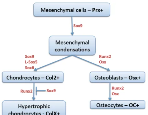
Figure 1. Schematic representation of endochondral bone formation. Adapted from Karsenty et al.,(2009). (A) Mesenchymal cells condense and (B) then become Col2 positive chondrocytes, (C) Chondrocytes at the centre of the condensation stop proliferating, become hypertrophic and express ColX (shown in pink). (D)
Perichondrial cells adjacent to hypertrophic chondrocytes become Col1 positive osteoblasts, which form the bone collar. (E) Hypertrophic chondrocytes direct the formation of mineralized matrix, attract blood vessels, and undergo apoptosis. (F) Osteoblasts of primary spongiosa accompany vascular invasion, forming the
primary spongiosa (ps). Chondrocytes continue to proliferate, lengthening the bone. At the end of the bone, the secondary ossification centre (soc) forms through cycles of chondrocyte hypertrophy, vascular invasion and osteoblast activity.
mature osteoblasts. Some markers that characterize the final stages of osteblast differentiation are ALP (Alkaline Phosphatase) and BSP (Bone Sialo-‐Protein).
Concurrently, the cells at the periphery of the chondrocyte template will become the perichondrium (Kronenberg, 2007). The perichondrial cells have specific functions during endochondral ossification. First, they are able to differentiate into either chondrocytes or osteoblasts, which will migrate to the trabecular and cortical bone. Second, they signal back and forth with the underlying cartilage. This specific signaling between the chondrocytes and perichondrial cells causes Col2 expressing chondrocytes to differentiate and mature at the
perichondrium into Col1 expressing osteoblasts, which will specifically form the bone collar (Maes et al., 2010) (Figure 1D). As bones grow in size, the secondary ossification centers (SOC) form, usually in the epiphysis of long bones. These regions form through cycles of chondrocyte hypertrophy, vascular invasion and osteoblast activity (Figure 1F). In the long bones, chondrocytes continue to proliferate and cartilage matrix is deposited, causing the bone to grow in length. This process, which starts in embryogenesis, is completed usually at puberty.
The alternate bone differentiation mechanism, intramembranous ossification is used for the cranial flat bones of the skull (frontal, parietal, occipital, and temporal bones) and the clavicles. In this process, condensed mesenchymal precursors
differentiate directly into osteoblasts, without a cartilage intermediate. Like endochondral ossification, this process initiates in utero, but is completed after birth. Once bones have formed during embryonic development, either through endochondral or intramembranous ossification, they continue to be actively
remodeled after birth during periods of growth or bone repair. This involves a tightly regulated interplay between the activities of chondrocytes, osteoblasts and osteoclasts, the three cell lineages involved in bone development.
I.3. Cell lineages involved in bone development
Three key players are involved in bone formation: osteoblasts and chondrocytes, which originate from common mesenchymal progenitors and
osteoclasts, which are of hematopoietic origin (Kronenberg, 2003; Zelzer and Olsen,
2003).
Chondrocytes
Early during endochondral ossification, mesenchymal progenitors differentiate into chondrocytes. This process is critically dependent upon the transcription factor Sox9 (Figure 2), which binds, and activates the transcription of chondrocyte-‐specific genes (Bell et al., 1997; Zelzer and Olsen, 2003). Sox9 belongs to a large family of transcription factors, which also includes Sox5 and Sox6. These two Sox family members also play important roles in chondrocyte differentiation. In particular, they actively cooperate to regulate the expression of cartilage-‐specific components encountered in the extracellular matrix – such as type II collagen (Col2a1). Consistent with this, Sox 5, 6 and 9 form complexes with other nuclear proteins in chondrocytes and are also co-‐expressed at sites of chondrogenesis (Zhou et al., 1998). Furthermore, in Sox5-‐ and Sox6-‐null mice the chondrocytes of the growth plate are unable to differentiate into hypertrophic chondrocytes (Lefebvre et al., 1998). Also, inactivation of Sox9 in limb buds prior to
Figure 2. Differentiation factors involved in chondrocytic and osteoblastic differentiation. Adapted from (Zelzer and Olsen, 2003). Mesenchymal precursors, which express Prx, form mesenchymal condensations at the location of the future bones. The cells of these condesations will differentiate either into Col2 expressing chondrocytes under the control of Sox9, Sox6 and Sox5, or into Osx expressing osteoblasts. The Col2 chondrocytes will differentiate into ColX expressing
hypertrohic chondrocytes in the presence of Runx2 and Sox9 inhibits this process. The Osx osteoblasts will differentiate into Osteocalcin (OC) expressing mature osteocytes, capable of secreting mineralized matrix.
the formation of mesenchymal condensations causes complete absence of both cartilage and bone (Akiyama et al., 2002).
Additionally, RUNX2, a transcription factor that was first identified through its role in osteoblast has also been linked to later stages of chondrocyte
differentiation. Runx2 is required for chondrocytes to exit the cell cycle and become hypertrophic, since Runx2-‐deficient mice lack
hypertrophic chondrocytes in their limbs (Kim et al., 1999). Runx2 positively regulates maturation to hypertrophic chondrocyte, while Sox9 negatively regulates this process (Figure 2).
Other important factors involved in proper chondrocyte differentiation are the signaling molecules Indian Hedgehog (Ihh) and Parathyroid hormone-‐related protein (PTHrP). These will be discussed in more detail in Section I.4 of this introduction.
Osteoblasts
Osteoblasts originate from mesenchymal progenitor cells and go through several stages of differentiation to become mineralizing mature osteoblasts.
Different proteins are expressed at different stages of osteoblast differentiation and are therefore used as markers to study the dynamics of osteoblast differentiation from osteoblast progenitors to mature osteoblasts (Figure 2). Early in development, osteoblast progenitors express RUNX2. This promotes differentiation, resulting in the formation of Osx-‐expressing osteoblast precursors that commit to an
osteoblastic fate through expression of alkaline phosphatase (ALP) and type I collagen (Col1). Mid-‐stage markers of osteoblast differentiation are osteopontin
(OPN) and osteonectin, while the late stage marker osteocalcin (OC) is expressed specifically by mature osteoblasts. Osteocytes, the most abundant bone cells (95%) in adult bone, are the terminal stage of osteoblast differentiation and they are embedded deep within the bone matrix during bone formation (Franz-‐Odendaal et al., 2006; Fulzele et al., 2012). These cells are important for regulating bone
remodeling, due to their ability to secrete RANKL, which promotes osteoclast differentiation (Xiong et al., 2011) and also expression of sclerostin, a suppressor of osteoblast proliferation (Winkler et al., 2003).
Runx2 is the earliest transcription factor absolutely required for the differentiation of mesenchymal progenitors into osteoblasts (Figure 2). Runx2 regulates the expression of several osteoblast specific proteins, such as osteocalcin, alkaline phosphatase and type I collagen (Ducy et al., 1997; Lee et al., 2007).
Interestingly, overexpression of Runx2 in other cell types in vitro, such as fibroblasts or myoblasts, is sufficient to induces the expression of osteoblasts specific markers (Ducy, 2000). Conversely, deletion of Runx2 in mice prevents expression of early or late osteoblast markers and formation of mature bone tissue (Ducy et al., 1997). As noted above, these Runx2 null mice also lack hypertrophic chondrocytes, despite other types of chondrocytes being present (Komori et al., 1997; Otto et al., 1997). Taken together, these observations prove that Runx2 is a critical factor required for proper osteoblast differentiation.
Osterix1 (OSX1) is the other transcription factor known to be essential for osteoblast differentiation (Figure 2). Osx1-‐null mice do not express any bone-‐ specific markers (Nakashima et al., 2002). However, it was shown by in situ
hybridization analysis that Osx-‐null mice express Runx2. At the same time Runx2-‐ null mice do not express Osx, indicating that Osx acts downstream Runx2 in the osteoblast differentiation lineage (Nakashima et al., 2002). As Runx2 has a role in both chondrocytic and osteoblastic differentiation, Osterix is likely the factor that provides specificity in osteoblastic differentiation. Osx expression is highly specific to the osteoblast lineage, but low-‐level expression has been detected in
chondrocytes in vivo (Nakashima et al., 2002; Rodda and McMahon, 2006; Yagi et al., 2003). In an in vitro setting, Osx is sufficient to induce expression of osteoblast markers (Tai et al., 2004).
Importantly, normal cartilage development is required for proper osteoblast differentiation. This is supported by in vivo mouse studies showing that mutation of chondrocyte specific factors, such as Ihh, cause defects in osteoblasts differentiation. In vivo bone development is also negatively impacted when mutations are
introduced into Ihh. Specifically, Ihh-‐null mice exhibit severe dwarfism in axial and appendicular skeletal elements and do not form endochondral bones (St-‐Jacques et al., 1999); (Long et al., 2004; Rodda and McMahon, 2006).
Osteoclasts
Osteoclasts originate from the hematopoietic lineage, specifically from mature monocytes and macrophages, and their role is to release specific enzymes that can lyse adjacent bone. The balance between osteoblast activity (which build bone) and osteoclast activity (which destroy it) is crucial for maintaining bone density and regulating proper bone remodeling. Notably, osteoblasts can influence osteoclast differentiation in either a positive or negative manner, through secretion
of regulatory factors, such as CSF-‐1, RANKL and Osteoprotegerin (OPG). CSF-‐1 and RANKL play a promoting role via activation of osteoclast specific genes. OPG is a receptor for RANKL that acts to sequester it, and thereby inhibits osteoclast
formation (Simonet et al., 1997). Animal studies have shown that mice without CSF-‐ 1 or the receptor for RANKL are unable to make osteoclasts (Kong et al., 1999; Yoshida et al., 1990) and they develop osteopetrosis. In cartilage, RUNX2 promotes expression of RANKL, the key ligand driving osteoclast differentiation. Hyperactive
osteoclasts are responsible for many adult skeletal diseases, including osteoporosis, rheumatoid arthritis and periodontal disease (Rodan and Martin, 2000). Reduced osteoclast activity leads to diseases where excessive bone fills out the bone marrow cavity, such as osteopetrosis.
I.4. Major signaling pathways involved in bone development
Several major signaling pathways are important for bone development, including the Indian Hedgehog (Ihh)/Parathyroid hormone-‐related protein (PTHrP), the fibroblast growth factor (FGF) and the bone morphogenetic protein (BMP) pathways (Kronenberg, 2003). In general, these pathways function to coordinate proper chondrocyte and osteoblast differentiation. The Ihh/PTHrP and the FGF pathways will be discussed in more detail in the next section.
Indian hedgehog/ Parathyroid hormone-‐related protein signaling Ihh and PTHrP signaling is complex, involving crosstalk and both negative and positive feedback loops (Figure 3). Perichondrial cells and chondrocytes at the end of long bones secrete PTHrP. (Figure 3.1). PTHrP acts on receptors found on
proliferating chondrocytes and keeps the cells in a proliferative state and delays their secretion of Ihh (Kronenberg, 2003; St-‐Jacques et al., 1999). Ihh is produced by cells that fail to receive enough PTHrP to suppress its expression. Ihh acts on its own receptor found on proliferating chondrocytes, and it stimulates their continuous proliferation and prevents differentiation to hypertrophic chondrocytes (Figure 3.2). Ihh also acts on chondrocytes at the end of the long bones and stimulates their production of PTHrP (Figure 3.3). Importantly, Ihh signals to perichondrial cells immediately adjacent to the prehypertrophic and hypertrophic cells, and it directs them to move away from the chondrocyte pathway and differentiate to osteoblasts. These osteoblasts end up forming the bone collar (Figure 3.4). In vivo studies have shown that Ihh-‐null mice exhibit severe dwarfism in axial and appendicular skeletal elements and do not form endochondral bones (St-‐Jacques et al., 1999). Null
mutations of PTHrP or its receptor decreases the number of proliferating growth plate chondrocytes and increases the size of the hypertrophic zone (Lanske et al., 1996).
Fibroblast growth factor (FGF) signaling
FGF signaling has an important role in chondrocyte proliferation and differentiation. The mechanism by which this signaling pathway works is complex and still not fully understood. This is mainly due to the fact that various
Figure 3. Indian hedgehog (Ihh)/parathyroid hormone-related protein (PTHrP) negative-feedback loop. Adapted from Kronenberg (2003). (1) Chondrocytes at the end of long bones secrete PTHrP. PTHrP acts on receptors found on proliferating chondrocytes to keep them in a proliferative state. (2) Ihh is produced by cells that fail to receive enough PTHrP to suppress its expression. Ihh acts on its own receptor found on proliferating chondrocytes, stimulates their continuous proliferation and prevents their differentiation to hypertrophic chondrocytes. (3) Ihh also acts on chondrocytes at the end of the long bones and stimulates their production of PTHrP. (4) Ihh signals to perichondrial cells immediately adjacent to the prehypertrophic and hypertrophic cells, and it directs them to differentiate to osteoblasts.
members of the FGF family, and also many FGF receptors, are expressed early in bone development and these have overlapping functions. Early during
endochondral bone development, FGFR-‐2 is expressed in the condensing
mesenchyme. In vitro studies in mesenchymal cell lines showed that FGFs stimulate expression of Sox9, the transcription factor essential for early stage chondrocyte differentiation (Figure 2). Thus, expression of FGFR-‐2 early in the limb mesenchyme is thought to induce the expression of Sox-‐9. At late times in development,
proliferating chondrocytes express FGFR-‐3, while pre-‐hypertrophic and
hypertrophic chondrocytes express FGFR-‐2. FGF signaling through FGFR-‐3 was studied in more depth and shown to have a negative effect on chondrocyte proliferation. This conclusion is based on the finding that activating mutations in FGFR-‐3 decrease the proliferation rate of chondrocytes, partially through the JAK-‐ STAT1 pathway (Sahni et al., 1999). Notably, Fgfr3 null mice show an increase in the rate of chondrocyte proliferation and expansion of chondrocyte columns (Colvin et al., 1996; Deng et al., 1996). Concomitantly, FGF signaling partially works through regulating the Ihh/ PTHrP negative loop, by suppressing Ihh expression and inducing a decrease in chondrocyte proliferation (Naski et al., 1998; Ornitz and Marie, 2002).
PART II. THE RAS ONCOGENES
Proper cellular proliferation is essential for normal development and growth of organisms. Tight regulation of the cell cycle machinery, through various
intracellular and extracellular signals, is very important for cells to know when to divide, or stop dividing, under the right conditions (Hanahan and Weinberg, 2000). In cancer, this regulation is disrupted and tumor cancer cells proliferate excessively. Mutations in the mitogenic signaling pathways, such as in members of the RAS family, are some of the most common mutations in cancer and result in an
uncoupling of cell cycle regulation from extracellular cues. Given the importance of these proteins in proliferation and tumorigenesis, it is not surprising that their deregulation also has profound effects on normal development. RASopathies are a group of developmental disorders caused by germline mutations in components of the Ras/MAPK pathway that result in increased signaling. These will be discussed in further detail in Part III of this introduction.
This thesis presents two novel mouse models in which a constitutive mutation of K-‐ras is expressed at various stages during limb development. These models are used to investigate how K-‐rasG12D affects skeletal development when
expressed in a spatial and temporal specific manner.
II. 1. Discovery
Initially, the Ras genes were discovered as the transforming sequences from the genomes of Harvey and Kirsten rat sarcoma viruses (Chien et al., 1979; Ellis et al., 1981). Independently, the same sequences, cloned from various cancer cells lines
(Pulciani et al., 1982; Shih and Weinberg, 1982) or isolated from chemically treated cells (Shih et al., 1981) were identified as inducers of transformation of NIH 3T3 cells. Soon after, it was realized that the sequences in the two different contexts, viruses (viral Ras genes) and cancer cells, are homologous (Parada et al., 1982). This led to the discovery of the 3 members of the Ras family: H-Ras, K-Ras and lastly N-ras (Capon et al., 1983; Johnson et al., 2001).
Further work has shown that a single point mutation in these genes can transform the normal cellular gene into an oncogene (Tabin et al., 1982;
Taparowsky et al., 1982). RAS genes represent frequent mutational targets in cancer and have been shown to be mutated in ~30% of human tumors (Johnson et al., 2001). Interestingly, mutations in the RAS genes are not randomly distributed among the different isoforms, and the majority of mutations occur in K-RAS,
especially in lung, pancreas and colon tumors (Bos, 1989; Johnson et al., 2001). It is clear that mutations in different members of the Ras family lead to different tumor types (Karnoub and Weinberg, 2008).
H-ras and N-ras are dispensable for development, both individually and in combination (Janssen et al., 2005). K-ras encodes two splice variants with KRASA expressed in a tissue specific and developmentally restricted fashion and KRASB being ubiquitously expressed. Mice with a homozygous null mutation in K-ras are not viable and die during embryogenesis due to a wide spectrum of defects (cardiac, liver, neurological and hematopoietic defects), indicating that K-ras is essential for embryonic development (Tuveson et al., 2004).
II.2. Ras proteins and the GTPase cycle
Early studies identified Ras proteins as small 21kD proteins localized to the cell membrane. These could both bind GTP, and hydrolyze it, proof of an internal GTP-‐ase activity (Shih et al., 1980). Soon after, these proteins were classified as small G-‐proteins (Hurley et al., 1984) that function as molecular switches in controlling the signaling from membrane receptors to intracellular effector
cascades. They are regulated by binding to either GTP (in their ON state) or to GDP (in their OFF state; Figure 4). Following the discovery that the mitogenic signaling molecule, induces GTP binding (Kamata and Feramisco, 1984), it was concluded that the intracellular effector cascades controlled by Ras proteins were involved in cell proliferation, differentiation, and apoptosis.
Subsequent studies and the discovery of many other players helped to establish the precise mechanism by which mitogenic signals are transmitted from the cell membrane to the cell machinery and a complex GTP-‐GDP cycle was
described (Figure 4; McCormick, 1993). Upon binding of mitogenic ligans to growth factor receptors, the intracellular domains of these receptors bind adaptor proteins (GRB2), which recruit guanine nucleotide exchange factors (GEFs, such as SOS) to the plasma membrane. This brings these in close proximity to the Ras proteins, which are tethered in the membrane, allowing interaction and to release GDP and bind GTP, which results in their activation and the activation of downstream pathways. This stimulates the Ras proteins as described in more detail below. The inactivation of the GTP-‐bound Ras proteins occurs through the hydrolysis of GTP to GDP, catalyzed by GTPase-‐activating proteins (GAPs)
Figure 4. Ras complex downstream signaling networks. Adapted form Karnoub and Weinberg (2008). The MAPK signaling pathway controls downstream effectors critically involved in cell proliferation, differentiation, motility, apoptosis and senescence. The binding of GTP to Ras protein locks it in an active state, which enables interactions with multiple downstream effectors. A slow intrinsic GTPase activity leads to Ras inactivation and signal termination. This ON–OFF cycle is tightly controlled by GTPase-‐activating proteins (GAPs – p120GAP or NF1) and guanine-‐ nucleotide exchange factors (GEFs – SOS).
GF=Growth Factor, RTK=Receptor Tyrosine Kinase.
including p210GAP and NF1 (Cawthon et al., 1990; Karnoub and Weinberg, 2008; Martin et al., 1990).
Not surprisingly, mutations that disrupt the GTP-‐GDP cycle of Ras proteins have been shown to be oncogenic. More specifically, these mutations cause an inhibition or reduction of the intrinsic GTPase activity or suppress their interaction with GAPs. This results in deregulated activation of the Ras proteins -‐ often in the absence of appropriate upstream signaling -‐ by increasing the ratio of the active GTP-‐bound state relative to the GDP-‐bound inactive state. The most common
oncogenic point mutations in the Ras genes are found in codons 12, 13 and 61 (Clark et al., 1985; Der et al., 1986; Trahey and McCormick, 1987). These mutation lock Ras protein in a constitutively active form, by preventing it from releasing the bound GTP.
II.3. Ras downstream signaling pathways – MAPK pathway
Ras downstream effectors can have a multitude of biological functions. Since growth factors such as EGF can stimulate Ras activity, it was concluded that one of the most important roles for Ras proteins is their role in mitogenic signaling (Mulcahy et al., 1985). Subsequently, it became clear that ras activates a multitude of downstream signaling pathways, and has numerous roles including controlling cell cycle and differentiation (MAPK pathway), cell survival (PI3K pathway), cell signaling (PLCε), endocytosis (Ral-‐GEF) and many others (Karnoub and Weinberg, 2008; Figure 4). It is important to understand how Ras can execute all these
downstream functions, as this can shed more light on the role of these proteins in development and cancer.
I will further focus my discussion on the MAPK pathway, because this is most relevant to this thesis project. The Mitogen Activated Protein Kinase (MAPK)
pathway is one of the best-‐characterized downstream signaling pathways of the Ras proteins. The Ras/MAPK pathway transduces extracellular signals in the form of growth factors and small molecules to the intracellular environment.
Active Ras leads to the activation of the Raf family of kinases: A-‐Raf, B-‐Raf and/or C-‐Raf. Raf, first identified as an oncogene (Vojtek et al., 1993), becomes localized to the plasma membrane and initiates activation of the MAPK signaling cascade (Figure 4). In this cascade, Raf kinase phosphorylates MEK1/2 (MAPK kinases), which then phosphorylate and activate ERK1/2 kinases (Hagemann and Blank, 2001). ERK1/2 are the ultimate effectors and exert their function on a large number of downstream molecules including nuclear factors, transcription factors, membrane proteins, and protein kinases. All these molecules control vital cellular functions including cell cycle progression, differentiation, and cell growth (Yoon and Seger, 2006).
Some of the better-‐characterized downstream ERK effectors are the Ets, ELK and AP-‐1 (c-‐Fos-‐c-‐Jun) complex of transcription factors. Activation of these
transcription factors leads to increased expression of cell cycle genes, such as cyclin D1 (Hitomi and Stacey, 1999), supporting the role of Ras as a mitogenic protein. The MAPK pathway can have other outputs as well, such as the p38 pathway and the Jun N-‐terminal kinase (JNK) pathway, with roles related to stress responses. Notably,
Ras can direct distinct outputs and have different effects depending on which MAPK pathways it activates.
ERK targets several core regulators of osteoblast differentiation. It phosphorylates the RUNX2 transcription factor and positively regulates early osteoblast differentiation in vivo (Ge et al., 2007; Xiao et al., 2000; Figure 5). Furthermore, active ERK, via its association with RUNX2, binds to the osteoblast specific promoters osteocalcin (Ocn) and bone sialoprotein (Bsp) (Li et al., 2009). Besides RUNX2, ERK also activates RSK2 (Dalby et al., 1998), which phosphorylates ATF4, a pro-‐collagen gene transcriptional regulator of late-‐stage osteoblasts (Yang et al., 2004; Figure 5).
Given that Ras has a direct role in cellular transformation and during development, an important question becomes which of its downstream signaling pathways are critical for its tumorigenic functions, versus its various developmental roles. The complexity of Ras signaling underscores the importance of focusing on specific cell types when studying the Ras effector pathways.
Figure 5. MAPK (ERK) pathway in osteoblast differentiation. Adapted from (Greenblatt et al., 2013) During early osteoblast differentiation, ERK phosphorylates RUNX2 to increase its transcriptional activity and induce osteoblast differentiation. Later in osteoblast differentiation, ERK activates the kinase RSK2, which in turn phosphorylates and activates ATF4.