HAL Id: hal-03089739
https://hal.archives-ouvertes.fr/hal-03089739
Submitted on 28 Dec 2020HAL is a multi-disciplinary open access archive for the deposit and dissemination of sci-entific research documents, whether they are pub-lished or not. The documents may come from teaching and research institutions in France or abroad, or from public or private research centers.
L’archive ouverte pluridisciplinaire HAL, est destinée au dépôt et à la diffusion de documents scientifiques de niveau recherche, publiés ou non, émanant des établissements d’enseignement et de recherche français ou étrangers, des laboratoires publics ou privés.
Solid-state NMR study of microalgal membranes
Alexandre Poulhazan, Alexandre Arnold, Dror Warschawski, Isabelle Marcotte
To cite this version:
Alexandre Poulhazan, Alexandre Arnold, Dror Warschawski, Isabelle Marcotte. Solid-state NMR study of microalgal membranes. Solid-State NMR, IOP Publishing, 2020, �10.1088/978-0-7503-2532-5ch4�. �hal-03089739�
Chapter X
Solid-state NMR study of microalgal membranes
Alexandre Poulhazan
1, Alexandre A. Arnold
1, Dror E. Warschawski
1,2and
Isabelle Marcotte
11Department of Chemistry, Université du Québec à Montréal, P.O. Box 8888, Montréal, QC,
H3C 3P8 Canada.
2Sorbonne Université, École Normale Supérieure, PSL University, CNRS, Laboratoire des
biomolécules, LBM, 75005 Paris, France.
Abstract
Microalgae are ubiquitous primary organisms that play key roles in the aquatic environment where they are the basis of the food web. These microorganisms also produce a variety of molecules with high added value, which find applications in human and animal nutrition, as therapeutic agents and as biofuels. Lipid membranes abound in microalgae where they protect the cell and are essential building blocks of organelles such as the chloroplast. To better understand the architecture and dynamics of microalgal cell membranes and their constituents, it is important that they are investigated in their natural environment. Solid-state nuclear magnetic resonance (SS-NMR) is emerging as a powerful tool to tackle these complex assemblies in situ on cell extracts and also in whole cells. Several nuclei can be studied to probe algal membranes and, moreover, microalgae can be fully 13C- or 15N-labeled in a cost-effective
manner. In the case of full organism labelling, the challenge of SS-NMR is to develop and identify experiments to filter specific constituent signals. In this chapter, we show how SS-NMR can provide a wealth of information on the structure, dynamics and composition of microalgal membranes. We also summarize the main SS-NMR approaches so far applied on lipid extracts or whole cells under near to native conditions and suggest potential avenues to improve our understanding of these systems.
X.1 Overview
Microalgae are microscopic algal cells also known as phytoplankton. The cell wall and plasma membrane protect the organelles in the cytoplasm, such as the nucleus, the chloroplast, the reticulum, as well as the sugar and lipid reserves (Figure X.1). About 30,000 species of microalgae are found in fresh- and saltwater, where they are at the basis of the aquatic food chain (Richmond and Hu, 2013). As such, global warming or pollution effects on this resource can have an impact on all higher organisms.
Microalgae bear an important socio-economical potential. They are already used in human and animal nutrition, and have been exploited for their therapeutic activity related to antimicrobial (Falaise et al., 2016), antiviral (de Jesus Raposo et al., 2015), antioxidant, anti-inflammatory (Rasool et al., 2007) or anti-cancer (Gutiérrez et al., 2010) properties. Another interesting characteristic of microalgae is their ability to sequestrate and bioaccumulate heavy metals (Posadas et al., 2017, Piccini et al., 2019). They thus stand as an elegant avenue for contaminated water treatment. Microalgae are also used in the development of third-generation
biofuels, which partially or completely come from solar instead of fossil energy (Abo et al., 2019).
On a more fundamental level, microalgae are great tools for the study of photosynthesis. For example, carbon metabolism was deciphered in Chlorella by Melvin Calvin, Nobel Prize in 1961. The dynamics of photosynthesis has been investigated in detail in Chlamydomonas (Eberhard et al., 2008). New transgene strategies are available, mainly studies of
Chlamydomonas reinhardtii (Doron et al., 2016, Baier et al., 2018), which has been called the
‘photosynthetic Escherichia coli’ because of its optional heterotrophic metabolism, allowing the use of non-photosynthetic mutants.
The microalgal cell wall, membranes and lipids are key players for both biochemical processes and biotechnological applications. Their selective permeability and mechanical or osmotic shock resistance make the cell interfaces and membranes a source of inspiration for biomaterial development. Furthermore, lipids in general are valuable nutritional ingredients with long-chain and ω-3 poly-unsaturated fatty acids (FAs), but also as precursors for biodiesel production from triacylglycerol (TAG). Understanding the composition, dynamics and architectures of these assemblies is essential to bioengineer new microalgal strains with efficient photosynthesis and easy lipid extraction from the biomass, or to design novel materials based on natural products with interesting biological/physical properties. Finally, microalgal membrane structure and composition (see Figure X.1) can vary depending on temperature, pH or interaction with exogenous molecules, making them excellent global warming reporters.
Solid-state nuclear magnetic resonance (SS-NMR) is an invaluable tool providing nanoscopic information on the structure, dynamics and interactions of large biological molecules with virtually no limit in molecular size. This is why SS-NMR has proven its capabilities to investigate whole cell constituents, as well as their organization and membrane interactions. However, the cell complexity leads to crowded NMR spectra in which many signals potentially overlap. Therefore, SS-NMR approaches are being developed to tackle such challenging systems (Warnet et al., 2015, Werby and Cegelski, 2019, Narasimhan et al., 2020). Considering the environmental and economical importance of microalgae, the objective of this chapter is to present how SS-NMR can provide information on the molecular composition and organization of microalgal membranes and the cell wall. Before presenting different NMR methodologies to address these cell compartments, the composition of microalgal membranes will first be summarized.
X.2 Microalgal cell membrane composition and architecture
Microalgal membranes are composed of lipids, proteins, carbohydrates, phytosterols as well as pigments. Although lipids ensure membrane integrity and efficient cell impermeability, the detailed lipidome of microalgae has been described for only a handful of species such as C.
reinhardtii and Nannochloropsis oceanica (Zhao et al., 2013, Nguyen et al., 2013). Membranes
are also rich in proteins that maintain the cell structure and metabolism, and in carbohydrates bound to both lipids and proteins, allowing key processes such as cell recognition or protein activity. Pigments are found in chloroplast membranes (Figure X.1) where they are involved in light energy capture and dissipation. Indeed, a lipid membrane also protects organelles in the cytoplasm, such as the reticulum and the nucleus. Moreover, lipid droplets are also present inside the cells.
Figure X.1. Microalgal cell with organelles and membrane structures. The cell wall is generally composed of glycoproteins, cellulosic material, carbonate or silica and the plasma membrane is rich in phospholipids. Chloroplast membranes have different galactolipid-to-phospholipid ratios, going from 19:81 in the chloroplast outer layer to 2:98 in thylakoid membranes (Harwood, 1998). Lipid droplets incorporate TAGs and ergosterol, as a dense energy storage, surrounded by a single phospholipid monolayer. Energy can also be stored as complex carbohydrates, namely starch or chrysolaminarin.
X.2.1 Microalgal cell wall and plasma membrane
The cell wall primarily acts as a shell and thus makes biomolecules such as TAGs harder to harvest and exploit. It also protects the cell from mechanical constraints and external stress, and is involved in cell-cell and cell-substrate adhesion, protection from bacteria as well as sexual reproduction (Domozych et al., 2012). Although the plasma membrane is not the largest membrane in the cell, it must be studied since it faces a wide range of stresses such as temperature changes and osmotic shocks in its environment. Moreover, multiple specific transporters are found within this membrane which is the last barrier crossed by molecules before accessing the cytosol. It is also where cell signalling is initiated. One of the first studies of the lipid composition of microalgal plasma membranes focused on the green algae
Hydrodictyon africanum. Phospholipids, such as phosphatidylcholine (PC), phosphatidylserine
(PS) and cardiolipin (CL), were mainly identified (Bailey and Northcote, 1976). The microalgal cell membrane is mostly composed of polar lipids (Harwood, 1998) and its composition can adapt to external stress or cell state (Mimouni et al., 2018).
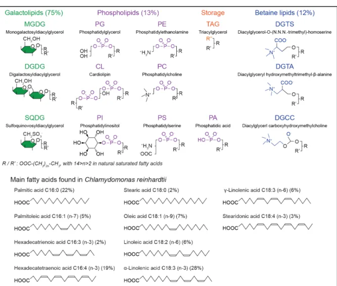
Figure X.2. Main lipids and fatty acids found in microalgae. Galactolipids and betaine lipids are the most abundant in microalgae, and mainly located in photosynthetic membranes. Phospholipids are found in other membranes. Triacylglycerols (TAGs) are stored in lipid droplets. The lipid profile and fatty acid chain composition are species- and medium-dependent. As a reference, values for C. reinhardtii are indicated (Vieler et al., 2007, Nguyen et al., 2013).
The relationship between the plasma membrane and the microalgal cell wall is so intimate that considering the membrane without the cell wall would make no sense in terms of dynamics and shape. Indeed, the cell wall synthesis and its remodelling proteins are associated to the membrane (Popper et al., 2014) and some fibrillar assemblies anchored in the cell wall traverse the plasma membrane (Domozych, 2019), making membrane and cell wall boundaries hard to define precisely (Figure X.1). For microalgae such as Chlorophyceae, the cell wall is composed of very organized carbohydrates and glycoproteins (Rashidi and Trindade, 2018), so that glycan patterns and structures are important to determine. While electron microscopy (EM) has provided micrometer resolution images important for taxonomic determination, SS-NMR can potentially provide additional structural and molecular-level dynamic information of the cell membrane on different timescales.
X.2.2 Photosynthetic organelles (thylakoids and chloroplast)
There are several organelles in the algal cell and a membrane borders them all. The chloroplast has, by far, the largest membrane surface, with three layers of densely packed lipid
membranes (Figure X.1). This explains why the lipid profile of other organelles is difficult to determine without getting any chloroplast membrane or pigment contamination during purification. The lipid and FA chain profiles of microalgae are usually given for the whole organism (Figure X.2). The membrane surface of each organelle has not been measured or estimated yet, even if a 3D reconstitution made by in situ cryo-electron tomography of C.
reinhardtii has been reported (Engel et al., 2015).
The lipid composition of chloroplasts membranes is very different from that of plasma membranes as they mostly contain galactolipids and few phospholipids. It varies according to the species but monogalactosyldiacylglycerol (MGDG, 40 to 55%), digalactosyldiacylglycerol (DGDG, 15-35%), sulfoquinovosyldiacylglycerol (SQDG, 10-20%) and phosphatidylglycerol (PG, 10-20%) are the main components (Harwood, 1998). In Chlamydomonas, the chloroplast envelopes and thylakoid membranes have slightly different lipid compositions (Mendiola-Morgenthaler et al., 1985). On the other hand, MGDG/DGDG and SQDG/PG ratios seem to be regulated in chloroplast membranes of various species and could be critical for proper thylakoid development (Boudière et al., 2014). Betaine lipids have been detected in a few brown algae, such as diacylglycreyl-O-(N,N,N-trimethyl) homoserine (DGTS) and diacylglyceryl-O-(hydroxymethyl)-(N,N,N-trimethyl)β-alanine (DGTA) (Murakami et al., 2018). Finally, recent work mentions glycolipids as microalgal membrane stabilizers because, during water or cold stress, the chloroplast membrane can be enriched in oligogalactolipids, produced by MGDG conversion (Hölzl and Dörmann, 2019).
X.2.3 Lipid droplets and free fatty acids
The lipid droplet (LD) is an important organelle for biofuel production due to the TAGs that they contain. Little is known about microalgae LDs but interest is growing due to their high potential as an alternative to fossil fuels. TAGs produce twice as much energy than starch and protein per unit weight. Moreover, microalgae are of great interest for nutrition purposes because of the essential long-chain and unsaturated FAs that are contained in the LDs (Spolaore et al., 2006).
The number, size and composition of LDs vary greatly among microalgal cell types but also according to the life cycle and metabolic stress. For example, nitrogen, iron, sulfur and phosphorus starvation are commonly used strategies to increase lipid storage in microalgae such as C. reinhardtii (Hu et al., 2008, Wang et al., 2009, Goold et al., 2014). The size of LDs can range from nm to µm and they are very dynamic in terms of molecular motion, anabolism and catabolism. LDs are easily observed in the cytosol using lipophilic dyes such as Nile red or Bodipy, thus facilitating their quantification by microscopy or flow cytometry, as well as transmission electron microscopy which has been used to further describe their structure (Goodson et al., 2011). Lipid droplets share the same overall structure with a hydrophobic oil core of storage lipids that mainly comprise TAGs (80-90% by weight) and sterol esters, surrounded by a phospholipid monolayer that also contains proteins (only 5% by weight) (Figure X.1). Biochemical and microscopy studies have shown that the endoplasmic reticulum and plastid membranes are involved in LD production (Fan et al., 2011, Goodson et al., 2011). LDs are also associated with lipid homeostasis and several cellular signalling processes (Farese and Walther, 2009).
The FA composition of TAGs is similar to that of whole cells (Goold et al., 2014). However, the profile of the LD enveloping monolayer is very specific. For example, in C. reinhardtii (Wang et al., 2009) and Haematococcus pluvialis (Peled et al., 2011), DGTS, sulfoquinovosyldiacylglycerol (SQDG) and traces of DGDG are its major component. The lipids around the LD core are thought to be intermediates for acyl chain production in TAGs. Specific proteins are associated with LDs (Yoneda et al., 2016, Wang et al., 2017, Wang et al.,
2019). In C. reinhardtii, the predominant protein is the very hydrophobic Major Lipid Droplet Protein (MLDP) (Goold et al., 2014), but other LD-associated proteins have been identified (Vieler et al., 2012), and complete proteomics describe more than 200 proteins linked to LDs (Nguyen et al., 2011).
X.3 Technical considerations
All the microalgal cell compartments presented in the previous section have a large diversity in molecular composition and structure. However, sophisticated techniques and appropriate stable isotope labelling allow the determination of molecular structures, dynamics and interactions in complex systems such as microalgae by SS-NMR.
X.3.1 Observable nuclei for microalgal membranes studies
Since protons (1H) are the most sensitive nuclei for biological applications in NMR, 1
H-based techniques have been used to study microalgae, as will be illustrated below. The large abundance of protons in biological samples is an advantage in terms of sensitivity, but a drawback in terms of resolution. Therefore, other nuclei with higher specificity are frequently exploited. Phosphorous-31 (31P), for example, has 100% natural abundance and high
gyromagnetic ratio. Phosphorus is mainly found in ATP, DNA, phosphorylated proteins but also in phospholipids and, therefore, 31P NMR is an excellent probe to determine the membrane
composition and dynamics (Overall et al., 2019). Carbon is present in all organic molecules of the cell but only the 13C isotope, which has a low (1%) natural abundance, generates an NMR
signal. Since its gyromagnetic ratio is one quarter that of 1H, 13C has a lower sensitivity and
isotopic enrichment is required for experiments to be time efficient. Finally, 15N NMR can also
be used to study microalgae, but is even less sensitive than 13C and with very low natural
abundance (0.4%), so 15N NMR can only be performed with isotopic labelling.
Uniform 13C- and/or 15N-labelling of microalgae is easily carried out with affordable carbon
and nitrogen sources, i.e., sodium bicarbonate (NaH13CO
3) and ammonium chloride (15NH4Cl),
respectively. The cost of growing uniformly labelled C. reinhardtii, for example, can be estimated to less than 100$/L of medium with an optimal cell concentration of 3 to 4×10⁶
cells/mL. Depending on metabolism or the targeted molecule, the medium might also require more expensive 13C-labelled glucose, sodium acetate (Venters et al., 1991) or CO
2 (Akhter et
al., 2016).
Targeted labelling of specific molecules of interest, such as lipids, amino acids, carbohydrates or pigments, within the whole organism is an interesting alternative approach. This strategy can be applied to 13C or 15N but also to other reporter nuclei, such as 2H (Pius et
al., 2012, Tardy-Laporte et al., 2013, Warnet et al., 2016, Molugu et al., 2017, Salnikov et al., 2019) (see Chapter X Booth) or 19F (Matsumori et al., 2008, Shi et al., 2012, Afonin et al.,
2014, Kozorog et al., 2018), often chosen to replace protons in key locations. These specific labelling approaches have rarely been applied to microalgae as they require a good knowledge of algal metabolism, but they hold great promise in particular for in vivo SS-NMR studies, as performed with intact bacteria (Booth et al., 2017).
X.3.2 Solution and solid-state NMR
The NMR line width of a given nucleus depends on its interactions with neighbouring nuclei or electrons, including dipolar and quadrupolar interactions, chemical shift anisotropy but also anisotropic magnetic susceptibility effects. Most of these interactions are orientation-dependent but averaged out under fast and isotropic tumbling, such as for small molecules in solution, resulting in very narrow NMR lines.
In solution NMR, molecules can be discriminated according to their diffusion correlation time. Such spectral filtering hence simplifies the spectra and has been applied to study whole microalgal cells (Courtier-Murias et al., 2012, Akhter et al., 2016). Among the available “diffusion filters”, diffusion editing (DE) removes the signal of mobile molecules, while
inverse diffusion editing (IDE) only shows the spectrum of fast diffusing molecules. Single
pulse experiments will obviously show both types of signals.
Macromolecules and complex assemblies such as cell extracts, or even whole cells such as microalgae, are considered as “solid” on the NMR time scale. The orientation dependence of the interactions presented above leads to broad lines that often overlap with neighbouring signals, resulting in the loss of spectral resolution. Such systems are thus studied using SS-NMR techniques. High resolution can be restored by using strategies that replace fast and isotropic tumbling by coherent averaging. One such strategy is magic-angle spinning (MAS), where the sample is spun at high frequency at an angle of 54.7° with respect to the magnetic field direction. Depending on the available spinning frequency, interactions can be completely averaged out, resulting in narrower lines. The required spinning frequency depends on the observed nuclei and the interactions that they experience. One may wonder if high spinning frequencies would be deleterious to soft eukaryotic cells. Microalgae are very resistant organisms, owing to their protective and rigid cell wall. It has been shown that C. reinhardtii can spend 24 h in a SS-NMR rotor spinning at 10 kHz, at room temperature and without any nutriments, with around 80% survival and cell morphology preserved (Poulhazan et al., 2018). In the case of protons in rigid solids, homonuclear dipolar interactions are very strong and often cannot be efficiently averaged out using standard MAS. Therefore, most 1H MAS
SS-NMR studies are restricted to very dynamic small molecules where the remaining dipolar couplings and anisotropic magnetic susceptibility can be cancelled by MAS at moderate frequencies (typically below 10 kHz). This is the field of high-resolution magic-angle spinning NMR (HRMAS). In 1H HRMAS, as in solution NMR, the lipid droplet signals dominate those
of membrane lipids whereas higher spinning frequencies would be required to observe intense membrane phospholipid, carbohydrate or protein signals. HRMAS allows for direct measurements of non-liquid soft tissues and cells, generally by 1H NMR, and is typically used
to characterize metabolites, even in intact cells, as is summarized in the in-cell section.
High-resolution SS-NMR developments over the last 30 years have provided robust methodologies to characterize the structure and dynamics of biological membrane components (lipids and proteins) by observing 13C signals under MAS, sometimes in combination with 15N
since these nuclei are found in all peptide bonds. Since the internuclear interactions involved are smaller than for 1H, standard MAS is often enough to generate high-resolution one- (1D)
and multidimensional spectra. In some cases, line narrowing by MAS results in membrane components spectra that are similar to those obtained by solution NMR.
While solution NMR and HRMAS are restricted to molecules with “fast” dynamic properties, SS-NMR can reveal and exploit the dynamic heterogeneity of biological membranes. To do so, adequate “dynamic filters” that select only rigid or dynamic components within the sample are utilized. This results not only in spectral simplification but also provides information regarding the molecular environment. The most common filters take advantage of proton-to-carbon polarization transfer schemes. For example, INEPT (nuclei enhancement by polarization transfer) will only be efficient for very dynamic molecules (with tumbling times from 10-15 to 10-9 s) and only detect carbons directly bound to protons. On the other hand, cross
polarization (CP) will only show carbons from more rigid molecules (with correlation times from 10-9 to 1 s). Single pulse 13C experiments will show both types of signals. Other types of
filters, that will not be detailed here, have also been developed (Reckel et al., 2012, Renault et al., 2012, Warnet et al., 2015, Matlahov and van der Wel, 2018).
Since cells are not pure solids or pure liquids, the research group of Simpson has developed tools to study such heterogeneous samples with an approach called comprehensive multiphase (CMP) NMR (Simpson A.J., 2013, Liaghati Mobarhan et al., 2019). Indeed, the dynamical heterogeneity of intact cells requires the use of both solution and SS-NMR approaches to obtain a complete picture of their composition and function. CMP-NMR probes thus combine lock, gradients and improved shimming capabilities, typical of solution NMR probes, with access to moderate magic-angle spinning frequencies (vide infra) of a few kHz and high-power pulses typical of SS-NMR probes. With this type of equipment, monitoring different types of molecules is possible: from dissolved metabolites up to very rigid storage molecules or proteins.
X.4. NMR of lipid extracts and model membranes
Considering the complexity of microalgal cells, simple and straightforward approaches are used to garner information on membrane lipids by NMR. First, lipids can be extracted, dissolved in organic solvents, and studied by simple 1H, 13C, or 31P solution NMR. This
approach has been applied, for example, on whole cell extracts of Pavlova spp. (Kobayashi et al., 2007) or Neochloris oleoabundans (Beal et al., 2010) for lipid assignment and quantification in combination with other techniques (Challagulla et al., 2017). The use of lipid extracts obviously neglects important information such as the lipid environment, collective properties and membrane dynamics.
Another strategy to study microalgal membrane properties is to employ model systems with lipid composition similar to the mimicked membranes. Although part of the living organism complexity is sacrificed, model membranes provide valuable information on specific biophysical properties and interactions with the lipids. Mixtures of galactolipids such as MGDG and DGDG have been studied by 2H and 31P static SS-NMR as early as 1985 to
investigate phase transitions in microalgae and chloroplast membranes (Brentel et al., 1985, Lindblom et al., 1986, Borovyagin et al., 1988). Galactolipid structures and dynamics were later probed by 13C SS-NMR using MAS (Adebodun et al., 1992, Gross et al., 1995, Castro et
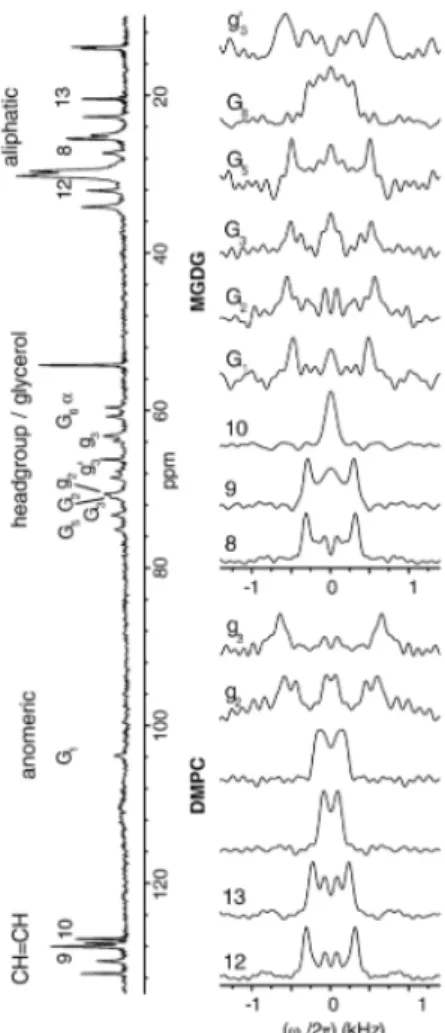
al., 2007) or in fast-tumbling bicelles by solution NMR (Howard and Prestegard, 1995, Ye et al., 2013). Figure X.3 shows a 1H-13C proton-detected local field (PDLF) study that allowed
measurement of the order parameters at various carbon positions in model unlabelled DMPC/MGDG membranes. This revealed that MGDG reduced the order parameters along DMPC molecules (Castro et al., 2007). Simplified thylakoid model membranes made of palmitoyl-oleoyl phosphocholine (POPC) and MGDG were used to determine the interactions of Hcf106 - a peptide found in the thylakoid of microalgae - with its host membrane lipids (Zhang et al., 2013, Zhang et al., 2014). Finally, the light harvesting complex II (LHCII) of C.
reinhardtii was also studied by 13C,15N-NMR reconstituted in a thylakoid-like membrane
Figure X.3. 1H-13C MAS (5 kHz) NMR PDLF spectra of a model membrane made of
unlabelled DMPC and MGDG, recorded at 42°C. The 1D 13C NMR spectrum is displayed on
the left. Reprinted from (Castro et al., 2007) with permission.
X.5.
In vivo, in-cell and in situ NMR
In-cell SS-NMR tackles complex samples that are very hard to characterize using other atomic-resolution techniques such as EM, X-ray crystallography or solution NMR. While in
vivo NMR addresses the challenge of studying living cells, in situ NMR examines cell extracts
- the cell is no longer intact but molecules are still in their native local environment. In vivo, in-cell and in situ SS-NMR have only recently been applied to microalgae but a wide range of applications are possible, as will be presented in the next sections.
X.5.1 1H-NMR based approaches
As previously explained, HRMAS provides insights into cellular metabolism by producing spectra with resolution sometimes comparable to that of solution NMR; and also allows spectral editing according to the dynamics of the constituents. HRMAS is applied to a variety of biological samples including human or mice cell lines, or tissues for clinical studies, allowing diagnosis and follow up of organism responses to treatments (Kaebisch et al., 2017). It is a common tool in metabolomics, especially for detecting metabolites associated with lipids such as inositol, phosphoryl choline, FAs and cholesterol. 1H HRMAS is also used in lipid
profiling and characterization of the saturated/unsaturated FA ratio (Griffin et al., 2003, Kabli et al., 2009, Berry et al., 2016). Terpenes found in chloroplast membranes are interesting molecules also detected by HRMAS (Gaysinski et al., 2015). Finally, because lipids are an important part of cell metabolism, several studies have focused on alterations of lipid
metabolism using HRMAS to detect lipid droplet perturbation, for example in cancer cell lines (Pan et al., 2012).
Considering the capabilities of HRMAS, this approach has been used to study microalgae, including their membranes and the in vivo intracellular C/N ratio (Bondu et al., 2008, Hashim et al., 2013). Native membranes are difficult to study because they are rigid structures on the NMR time scale, thus leading to broad and overlapping lines, with notable exceptions (Simon et al., 2015). Merkley and Syvitsk (Merkley and Syvitski, 2011) successfully assigned lipids and carbohydrates in whole Nannochloropsis granulata and monitored the unsaturated/saturated FA ratio as a function of cell harvest time using multidimensional 1 H-13C HRMAS. Also, Chauton et al. published several studies on different microalgae species,
such as C. mülleri (Webb et al., 2006), Dunaliella sp. (Chauton and Størseth, 2008),
Chaetoceros muelleri (Chauton et al., 2004) and Thalassiosira pseudonana (Chauton et al.,
2003). In these works, detailed assignments of lipids were reported, allowing quantification in response to growth conditions. HRMAS was also used to classify microalgae using statistical data treatments such as Principal Component Analysis (PCA). NMR studies performed on other cells and organisms will certainly inspire future investigations of microalgal lipids. For example, HRMAS has been used to characterize erythrocyte membranes and perform permeability studies in Daphnia magna (Bruno et al., 2006, Kovacevic et al., 2018). Recent work on living earthworms (Eisenia fetida) and freshwater shrimps (Hyalella azteca) have improved the lipid signal treatment based on relaxation and diffusion spectral filtering (Mobarhan et al., 2016, Hassan et al., 2019).
An alternative to HRMAS studies for 1H detection is the emerging use of ultrahigh
frequency MAS. Although membrane lipids and proteins can be studied at intermediate frequencies (15 kHz (Gopinath et al., 2017)), ultra-fast MAS (> 50 kHz) allows detection of protons in rigid molecules with increased sensitivity and a resolution comparable to that of solution NMR. Combined with 13C and 15N labelling of samples, sequential assignment of
membrane proteins or other complex molecules is possible, even in their native environment, e.g. proteins involved in photosynthesis (Beal et al., 2010, Medeiros-Silva et al., 2016). New information on biomembrane structure is expected to emerge from this approach, in particular for the study of membrane proteins in microalgae.
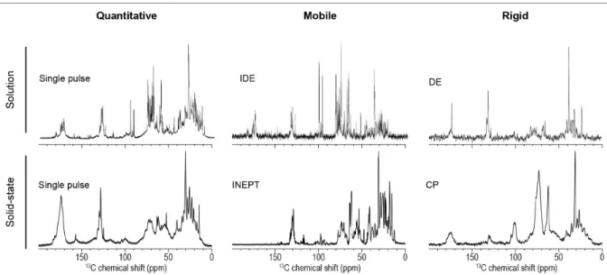
X.5.2 1D 13C solid-state NMR methods
As stated above, SS-NMR can be exploited to obtain valuable information on cell samples. Depending on the spectral editing used, both rigid and dynamic molecules can be identified and their dynamics in situ revealed - information that is lost using solution NMR of cell extracts, for example. Whole uniformly 13C-labelled C. reinhardtii were studied by our group
in 2015 by 1D 13C SS-NMR with MAS, along with Pavlova lutheri and Nannochloropsis
oculata (Arnold et al., 2015). This work allowed the assignment of different lipids,
carbohydrates including glycans, as well as rigid cell-wall proteins using different dynamic filters. As shown in Figure X.4, the more mobile molecules of C. reinhardtii are revealed by
13C SS-NMR using the INEPT filter, the most intense peaks being ascribed to methyl groups
from the lipid acyl chains around 30 ppm. This spectrum is comparable to a 13C solution NMR
Figure X.4. Comparison of 13C labelled C. reinhardtii whole cell spectra by solution and
solid-state NMR using either single pulse or filtered experiments. Solid-solid-state experiments were performed at 600 MHz at 25°C with a 10 kHz MAS frequency (adapted from Arnold et al. (Arnold et al., 2018)). Solution NMR experiments were performed at 500 MHz at room temperature (adapted from Akhter et al. (Akhter et al., 2016)).
On the exact same sample, a CP filter revealed the more rigid molecules such as starch, with C1 carbon at 101 ppm. Starch is invisible in solution NMR, even with a DE filter. These experiments highlight that many lipids are very mobile, while proteins in the cell wall are much more rigid assemblies. In 2018, Azadi-Chegeni et al. published similar work on C. reinhardtii whole cells (Azadi-Chegeni et al., 2018a, Azadi-Chegeni et al., 2018b). For example, they assigned several component resonances in whole cell samples and quantified the dynamics in terms of correlation times and order parameters of these molecules at different temperatures (Azadi-Chegeni et al., 2018a). They also investigated LHCII in native membranes and showed that they favored protein conformational stability. These works represent a step toward the understanding of chloroplast complexity in terms of molecular composition and dynamics.
Figure X.5. 13C CMP NMR (15°C and 1.1 kHz spinning frequency) spectra of uniformly 13
C-labelled Chlorella vulgaris microalgae with and without H. azteca daphnia. The pink region highlights the glucose peaks that increase when H. azteca is in contact with the algae. Reprinted from (Liaghati Mobarhan et al., 2019) with permission.
Another interesting study reports the consumption of Chlorella vulgaris lipids by the daphnia Hyalella azteca using comprehensive multiphase NMR and 13C-labelled microalgae.
As shown in Figure X.5, good resolution was obtained and lipid consumption could be monitored with this new probe. Multidimensional solid-state experiments were also recorded with 1H-13C correlations from 13C-labelled algae and 2H-13C correlations from 2H-13C
uniformly labelled algae.
X.5.3 2D-13C and 15N-NMR experiments
With isotopically-labelled samples sensitivity is greatly increased, enabling the use of multidimensional SS-NMR which also significantly enhances spectral resolution. 2D SS-NMR NMR has already proven successful in studying biological membranes in situ or in whole cells, mainly bacteria (Andronesi et al., 2005, Schanda et al., 2014, Romaniuk and Cegelski, 2015, Booth et al., 2017, Bouhlel et al., 2019) but also bovine retinas (Verhoeven et al., 2001). More closely related to algal membranes, the work on bacterial chlorosomal antenna by Van Rossum
et al. is the first to mention and assign galactolipid signals in a native thylakoid membrane
protein study (van Rossum et al., 2001).
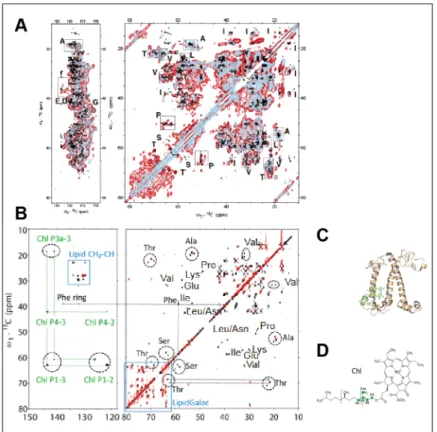
Figure X.6. 13C-13C solid-state NMR spectra of LHCII from C. reinhardtii in: (A) its native
thylakoid environment (red contours), reconstituted in β-n-dodecyl-D-maltoside detergent buffer (light blue contours); and (B) pure extracted protein (black contours) and in a reconstituted thylakoid membrane (red contours). (C) The LHCII crystal structure, and (D) chlorophyll molecular structure are also given. A is adapted from (Pandit et al., 2013), and B-D are adapted from (Azadi-Chegeni et al., 2018b), with permission.
As proteins are part of the membrane architecture, it is important to present some of the SS-NMR studies carried out on these proteins in microalgal membranes. Protein conformations, dynamics and interactions with different chloroplast partners in photosynthesis have been described in several model organisms in a membrane environment using multidimensional SS-NMR, for example by the De Groot group, for green and purple bacteria light harvesting
complexes (Huang and McDermott, 2008, Alia et al., 2009) but also bacteriorhodopsin (Varga et al., 2008). They have also shown the interaction between photosynthesis-associated proteins and their chromophore, using a single amino acid labelling approach (Alia et al., 2001). To better understand the photosynthesis machinery in an environment close to native, the LHCII protein complex was purified from C. reinhardtii with some residual lipids and examined by multidimensional SS-NMR (Pandit et al., 2011, Pandit et al., 2013, Sunku et al., 2013). LHCII switching its conformation from light harvesting to photoprotective mode through non-photochemical quenching was studied under MAS (<15 kHz) NMR and at temperatures below -30°C.
The only multidimensional SS-NMR study of a light harvesting system in native microalga membranes, without any purification or reconstitution steps, was performed on isolated C.
reinhardtii thylakoid membranes (Pandit et al., 2013). The resolution was sufficient to report
LHCII conformational dynamics - a phenomenon impossible to observe using detergents or protein crystals. The results are thus specific to the dynamics in membranes of a photosynthetic organism, which prevents photo-damage. Figure X.6 compares 13C-13C spectra of LHCII in its
native thylakoid environment and reconstituted in detergent. This can be compared also to 13 C-13C spectra of the same protein extracted from its membrane or reconstituted in a model
thylakoid-like membrane, as done by Azadi-Chegeni et al. (Azadi-Chegeni et al., 2018b).
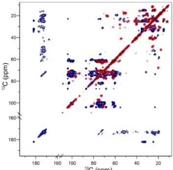
Figure X.7. 13C-13C in vivo solid-state NMR spectra of uniformly 13C-labelled C. reinhardtii
recorded at a MAS frequency of 26.7 kHz using either INEPT- (in red) or CP-based (in blue) filters. From (Arnold et al., 2018) with permission.
By using in vivo 2D solid-state NMR and dynamic spectral edition on 13C-labelled
microalgae, we improved our previous 1D NMR results for C. reinhardtii, showing that precise assignments of carbohydrates, lipids and proteins are possible (Arnold et al., 2018). Figure X.7 compares rigid and mobile molecules detected by 13C-13C SS-NMR using either INEPT- or
CP-based filters. A large range of microalgal membrane lipid classes could thus be identified
in situ, with those from thylakoid membranes dominating due to their abundance. In addition
necessary step towards a better understanding of carbohydrates architecture in the cell walls of microalgae, and their ability to face osmotic shocks, for example.
X.6. Conclusion and future prospects
In this chapter, we have reviewed the NMR techniques that can provide molecular-level information on the structure, as well as dynamics, of microalgal membranes constituents. So far, little is known about these membranes reflecting their overall complexity but they are a key component to understanding the photosynthetic machinery, and to bioengineer new strains for biofuel or food supplements.
We have shown the versatility of NMR, which can detect nuclei, such as 1H, 31P, 13C, 15N, 2H and 19F, in either single or multidimensional experiments. It is applicable to small or large,
dynamic or rigid molecules, to metabolites, model membranes, cell extracts, whole cells and even living cells. For NMR, isotopic labelling is generally required. While uniform labelling is routinely performed, as demonstrated here with examples of fully 13C-labelled microalgae,
new approaches can certainly be developed to specifically label molecules of interest within whole microalgae, providing that the biosynthetic pathways are known. This strategy has notably been applied to bacteria and to 2H in vivo SS-NMR study of their membranes (Booth
et al., 2017).
Depending on the sample and cellular constituent studied, solution or SS-NMR approaches are available. In the solid state, they include static or MAS samples, spinning at moderate or high frequencies. New techniques continue to emerge, including the use of dynamic filters and comprehensive multiphase NMR, which are useful to discriminate membrane lipids from lipid droplets, for example.
Beside broad molecular identification, NMR focuses on high-resolution structure determination of biomolecules. Microalgal membrane proteins and photosynthetic systems are important targets since they are involved in metabolism and transport. Their structures will need to be determined to understand these mechanisms (Ladizhansky, 2017). Revealing the structure of microalgae cell walls (including their connexion to the membrane) is also an area where SS-NMR can contribute, either within whole cells or extracts, as has been done with higher plants to obtain atomic resolution of cellulose nanostructures (Dick-Pérez et al., 2011, Wang and Hong, 2015). Such information is essential for a fundamental understanding of microalgal species and also for commercial exploitation of this aquatic resource.
Finally, Dynamic Nuclear Polarization (DNP) coupled with SS-NMR has been shown to be very powerful for studies of biological assemblies, such as bacterial cell walls (Wang et al., 2016), or membrane proteins (Becker-Baldus et al., 2015), with unprecedented sensitivity. Using this approach on microalgae should improve our knowledge of the complex entanglement of proteins, lipids and carbohydrates in cellular external matrices. It is exciting to see that, while in-cell SS-NMR is still the source of many new developments, it has now reached the maturity to be applied to whole microalgae to give better understanding of living organisms at an atomic level.
X.7 Acknowledgements
This work was supported by the Natural Sciences and Engineering Research Council (NSERC) of Canada (grant RGPIN-2018-06200 to I.M.) and the Centre National de la Recherche Scientifique (UMR 7203 to D.E.W.). A.P. would like to thank the PROTEO network, the Faculty of Science of the Université du Québec à Montréal (UQAM) and the Fonds Québécois de la Recherche sur la Nature et les Technologies for the award of
scholarships. I.M. is a member of Ressources Aquatiques Québec (RAQ) and PROTEO research networks.
References
ABO, B. O., ODEY, E. A., BAKAYOKO, M. & KALAKODIO, L. 2019. Microalgae to biofuels production: a review on cultivation, application and renewable energy. Rev. Environ.
Health, 34, 91.
ADEBODUN, F., CHUNG, J., MONTEZ, B., OLDFIELD, E. & SHAN, X. 1992. Spectroscopic studies of lipids and biological membranes: carbon-13 and proton magic-angle sample-spinning nuclear magnetic resonance study of glycolipid-water systems. Biochem., 31, 4502-4509.
AFONIN, S., GLASER, R. W., SACHSE, C., SALGADO, J., WADHWANI, P. & ULRICH, A. S. 2014. 19F NMR screening of unrelated antimicrobial peptides shows that membrane interactions are largely governed by lipids. Biochim. Biophys. Acta Biomembr., 1838, 2260-2268.
AKHTER, M., DUTTA MAJUMDAR, R., FORTIER-MCGILL, B., SOONG, R., LIAGHATI-MOBARHAN, Y., SIMPSON, M., ARHONDITSIS, G., SCHMIDT, S., HEUMANN, H. & SIMPSON, A. J. 2016. Identification of aquatically available carbon from algae through solution-state NMR of whole 13C-labelled cells. Anal. and Bioanal. Chem., 408, 4357-70.
ALIA, A., GANAPATHY, S. & DE GROOT, H. J. 2009. Magic Angle Spinning (MAS) NMR: a new tool to study the spatial and electronic structure of photosynthetic complexes.
Photosynth. Res., 102, 415-25.
ALIA, A., MATYSIK, J., SOEDE-HUIJBREGTS, C., BALDUS, M., RAAP, J., LUGTENBURG, J., GAST, P., VAN GORKOM, H. J., HOFF, A. J. & DE GROOT, H. J. M. 2001. Ultrahigh Field MAS NMR Dipolar Correlation Spectroscopy of the Histidine Residues in Light-Harvesting Complex II from Photosynthetic Bacteria Reveals Partial Internal Charge Transfer in the B850/His Complex. J. Am. Chem. Soc., 123, 4803-4809.
ANDRONESI, O. C., BECKER, S., SEIDEL, K., HEISE, H., YOUNG, H. S. & BALDUS, M. 2005. Determination of Membrane Protein Structure and Dynamics by Magic-Angle-Spinning Solid-State NMR Spectroscopy. J. Am. Chem. Soc., 127, 12965-12974.
ARNOLD, A. A., BOURGOUIN, J. P., GENARD, B., WARSCHAWSKI, D. E., TREMBLAY, R. & MARCOTTE, I. 2018. Whole cell solid-state NMR study of Chlamydomonas reinhardtii microalgae. J. Biomol. NMR, 70, 123-131.
ARNOLD, A. A., GENARD, B., ZITO, F., TREMBLAY, R., WARSCHAWSKI, D. E. & MARCOTTE, I. 2015. Identification of lipid and saccharide constituents of whole microalgal cells by 13C solid-state NMR. Biochim. Biophys. Acta Biomembr., 1848, 369-377.
AZADI-CHEGENI, F., SCHIPHORST, C. & PANDIT, A. 2018a. In vivo NMR as a tool for probing molecular structure and dynamics in intact Chlamydomonas reinhardtii cells.
Photosynth. Res., 135, 227-237.
AZADI-CHEGENI, F., WARD, M. E., PERIN, G., SIMIONATO, D., MOROSINOTTO, T., BALDUS, M. & PANDIT, A. 2018b. Conformational dynamics of photosynthetic Light-Harvesting Complex II in a native membrane environment. bioRxiv, 288860 (Article retrieved June 9, 2020).
BAIER, T., WICHMANN, J., KRUSE, O. & LAUERSEN, K. J. 2018. Intron-containing algal transgenes mediate efficient recombinant gene expression in the green microalga Chlamydomonas reinhardtii. Nucleic Acids Res., 46, 6909-6919.
BAILEY, D. S. & NORTHCOTE, D. H. 1976. Phospholipid composition of the plasma membrane of the green alga, Hydrodictyon africanum. Biochem. J., 156, 295-300.
BEAL, C. M., WEBBER, M. E., RUOFF, R. S. & HEBNER, R. E. 2010. Lipid analysis of Neochloris oleoabundans by liquid state NMR. Biotechnol. Bioeng., 106, 573-83.
BECKER-BALDUS, J., BAMANN, C., SAXENA, K., GUSTMANN, H., BROWN, L. J., BROWN, R. C., REITER, C., BAMBERG, E., WACHTVEITL, J., SCHWALBE, H. & GLAUBITZ, C. 2015. Enlightening the photoactive site of channelrhodopsin-2 by DNP-enhanced solid-state NMR spectroscopy. Proc. Natl. Acad. Sci. U.S.A., 112, 9896-901.
BERRY, J. P., ROY, U., JAJA-CHIMEDZA, A., SANCHEZ, K., MATYSIK, J. & ALIA, A. 2016. High-resolution magic angle spinning nuclear magnetic resonance of intact zebrafish embryos detects metabolic changes following exposure to teratogenic polymethoxyalkenes from algae. Zebrafish, 13, 456-465.
BONDU, S., KERVAREC, N., DESLANDES, E. & PICHON, R. 2008. The use of HRMAS NMR spectroscopy to study the in vivo intra–cellular carbon/nitrogen ratio of Solieria chordalis (Rhodophyta). J. Appl. Phycol., 20, 673-679.
BOOTH, V., WARSCHAWSKI, D. E., SANTISTEBAN, N. P., LAADHARI, M. & MARCOTTE, I. 2017. Recent progress on the application of 2H solid-state NMR to probe the interaction of antimicrobial peptides with intact bacteria. Biochim. Biophys. Acta Proteins Proteom., 1865, 1500-1511.
BOROVYAGIN, V. L., TARAKHOVSKY, Y. S. & VASILENKO, I. A. 1988. Polymorphism in galactolipid/phosphatidylglycerol model membranes initiated by chlorophylls: 31P-NMR and electron-microscopy studies. Biochim. Biophys. Acta, 939, 111-123.
BOUDIÈRE, L., MICHAUD, M., PETROUTSOS, D., RÉBEILLÉ, F., FALCONET, D., BASTIEN, O., ROY, S., FINAZZI, G., ROLLAND, N., JOUHET, J., BLOCK, M. A. & MARÉCHAL, E. 2014. Glycerolipids in photosynthesis: Composition, synthesis and trafficking. Biochim.
Biophys. Acta Bioenerg., 1837, 470-480.
BOUHLEL, Z., ARNOLD, A. A., WARSCHAWSKI, D. E., LEMARCHAND, K., TREMBLAY, R. & MARCOTTE, I. 2019. Labelling strategy and membrane characterization of marine bacteria Vibrio splendidus by in vivo 2H NMR. Biochim. Biophys. Acta Biomembr., 1861, 871-878.
BRENTEL, I., SELSTAM, E. & LINDBLOM, G. 1985. Phase equilibria of mixtures of plant galactolipids. The formation of a bicontinuous cubic phase. Biochim. Biophys. Acta
Biomembr., 812, 816-826.
BRUNO, E., DIGILIO, G., CABELLA, C., DE REGGI, A., BARONI, S., MAINERO, V. & AIME, S. 2006. Water exchange across the erythrocyte plasma membrane studied by HR-MAS NMR spectroscopy. Magn. Reson. Med., 56, 978-985.
CASTRO, V., DVINSKIKH, S. V., WIDMALM, G., SANDSTRÖM, D. & MALINIAK, A. 2007. NMR studies of membranes composed of glycolipids and phospholipids. Biochim. Biophys.
Acta Biomembr., 1768, 2432-2437.
CHALLAGULLA, V., NAYAR, S., WALSH, K. & FABBRO, L. 2017. Advances in techniques for assessment of microalgal lipids. Crit. Rev. Biotechnol., 37, 566-578.
CHAUTON, M., STØRSETH, T. & JOHNSEN, G. 2003. High-resolution magic angle spinning 1H NMR analysis of whole cells of Thalassiosira pseudonana (Bacillariophyceae): Broad range analysis of metabolic composition and nutritional value. J. Appl. Phycol., 15, 533-542.
CHAUTON, M., STØRSETH, T. & KRANE, J. 2004. High-resolution magic angle spinning NMR analysis of whole cells of Chaetoceros muelleri (Bacillariophyceae) and comparison
with C-13-NMR and distortionless enhancement by polarization transfer C-13-NMR analysis of lipophilic extracts. J. Phycol., 40, 611-618.
CHAUTON, M. & STØRSETH, T. F. 2008. HR MAS NMR spectroscopy of marine microalgae, Part 1: Classification and metabolite composition from HR MAS 1H NMR spectra and multivariate analysis. In: WEBB, G. A. (ed.) Modern Magnetic Resonance. Dordrecht: Springer.
COURTIER-MURIAS, D., FAROOQ, H., MASOOM, H., BOTANA, A., SOONG, R., LONGSTAFFE, J. G., SIMPSON, M. J., MAAS, W. E., FEY, M., ANDREW, B., STRUPPE, J., HUTCHINS, H., KRISHNAMURTHY, S., KUMAR, R., MONETTE, M., STRONKS, H. J., HUME, A. & SIMPSON, A. J. 2012. Comprehensive multiphase NMR spectroscopy: Basic experimental approaches to differentiate phases in heterogeneous samples. J. Magn.
Reson., 217, 61-76.
DE JESUS RAPOSO, M. F., DE MORAIS, A. M. & DE MORAIS, R. M. 2015. Marine polysaccharides from algae with potential biomedical applications. Mar. Drugs, 13, 2967-3028.
DICK-PÉREZ, M., ZHANG, Y., HAYES, J., SALAZAR, A., ZABOTINA, O. A. & HONG, M. 2011. Structure and interactions of plant cell-wall polysaccharides by two- and three-dimensional magic-angle-spinning solid-state NMR. Biochem., 50, 989-1000.
DOMOZYCH, D., CIANCIA, M., FANGEL, J., MIKKELSEN, M., ULVSKOV, P. & WILLATS, W. 2012. The cell walls of green algae: a journey through evolution and diversity. Front. Plant
Sci., 3, 82.
DOMOZYCH, D. S. 2019. Algal cell walls. In: SONS, J. W. (ed.) eLS. Chichester.
DORON, L., SEGAL, N. A. & SHAPIRA, M. 2016. Transgene expression in microalgae - from tools to applications. Front. Plant Sci., 7, 505.
EBERHARD, S., FINAZZI, G. & WOLLMAN, F. A. 2008. The dynamics of photosynthesis. Annu.
Rev. Genet., 42, 463-515.
ENGEL, B. D., SCHAFFER, M., KUHN CUELLAR, L., VILLA, E., PLITZKO, J. M. & BAUMEISTER, W. 2015. Native architecture of the Chlamydomonas chloroplast revealed by in situ cryo-electron tomography. eLife, 4, e04889.
FALAISE, C., FRANCOIS, C., TRAVERS, M. A., MORGA, B., HAURE, J., TREMBLAY, R., TURCOTTE, F., PASETTO, P., GASTINEAU, R., HARDIVILLIER, Y., LEIGNEL, V. & MOUGET, J. L. 2016. Antimicrobial compounds from eukaryotic microalgae against human pathogens and diseases in aquaculture. Mar. Drugs, 14, 159.
FAN, J., ANDRE, C. & XU, C. 2011. A chloroplast pathway for the de novo biosynthesis of triacylglycerol in Chlamydomonas reinhardtii. FEBS Lett., 585, 1985-91.
FARESE, R. V., JR. & WALTHER, T. C. 2009. Lipid droplets finally get a little R-E-S-P-E-C-T. Cell, 139, 855-60.
GAYSINSKI, M., ANNICK, O.-M., THOMAS, O. & CULIOLI, G. 2015. Extraction, purification, and NMR analysis of terpenes from brown algae. Methods Mol. Biol., 1308, 207-23. GOODSON, C., ROTH, R., WANG, Z. T. & GOODENOUGH, U. 2011. Structural correlates of
cytoplasmic and chloroplast lipid body synthesis in Chlamydomonas reinhardtii and stimulation of lipid body production with acetate boost. Eukaryot. Cell, 10, 1592-606. GOOLD, H., BEISSON, F., PELTIER, G. & LI-BEISSON, Y. 2014. Microalgal lipid droplets:
Composition, diversity, biogenesis and functions. Plant Cell Rep., 34, 545-55.
GOPINATH, T., NELSON, S. E. D., SOLLER, K. J. & VEGLIA, G. 2017. Probing the conformationally excited states of membrane proteins via 1H-detected MAS solid-State NMR spectroscopy. J. Phys. Chem. B, 121, 4456-4465.
GRIFFIN, J. L., LEHTIMAKI, K. K., VALONEN, P. K., GROHN, O. H., KETTUNEN, M. I., YLA-HERTTUALA, S., PITKANEN, A., NICHOLSON, J. K. & KAUPPINEN, R. A. 2003. Assignment of 1H nuclear magnetic resonance visible polyunsaturated fatty acids in BT4C gliomas undergoing ganciclovir-thymidine kinase gene therapy-induced programmed cell death. Cancer Res., 63, 3195-201.
GROSS, J. D., COSTA, P. R., DUBACQ, J. P., WARSCHAWSKI, D. E., LIRSAC, P. N., DEVAUX, P. F. & GRIFFIN, R. G. 1995. Multidimensional NMR in lipid systems. Coherence transfer through J couplings under MAS. J. Magn. Reson. B, 106, 187-90.
GUTIÉRREZ, M., TIDGEWELL, K., CAPSON, T. L., ENGENE, N., ALMANZA, A., SCHEMIES, J., JUNG, M. & GERWICK, W. H. 2010. Malyngolide dimer, a bioactive symmetric cyclodepside from the panamanian marine cyanobacterium Lyngbya majuscula. J. Nat. Prod., 73, 709-711.
HARWOOD, J. L. 1998. Membrane Lipids in Algae. In: PAUL-ANDRÉ, S. & NORIO, M. (eds.)
Lipids in photosynthesis: structure, function and genetics. Dordrecht: Springer
Netherlands.
HASHIM, F., DENIS, C.-M., RONALD, S., WOLFGANG, B., WILLIAM, M. K. & ANDRÉ, J. S. 2013. HR-MAS NMR spectroscopy: a practical guide for natural samples. Curr. Org. Chem., 17, 3013-3031.
HASSAN, Q., DUTTA MAJUMDAR, R., WU, B., LANE, D., TABATABAEI-ANRAKI, M., SOONG, R., SIMPSON, M. J. & SIMPSON, A. J. 2019. Improvements in lipid suppression for 1H NMR-based metabolomics: Applications to solution-state and HR-MAS NMR in natural and in vivo samples. Magn. Reson. Chem., 57, 69-81.
HÖLZL, G. & DÖRMANN, P. 2019. Chloroplast lipids and their biosynthesis. Annu. Rev. Plant
Biol., 70, 51-81.
HOWARD, K. P. & PRESTEGARD, J. H. 1995. Membrane and solution conformations of monogalactosyldiacylglycerol using NMR/molecular modeling methods. J. Am. Chem.
Soc., 117, 5031-5040.
HU, Q., SOMMERFELD, M., JARVIS, E., GHIRARDI, M., POSEWITZ, M., SEIBERT, M. & DARZINS, A. 2008. Microalgal triacylglycerols as feedstocks for biofuel production: perspectives and advances. Plant J., 54, 621-39.
HUANG, L. & MCDERMOTT, A. E. 2008. Partial site-specific assignment of a uniformly (13)C, (15)N enriched membrane protein, light-harvesting complex 1 (LH1), by solid state NMR. Biochim. Biophys. Acta, 1777, 1098-108.
KABLI, S., SPAINK, H. P., DE GROOT, H. J. & ALIA, A. 2009. In vivo metabolite profile of adult zebrafish brain obtained by high-resolution localized magnetic resonance spectroscopy. J. Magn. Reson. Imaging, 29, 275-81.
KAEBISCH, E., FUSS, T. L., VANDERGRIFT, L. A., TOEWS, K., HABBEL, P. & CHENG, L. L. 2017. Applications of high-resolution magic angle spinning MRS in biomedical studies I-cell line and animal models. NMR Biomed., 30.
KOBAYASHI, Y., TORII, A., KATO, M. & ADACHI, K. 2007. Accumulation of cyclitols functioning as compatible solutes in the haptophyte alga Pavlova sp. Phycol. Res., 55, 81-90. KOVACEVIC, V., SIMPSON, A. J. & SIMPSON, M. J. 2018. Evaluation of Daphnia magna
metabolic responses to organic contaminant exposure with and without dissolved organic matter using 1H nuclear magnetic resonance (NMR)-based metabolomics.
Ecotoxicol. Environ. Saf., 164, 189-200.
KOZOROG, M., SANI, M.-A., LENARČIČ ŽIVKOVIĆ, M., ILC, G., HODNIK, V., SEPAROVIC, F., PLAVEC, J. & ANDERLUH, G. 2018. 19F NMR studies provide insights into lipid
membrane interactions of listeriolysin O, a pore forming toxin from Listeria monocytogenes. Sci. Rep., 8, 6894.
LADIZHANSKY, V. 2017. Applications of solid-state NMR to membrane proteins. Biochim.
Biophys. Acta Proteins Proteom., 1865, 1577-1586.
LIAGHATI MOBARHAN, Y., SOONG, R., LANE, D. & SIMPSON, A. J. 2019. In vivo comprehensive multiphase NMR. Magn. Reson. Chem., 58, 427-444.
LINDBLOM, G., BRENTEL, I., SJOLUND, M., WIKANDER, G. & WIESLANDER, A. 1986. Phase equilibria of membrane lipids from Acholeplasma laidlawii: importance of a single lipid forming nonlamellar phases. Biochem., 25, 7502-10.
MATLAHOV, I. & VAN DER WEL, P. C. A. 2018. Hidden motions and motion-induced invisibility: Dynamics-based spectral editing in solid-state NMR. Methods, 148, 123-135.
MATSUMORI, N., KASAI, Y., OISHI, T., MURATA, M. & NOMURA, K. 2008. Orientation of fluorinated cholesterol in lipid bilayers analyzed by 19F tensor calculation and solid-state NMR. J. Am. Chem. Soc., 130, 4757-66.
MEDEIROS-SILVA, J., MANCE, D., DANIËLS, M., JEKHMANE, S., HOUBEN, K., BALDUS, M. & WEINGARTH, M. 2016. 1H-detected solid-state NMR studies of water-inaccessible proteins in vitro and in situ. Angew. Chem. Int. Ed., 55, 13606-13610.
MENDIOLA-MORGENTHALER, L., EICHENBERGER, W. & BOSCHETTI, A. 1985. Isolation of chloroplast envelopes from Chlamydomonas. Lipid and polypeptide composition.
Plant Sci., 41, 97-104.
MERKLEY, N. & SYVITSKI, R. 2011. Profiling whole microalgal cells by high-resolution magic angle spinning (HR-MAS) magnetic resonance spectroscopy. J. Appl. Phycol., 24, 535-40.
MIMOUNI, V., COUZINET-MOSSION, A., ULMANN, L. & WIELGOSZ-COLLIN, G. 2018. Lipids from microalgae. In: LEVINE, I. A. & FLEURENCE, J. (eds.) Microalgae in Health and
Disease Prevention. Academic Press.
MOBARHAN, Y. L., FORTIER-MCGILL, B., SOONG, R., MAAS, W. E., FEY, M., MONETTE, M., STRONKS, H. J., SCHMIDT, S., HEUMANN, H., NORWOOD, W. & SIMPSON, A. J. 2016. Comprehensive multiphase NMR applied to a living organism. Chem. Sci., 7, 4856-4866.
MOLUGU, T. R., LEE, S. & BROWN, M. F. 2017. Concepts and methods of solid-state NMR spectroscopy applied to biomembranes. Chem. Rev., 117, 12087-12132.
MURAKAMI, H., NOBUSAWA, T., HORI, K., SHIMOJIMA, M. & OHTA, H. 2018. Betaine lipid is crucial for adapting to low temperature and phosphate deficiency in Nannochloropsis.
Plant Physiol., 177, 181-193.
NARASIMHAN, S., FOLKERS, G. & BALDUS, M. 2020. When small becomes too big: expanding the use of in-cell solid-state NMR spectroscopy. ChemPlusChem, 85, 760-768.
NGUYEN, H. M., BAUDET, M., CUINE, S., ADRIANO, J. M., BARTHE, D., BILLON, E., BRULEY, C., BEISSON, F., PELTIER, G., FERRO, M. & LI-BEISSON, Y. 2011. Proteomic profiling of oil bodies isolated from the unicellular green microalga Chlamydomonas reinhardtii: with focus on proteins involved in lipid metabolism. Proteomics, 11, 4266-73.
NGUYEN, H. M., CUINE, S., BEYLY-ADRIANO, A., LEGERET, B., BILLON, E., AUROY, P., BEISSON, F., PELTIER, G. & LI-BEISSON, Y. 2013. The green microalga Chlamydomonas reinhardtii has a single omega-3 fatty acid desaturase that localizes to the chloroplast and impacts both plastidic and extraplastidic membrane lipids. Plant Physiol., 163, 914-28.
OVERALL, S. A., ZHU, S., HANSSEN, E., SEPAROVIC, F. & SANI, M. A. 2019. In Situ Monitoring of Bacteria under Antimicrobial Stress Using 31P Solid-State NMR. Int. J. Mol. Sci., 20, 181.
PAN, X., WILSON, M., MCCONVILLE, C., ARVANITIS, T. N., KAUPPINEN, R. A. & PEET, A. C. 2012. The size of cytoplasmic lipid droplets varies between tumour cell lines of the nervous system: a 1H NMR spectroscopy study. MAGMA, 25, 479-85.
PANDIT, A., MOROSINOTTO, T., REUS, M., HOLZWARTH, A. R., BASSI, R. & DE GROOT, H. J. M. 2011. First solid-state NMR analysis of uniformly 13C-enriched major light-harvesting complexes from Chlamydomonas reinhardtii and identification of protein and cofactor spin clusters. Biochim. Biophys. Acta Bioenerg., 1807, 437-443.
PANDIT, A., REUS, M., MOROSINOTTO, T., BASSI, R., HOLZWARTH, A. R. & DE GROOT, H. J. M. 2013. An NMR comparison of the light-harvesting complex II (LHCII) in active and photoprotective states reveals subtle changes in the chlorophyll a ground-state electronic structure. Biochim. Biophys. Acta Bioenerg., 1827, 738-744.
PELED, E., LEU, S., ZARKA, A., WEISS, M., PICK, U., KHOZIN-GOLDBERG, I. & BOUSSIBA, S. 2011. Isolation of a novel oil globule protein from the green alga Haematococcus pluvialis (Chlorophyceae). Lipids, 46, 851-61.
PICCINI, M., RAIKOVA, S., ALLEN, M. J. & CHUCK, C. J. 2019. A synergistic use of microalgae and macroalgae for heavy metal bioremediation and bioenergy production through hydrothermal liquefaction. Sustain. Energy Fuels, 3, 292-301.
PIUS, J., MORROW, M. R. & BOOTH, V. 2012. 2H solid-state nuclear magnetic resonance investigation of whole Escherichia coli interacting with antimicrobial peptide MSI-78.
Biochem., 51, 118-125.
POPPER, Z. A., RALET, M.-C. & DOMOZYCH, D. S. 2014. Plant and algal cell walls: diversity and functionality. Ann. Bot., 114, 1043-1048.
POSADAS, E., ALCÁNTARA, C., GARCÍA-ENCINA, P. A., GOUVEIA, L., GUIEYSSE, B., NORVILL, Z., ACIÉN, F. G., MARKOU, G., CONGESTRI, R., KOREIVIENE, J. & MUÑOZ, R. 2017. Microalgae cultivation in wastewater. In: GONZALEZ-FERNANDEZ, C. & MUÑOZ, R. (eds.) Microalgae-Based Biofuels and Bioproducts. Woodhead Publishing.
POULHAZAN, A., ARNOLD, A. A., WARSCHAWSKI, D. E. & MARCOTTE, I. 2018. Unambiguous ex situ and in cell 2D (13)C solid-state NMR characterization of starch and its constituents. Int. J. Mol. Sci., 19.
RASHIDI, B. & TRINDADE, L. M. 2018. Detailed biochemical and morphologic characteristics of the green microalga Neochloris oleoabundans cell wall. Algal Res., 35, 152-159. RASOOL, M., SABINA, E. & LAVANYA, B. 2007. Anti-inflammatory effect of Spirulina fusiformis
on adjuvant-induced arthritis in mice. Biol. Pharm. Bull., 29, 2483-7.
RECKEL, S., LOPEZ, J. J., LOHR, F., GLAUBITZ, C. & DOTSCH, V. 2012. In-cell solid-state NMR as a tool to study proteins in large complexes. Chembiochem, 13, 534-7.
RENAULT, M., TOMMASSEN-VAN BOXTEL, R., BOS, M. P., POST, J. A., TOMMASSEN, J. & BALDUS, M. 2012. Cellular solid-state nuclear magnetic resonance spectroscopy. Proc.
Natl. Acad. Sci. U.S.A., 109, 4863-8.
RICHMOND, A. & HU, Q. 2013. Handbook of microalgal culture: applied phycology and
biotechnology, John Wiley and Sons.
ROMANIUK, J. A. H. & CEGELSKI, L. 2015. Bacterial cell wall composition and the influence of antibiotics by cell-wall and whole-cell NMR. Philos. Trans. R. Soc. Lond., B, Biol. Sci., 370, 20150024.
SALNIKOV, E. S., AISENBREY, C., POKRANDT, B., BRÜGGER, B. & BECHINGER, B. 2019. Structure, topology, and dynamics of membrane-inserted polypeptides and lipids by solid-state NMR spectroscopy: investigations of the transmembrane domains of the DQ beta-1 subunit of the MHC II receptor and of the COP I protein p24. Front. Mol.
Biosci., 6.
SCHANDA, P., TRIBOULET, S., LAGURI, C., BOUGAULT, C. M., AYALA, I., CALLON, M., ARTHUR, M. & SIMORRE, J.-P. 2014. Atomic model of a cell-wall cross-linking enzyme in complex with an intact bacterial peptidoglycan. J. Am. Chem. Soc., 136, 17852-17860.
SHI, P., LI, D., CHEN, H., XIONG, Y., WANG, Y. & TIAN, C. 2012. In situ 19F NMR studies of an E. coli membrane protein. Protein Sci., 21, 596-600.
SIMON, G., KERVAREC, N. & CERANTOLA, S. 2015. HRMAS NMR analysis of algae and identification of molecules of interest via conventional 1D and 2D NMR: sample preparation and optimization of experimental conditions. Methods Mol. Biol., 1308, 191-205.
SIMPSON A.J., C.-M. D., FAROOQ H., MASOOM H., BOTANA A., SOONG R., LONGSTAFFE J.G., LAM L., SUTRISNO A., FAROOQ H., SIMPSON M.J., MAAS W.E., FEY M., ANDREW B., STRUPPE J., HUTCHINS H., KRISHNAMURTHY S., KUMAR R., MONETTE, M., STRONKS H.J., HUME A. 2013. Environmental comprehensive multi-phase NMR. Emagres, 2, 399-414.
SPOLAORE, P., JOANNIS-CASSAN, C., DURAN, E. & ISAMBERT, A. 2006. Commercial applications of microalgae. J. Biosci. Bioeng., 101, 87-96.
SUNKU, K., DE GROOT, H. J. & PANDIT, A. 2013. Insights into the photoprotective switch of the major light-harvesting complex II (LHCII): a preserved core of arginine-glutamate interlocked helices complemented by adjustable loops. J. Biol. Chem., 288, 19796-804. TARDY-LAPORTE, C., ARNOLD, A. A., GENARD, B., GASTINEAU, R., MORANÇAIS, M., MOUGET, J.-L., TREMBLAY, R. & MARCOTTE, I. 2013. A 2H solid-state NMR study of the effect of antimicrobial agents on intact Escherichia coli without mutating. Biochim. Biophys.
Acta Biomembr., 1828, 614-622.
VAN ROSSUM, B. J., STEENSGAARD, D. B., MULDER, F. M., BOENDER, G. J., SCHAFFNER, K., HOLZWARTH, A. R. & DEGROOT, H. J. 2001. A refined model of the chlorosomal antennae of the green bacterium Chlorobium tepidum from proton chemical shift constraints obtained with high-field 2-D and 3-D MAS NMR dipolar correlation spectroscopy. Biochem., 40, 1587-95.
VARGA, K., ASLIMOVSKA, L. & WATTS, A. 2008. Advances towards resonance assignments for uniformly--13C, 15N enriched bacteriorhodopsin at 18.8 T in purple membranes. J.
Biomol. NMR, 41, 1-4.
VENTERS, R. A., CALDERONE, T. L., SPICER, L. D. & FIERKE, C. A. 1991. Uniform 13C isotope labeling of proteins with sodium acetate for NMR studies: application to human carbonic anhydrase II. Biochem., 30, 4491-4.
VERHOEVEN, M. A., CREEMERS, A. F. L., BOVEE-GEURTS, P. H. M., DE GRIP, W. J., LUGTENBURG, J. & DE GROOT, H. J. M. 2001. Ultra-high-field MAS NMR assay of a multispin labeled ligand bound to its G-protein receptor target in the natural membrane environment: electronic structure of the retinylidene chromophore in rhodopsin. Biochem., 40, 3282-3288.
VIELER, A., BRUBAKER, S. B., VICK, B. & BENNING, C. 2012. A lipid droplet protein of Nannochloropsis with functions partially analogous to plant oleosins. Plant Physiol., 158, 1562-9.
VIELER, A., WILHELM, C., GOSS, R., SÜSS, R. & SCHILLER, J. 2007. The lipid composition of the unicellular green alga Chlamydomonas reinhardtii and the diatom Cyclotella meneghiniana investigated by MALDI-TOF MS and TLC. Chem. Phys. Lipids, 150, 143-55.
WANG, T. & HONG, M. 2015. Solid-state NMR investigations of cellulose structure and interactions with matrix polysaccharides in plant primary cell walls. J. Exp. Bot., 67, 503-514.
WANG, T., YANG, H., KUBICKI, J. D. & HONG, M. 2016. Cellulose structural polymorphism in plant primary cell walls investigated by high-field 2D solid-state NMR spectroscopy and density functional theory calculations. Biomacromolecules, 17, 2210-2222. WANG, X., HAO, T.-B., BALAMURUGAN, S., YANG, W.-D., LIU, J.-S., DONG, H.-P. & LI, H.-Y.
2017. A lipid droplet-associated protein involved in lipid droplet biogenesis and triacylglycerol accumulation in the oleaginous microalga Phaeodactylum tricornutum.
Algal Res., 26, 215-224.
WANG, X., WEI, H., MAO, X. & LIU, J. 2019. Proteomics analysis of lipid droplets from the oleaginous alga Chromochloris zofingiensis reveals novel proteins for lipid metabolism. Genomics Proteomics Bioinformatics, 17, 260-272.
WANG, Z. T., ULLRICH, N., JOO, S., WAFFENSCHMIDT, S. & GOODENOUGH, U. 2009. Algal lipid bodies: stress induction, purification, and biochemical characterization in wild-type and starchless Chlamydomonas reinhardtii. Eukaryot. cell, 8, 1856-1868.
WARNET, X. L., ARNOLD, A. A., MARCOTTE, I. & WARSCHAWSKI, D. E. 2015. In-cell solid-state NMR: an emerging technique for the study of biological membranes. Biophys. J., 109, 2461-2466.
WARNET, X. L., LAADHARI, M., ARNOLD, A. A., MARCOTTE, I. & WARSCHAWSKI, D. E. 2016. A 2H magic-angle spinning solid-state NMR characterisation of lipid membranes in intact bacteria. Biochim. Biophys. Acta Biomembr., 1858, 146-152.
WEBB, G., STØRSETH, T., CHAUTON, M. & KRANE, J. 2006. HR MAS NMR spectroscopy of marine microalgae, Part 2: 13C and 13C HR MAS NMR analysis used to study fatty acid composition and polysaccharide structure. In: G.A., W. (ed.) Modern Magnetic
Resonance. Dordrecht: Springer.
WERBY, S. H. & CEGELSKI, L. 2019. Spectral comparisons of mammalian cells and intact organelles by solid-state NMR. J. Struct. Biol., 206, 49-54.
YE, W., LIEBAU, J. & MALER, L. 2013. New membrane mimetics with galactolipids: lipid properties in fast-tumbling bicelles. J. Phys. Chem. B, 117, 1044-50.
YONEDA, K., YOSHIDA, M., SUZUKI, I. & WATANABE, M. M. 2016. Identification of a major lipid droplet protein in a marine diatom Phaeodactylum tricornutum. Plant Cell Physiol., 57, 397-406.
ZHANG, L., LIU, L., MALTSEV, S., LORIGAN, G. A. & DABNEY-SMITH, C. 2013. Solid-state NMR investigations of peptide-lipid interactions of the transmembrane domain of a plant-derived protein, Hcf106. Chem. Phys. Lipids, 175-176, 123-30.
ZHANG, L., LIU, L., MALTSEV, S., LORIGAN, G. A. & DABNEY-SMITH, C. 2014. Investigating the interaction between peptides of the amphipathic helix of Hcf106 and the phospholipid bilayer by solid-state NMR spectroscopy. Biochim. Biophys. Acta, 1838, 413-8.
ZHAO, L. X., LIU, A. C., YU, S. W., WANG, Z. X., LIN, X. Q., ZHAI, G. X. & ZHANG, Q. Z. 2013. The permeability of puerarin loaded poly(butylcyanoacrylate) nanoparticles coated with polysorbate 80 on the blood-brain barrier and its protective effect against cerebral ischemia/reperfusion injury. Biol. Pharm. Bull., 36, 1263-70.