HAL Id: hal-02659448
https://hal.inrae.fr/hal-02659448
Submitted on 30 May 2020HAL is a multi-disciplinary open access archive for the deposit and dissemination of sci-entific research documents, whether they are pub-lished or not. The documents may come from teaching and research institutions in France or abroad, or from public or private research centers.
L’archive ouverte pluridisciplinaire HAL, est destinée au dépôt et à la diffusion de documents scientifiques de niveau recherche, publiés ou non, émanant des établissements d’enseignement et de recherche français ou étrangers, des laboratoires publics ou privés.
Distributed under a Creative Commons Attribution - ShareAlike| 4.0 International
Mucosal immunity in Toxoplasma gondii infection
J Schulthess, D Fourreau, S Darche, B Meresse, L Kasper, N Cerf-Bensussan,
Dominique Buzoni
To cite this version:
J Schulthess, D Fourreau, S Darche, B Meresse, L Kasper, et al.. Mucosal immunity in Toxoplasma gondii infection. Parasite, EDP Sciences, 2008, 15 (3), pp.389-395. �10.1051/parasite/2008153389�. �hal-02659448�
M
UCOSAL IMMUNITY INT
OXOPLASMA GONDII INFECTIONSCHULTHESS J.*, FOURREAU D.**, DARCHE S.*, MERESSE B.***, KASPER L**, CERF-BENSUSSAN N.*** & BUZONI-GATEL D.****
Summary :
Toxoplasma gondii is an intracellular parasite that frequently infects
a large spectrum of warm-blooded animals. This parasite induces abortion and establishes both chronic and silent infections, particularly in the brain. Parasite penetration into the host activates a strong anti-parasite immune response. In the present paper, we will discuss the interplay between innate and adaptive immunity that occurs within the infected intestine to clear the parasite and to maintain intestinal homeostasis despite the exacerbation of an inflammatory immune response.
KEY WORDS : Toxoplasma gondii, immunity, mucosal.
* RPPI, Institut Pasteur-INRA, 28, rue du Dr Roux, 75724 Paris cedex 15, France.
** Department of Microbiology, Dartmouth Medical School, Lebanon, NH 03756, USA.
*** INSERM U793, Hopital Necker, 75115, Paris, France. **** INRA, IASP, 37380 Nouzilly, France.
Correspondence: Dominique Buzoni-Gatel.
Tel.: + 33 (0)2 47 42 7314 – Fax: + 33 (0)2 47 71 17 62. E-mail: [email protected]
plasma initially crosses the intestinal epithelium, dis-seminates into the deep tissues and traverses biological barriers in the placenta, the brain and the blood-retina barrier (Barragan, 2003). The tight junctions that provide complex enterocyte-enterocyte interaction constitute a physical barrier against the penetration of intestinal microorganisms. However, despite this phy-sical barrier, Toxoplasma actively crosses polarized cell monolayers (such as intestinal epithelium) and this ability is linked to parasite motility and virulence in the mouse model (Barragan, 2002). Parasite transmigration required viable and actively motile parasites but the integrity of host cell barriers is not altered during parasite transmigration. In all likelihood, enterocytes play a crucial role as sentinel against parasite invasion. Alpha-defensins (or Cryptdins – Crps) are a group of cationic antimicrobial peptides, harbouring a broad spectrum of microbicidal activity against microbes. In mouse small intestine, epithelial cells and more spe-cifically Paneth cells produce Crps as a component of secretory granules released in the lumen. Infection of B6 mice with T. gondii, can regulate Crps mRNAs expres-sion by intestinal epithelial cells. This response appears mediated via TLR9-dependant production of IFN-βthat may be involved in the blockade of T. gondii host penetration (David Fourreau, manuscript in prepara-tion).
T. gondii infection of the intestine following oral chal-lenge in certain strains of inbred and outbred mice as well as rodents, pigs and non-human primates can induce a severe form of intestinal inflammation. In C57BL/6 mice, this pathology shares both morphologic and histologic characteristics with human IBD, such as loss of intestinal epithelial architecture, shortened villi, massive influx of inflammatory cells into the lamina propria and scattered patches of necrosis. When unre-gulated, this inflammatory process results in the early mortality of the susceptible hosts (Lisenfeld, 1996). Migration of CD11c+and CD11b+monocytes, DCs,
macro-phages and PMNs into the lamina propria at day 7 after infection has been reported following oral infection with parasite tissue cysts (Courret, 2005).
At day 5 after infection with T. gondii most of these lamina propria DCs are mature, as indicated by
high-T
he gastrointestinal tract is the largest mucosalarea of the body in contact with the external environment. The intestinal epithelium protects the host against microbial infection not only by for-ming a physical barrier, but also by active participa-tion in host innate defence via the producparticipa-tion of cyto-kines, chemokines and antimicrobial peptides. The gastrointestinal tract is populated by a resident micro-flora, essential for immunological intestinal homeostasis and as a source for nutrients. Maintenance of resident bacterial number and simultaneous protection against potential pathogens, including Toxoplasma gondii, acquired by the oral route, is provided by numerous non immunological and immunological factors. In the present review the role of gut associated immune system in protection against T. gondii is discussed.
INNATE IMMUNITY
E
pithelial cells provide the first line of innate immunological host defense against oral T. gon-dii infection. During natural infections,Toxo-level expression of MHC class II, CD40, CD80, and CD86. It has been proposed that DCs from the lamina propria might gain access to the intestinal contents by using unique proteins to separate the tight junctional border between the enterocytes without disrupting the monolayer integrity (Rescigno, 2001). This process would allow for the direct sampling by DCs of patho-gens within the gut lumen. Alternatively pathopatho-gens that cross the epithelium may also be captured directly by the DCs that process antigen for presentation. An addi-tional way for the DCs from the lamina propria to sample the antigens is via the infected enterocytes. In this model, apoptotic enterocytes are digested by the DCs and processed for antigen expression.
When hosts, including humans, ingest tissue cysts or oocysts containing T. gondii, free parasites are released in the gut lumen. They subsequently enter enterocytes
where they multiply and initiate the infection. Entero-cytes loaded with parasites secrete chemokines that recruit leukocytes in the lamina propria (LP) extravas-cular space. Parasites then disseminate to several dis-tant tissues including the brain, a major site supporting parasite latency (Dubey, 1997). This event has impor-tant clinical implications since T. gondii as a chronic infection is associated with the encysted bradyzoite that slowly replicates under the control of unique host depen-dent immune signals. T. gondii can efficiently enter and survive within DCs (Channon, 2000). The functional plasticity as well as the migratory property of DCs (mostly CD11c+ cells) can then be utilized by
patho-gens for dissemination through the body.
Studies in one of our laboratories (DBG) have demons-trated that following intragastric inoculation of cysts in mice, CD11c+dendritic cells from the intestinal lamina
SCHULTHESS J., FOURREAU D., DARCHE S. ET AL.
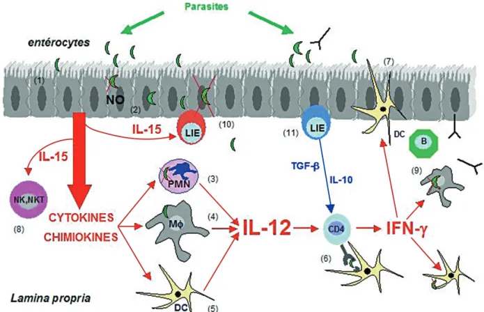
Fig. 1. – A model of the GI mucosal immune response to Toxoplasma gondii.
When parasites invade the mucosal intestinal epithelium, they first face a physical barrier brought by the enterocytes bound together by tight junctions (1).
Parasites have developed multiple strategies to adhere, sometimes to invade the enterocytes and to spread beyong the epithelium. When enterocytes are infected by the parasites, physiological and morphological disturbances occur, and enterocytes might secrete cytotoxic mole-cules such as nitric oxyde (NO) (2).
In addition, enterocytes respond to the infection by secretion of chemokines and cytokines that attract polymorphonuclear leukocytes (PMNs) (3), macrophages MΦ(4) and dendritic cells (DCs) (5).
When stimulated, these cells from the innate immune system can be directly microbicidal. They are also source of cytokines such as IL-12 that triggers the adaptive CD4 immune response (6).
To be elicited specific immune response needs antigen presentation, mainly through DCs. DCs sample the antigen by different pathways, one of them, is direct antigen capture into the lumen by elongation of the dendrites through the tight junctions (7).
Activated T cells, in addition to NK and NKT cells (8) stimulated by cytokines produced by infected enterocytes such as IL-15, secrete
IFN-γthat activates MΦ, DCs, enterocytes for parasite clearance.
B cells (9) are also triggered to secrete antibodies that can cross the epithelial barrier by active transcytose and reach parasite into the lumen. Besides microbicidal activities, IFN-γif not controlled, might damage the intestinal integrity.
propria, the Peyer’s patches and the mesenteric lymph nodes were parasitized whereas parasites were asso-ciated with the CD11c-CD11b+monocytes in the
peri-pheral circulation. These parasitized cells are involved in disease induction in the brain of naive recipient mice as demonstrated by adoptive transfer experiment. Ex vivo analysis of parasitized cells showed that single tachyzoites, non-replicating parasites could be identi-fied at the cell periphery often surrounded by the host cell plasma membrane. By several approaches inclu-ding vital staining of leukocytes, antibody labeling or chimeric mice in which the hematopoietic cells expressed the green fluorescent protein, it was deter-mined that T. gondii infected CD11b+leukocytes can
traffic to the brain extra-vascular space (Courret, 2005). Additional studies identified CD11c+and 33D1+cells
loca-lized at inflammatory sites in infected brain (Fisher, 2000).
Parasite infection of enterocytes results in the upre-gulation and expression of CD40, CD1d, Class II sug-gesting that enterocytes may act as APCs and be involved in the activation of lymphocytes. Because of the limited expression of CD80 and CD86 mandatory for efficient lymphocyte stimulation, DCs and macro-phages that infiltrate the infected epithelium are likely to be principally involved as APCs for CD4+T cells
acti-vation from the lamina propria. In addition to their cru-cial role as APC, DCs display an anti microbial func-tion. IFN-γactivation of DCs triggers oxygen dependant inhibition of T. gondii (Aline, 2002).
In addition to PMNs, both DCs (Aliberti, 2003) as well as macrophages (Oliviera, 2000) produce IL-12 follo-wing parasite infection. Interleukin-12 (IL-12) is the major cytokine triggering IFN-γ synthesis by NK and T lymphocytes during T. gondii infection. IL-12 is the major initiation signal for host resistance to the para-site. IL-12 is also assumed to be responsible for T-hel-per 1 (Th1) effector choice in T. gondii infection. CD40/CD154 interaction is involved in the regulation of macrophage production of interleukin 12 (IL-12) and T-cell production of IFN-γ. Infection of C57BL/6 mice with T. gondii results in an upregulation of CD40 expression on accessory cell populations at local sites of infection as well as in lymphoid tissues. CD40/ CD154 ligation is essential to initiate the intestinal inflammatory response observed after oral infection of C57BL/6 mice (Li, 2002) and CD40/CD40L interaction is crucial in resistance to T. gondii (Reichmann, 2000; Andrade, 2005).
Aside from being the precursors of the antibody secre-ting cells, B cells are engaged in other immune func-tions such as antigen presentation to T cells or cyto-kine production. These functions may contribute to the pathogenic role of B cells in a wide range of autoim-mune diseases. We demonstrate that B cells acquire the capacity to amplify IFN-γproduction by CD4 and CD8
T cells during the course of the Th1 inflammatory res-ponse to T. gondii infection. Using two different stra-tegies: i) reconstitution of B cell-deficient mice with B cells expressing an alloantigen different from the reci-pients, and ii) adoptive transfer of B and T cells into RAG-/-mice, we observed that B cells from T. gondii-infected mice, but not from naïve mice, induce higher IFN-γexpression by splenic host T cells. In vitro assays allowing the physical separation of T cells and B cells demonstrate that antigen-primed B cells enhance IFN-γ production by T cells in a contact-dependent fashion. Using an transgenic strain of T. gondii and OVA-specific CD4 T cells, we observed that the pro-inflam-matory effect of B cells is neither antigen-specific nor requires MHCII expression. However, TNF-αexpressed on the surface of B cells appears to mediate in part the upregulation of IFN-γby the effector T cells (Men-nard, 2007).
Because of their major role in microbial recognition, the involvement of receptor pathways involving the Toll-like receptor (TLR)/IL-1R superfamily in triggering both DC IL-12 production and host resistance to T. gondii has recently been addressed. Innate immune recogni-tion relies on a limited number of germ-line encoded receptors, such as TLRs that recognize Pathogen Asso-ciated Molecular Patterns (PAMPs) of microbial origin. Although TLRs are expressed in a broad range of tis-sues the greatest variety of TLR mRNAs is found in pro-fessional APCs suggesting a key role of TLRs in innate immunity that is essential in the development of the acquired immune response characterized by polariza-tion of naive CD4+ helper T cells toward the TH1 or
TH2 phenotype. Mice lacking myeloid differentiation factor 88 (MyD88), an adapter molecule used by all TLRs as well as IL-1R and IL-18R exhibited a near com-plete abrogation of the parasite-induced IL-12 response, and when challenged with T. gondii, the knockout (KO) animals displayed a loss in resistance to infection equivalent to that of IL-12-deficient mice (Scanga, 2002). Recently we have identified a role for TLR9 in the Th1-type inflammatory response that ensues following oral infection with T. gondii.
Following oral infection with T. gondii, susceptible B6 but not TLR9-/- (B6 background) mice develop a Th1
dependent acute ileitis compared to TLR9-/- mice (B6
background) that are free of gut inflammation. TLR9
-/-mice have higher parasite burdens than control WT mice suggesting depressed IFN-γ dependent parasite killing. IL-12 producing DCs were reduced in TLR9
-/-mice compared with the WT controls which correspon-ded with a reduction in total T cell frequency as well as IFN-γ producing T cells from the Lamina Propria. Infection of chimeric mice deleted of TLR9 in either the hematopoietic or non-hematopoietic compartments indicates that TLR9 expression in both compartments is involved (Minns, 2006).
In addition to IL-12, macrophages and dendritic cells produced IL-15. IL-15 exhibits pleiotropic functions at the interface between innate and adaptive immunity. IL-15 is a 14 kD cytokine that shares common features with IL-2 and exhibits stimulatory effects on T cell pro-liferation. However, IL-15 can be distinguished from IL-2 by its broad cell-type distribution, its potent in vivo anti-apoptotic effects and as most relevant to this review a bridge between the innate and adaptive immune res-ponse. IL-15 is necessary for the differentiation and/or homeostatic maintenance of the three subsets of lym-phocytes linked to innate immune response, NK, NK/T and CD8ααintraepithelial lymphocytes (IEL). IL-15 is also mandatory for the survival of memory CD8 T lym-phocytes. IL-15 induces the effector functions of NK and CD8 T lymphocytes, promotes the selection of high avidity cytotoxic CD8 T cells and their expression of the co-receptor CD8αβ(reviewed in Fehinger, 2001). Finally, IL-15 stimulates the maturation of dendritic cells and thereby promotes antigen presentation.
The role of IL-15 produced by hematopietic cells is controversial after infection by T. gondii (Doherty, 1996; Lieberman, 2004; Khan, 2002). In contrast, we have recently observed, using hematopoietic chimeric mice that IL-15 produced by infected enterocytes, initiates the inflammatory immune response that leads to the development of the lethal ileitis in C57BL/6 mice (Julie Shulthess, manuscript in preparation). IL-15 is critical for the differentiation and or homeostasis of several murine innate immune cell subsets, including natural killer, NK/T cells and CD8αα intraepithelial lympho-cytes as well as the generation and maintenance of spe-cific memory CD8 TCRαβcells. In addition, IL-15 plays redundant functions with other cytokines to promote maturation of dendritic cells, proliferation of T and B cells, cytotoxicity of NK and CD8+T cells and
produc-tion of proinflammatory cytokines.
Among cells targeted by IL-15, NK and NKT cells plays a major role during the early phase of the T. gon-dii infection. Natural killer T cells represent a minor subset of T lymphocytes that share receptor structures with conventional T cells and NK cells. Murine NKT cells express intermediate levels of a TCR using a semi-invariant Vα14-Jα281 TCR-chain paired with a limited number of β chain such as Vβ8, -7, or -2 TCR toge-ther with NK cell receptors (NKR-P1, Ly-49, and NK1.1 in C57BL/6 mice). These cells are located mainly in the liver, spleen, thymus, and bone marrow and recognize Ag in the context of the monomorphic CD1d Ag-pre-senting molecule. Our findings suggest a potentially cri-tical role for these early responder cells in the initia-tion and regulainitia-tion of the lethal inflammatory process. The implication of NKT cells was demonstrated by the observation that NKT cell-deficient mice (Jα281-/-) are
more resistant than C57BL/6 mice to the development of lethal ileitis. Jα281-/-mice failed to overexpress
IFN-γin the intestine early after infection. This detrimental effect of NKT cells is blocked by treatment with α -galac-tosylceramide, which prevents death in C57BL/6, but not in Jα281-/-, mice. This protective effect is
charac-terized by a shift in cytokine production by NKT cells toward a Th2 profile and correlates with an increased number of mesenteric Foxp3 lymphocytes. These results highlight the participation of NKT cells in the parasite clearance by shifting the cytokine profile toward a Th1 pattern and simultaneously to immunopathological manifestation when this Th1 immune response remains uncontrolled and give the evidence that NKT cells are important in regulation of Th1/Th2 differentiation (Ronet, 2005). The parasite antigen able to trigger NKT func-tion is not yet determined, but it is well known that NKT recognize Ag in the context of the monomorphic CD1d Ag-presenting molecule. CD1d and the invariant TCR-chain are essential for the normal development of NKT. CD1 molecules present hydrophobic lipid Ags. How-ever in contrast to mice that are genetically impaired for NKT cell (Jα281-/-mice) and that exhibit resistance
to the development of lethal ileitis in C57B6 mice, CD1d deficient mice were more susceptible to the infection and apparently do not control their inflammatory res-ponse (Smiley, 2005). In C57BL/6 mice, CD4+cells can
cause intestinal pathology during T. gondii infection. Compared with WT mice, infected CD1d-deficient C57BL/6 mice had higher frequencies and numbers of activated (CD44high) CD4+ cells in mesenteric lymph
nodes. Depletion of CD4+cells from CD1d-deficient mice
reduced weight loss and prolonged survival, demons-trating a functional role for CD4+cells in their increased
susceptibility to T. gondii infection. An other explana-tion might be that in addiexplana-tion to NKT depleexplana-tion, regu-latory cells, such as IEL and B cells, are also reduced in CD1d-/- mice (Allez, 2004; Mizoguchi, 2002).
INTESTINAL ADAPTIVE IMMUNE RESPONSE
A
ctivation of the innate immune system results in antigen presentation and activation of the antigen specific T and B cell intestinal response. Intraepithelial lymphocytes (IELs) are located inbet-ween epithelial cells, below the intercellular tight junc-tions. Most of the IELs are CD8+T lymphocytes and bearan oligoclonal repertoire of T-cell antigen receptor (TCR) and express the unusual integrin αEβ7, which is invol-ved in adherence to epithelial cells by binding to E-cadherin.
Infection of the gut with mucosal pathogens can result in the migration and activation of IELs. IEL migration towards the T. gondii infected enterocytes requires the expression of the chemokine receptor CCR5 in res-ponse to the secretion of MIP-1α (Luangsay, 2003). SCHULTHESS J., FOURREAU D., DARCHE S. ET AL.
IELs provide a number of important immunological func-tions, including cytotoxic activity, secretion of cytokines and modulation of epithelial cell death and regenera-tion. T. gondii antigen-primed IELs are cytotoxic for T. gondii-infected enterocytes (Chardès, 1994; Buzoni-Gatel, 1997). We showed that IL-15KO mice failed to develop the lethal ileitis after infection by the parasite. Our preliminary data indicate that IEL boosted by IL-15 might participate to the deleterious role of this cyto-kine in the loss of intestinal homeostasis although parasite killing might require IL-15 (Schulthess, manus-cript in preparation).
However IELs may display a dual role depending on the phenotype. Adoptive transfer of antigen primed CD8 α/β TCR α/β IELs into naive mice prior to infec-tion, rescues the recipient mice from death (Buzoni-Gatel, 2001) in contrast to IELs that exhibit αTCR γ/δ phenotype. T. gondii antigen-primed IELs produce subs-tantial amounts of TGF-β that down regulate the pro-duction of IFN-γ from the CD4 lymphocytes in the lamina propria through a Smad 2, 3 dependant path-way (Mennechet, 2004).
In intestinal toxoplasmosis the development of a Th1 like T cell response, orchestrated by IFN-γ producing CD4 T cell from the lamina propria leads to the inhi-bition of parasite replication, but also may damage the intestinal barrier. CD4 T cells from the T. gondii infec-ted lamina propria produce copious amount of IFN-γ and TNF-αthat enhance the production of chemokines by infected enterocytes and increase the inflammatory response (Mennechet, 2002).
Studies indicate that production of secretory IgA anti-bodies are associated with early infection in mice (Char-dès, 1992, 1993). The lamina propria is indeed popu-lated with numerous B cells that differentiate into IgA plasmacytoid cells. In addition the natural presence of TGF−βinto the intestine contributes to IgA switch. How-ever, the protective role of these secretory IgA are still debated. Specific antibodies are not considered to be the major factor in recovery from infection, although they may play a role in protection against re-infection and are useful for an early diagnostic.
PARASITE ANTIGENS THAT TRIGGER
THE INNATE RESPONSE
T
he role of specific Toxoplasma antigens in the induction of the innate response is only partially understood. The surface of T. gondii comprises of a family of developmentally regulated glycosylphos-phatidylinositol (GPI)-linked proteins (SRSs), of which surface antigen 1 (SAG1) is the prototypic member. SAG1 protein is exclusively expressed on the tachyzoite. The biological role for this superfamily of surfacepro-teins remains mostly enigmatic although there is evi-dence for a role in parasite attachment. SAG1 induces the dominant antibody response during infection and a strong, systemic Th1-like T cell response characte-rized by high-titer IFN-γ production by CD4 and CD8 T lymphocyte.
A SAG1 null mutant was engineered by homologous recombination and used to infect C57BL/6 mice. This mutant was shown in vitro to adhere and to replicate in fibroblasts at the same or even at a better rate than the control parental strain. In vivo, we were able to demonstrate that this antigen deficient parasite is una-ble to induce ileitis following intraluminal infection. Although this mutant can replicate in both the host and in vitro cell culture, infection is associated with a decrease in both innate and adaptive inflammatory immune responses (Rachinel, 2004).
Parasite penetration into the host activates a strong anti-parasite immune response, but is also used by the para-site to chronically persist. John Boothroyd reports (Saeij, 2007) the molecular cross talk between the parasite rhoptry proteins and the host cell. During host cell invasion, rhoptries participate to the constitution of the mobile junction that drives the parasite into the host cell, while building the parasitophorus vacuole in which the parasite grows (El Hajj, 2007). Some soluble rhoptries, such as Rop16, are shed into the cytoplasm, and then reach the nucleus where they can eventually impact different signalling pathways such as STAT3/6, key molecules in the immune response establishment. Whatever the signals and the pathways used to shift the immune response, a Th1 like immune response is absolutely necessary to control parasite replication. If left unmodulated this Th1 like immune response can lead to lethal host damage such as the ileitis seen in C57BL/6 mice.
REFERENCES
ALIBERTIJ., VALENZUELAJ. G., CARRUTHERSV.B., HIENYS., ANDER -SENJ., CHARESTH., REIS ESOUSAC., FAIRLAMBA., RIBEIROJ.M. & SHERA. Molecular mimicry of a CCR5 binding-domain in the microbial activation of dendritic cells. Nat.
Immu-nol., 2003, 4, 485-490.
ALINE F., BOUT D. & DIMIER-POISSON I. Dendritic cells as effector cells: gamma interferon activation of murine den-dritic cells triggers oxygen-dependent inhibition of
Toxo-plasma gondii replication. Infect. Immun., 2002, 70,
2368-2374.
ALLEZ M. & MAYER L. Regulatory T cells: peace keepers in the gut. Inflamm. Bowel. Dis., 2004, 10, 666-676. ANDRADER.M., WESSENDARPM., PORTILLOJ.A., YANGJ.Q., GOMEZ
F. J., DURBINJ. E., BISHOPG. A. & SUBAUSTEC.S. TNF recep-tor-associated factor 6-dependent CD40 signaling primes macrophages to acquire antimicrobial activity in response to TNF-α. J. Immunol., 2005, 175, 6014-6021.
BARRAGANA. & SIBLEY L.D. Migration of Toxoplasma gondii across biological barriers. Trends Microbiol., 2003, 11, 426-430.
BARRAGANA. & SIBLEYL.D. Transepithelial migration of
Toxo-plasma gondii is linked to parasite motility and virulence. J. Exp. Med., 2002, 195, 1625-1633.
BUZONI-GATELD., DEBBABIH., MENNECHETF.J., MARTINV., LEPAGE A.C., SCHWARTZMANJ.D. & KASPERL.H. Murine ileitis after intracellular parasite infection is controlled by TGF-beta-producing intraepithelial lymphocytes. Gastroenterology, 2001, 120, 914-924.
BUZONI-GATELD., LEPAGEA.C., DIMIER-POISSONI.H., BOUTD.T. & KASPERL.H. Adoptive transfer of gut intraepithelial lym-phocytes protects against murine infection with Toxoplasma
gondii. J. Immunol., 1997, 158, 5883-5889.
CHANNONJ.Y., SEGUINR.M. & KASPERL.H. Differential infecti-vity and division of Toxoplasma gondii in human peri-pheral blood leukocytes. Infect. Immun., 2000, 68, 4822-4826.
CHARDEST., BUZONI-GATELD., LEPAGEA., BERNARDF. & BOUTD.
Toxoplasma gondii oral infection induces specific cytotoxic
CD8 alpha/beta+ Thy-1+ gut intraepithelial lymphocytes,
lytic for parasite-infected enterocytes. J. Immunol., 1994,
153, 4596-4603.
CHARDEST., VELGE-ROUSSELF., MEVELECM.N., BLAISEF. & BOUTD. Local and systemic cellular immunity following oral infec-tion of mice with Toxoplasma gondii cysts. Ann. Rech. Vet., 1992, 23, 290-291.
CHARDES T., VELGE-ROUSSEL F., MEVELEC, P., MEVELEC M.N., BUZONI-GATELD. & BOUTD. Mucosal and systemic cellular immune responses induced by Toxoplasma gondii antigens in cyst orally infected mice. Immunology, 1993, 78, 421-429.
COURRETN., DARCHES., SONIGOP., MILONG., BUZONI-GATELD. & TARDIEUX I. CD11c and CD11b expressing mouse leu-kocytes transport single Toxoplasma gondii tachyzoites to the brain. Blood, 2006, 107 (1), 309-316.
DOHERTYT.M., SEDERR.A. & SHERA. Induction and regulation of IL-15 expression in murine macrophages. J. Immunol., 1996, 156, 735-741.
DUBEY J.P. Distribution of tissue cysts in organs of rats fed
Toxoplasma gondii oocysts. J. Parasitol., 1997, 83, 755-757.
DUBEY J.P., SPEER C.A., SHEN S.K., KWOK O.C. & BLIXT J.A. Oocyst-induced murine toxoplasmosis: life cycle, patho-genicity, and stage conversion in mice fed Toxoplasma
gondii oocysts. J. Parasitol., 1997, 83, 870-882.
ELHAJJH., LEBRUNM., AROLDS., VIALH., LABESSEG. & DUBRE -METZJ.F. ROP18 is a rhoptry kinase controlling the intracellu-lar proliferation of Toxoplasma gondii. Plos. Pathogen., 2007,
3 (2), e14.
FEHNIGERT.A. & CALIGIURI M.A. Interleukin 15: biology and relevance to human disease. Blood, 2001, 97, 14-32. FISCHERH.G., BONIFASU. & REICHMANNG. Phenotype and
func-tions of brain dendritic cells emerging during chronic infec-tion of mice with Toxoplasma gondii. J. Immunol., 2000,
164, 4826-4834.
KHANI.A., MORETTOM., WEIX.Q., WILLIAMSM., SCHWARTZMAN J.D. & LIEW F.Y. Treatment with soluble
interleukin-15Ralpha exacerbates intracellular parasitic infection by blocking the development of memory CD8+ T cell
res-ponse. J. Exp. Med., 2002, 195, 1463-1470.
LIW., BUZONI-GATELD., DEBBABIH., HUM.S., MENNECHETF.J., DURELLB.G., NOELLER.J. & KASPERL.H. CD40/CD154 liga-tion is required for the development of acute ileitis follo-wing oral infection with an intracellular pathogen in mice.
Gastroenterology, 2002, 122, 762-773.
LIEBERMANL.A., VILLEGAS E.N. & HUNTERC.A. Interleukin-15-deficient mice develop protective immunity to Toxoplasma
gondii. Infect. Immun., 2004, 72, 6729-6732.
LIESENFELDO., KOSEKJ., REMINGTONJ. S. & SUZUKIY. Associa-tion of CD4+T cell-dependent, interferon-gamma-mediated
necrosis of the small intestine with genetic susceptibility of mice to peroral infection with Toxoplasma gondii. J. Exp.
Med., 1996, 184, 597-607.
LUANGSAYS., KASPERL.H., RACHINELN., MINNSL.A., MENNECHET F.J., VANDEWALLEA. & BUZONI-GATELD. CCR5 mediates spe-cific migration of Toxoplasma gondii-primed CD8 lympho-cytes to inflammatory intestinal epithelial cells.
Gastroen-terology, 2003, 125, 491-500.
MENNARDL., MINNSL.A., DARCHES., MIELCARZD.W., FOURREAU D.M., ROOSD., DZIERSZINSKIF., KASPERL.H. & BUZONI-GATELD. B cells amplify IFN-γProduction by T cells via a TNF-α mediated mechanism. J. Immunol., 2007, 179, 4857-4866. MENNECHETF.J., KASPERL.H., RACHINELN., LIW., VANDEWALLEA. & BUZONI-GATELD. Lamina propria CD4+ T lymphocytes
synergize with murine intestinal epithelial cells to enhance proinflammatory response against an intracellular patho-gen. J. Immunol., 2002, 168, 2988-2996.
MENNECHETF.J., KASPERL.H., RACHINELN., MINNSL.A., LUANGSAYS., VANDEWALLEA. & BUZONI-GATELD. Intestinal intraepithelial lymphocytes prevent pathogen-driven inflammation and regulate the Smad/T-bet pathway of lamina propria CD4+
T cells. Eur. J. Immunol., 2004, 34, 1059-1067.
MINNSL.A., MENARDL.C., FOUREAUD.M., DARCHES., RONETC., MIELCARZ D.W., KASPER L.H. & BUZONI-GATEL D. TLR 9 is required for the GALT response following oral infection of T. gondii. J. Immunol., 2006, 176 (12), 7589-7597. MIZOGUCHIA., MIZOGUCHIE., TAKEDATSUH., BLUMBERGR.S. &
BHANA.K. Chronic intestinal inflammatory condition gene-rates IL-10-producing regulatory B cell subset characterized by CD1d upregulation. Immunity, 2002, 16, 219-230. OLIVEIRAM.A., SANTIAGOH.C., LISBOAC.R., CERAVOLLOI.P., TRIN
-CHIERIG., GAZZINELLIR.T. & VIEIRAL.Q. Leishmania sp: com-parative study with Toxoplasma gondii and Trypanosoma
cruzi in their ability to initialize IL-12 and IFN-γsynthesis.
Exp. Parasitol., 2000, 95, 96-105.
RACHINEL N., BUZONI-GATEL D., DUTTA C., MENNECHET F.J., LUANGSAYS., MINNSL.A., GRIGGM.E., TOMAVOS., BOOTHROYD J.C. & KASPERL.H. The induction of acute ileitis by a single microbial antigen of Toxoplasma gondii. J. Immunol., 2004,
173, 2725-2735.
REICHMANN G., WALKER W., VILLEGAS E.N., CRAIG L., CAI G., ALEXANDERJ. & HUNTERC.A. The CD40/CD40 ligand inter-action is required for resistance to toxoplasmic encephali-tis. Infect. Immun., 2000, 68, 1312-1318.
RESCIGNOM., URBANOM., VALZASINAB., FRANCOLINIM., ROTTAG., BONASIOR., GRANUCCIF., KRAEHENBUHLJ.P. & RICCIARDI-CAS -SCHULTHESS J., FOURREAU D., DARCHE S. ET AL.
TAGNOLI P. Dendritic cells express tight junction proteins and penetrate gut epithelial monolayers to sample bacteria.
Nat. Immunol., 2001, 2, 361-367.
RONET C., DARCHE S., LEITE DEMORAESM., MIYAKE S., YAMA -MURA T., LOUIS J.A., KASPER L.H. & BUZONI-GATEL D. NKT cells are critical for the initiation of an inflammatory bowel response against Toxoplasma gondii. J. Immunol., 2005,
175, 899-908.
SAEIJJ.P., COLLER S., BOYLEJ.P., JEROMEM.E., WHITE M.W. & BOOTHROYDJ.C. Toxoplasma co-opts host gene expression by injection of a polymorphic kinase homologue. Nature, 2007, 445, 324-327.
SCANGAC.A., ALIBERTIJ., JANKOVICD., TILLOYF., BENNOUNAS., DENKERSE.Y., MEDZHITOVR. & SHERA. Cutting edge: MyD88 is required for resistance to Toxoplasma gondii infection and regulates parasite-induced IL-12 production by den-dritic cells. J. Immunol., 2002, 168, 5997-6001.
SMILEYS.T., LANTHIERP.A., COUPER K.N., SZABAF.M., BOYSON J.E., CHENW. & JOHNSONL.L. Exacerbated susceptibility to infection-stimulated immunopathology in CD1d-deficient mice. J. Immunol., 2005, 174, 7904-7911.