Sucrose transport through maltoporin mutants of Escherichia coli
6
0
0
Texte intégral
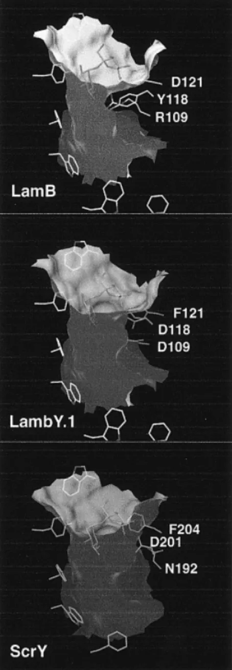
(2) P.Van Gelder et al.. Fig. 1. (a) Stereo view of the superposition of the crystal structures of LamB (Schirmer et al., 1995) and the ScrY–sucrose complex (carbon atoms shown in green) (Forst et al., 1998). Shown is a cross-section of the channel at the height of the channel constriction. The view is from the extracellular side. Part of the greasy slide can be seen at the lower left. Loop L3 extends at the top of the figure. Most of the residues of the channel lining are identical except those at site *1, R109 (LamB), N192 (ScrY, partly buried within L3); site *2, F118 (LamB), D201(ScrY); and site *3, D121 (LamB), F204 (ScrY). (b) Superposition of the crystal structures of LamB mutant LamBY.1 and ScrY (carbon atoms in green). The cyclic averaged electron density of the mutant (contoured at 1.5σ) is shown superimposed on the structures. The density within the channel has not been modelled and may represent a cation and a water molecule.. interact with their respective substrates in LamB and ScrY (Dutzler et al., 1996; Forst et al.; 1998). Recently, the above-mentioned residues have been replaced by their LamB equivalents in ScrY (Ulmke et al., 1999). The in vivo analysis showed that maltose transport was not significantly improved, while sucrose translocation was impaired. This study aimed to render LamB permeable for sucrose. The initial mutant design was based on the knowledge of the LamB structure and a LamB–ScrY sequence alignment resulting in the triple mutant LamBY.1 (R109D, Y118D, D121F). Structural superposition of the two wild-type proteins (Figure 1a) performed later when the ScrY structure became available revealed that ScrY residue N192 instead of D191 is the structural homologue of R109. Therefore, a second triple mutant (LamBY.2; R109N, Y118D, D121F) was generated. Finally, in a third mutant (LamB.RY; R109A, Y118A), two bulky residues that contribute substantially to the LamB channel constriction were pruned to alanines. Materials and methods Strains and plasmids All porins were expressed in E.coli strain BL21(DE3)omp5(∆lamB, ompR) (Prilipov et al., 1998). Construction of 944. plasmid pGLamB and pGOmpF, containing the pMB1 ori of replication and ampicillin resistance gene, was described previously (Prilipov et al., 1998). Site-directed mutagenesis of LamB was performed by the mutagenesis method as described (Prilipov et al., 1998), resulting in plasmid pGLamBY.1 (changes R109D, Y118D, D121F), pGLamBY.2 (changes R109N, Y118D, D121F) and pGLamB.RY (changes R109A, Y118A). Mutations were confirmed by DNA sequence analysis (PerkinElmer ABI prism, 310 genetic analyser). Plasmid pGScrY was constructed by cloning a PCR fragment containing the scrY gene from pCH186 (Hardesty et al., 1991) into SalI/BamHI-digested pGLamB. All DNA manipulations were performed according to Sambrook et al. (Sambrook et al., 1989). Protein purification, crystallization and crystallography Expression and purification of the proteins were performed as described for LamB (Prilipov et al., 1998). Crystallization of the LamBY.1 mutant was performed according to Keller et al. (Keller et al., 1994) with the protein concentration for crystallization raised from 6 to 8 mg/ml. Crystallization of mutant LamBY.2 (based on structural alignment with ScrY, when the structure became available) was not attempted, since it is functionally very similar to LamBY.1. Diffraction data were collected with a MARRESEARCH.
(3) Sugar transport through maltoporin of E.coli. Table I. Crystallographic data for the LamBY.1 mutant Number of crystals X-ray source Resolution range (Å) Unique reflections Rmerge Completeness (%) R (%) Rfree (%) R.m.s.d. bonds (Å) R.m.s.d. angles (°). 1 Rotating anode 20–3.2 43576 (3454) 13.1 (35.3) 88.5 (71.3) 19.3 (28.0) 21.8 (33.7) 0.016 2.2. The space group is C2221, with cell constants a ⫽ 129.8 Å, b ⫽ 211.9 Å and c ⫽ 217.3 Å. Values in parentheses are for the highest resolution shell (3.31–3.20 Å).. image plate at a rotating anode X-ray generator (Table I). Data were processed and scaled using the programs DENZO and SCALEPACK (Otwinowski, 1993). The crystal structure was solved by difference Fourier methods using the phases of native LamB. After rigid body refinement, cyclic averaging over the three monomers of the asymmetric unit was performed using the program DM (Cowtan and Main, 1996). Subsequently, the model was rebuilt using program O (Jones and Kjeldgaard,1993) and refined by torsion angle simulated annealing including a bulk solvent contribution using program XPLOR (Bru¨ nger, 1992). The pore cross-sections of the various channels were calculated using a 1⫻1⫻1 Å grid. The channel coordinate of LamB was approximated by the crystallographic z-axis (the molecular 3-fold axis forms an angle of 7° with the z-axis). For a given coordinate z, the number of grid points within the channel having a distance ⬎1.4 Å from the protein were counted. The channel of the double alanine mutant LamB.RY was modelled by truncation of the respective side chains of wild-type model. For the calculation of the cross-sectional area of ScrY, the model was first superimposed onto the LamB model. Current fluctuation analysis and liposome swelling assay Lipid bilayers were formed as described previously (Van Gelder et al., 2000). Current fluctuation measurements were performed to derive the kinetic parameters of sugar transport. In the absence of sugar the ion current through open channels fluctuates around an average value called the 1/f noise contribution. Upon addition of sugar, additional current fluctuations will be introduced which reflect the on and off rates of sugar binding. Assuming a two-state channel (sugar bound or unbound), the fluctuations around the average ionic current value yield the Lorentzian power spectrum (Nekolla et al., 1994; Andersen et al., 1995) which is given by Sω ⫽ S0/1⫹ (ω/ωc)2, where S0, the plateau value at zero frequency, is given by S0 ⫽ (4i2/koff2)K[M]/(1 ⫹ K[M])3, with i ⫽ current through a single channel, [M] ⫽ sugar concentration, koff ⫽ dissociation constant and K ⫽ equilibrium constant given by K ⫽ kon/koff. The corner frequency, ωc, the second parameter determining the Lorentzian, is the frequency at which the amplitude decays to 21S0. Fluctuation theory shows that this frequency is related to the inverse of the decay time and to the on and off rates of sugar binding (Nekolla et al., 1994; Andersen et al., 1995). The filtered output voltage was recorded on paper and also on a storage oscilloscope (LeCroy) equipped with a fast Fourier transform (FFT) module. FFT was performed with a rectangular window on the oscilloscope. Power spectrum densi-. ties were recorded with a resolution of 1 Hz from 1 to 5000 Hz and averaged 50–100 times. Prior to sugar addition, we recorded the background contribution (1/f) which was subtracted from the Lorentzian-type power spectrum. One disadvantage of this method is that productive binding of the solute cannot be distinguished from non-productive binding. The latter situation would involve binding and release of the solute from/to the same side of the channel without translocation of the solute. Liposome swelling assays with 2 µg of purified protein were performed as described (Luckey and Nikaido, 1980). Raffinose was used to determine the isotonic sugar concentration, which was typically between 60 and 100 mM. Liposome swelling monitors translocation directly. Since these experiments are performed at saturating sugar concentrations, the off rate towards the interior of the liposome is measured. When comparing translocation rates through different channels, it has to be considered that the individual proteoliposome preparations may differ in the size distribution of the vesicles and in the number of incorporated channels. Sugar transport assays LB medium supplemented with ampicillin (100 µg/ml) was inoculated with E.coli strain BL21(DE3)omp5 expressing the various maltoporin mutants. The bacteria were harvested at late log phase, collected by a quick spin, extensively washed in M9 medium and redissolved in M9 medium. For the transport assay, 10 µl of [14C]sucrose (ARC Chemical, specific activity 600 mCi/mmol) was added to 1.5 ml of the cell suspension and the final sugar concentration was adjusted to 1 µM with cold sucrose. At different time points after the addition of the sugar, 150 µl of the suspension were filtered through a glass microfibre filter (Whatman GF/C) and washed with 5 ml of M9 medium. The filters were dried for 10 min at 60°C and counted in a scintillation counter. The in vivo sugar uptake assay measures translocation rates directly, is performed at low (physiological) sugar concentration and, therefore, is sensitive to the on rate of the process. It has the advantage that the channels are obviously oriented unidirectionally. Results Crystal structure of the triple mutant LamBY.1 The crystal structure of LamBY.1 has been determined to 3.2 Å resolution by X-ray crystallography (Table I). The structure is virtually identical with that of wild-type LamB. Despite the moderate resolution of the data, the electron density at the pore constriction clearly accounts for the three side chain replacements (Figure 1b). The side chains of D109 and D118 are within H-bonding distance, and consequently one of the two residues has to be protonated. There is residual density found in the lumen of the channel that extends into the space occupied by Y118 in wild-type LamB. This density may be attributed to a cation, which would be stabilized by the adjacent carboxylates and a water molecule. Superposition of the wild-type structures of LamB and ScrY (overall root-mean-square deviation 1.3 Å) (Forst et al., 1998) shows that the loop L3 segment Y107QRH (LamB) corresponds to D189RDNF (ScrY), with the first and the last residues being structurally equivalent (Figure 1a). Thus, ScrY carries a oneresidue insertion which causes R109 to have no close equivalent [the Cα-position of R109 (LamB) has a distance of 5.0 and 1.4 Å to D191 and N192 of ScrY, respectively]. Accordingly, 945.
(4) P.Van Gelder et al.. Fig. 3. Cross-sectional area of the central part of the porin channels measured as a function of the channel axis coordinate z. Squares, LamB; triangles, LamBY.1; circles, ScrY; diamonds, LamB.RY.. Fig. 2. Comparison of the channel shapes (visualized by the molecular surfaces) of LamB, LamBY.1 and ScrY. The outer surface of the molecules has been clipped off. The extracellular entrance to the channels is at the top of the figures. Residues of the ‘greasy slide’ and of the channel constriction are shown as stick models. The sucrose molecule is placed at the position as identified in the LamB-sucrose complex (Wang et al., 1997). Figures 1 and 2 were generated with the program Dino (A.Philippsen, http://www.bioz.unibas.ch/~xray/dino).. residue D109 of the mutant, which protrudes into the channel lumen, is at a different position compared with D191 or N192 of ScrY (Figure 1b). Side chains D118 and F121 of the mutant, however, show conformations similar to their counterparts in ScrY. The distances between the centres of the equivalent side chains are 1.8 and 1.0 Å, respectively. In Figure 2, the channel shapes of the two wild-type proteins are compared with that of the mutant, and in Figure 3 the channel cross-sections are quantified. Compared with LamB, the pore cross-section of LamBY.1 is clearly increased. Replacement of D121 by the more bulky phenylalanine residue has no effect on the channel size, since the side chain is found remote from the constriction. At the constriction, the triple mutant pore is smaller than that of ScrY, mainly owing to the 946. protruding residue D109 which is stabilized by an H-bond to D118 (Figure 1b). The structure of the double alanine mutant has been modelled by simply truncating the relevant side chains (R109A, Y118A). Based on this, the cross-section of the mutant channel even exceeds that of ScrY (Figure 3). The reverse operation, replacement of ScrY porin residues by structurally equivalent LamB residues, has been performed recently (Ulmke et al., 1999). As analyzed in vivo, the ScrY mutants did not exhibit an improved apparent transport Km for maltose. Transport of sucrose, however, was considerably impeded, mainly owing to the D201Y mutation (which is the reverse of the Y118D mutation in LamB). The neutral effect of mutation N192R (which is the reverse of the R109D mutation in LamB) found by the authors may be due to a different side chain conformation which does not cause channel narrowing. Transport characteristics of the various channels Purified proteins were reconstituted into liposomes and the relative permeation rate of maltose and sucrose was determined by liposome swelling assay (Table II). Both wild-type channels, LamB and ScrY, show clear specificity for their respective substrates, with the permeation rates of sucrose through LamB and of maltose through ScrY at the detection limit as observed previously (Hardesty et al., 1991). Both triple mutants, LamBY.1 and LamBY.2, showed a considerable increase in the relative sucrose permeation rate, approaching 20–25% of the maltose rate (Table II). Through the double mutant LamBRY, sucrose permeation was even almost as efficient as maltose permeation. Also, the absolute swelling rates are drastically increased for sucrose, with the LamB.RY rate exceeding the ScrY rate (Table II). This demonstrates that the relative gain in sucrose permeation is not merely due to impaired maltose permeation, although translocation of maltose appears to be less efficient in the LamB mutants, probably owing to a slightly perturbed binding site at the channel constriction. In vivo transport of sucrose In vivo uptake of radiolabeled sucrose was measured to investigate the physiological relevance of the in vitro data. Since the ScrA and ScrB proteins that are required for sucrose transport across the cytoplasmic membrane and sucrose metabolism (Schmid et al., 1988) were not provided, the assay reflects sugar permeation through the porins into the.
(5) Sugar transport through maltoporin of E.coli. Table II. Thermodynamic and kinetic data for maltose and sucrose transport through LamB, LamB mutants, ScrY and OmpF Mutant. Sugar. kon (103 M–1 s–1). koff (103 s–1). LamB. Maltose Sucrose Maltose Sucrose Maltose Sucrose Maltose Sucrose Maltose Sucrose Maltose Sucrose. 270 ⫾ 77 1/f 40 ⫾ 26 1/f 130 ⫾ 25 1/f 110 ⫾ 32 120 ⫾ 43 870 ⫾ 148 650 ⫾ 135 1/f 1/f. 7.0 1/f 4.1 1/f 7.9 1/f 6.3 3.4 6.9 9.0 1/f 1/f. LamBY.1 LamBY.2 LamB.RY ScrY OmpF. ⫾ 1.5 ⫾ 0.4 ⫾ 0.7 ⫾ ⫾ ⫾ ⫾. 1.6 0.6 0.3 2.0. Kd (mM). d(A/Ai)/dt (min–1). 25 15a 105 – 62 – 57 28 8 13 – –. 0.234 0.006 0.124 0.031 0.098 0.020 0.171 0.133 0.004 0.093 ndb ndb. ⫾ ⫾ ⫾ ⫾ ⫾ ⫾ ⫾ ⫾ ⫾ ⫾. 0.086 0.008 0.012 0.005 0.015 0.007 0.006 0.004 0.008 0.025. φrel (%) 100 2.7 100 25 100 21 100 78 4.5 100 75c 100c. The rate constants kon and koff and the dissociation constant Kd ⫽ koff/kon are the means of at least three measurements and d(A/Ai)dt (with Ai ⫽ initial absorbance) and φrel values are the means of at least 10 measurements from at least three different proteoliposome preparations. aValue derived from the stability constant K⫽ 67 M–1 reported by Benz et al. (Benz et al., 1987). bNot determined. cValues taken from Nikaido and Rosenberg (Nikaido and Rosenberg, 1981) with sucrose permeation set to 100%.. Fig. 4. Uptake of [14C]sucrose into whole cells containing plasmids expressing the porins ScrY (open circles), LamB (crosses), LamBY.1 (closed circles), LamBY.2 (squares) and LamB.RY (diamonds). The concentration of sucrose in the medium was 1 µM.. periplasmic space. SDS–PAGE analysis showed that the various porins were equally well expressed (data not shown). As expected, the bacteria expressing wild-type LamB showed no sucrose uptake (Figure 4). In contrast, all three LamB mutants exhibited significant sucrose uptake, with the double mutant LamB.RY performing best and almost approaching the efficiency of the bacteria expressing wild-type ScrY. Determination of kinetic parameters and binding affinities Ion flow through LamB and ScrY is blocked upon sugar binding. Based on this, apparent on and off rates (kon, koff) of sugar binding can be derived by analysis of sugar-induced ion current noise (Nekolla et al., 1994). The kinetic constants and the derived dissociation constants (Kd ⫽ koff/kon) of maltose and sucrose binding to the various porins are summarized in Table II. For wild-type LamB and the two triple mutants, the noise spectrum did not change upon addition of sucrose and no kinetic data could be derived (see Discussion). The same was observed in a control experiment with the non-specific OmpF porin. Discussion The outer membrane of Gram-negative bacteria contains several channel-forming proteins which allow the passage of solutes by passive diffusion. Most of them are non-specific and exclude solutes by size, whereas others contain a binding. site for a particular compound. In this study, two such proteins were investigated, the maltooligosaccharide-specific LamB and the sucrose-specific ScrY porin. To confer sucrose specificity to LamB, two triple mutants were designed based on sequence and structural comparisons between LamB and ScrY, while the design of the double alanine mutant aimed for a channel with a large cross-section. In agreement with earlier results (Luckey and Nikaido, 1980; Ulmke et al., 1999), both translocation assays demonstrate that there is virtually no sucrose permeation through LamB. It has been demonstrated by X-ray structure analysis (Wang et al., 1997) that the fructose moiety of sucrose is indeed too large to translocate through the LamB channel, whereas the glucose moiety inserts partly into the channel constriction and blocks the channel. The affinity constant of sucrose to LamB was measured to be 67 M–1 by sugar titration (Benz et al., 1987). Sugar-induced noise analysis (Table II), however, did not reveal a change in the noise spectrum upon addition of the sugar. It appears likely that association/dissociation with and from the binding site, which is well accessible from the extracellular side, is too fast to be resolved by the instrumentation. Andersen et al. (1995, 1998) reported (very slow) rates for this process, which may reflect permeation events. It is not clear, however, how they were able to reveal sucrose-induced noise, while the sensitivity of their measurements was insufficient to measure maltose or even maltotriose rates. The noise spectrum of the ion current through mutants LamBY.1 and LamBY.2 was not changed upon addition of sucrose. This may have the same reason as in the case above, i.e. that the kinetics are too fast to be resolved or may be due to absence of a binding site. The former possibility is contradicted by the significant permeation (Table II, Figure 4) measured in both translocation assays. The latter interpretation is supported by the fact that both R109 and D121, which form H-bonds with sucrose in the respective LamB complex (Wang et al., 1997) and have been replaced in the mutants, are not available for sugar binding (Figure 2a). Also, the putative cation bound to D118 may hinder insertion of the glucose moiety into the constriction. Moreover, the newly introduced D109 may well interfere with binding, since it protrudes into the lumen, whereas the corresponding ScrY residue N192 is interacting with loop L3 and is not obstructing the channel. 947.
(6) P.Van Gelder et al.. Furthermore, D118 is extending more into the channel lumen than D201 of ScrY owing to a shift in the backbone. We propose that for steric reasons (obstruction by residues 109 and 118), the sugar has to tilt and its glucosyl moiety cannot stay in contact with the greasy slide, thus explaining the absence of defined kinetic constants in the current noise measurements. Hence the triple mutants behave more like general diffusion channels. Also in the case of the non-specific diffusion transport of maltose through OmpF no defined binding kinetics could be determined (Table II). These results nicely show the importance of the greasy slide as a key component of the binding site and are in line with recent results obtained by mutagenesis of greasy slide residues (unpublished data). It also demonstrates that the binding site for a non-permeable sugar such as sucrose is still present (see also crystal structure of the wild-type LamB-sucrose complex) (Wang et al., 1997) and suggests that discrimination of solute transport is mainly based determined by the steric barrier at the constriction site. If it were true that residues 109 and 118 of the triple mutants are interfering with sucrose binding, their truncation should result in free access to the greasy slide. Indeed, truncation of these residues to alanine in mutant LamB.RY appears to restore a binding site for sucrose with well-defined kinetics (Table II). The channel binds sucrose and maltose with comparable affinity, probably mainly via interactions between the glucose moiety and the channel lining. The dissociation constants are larger than the corresponding wild-type LamB values by only a factor of ~2 (Table II). Under liposome swelling conditions, the mutations render the channel about equally permeable to maltose and sucrose. The in vivo assay reveals an uptake rate as high as ~75% of the rate through ScrY. In conclusion, the results show that wild-type LamB is impermeable to sucrose owing to steric hindrance at the channel constriction as directly revealed by the crystal structure of the complex (Wang et al., 1997). In the triple mutants, this block is partly removed, but association of the sugar with the channel is still impeded, as evidenced by the absence of defined binding kinetics. However, in the truncation mutant, all obstacles are cleared and sucrose is transported almost as efficiently as maltose. Binding kinetics are now observed, most probably reflecting association with the greasy slide. For efficient transport of solutes comprised (partly) of glucosyl residues, unimpaired interaction with the greasy slide appears to be crucial, whereas the lack of ionizable residues may be tolerated in certain instances (the translocation efficiency of maltose through the truncation mutant is almost wild-type like). In most cases, however, mutagenesis of ionic track residues severely impedes the efficiency of the channel (Dumas et al., 2000). In this context, the poor efficiency of ScrY for maltose (Table II) is intriguing. Probably, owing to a non-optimal arrangement, the polar ScrY–maltose interactions are not strong enough to promote shedding of the sugar hydration shell. An analogous mechanism has been proposed for ion channels to explain ion selectivity (Doyle et al., 1998). Acknowledgements We thank J.DiRienzo for providing plasmid pCH186, I.Bartoldus for the experiments with OmpF, A.Prilipov for construction of plasmid pGLamBY.1 and M.Winterhalter for technical support with the noise measurements. This work was sponsored by grant No. 31-53727.98 from the Swiss National. 948. Science Foundation to T.S. R.K was supported by grants Ko1686/3-1 and /32 from the Deutsche Forschungsgemeinschaft.. References Andersen,C., Jordy,M. and Benz,R. (1995) J. Gen. Physiol., 105, 385–401. Andersen,C., Cseh,R., Schu¨ lein,K. and Benz,R. (1998) J. Membr. Biol., 164, 263–274. Benz,R., Schmid,A. and Vos-Scheperkeuter,G.H. (1987) J. Membr. Biol., 100, 21–29. Bru¨ nger,A.T. (1992) X-plor Version 3.1. A System for X-ray Crystallography and NMR. Yale University Press, New Haven, CT. Cowtan,K.D. and Main,P. (1996) Acta Crystallogr., D52, 43–48. Doyle,D.A., Cabral,J.M., Pfuetzner,R.A., Kuo,A., Gulbis,J.M., Cohen,S.L., Chait,B.T. and MacKinnon,R. (1998) Science, 280, 69–77. Dumas,F., Koebnik,R., Winterhalter,M. and Van Gelder,P. (2000). J. Biol. Chem., 275, 19747–19751. Dutzler,R., Wang,Y.-F., Rizkallah,P.J., Rosenbusch,J.P. and Schirmer,T. (1996) Structure, 4, 127–134. Forst,D., Welte,W., Wacker,T. and Diederichs,D. (1998) Nature Struct. Biol., 5, 37–46. Hardesty,C., Ferran,C. and DiRienzo,J.M. (1991) J. Bacteriol., 173, 449–456. Jones,T.A. and Kjeldgaard,M. (1993) O Version 5.9, The Manual. Uppsala University, Uppsala. Keller,T.A., Ferenci,T., Prilipov,A. and Rosenbusch,J.P. (1994) Biochem. Biophys. Res. Commun., 199, 767–771. Koebnik,R., Locher,K.P. and Van Gelder,P. (2000) Mol. Microbiol., 37, 239–253. Luckey,M. and Nikaido,H. (1980) Proc. Natl Acad. Sci. USA, 77, 165–171. Nekolla,S., Andersen,C. and Benz,R. (1994) Biophys. J., 66, 1388–1397. Nikaido,H. and Rosenberg,E.Y. (1981) J. Gen. Physiol., 77, 121–135. Nikaido,H. and Vaara,M. (1985) Microbiol. Rev., 49, 1–32. Otwinowski,Z. (1993) In Sawyer,L. and Bailey,S. (eds), Data Collection and Processing. Science and Engineering Research Council Daresbury Laboratory, Daresbury, pp. 56–62. Prilipov,A., Phale,P.S., Van Gelder,P., Rosenbusch,J.P. and Koebnik,R. (1998) FEMS Microbiol. Lett., 163, 65–72. Randall-Hazelbauer,L. and Schwartz,M. (1973) J. Bacteriol., 116, 1436–1446. Sambrook,J., Fritsch,E.F. and Maniatis,T. (1989) Molecular Cloning. A Laboratory Manual. 2nd edn. Cold Spring Harbor Laboratory Press, Cold Spring Harbor, NY. Schirmer,T., Keller,T.A., Wang,Y.-F. and Rosenbusch,J.P. (1995) Science, 267, 512–514. Schmid, K, Ebner,R., Altenbuchner,J., Schmitt,R. and Lengeler,J.W. (1988) Mol. Microbiol., 2, 1–8. Schmid,K., Ebner,R., Jahreis,K., Lengeler,J.W. and Titgemeyer,J. (1991) Mol. Microbiol., 5, 941–950. Schu¨ lein,K., Schmid,K. and Benzl,R. (1991) Mol. Microbiol., 5, 2233–2241. Schu¨ lein,K., Andersen,C. and Benz,R. (1995) Mol. Microbiol., 17, 757–767. Szmelcman,S. and Hofnung,M. (1975) J. Bacteriol., 124, 112–118. Szmelcman,S., Schwartz,M., Silhavy,T.J. and Boos,W. (1976) Eur. J. Biochem., 65, 13–19. Ulmke,C., Kreth,J., Lengeler,J.W., Welte,W. and Schmid,T. (1999) J. Bacteriol., 181, 1920–1923. Van Gelder,P., Dumas,F., Rosenbusch,J.P. and Winterhalter,M. (2000) Eur. J. Biochem., 267, 79–84. Wang,Y.-F., Dutzler,R., Rizkallah,P.J., Rosenbusch,J.P. and Schirmer,T. (1997) J. Mol. Biol., 272, 56–63. Received March 16, 2001; revised August 2, 2001; accepted August 9, 2001.
(7)
Figure

![Fig. 4. Uptake of [ 14 C]sucrose into whole cells containing plasmids expressing the porins ScrY (open circles), LamB (crosses), LamBY.1 (closed circles), LamBY.2 (squares) and LamB.RY (diamonds)](https://thumb-eu.123doks.com/thumbv2/123doknet/14901133.653689/5.918.67.848.113.329/uptake-sucrose-containing-plasmids-expressing-circles-crosses-diamonds.webp)
Documents relatifs
(A) Etude de l’activité transcriptionnelle des différents mutants de RCXMyT1 : Tous les mutants continuent à réprimer l'activité luciférase par rapport à l'activité
Interesting segregation for amylose content (ranging from 19 to 42%) was found among different small granule genotypes, suggesting that there may actually be two
For all varieties, nodal cuttings were induced to produce nodes and microtubers on modified MS medium supplemented with various concentrations of sucrose (3, 5 and 8 % w/v). In a
Cette quatorzième édition fournit des informations sur l’activité partielle (ou chômage partiel), les restructurations, les inscriptions à Pôle emploi, les entrées en formation
We started with a full model including the extrinsic var- iables {standing biomass of the grassland, soil disturbance (yes or no), propagule pressure [continuous (log-transformed):
To demonstrate how the “traditional” and mutant classification approaches work consider the following scenario. We have a program that has been tested by three test cases named
We demonstrated using an in vitro pharmacodynamic model that the likelihood of selection of Escherichia coli resistant mutants to a fluoroquinolone was increased when the initial
En effet, le xyloglucane ayant un rôle prépondérant dans la cohésion des microfibrilles de cellulose entre elles, les enzymes impliquées dans son métabolisme in muro sont des
