HAL Id: hal-03079312
https://hal.archives-ouvertes.fr/hal-03079312
Submitted on 17 Dec 2020
HAL is a multi-disciplinary open access
archive for the deposit and dissemination of
sci-entific research documents, whether they are
pub-lished or not. The documents may come from
teaching and research institutions in France or
abroad, or from public or private research centers.
L’archive ouverte pluridisciplinaire HAL, est
destinée au dépôt et à la diffusion de documents
scientifiques de niveau recherche, publiés ou non,
émanant des établissements d’enseignement et de
recherche français ou étrangers, des laboratoires
publics ou privés.
and ciliogenesis but not for Wg signaling
Camille Enjolras, Joëlle Thomas, Brigitte Chhin, Elisabeth Cortier, Jean-Luc
Duteyrat, Fabien Soulavie, Maurice Kernan, Anne Laurençon, Bénédicte
Durand
To cite this version:
Camille Enjolras, Joëlle Thomas, Brigitte Chhin, Elisabeth Cortier, Jean-Luc Duteyrat, et al..
Drosophila chibby is required for basal body formation and ciliogenesis but not for Wg signaling.
Jour-nal of Cell Biology, Rockefeller University Press, 2012, 197 (2), pp.313-325. �10.1083/jcb.201109148�.
�hal-03079312�
The Rockefeller University Press $30.00
Correspondence to Bénédicte Durand: [email protected]
Abbreviations used in this paper: CO, chordotonal organ; ES, external sensory; IFT, intraflagellar transport; MKS, Meckel syndrome; NPHP, nephronophthisis; PLP, pericentrin-like protein; PNS, peripheral nervous system; SEP, sound-evoked potential; TZ, transition zone; UTR, untranslated region.
Introduction
The structure and protein composition of cilia are highly
con-served across eukaryotes. A cilium is built on a basal body
derived from the mother centriole. Centriole maturation into a
basal body is a complex process that includes the progressive
addition of different protein structures including transition
fibers, basal feet, and ciliary rootlets (Hoyer-Fender, 2010;
Ishikawa and Marshall, 2011). The prototypical centriole found
in most organisms is a radially symmetric arrangement of nine
microtubule triplets. However, Ecdysozoa such as Caenorhabditis
elegans
and Drosophila melanogaster show major differences
in the ultrastructure of centriole/basal bodies. In C. elegans,
the centriole is composed of singlet microtubules, not triplets.
In Drosophila, centrioles in embryos and most tissues are
composed of microtubule doublets. In spermatocytes of the
Drosophila
male germ line, centrioles do show the prototypical
nine triplets but lack some of the maturation structures found
in other eukaryotes.
Ciliogenesis starts with the docking of the basal body,
either directly to the membrane at the cell surface or to
cyto-solic vesicles that will eventually fuse to the plasma membrane
(Sorokin, 1968). An initial extension of microtubule doublets
from the basal body constitutes a specialized transition zone
(TZ) delineated by specific localized proteins and by
struc-tures such as transition fibers, which link the basal body to
the membrane and form a selective barrier to transport into
the cilium (Ishikawa and Marshall, 2011). Further extension
of the axoneme and ciliary membrane above the cell surface
requires intraflagellar transport (IFT), in which specialized
kinesin and dynein motors move an IFT protein complex and
associated cargoes from the base to the tip of the cilium and
back (Rosenbaum and Witman, 2002; Ishikawa and Marshall,
2011). Two notable exceptions to the IFT mode of assembly
are the flagella of Plasmodium and of Drosophila sperm (Han
et al., 2003; Sarpal et al., 2003; Briggs et al., 2004). Drosophila
sperm axonemes are first fully extended in a cytoplasmic
syncy-tium, independent of IFT, and only then ensheathed by a plasma
C
entriole-to–basal body conversion, a complex
process essential for ciliogenesis, involves the
progressive addition of specific proteins to
cen-trioles. CHIBBY (CBY) is a coiled-coil domain protein first
described as interacting with -catenin and involved in
Wg-Int (WNT) signaling. We found that, in Drosophila
melanogaster, CBY was exclusively expressed in cells
that require functional basal bodies, i.e., sensory
neu-rons and male germ cells. CBY was associated with the
basal body transition zone (TZ) in these two cell types.
Inactivation of cby led to defects in sensory transduction
and in spermatogenesis. Loss of CBY resulted in altered
ciliary trafficking into neuronal cilia, irregular
deposi-tion of proteins on spermatocyte basal bodies, and,
con-sequently, distorted axonemal assembly. Importantly,
cby
1/1flies did not show Wingless signaling defects.
Hence, CBY is essential for normal basal body structure
and function in Drosophila, potentially through effects
on the TZ. The function of CBY in WNT signaling in
vertebrates has either been acquired during vertebrate
evolution or lost in Drosophila.
Drosophila chibby is required for basal body
formation and ciliogenesis but not for Wg signaling
Camille Enjolras,
1Joëlle Thomas,
1Brigitte Chhin,
1Elisabeth Cortier,
1Jean-Luc Duteyrat,
1Fabien Soulavie,
1Maurice J. Kernan,
2,3Anne Laurençon,
1and Bénédicte Durand
11Centre de Génétique et de Physiologie Moléculaire et Cellulaire, Centre National de la Recherche Scientifique UMR 5534, Université Claude Bernard Lyon 1,
Villeurbanne, Lyon F69622, France
2Department of Neurobiology and Behavior and 3Center for Developmental Genetics, Stony Brook University, Stony Brook, NY 11794
© 2012 Enjolras et al. This article is distributed under the terms of an Attribution– Noncommercial–Share Alike–No Mirror Sites license for the first six months after the pub-lication date (see http://www.rupress.org/terms). After six months it is available under a Creative Commons License (Attribution–Noncommercial–Share Alike 3.0 Unported license, as described at http://creativecommons.org/licenses/by-nc-sa/3.0/).
THE
JOURNAL
OF
CELL
BIOLOGY
on February 6, 2017
Downloaded from
/content/suppl/2012/04/12/jcb.201109148.DC1.html Supplemental Material can be found at:with one notable exception: the CHIBBY (CBY) protein. CBY
was first identified as an interactor with -catenin in humans,
and RNAi experiments targeting cby expression implied that
CBY negatively regulates WNT signaling both in mammalian
cell culture and in Drosophila (Takemaru et al., 2003).
Iden-tified as PIGEA-14, CBY was also shown to be associated
with polycystin-2, a ciliary ion channel (Hidaka et al., 2004).
Last, cby
/mutant mice showed defective airway function
associated with impaired basal body docking, reduced number
of motile cilia (Voronina et al., 2009; Love et al., 2010), and
increased expression of -catenin regulatory targets (Love
et al., 2010).
We have identified Drosophila cby as a putative target
of the RFX transcription factor, which activates transcription
of many genes required for the assembly of cilia (Dubruille
et al., 2002; Laurençon et al., 2007). Together, these observations
suggested a possible link between Wg signaling components
and cilia in Drosophila. We show here that Drosophila cby is
expressed only in cells that harbor cilia, namely in ciliated type I
sensory neurons and in male germ cells. CBY protein is
associ-ated with the maturing basal bodies and TZ in these two cell
types. cby-null mutant flies show sensory transduction defects
and male hypofertility but no evidence of Wg signaling defects.
We show that CBY is required in ciliated neurons for proper
TZ and axonemal assembly and for protein trafficking into or
membrane, during spermatid individualization (Fuller, 1993).
However, a form of centriole maturation does occur earlier
in Drosophila spermatogenesis: in primary spermatocytes, all
four centrioles extend a distal zone formed of nine doublets
and ensheathed by a membrane cap. The elongated centrioles
migrate to the plasma membrane, where the distal segment
protrudes from the cell surface and hence resembles a short
cilium (Tates, 1971; Fuller, 1993). The function of this
tran-sient primary cilium is unknown. unc mutants, in which it is
shortened, can proceed through an apparently normal meiosis
(Baker et al., 2004). At the onset of meiosis I, the pairs of
elongated centrioles are engulfed together with their
mem-brane caps and relocalized to the meiotic spindle poles (Tates,
1971). These observations strongly suggest that basal body
and TZ-like structures assemble in Drosophila spermatocytes
before meiosis and flagellar elongation.
In vertebrates, cilia are required during development to
mediate signal transduction pathways such as the sonic
hedge-hog pathway (Goetz and Anderson, 2010). Defects in cilia
or cilia-associated proteins are also associated with defective
Wg-Int (WNT) and planar cell polarity signaling, although some
of the data are contradictory, and the precise mechanism is
far from understood (Ajima and Hamada, 2011; Wallingford and
Mitchell, 2011). To date, reports of an association between
cilia and Wingless (Wg) signaling are limited to vertebrates,
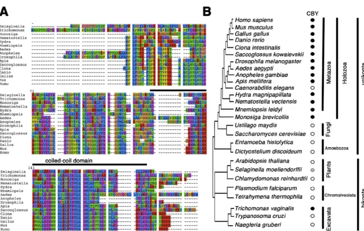
Figure 1. CBY conservation in animals. (A) CBY is a small protein with a conserved coiled-coil domain close to the C terminus, highly conserved in animals (black bar). Each of the eight classes of amino acids is represented by one color. (B) Orthologs of CBY are found in animals with motile cilia (black filled circles) and in the closest unicellular relatives of animal, the marine choanoflagellate Monosiga brevicollis, but not in nematode genomes or in most unicel-lular organisms (open circles). A probable homolog is found in the parabasalid protist Trichomonas, and a more distant relative is found in the club moss
S. moellendorffii (gray circle). CBY is not found in most bikonts (open circles).
on February 6, 2017
constructed two transgenes including the cby promoter and
entire coding sequence, fused at the C terminus to GFP or to
Tomato (see Materials and methods). In transgenic embryos,
expression of both constructs was restricted to type I sensory
neurons of the peripheral nervous system (PNS), the only
cili-ated cells in the embryo. The fusion protein was found in the
cell body and as a dot at the tip of the sensory process in all
type I sensory neurons (Fig. 2 A). No expression was detectable
in any other parts of the embryo (unpublished data). In an Rfx
mutant background, cby expression was lost, confirming that in
the PNS, cby is regulated by RFX (Fig. 2 A).
By coimmunolabeling with the centriole/basal body
mark-ers Asterless (ASL), pericentrin-like protein (PLP), and SAS-4,
CBY was located more precisely at the tip of the basal body to
a region that likely corresponds to the TZ between the basal
body and the cilium proper (Fig. 2 B). To confirm this
hypothe-sis, we looked at CBY location relative to B9D1/Meckel
syn-drome (MKS) 1–related (MKSR1), one of three interdependent
B9 domain proteins involved in assembling the TZ (Bialas
et al., 2009; Williams et al., 2011). Its Drosophila ortholog is
encoded by CG14870 and is expressed in ciliated cells
(Avidor-Reiss et al., 2004). We made a CG14870-MYC reporter
con-struct, with the promoter and complete coding sequences fused
to a MYC epitope, and found that CBY-Tomato and
CG14870-MYC were indeed colocalized at the distal end of the basal body
(Fig. 2 B). Hence, CBY is associated with the TZ of Drosophila
sensory cilia. UNC, a basal body–associated protein required
for axoneme structural integrity (Baker et al., 2004), also
co-localized with CBY at the distal end of basal bodies in type I
sensory neurons (Fig. 2 B).
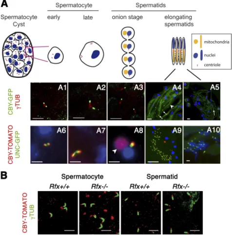
cby
was also expressed in larval and adult testis. CBY was
associated with the duplicated elongating centrioles in G2
sper-matocytes and with basal bodies in early spermatids (Fig. 3,
A1–3 and 6–8). No cby expression was found associated with
out of the cilia. In spermatogenesis, CBY is also required for
centriole-to–basal body conversion, as revealed by defective
basal body–associated uncoordinated (UNC) protein
deposi-tion. Last, we show that cby is required for axonemal
assem-bly during spermiogenesis. Altogether our results imply that,
in Drosophila, CBY functions in basal body/TZ biogenesis but
not in WNT signaling.
Results
The RFX target gene cby is expressed in Drosophila ciliated cells, and CBY is associated with maturing basal bodies
CBY, a small protein with a C-terminal coiled-coil domain,
is found in all ciliated unikont animals, with the exception
of C. elegans and other nematodes (Fig. 1). CBY is also
found in choanoflagellates, which can have a colonial life
and are closely related to animals (King et al., 2008). Two
prob-able CBY homologs are also present in the Excavate protist
Trichomonas vaginalis
and two possibly related proteins in
the lower plant Selaginella moellendorffii (which has
flagel-lated zoospores). Their occurrence suggests that a CBY homolog
was present in the universal ancestral eukaryote. However,
no CBY-related protein could be identified in any other
bikont genomes, including well-studied ciliated organisms
such as Chlamydomonas reinhardtii, Trypanosoma, or
Parame-cium
. The Excavates have been proposed to stem from a very
deep branching of the eukaryotic tree but with a closer
associa-tion with bikonts than with unikonts (Hampl et al., 2009); the
Trichomonas cby
homolog instead favors a closer association
with the unikonts.
Drosophila cby
was identified in a screen for potential
regulatory targets of the transcription factor RFX (Laurençon
et al., 2007). To analyze cby expression in Drosophila, we
Figure 2. cby is expressed in ciliated neurons of the Drosophila PNS. (A) Transgenic flies expressing cby coding sequences fused to GFP under cby regulatory sequences were analyzed for GFP expression. GFP expression is detected only in type I ciliated cells of the PNS in Rfx+/+ flies (left). GFP staining is observed in
the cytosol and mainly as a dot at the tip of the dendrite. The PNS is labeled (red) with an anti-Futsch antibody (22C10). In
Rfx/ flies, the cby reporter construct is not expressed (right).
(B) Double labeling of a transgenic fly strain expressing cby coding sequences fused to the fluorescent reporters GFP or Tomato under cby regulatory sequences with different markers of the centriole. CBY is localized at the tip of the centriole/basal body, as observed with ASL, PLP, SAS4, UNC fused to GFP, and CG14870 fused to a MYC epitope. CBY colocalizes with the TZ marker CG14870.
on February 6, 2017
cby is required for normal behavior, mechanosensation, and male fertility but not for Wg signaling
To understand the precise function of CBY, we constructed a
null mutation, cby
1, using homologous recombination (Maggert
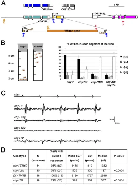
et al., 2008) to delete most of the cby coding region (Fig. 4 A).
PCR performed with primers designed on Fig. 4 A show that
recombination occurred correctly on the left and right arms of
the construct and that cby sequences are deleted in the cby
1allele (
Fig. S1
). In addition, we sequenced the entire
recombi-nant region and confirmed that coding sequences of exon 2 are
removed without affecting the adjacent genes.
RT-PCR on mRNA extracted from cby
1/1flies detected
no cby mRNA (Fig. S1), whereas expression of the two
adja-cent genes was not affected. In addition, cby
1/1homozygotes
and heterozygotes for cby
1and Df(3R)BSC805, a complete
de-ficiency of the region, showed similar survival, overall
behav-ior, and fertility (see the following three paragraphs; Figs. 4
and S1), confirming that cby
1is a null allele.
Homozygous cby
1/1and cby
1/Df(3R)BSC805 mutants were
viable, but their behavior was altered, and males appeared to
be hypofertile (Figs. 4 and S1). Wild-type flies show negative
geotaxis; they move upward on vertical surfaces. This can be
assayed with a simple climb test in which the percentages of
flies that climb to different heights in a tube are measured after
tapping them to the bottom. Whereas most control flies reached
and stayed at the top of the tube in the first minute, most cby
1/1or cby
1/Df(3R)BSC805
flies stayed at the bottom of the tube
centrioles in dividing spermatogonia before the spermatocyte
stage (unpublished data). In contrast to sensory neurons, cby
expression in the testis does not require RFX (Fig. 3 B),
consis-tent with RFX expression in spermatids only after the onset of
cby
expression (Vandaele et al., 2001).
During spermatid elongation, CBY was not maintained at
the basal body but was observed at the tip of the growing
axo-neme until late spermatid differentiation (Fig. 3, A4, 5, 9, and 10).
CBY was also found transiently associated with the
mitochon-dria in onion-stage spermatids and early elongating spermatids
(white arrowheads in Fig. 3, A8 and A4).
UNC is also located at both the basal body and growing
tip of the axoneme (Baker et al., 2004; Wei et al., 2008). We
observed that CBY-Tomato and UNC-GFP appeared together
at the tip of the elongating centrioles in early G2
spermato-cytes and remained there during spermatocyte maturation to
spermatids, as the centrioles converted to basal bodies, with
CBY localized in a zone that overlaps and extends more distal
to UNC (Fig. 3, A6–8). However, during axoneme elongation,
when CBY and UNC were both found at the tip of the growing
axoneme, only UNC was maintained at the basal body (Fig. 3,
A9 and 10).
Our observations show that CBY is localized at the distal
end of maturing basal bodies in Drosophila. Interestingly, this
localization is conserved in mammals in which CBY was found
at the tip of the basal body of primary cilia in inner medullary
collecting duct 3 (IMCD3) cells and above basal bodies of
mo-tile cilia in primary ependymal cells (
Fig. S3
).
Figure 3. cby is expressed during spermatogenesis. (A) The diagram shows the cycle of the centriole dur-ing spermatogenesis. In G2 spermatocytes, the two pairs of replicated centrioles elongate and reach the membrane before meiosis entry, and single centri-oles are distributed to each of the haploid sperma-tids after meiosis. (A1–3) CBY-GFP is localized at the tip of the elongating centrioles labeled for -tubulin (TUB) from early G2 spermatocytes to early sperma-tids. (A4 and 5) As sperm axoneme elongates, CBY is found at the tip of the axoneme and lost from the basal body (white arrows). Note that some CBY-GFP is found associated with mitochondria during sper-matid elongation (white arrowhead). (A6–10) CBY-TOMATO localization in live squashes of Drosophila testis expressing UNC-GFP. (A6–8) CBY is localized at the tip of the elongating centriole adjacent to the UNC-GFP staining from spermatocytes to early sper-matid stage. CBY-TOMATO is transiently associated with the mitochondria (arrowhead). (A9 and 10) In elongating spermatids, CBY-TOMATO is associated with UNC-GFP only at the tip of the axoneme (A9) and is no longer present with UNC-GFP at the basal body apposed to the nuclei at the spermatid heads (A10). (A4–10) Nuclei are labeled with Hoechst. (A1 and 6) Early G2 spermatocytes. (A2 and 7) Late G2 spermatocytes. (A3 and 8) Round spermatids. (A4, 5, 9, and 10) Elongating spermatids. (B) CBY-TOMATO expression is similar in Rfx+/+ and Rfx/
spermatocytes and spermatids. Centrioles are labeled with -tubulin antibody. Bars, 5 µm.
on February 6, 2017
Boekhoff-Falk, 2007). For each antenna, we measured the
maxi-mum amplitude of the averaged responses to 10 standard stimuli
that mimic the pulse phase of courtship song. The antennal
re-sponse amplitude in cby
1/1and in cby
1/Df(3R)BSC805 was strongly
reduced compared with controls (Fig. 4 C). Further, whereas 95%
of the control antennae showed a pulsatile response, many of the
cby
1/1and cby
1/Df(3R)BSC805 antennae lacked any response.
These observations lead us to conclude that cby
1mutants are
par-tially defective in chordotonal-mediated mechanotransduction.
Because CBY was initially described as a negative
regu-lator of Wg signaling in Drosophila (Takemaru et al., 2003),
we carefully checked cby
1/1flies for phenotypes associated with
loss or gain of Wg signaling. We did not observe any
expan-sion of engrailed expresexpan-sion domains (n = 21) nor any cuticular
defects (n = 119) in offspring embryos from homozygous cby
1/1male and female parents, which lack any parental or zygotic
contributions (
Fig. S2
). Because Wg gain-of-function
signal-ing leads to characteristic bristle phenotypes on the wsignal-ing and
the notum (Treisman et al., 1997), we also looked carefully at
bristle number and distribution in cby
1/1adult flies (n = 31) but
found no significant alterations (Fig. S2 D). Together, these
(Fig. 4 B). This phenotype was rescued by one copy of the
CBY-Tomato transgene, confirming that CBY is required for
negative geotaxis.
This change in geotaxis behavior could be a result either
of a defect in gravity perception or locomotor coordination or
both. Gravity is mainly sensed in Drosophila by antennal
chor-dotonal organs (COs; Kamikouchi et al., 2009; Sun et al., 2009),
whereas general locomotor coordination relies principally on
external mechanosensory organs, with some contributions by
COs. The cby
1/1behavioral phenotype was milder than the one
observed in atonal mutants, which lack all COs (Jarman et al.,
1993), or in transient receptor potential vanilloid channel or tilB
mutants, which lack CO function (Eberl et al., 2000; Kavlie et al.,
2010), and was much less severe than the extreme
uncoor-dination seen in flies that lack mechanotransduction in bristles
(Kernan et al., 1994) or lack all cilia (Sas-4 or Rfx mutant flies;
Dubruille et al., 2002; Basto et al., 2006). This indicates that
mechanosensation is not completely abolished in cby
1/1flies.
We confirmed a defect in mechanosensation by recording
antennal sound-evoked potentials (SEPs), which depend on
mech-anotransduction in the antennal CO (Eberl et al., 2000; Eberl and
Figure 4. Inactivation of cby leads to defective be-havior and mechanosensation. (A) Diagram of the
cby genomic locus. The second coding exon was
re-placed by homologous recombination with a miniwhite gene flanked by two LoxP sites. The primers designed to screen for the recombinant events are indicated in yellow for the left arm and red for the right arm. (B) Diagram representing the bang assay to evaluate fly coordination. The number of flies that reach a de-fined level in the tube is counted 1 min after tapping the tube. cby1/1 flies and cby1/Df(3R)BSC805 (cby1/Df)
stay at the bottom of the tube compared with control flies. The phenotype is rescued by adding one copy of the cby-Tomato transgene. n = 30 flies per assay. Error bars represent the SD of five assays. (C) Anten-nal nerve potentials recorded from age-matched sib
cby/TM6C heterozygotes (cby/+), cby homozygotes
(cby/cby), and from Df(3R)BSC805/TM6B (Df/TM6B) and cby/Df(3R)BSC805 (cby/Df) sibs in response to a five-pulse sound stimulus (stim). Each trace represents the averaged responses to 10 stimuli. Traces shown are those closest to the mean values for their respective genotypes. The third trace is representative of mutant recordings that do not show discernible SEP peaks. (D) Summary of sound-evoked nerve potential ampli-tudes. Mean and median peak amplitudes of the anten-nal SEPs for each indicated genotype. Mean values are derived only from those recordings showing discernible peaks; medians are from all recordings.
on February 6, 2017
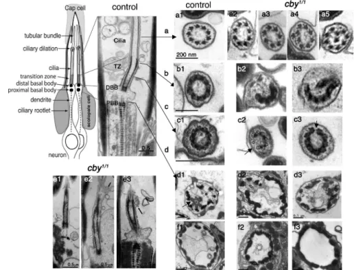
(22%; Fig. 5, d2 and 3). At the level of the TZ, which we believe
to correspond to the Y link region (Fig. 5, cross-sections b and c),
we could observe either completely interrupted symmetry (Fig. 5,
b2 and 3) or more subtle discontinuities in the microtubule-
associated structures of the TZ (black arrows in Fig. 5, c2
and 3). Discontinuities in the TZ could also be observed on
longitudinal sections (white arrows in Fig. 5, e1 and 2) in
addi-tion to membrane bulges (black arrows in Fig. 5, e2 and 3)
that were never observed in controls. Together, these results
show that cby is required for correct axoneme assembly in
Drosophila
and that its absence leads to improper TZ formation.
As we never observed missing basal bodies by -tubulin
staining of ciliated neurons (unpublished data), the EM
ob-servations also suggest that basal bodies, though present in
all neurons, are not positioned properly at the base of the cilia
in some neurons.
Because ultrastructural defects were incompletely
pen-etrant in cby
1/1flies, we hypothesized that cby has a more
subtle role in cilia biogenesis, as described for TZ proteins in
other organisms (Ishikawa and Marshall, 2011). In C. elegans,
TZ-associated proteins play an important role in admitting
specific proteins into the cilia (Williams et al., 2011). Thus,
we looked at the distribution of proteins known to traffic into the
cilia. The first, NOMPB, is an IFT component (IFT88/Polaris)
required for anterograde transport into the cilia (Han et al.,
2003). We observed that NOMPB strongly accumulated in cby
1/1embryonic cilia compared with discontinuous accumulation of
NOMPB in control cilia (Fig. 6, A and B). Conversely, CG11356,
the Drosophila ortholog of Arl13b, which couples anterograde
and retrograde IFT (Caspary et al., 2007; Li et al., 2010b),
observations demonstrate that Drosophila CBY is needed for
basal body–associated functions but not to mediate or regulate
Wg signaling.
CBY is required for axonemal integrity and protein trafficking into and out of sensory neuronal cilia
For a detailed understanding of the role of CBY in basal bodies,
we examined ciliary ultrastructure in cby
1/1flies and controls.
CO units or scolopidia are composed of two or three neurons
that each extends a cilium from the tip of their sensory process
(Fig. 5, top left). EM of COs in adult fly legs or antenna showed
that cilia were missing in 26% of the mutant scolopidia (Fig. 5,
f2 and 3). When ciliary profiles were present, we observed
characteristic ultrastructural defects that were never seen in
controls. The most frequent defect was a reduced number of
microtubule doublets (19%; Fig. 5, a2 and 3). In rare cases (1%),
extra doublets were observed, or the symmetry of the axoneme
was abnormal (Fig. 5, a4 and 5). The remaining 80% of cby
1/1chordotonal cilia were ultrastructurally normal, which is in
agreement with the relatively mild behavioral defects observed
in cby
1/1flies. We also investigated the TZ and basal body region
in cby
1/1flies. The two basal bodies in each chordotonal neuron
are aligned along their center axes, one distal to the other (Fig. 5).
However, basal bodies are embedded in electron-dense
mate-rial, and individual microtubules cannot be visualized by EM.
In control flies, all basal bodies of chordotonal neurons appear
as doughnut-shaped profiles with fibers connecting the basal
body to the dendritic membrane (Fig. 5, d1). In cby
1/1mutant
flies, some of the basal body structures were completely missing
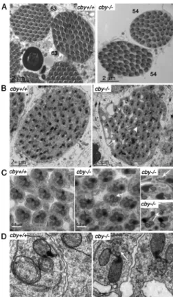
Figure 5. Ultrastructural defects of the chor-dotonal cilia in cby-deficient flies. (top left) Scheme and longitudinal section of a typical leg or antennal CO composed of two neurons ensheathed by a scolopale cell and linked to the cap cell at the tip of the ciliary endings. The cilia are anchored on a distal basal body (DBB) aligned above the proximal basal body (PBB) at the end of the dendrite. (a1–5) Sec-tions at the level of the ciliary axoneme. In con-trol legs (a1), axonemes are composed of nine peripheral microtubule doublets. (a2–5) cby1/1
axonemes show a reduced number of micro-tubule doublets (a2 and 3), altered symmetry (a4), or extra microtubules (a5). (b1–3) Sec-tions at the distal TZ. (b1) In control samples, a dense ring structure of ninefold symmetry is observed. (b2 and 3) In cby1/1 antennae,
interrupted, incomplete, and misformed ring structures are observed. (c1–3) Sections at the proximal TZ level. (c1) In control TZ, decora-tions connecting the membrane can be seen and likely correspond the Y links. (c2 and 3) In cby1/1 antennae, decoration are interrupted
(black arrows; c2), or the ninefold symmetry of the TZ decorations is interrupted (c3). (d1–3) Sections at the level of the proximal basal body. Bars, 0.5 µm. (d1) Basal bodies are an-chored to the cell membrane by dense fibers in control flies (black arrow). Tight junctions
connect the two dendrites. (d2 and 3) Two examples showing missing basal bodies in cby1/1 flies. (e1–3) Longitudinal sections of cby1/1 ciliary endings.
In several cases (e1 and 2), the TZ is discontinuous (white arrows). (e2 and 3) Membrane bulges are observed along the cilia (black arrows). (f1–3) Low magnifications of transverse sections of scolopidia. Bars, 0.5 µm. (f1) Two axonemes are present in control antennae. (f2 and 3) Only one or no axoneme is sometimes observed in cby1/1 antennae.
on February 6, 2017
Altogether, our observations indicate that CBY plays an
impor-tant function during spermatid differentiation in Drosophila.
Because UNC is a basal body–associated protein that is
also required for axonemal assembly in Drosophila, we looked
at UNC distribution in cby mutant testis. Remarkably, we
ob-served that UNC deposition during spermatocyte maturation
was severely affected, with UNC being deposited on a longer
and more variable length of the elongating centrioles compared
with controls (Fig. 8, A and B). The paired elongating centrioles
in cby
1/1testes often showed unequal extents of UNC protein
(Fig. 8 B, white arrow) in contrast to the symmetrical UNC
deposition in the controls. This aberrant UNC distribution is
maintained in early spermatids (Fig. 8, C, D [white arrows], E,
and F). Later, during spermatid differentiation, UNC labeling
at the basal body was reduced in cby
1/1testis compared with
controls (Fig. 8, G and H). In contrast, no differences were
ob-served between cby mutants and controls in -tubulin labeling
(unpublished data). Hence, altered UNC deposition could either
reflect a direct action of CBY on UNC deposition or indirectly
reflect a more general basal body formation defect. Altogether,
these results show that CBY is required for proper
centriole-to–basal body conversion in Drosophila spermatocytes.
showed reduced accumulation in cby
1/1cilia compared with
controls (Fig. 6, C and D). These results show that
ciliary-directed protein trafficking is altered in cby
1/1flies.
CBY is required for basal body maturation in Drosophila spermatogenesis
cby
1/1males are hypofertile (Fig. S1), and CBY is associated
with the maturing basal body during spermatogenesis (Fig. 3).
To understand its function during spermatogenesis, we looked
at the structure of mutant sperm cysts. Differential interference
contrast microscopy of live squashes of Drosophila testis showed
no cellular defects in spermatocyte-stage cysts; all were
com-posed of 16 cells showing that cby is not needed for
spermato-gonial divisions (n = 12; unpublished data).
When cysts in cby
1/1testes were examined by EM, we
fre-quently observed (88%) a reduced number of spermatids in each
mature cyst. Each set of 16 spermatocytes should give rise to 64
spermatids, and all control cyst sections do show 63–64 spermatid
profiles (Fig. 7 A). An altered number of spermatids was sometimes
seen in early spermatid cysts, suggesting a possible meiotic defect
(four out of six cby
1/1cysts had 62 or fewer spermatids, whereas
five out of five control cysts had 63–64 cells; Fig. 7 B). But no
sig-nificant variations were seen in the ratio of nuclei-to-mitochondrial
(Nebenkern) derivatives in onion-stage cysts, even though some
aberrant spermatids could sometimes be observed (two or three
aberrant cells in 3 out of 14 cysts; unpublished data). Hence,
meiosis defects do not seem to be prevalent in cby
1/1testis.
In early cby
1/1spermatid sections, mitochondrial
deriva-tives with no axoneme could be observed (2%; n = 294; Fig. 7 B,
white arrow) as well as axonemes with no visible mitochondria
derivatives (3.4%; n = 294; Fig. 7 B, arrowhead), which were
never observed in controls. We also observed partially penetrant
(14%; n = 294) but reproducible defects of the axonemal
ultra-structure. Incomplete axonemal structures were found (white arrows
in Fig. 7, C and D) with a reduced number of microtubule
dou-blets. Because these defects were visible in early spermatids, we
hypothesize that CBY is required for normal axoneme assembly.
Figure 6. CBY is required for IFT protein distribution. (A–D) Expression of NompB/IFT88 (A and B) and CG11356/ARL13b (C and D) in either control (A and C) or cby1/1 embryonic chordotonal cilia (B and D).
(A and B) NompB-GFP accumulation is increased in cby1/1 cilia compared
with control cilia (white arrow). (C and D) CG11356-GFP accumulates continuously in control cilia, but its accumulation is punctuated in cby1/1
cilia (white arrow). The overall abundance of CG11356 is also reduced in mutant cilia.
Figure 7. CBY is required for axonemal organization in the testis. (A) cby1/1 mutant testes still produce mature spermatozoids, but their number
is reduced. The spermatid cysts are disorganized, and the number of sper-matid per cysts is often compromised. (B) Young spersper-matid cysts are dis-organized and contain a reduced number of spermatids. Some axonemes are not associated with mitochondrial derivatives (white arrowhead), and some mitochondrial derivatives are not associated with an axoneme (white arrow). (C) In addition, a few axonemes are misorganized in mutant flies (white arrows) with mainly missing doublets. Bars, 0.5 µm. (D) High magni-fication of young spermatids highlighting the ultrastructural defects that can be observed on growing axonemes (white arrow). Bars, 200 nm.
on February 6, 2017
MKS, and Joubert syndrome (Hodges et al., 2010). TZ proteins
can be grouped in functional modules by their pattern of
cooc-currence across species and because mutations in each group
cause similar sets of symptoms in humans. The MKS functional
module is conserved in most of the ciliated organisms, but the
NPHP module is not found in all species and in particular not
in Drosophila (Hodges et al., 2010). We show here that CBY is
a novel component of the TZ, which colocalizes with CG14870
(B9D1/MKSR1), a component of the MKS module (Bialas
et al., 2009). CBY is found in animals with motile cilia but not
in C. elegans, suggesting a specific function in TZs of motile
cilia in animals. In agreement with this hypothesis, cby mutant
mice show defects in biogenesis of motile cilia as a result of
defective docking of the basal bodies at the apical membrane
(Voronina et al., 2009). However, in Drosophila, CBY is found
at the tip of the basal bodies, both in cells with motile cilia or
flagella (chordotonal neurons and spermatids) and also in
neu-rons that innervate external sensory (ES) organs such as bristles.
ES cilia lack the canonical axonemal cytoskeleton, and dynein
arms are tightly ensheathed by a surrounding matrix and, so,
are unlikely to be motile. We do not know whether CBY has
any function in ES cells. No behavioral defects were seen when
cby
mutant larvae were challenged with chemical or olfactory
stimuli (unpublished data), nor did mutant adults show the
se-vere uncoordination that characterizes bristle loss-of-function
mutations, suggesting that CBY is not essential for the function
of ES cilia. CBY may be more essential in motile CO cilia than
in ES organs. Or, the moderate structural defects in cby mutant
axonemes may be more consequential for the function of motile
cilia than for static ES cilia. Perhaps, as for the MKS module
proteins in nematode sensory cilia (Williams, et al., 2011), ES
phenotypes may become evident when other TZ proteins are
also mutated.
In mouse, a ciliary requirement for CBY may also be
lim-ited to motile cilia because cby-deficient embryos, unlike
mu-tants with complete ciliogenesis defects, survive until birth and
mostly show defects in airway motile ciliogenesis. However,
we observed that CBY was localized at the TZ of primary cilia
in IMCD3 cells (Fig. S1), suggesting a more general function of
CBY at the TZ. A function for CBY in primary cilia could
ex-plain the observation that cby mutant mice show defects in lung
development that are thought to result from WNT signaling
de-fects (Love et al., 2010). In agreement with a function for CBY
in primary cilia, injection of a truncated dominant form of CBY
in LLC-PK1 cells reduces the number of cells with primary
cilia (Hoffmeister et al., 2011).
CBY is required for protein trafficking into or out IFT-dependent cilia
Several TZ proteins have been shown to be required for protein
trafficking into the cilia and for IFT-dependent processes. This
has clearly been demonstrated for NPHP1, NPHP4, and NPHP6
in C. elegans for several of the MKS-associated proteins and for
NPHP1 in mammalian photoreceptors (Jauregui et al., 2008;
Jiang et al., 2009; Williams et al., 2011). We show here that at
least two proteins involved in IFT are abnormally distributed in
cby
1/1chordotonal cilia, NOMPB/IFT88 accumulating in cilia
Discussion
We show that cby plays an important function in the two
Dro-sophila
cell types in which centrioles mature to basal bodies. In
sensory cilia, CBY is required for proper TZ assembly and
modulates protein trafficking into or out of the cilium. In the
testis, we observed that CBY is required for centriole
conver-sion to a basal body and for proper completion of
spermatogen-esis. In conclusion, our work demonstrates that CBY has an
ancestral function in basal body biogenesis. Notably, we find no
evidence for a CBY function in Wg signaling in Drosophila.
CBY is a novel component of the sensory cilium TZCBY is associated with the TZ in cilia. The TZ is a molecularly
and ultrastructurally distinct axoneme domain located just distal
to the basal body. The ultrastructure of the TZ differs between
primary cilia and motile cilia and also between species (for a
comprehensive review, see Fisch and Dupuis-Williams [2011]).
Nevertheless, several components of the TZ are conserved in
many ciliated organisms. Several groups of TZ proteins are
asso-ciated with human diseases, such as nephronophthisis (NPHP),
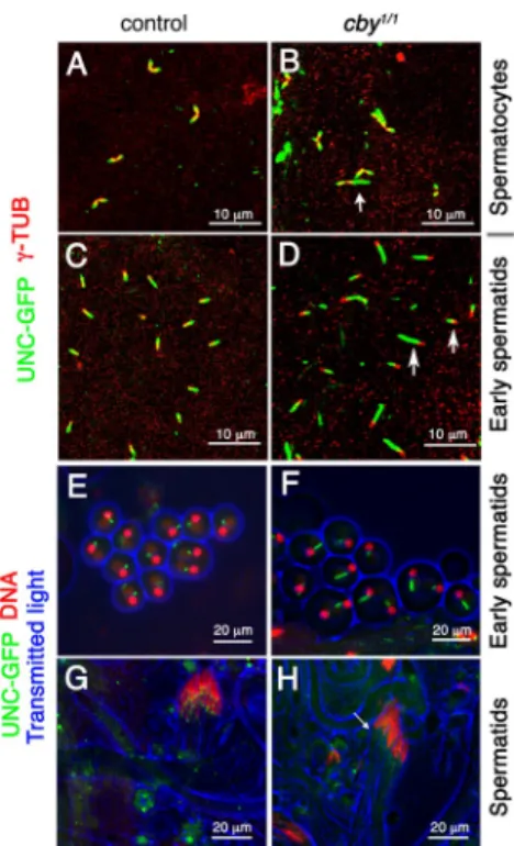
Figure 8. UNC distribution associated with basal body function is altered in cby1/1 testis. (A–H) Distribution of UNC-GFP and -tubulin (-TUB) was
observed by antibody staining in fixed testis from control (A and C) or
cby1/1 (B and D) flies or by GFP fluorescence in live squashed
prepara-tions of control (E and G) or cby1/1 testis (F and H). (A and B) The extent
of UNC protein deposition is irregular in cby1/1 spermatocytes compared
with control. The two apposed centrioles stained for -tubulin show equiva-lent UNC labeling in control but not in cby1/1 spermatocytes (white arrow).
(C–F) UNC labeling marks an increased length segment in cby1/1 early
spermatids (white arrows). (G and H) In cby1/1 elongating spermatids,
UNC staining at the basal body is diffuse and not limited to a dot, as shown in controls. Some faint UNC staining, not observed in controls, is also observed along the axoneme. The arrow points to the reduced and diffuse UNC staining at the base of the nucleus in cby mutant testis. (E–H) DNA stained with DAPI.
on February 6, 2017
genetic and biochemical interactions between cby and dila
will help dissect the pathway by which basal bodies mature
in Drosophila and in other animals in which both cby and dila
are conserved.
Function of the TZ and primary cilium-like extension in Drosophila spermatocytes
We observed that CBY, like UNC and DILA, is deposited at the
tip of the maturing centriole in Drosophila spermatocytes. The
maturing centrioles are associated with cytoplasmic vesicles
be-fore reaching the plasma membrane (Tates, 1971; unpublished
data), where they grow small cilium-like extensions. Altogether,
these observations suggest that a structure molecularly similar
to the TZ assembles during centriole maturation in Drosophila
spermatocytes and is maintained during meiosis and spermatid
elongation. Indeed, both UNC and CBY are found to be
associ-ated with the tip of the growing axoneme, suggesting that this
TZ-like structure is maintained during flagellar assembly. TZ
assembly does not require IFT (Williams et al., 2011), which
is consistent with the observations that IFT is not required for
Drosophila
spermatogenesis (Han et al., 2003; Sarpal et al.,
2003). Therefore, we propose that the primary cilium-like
struc-ture assembled in Drosophila spermatocytes more likely
corre-sponds to an elongated TZ from the maturing centriole, which
hence behaves like a basal body. Therefore, we hypothesize that
flagellar growth in Drosophila is similar to TZ assembly, which
would explain also why flagella assembly is IFT independent
in Drosophila.
Interestingly, in UNC-deficient spermatocytes, the primary
cilia/TZs are shorter, showing that UNC is involved in
elongat-ing these primary cilia/TZ (Baker et al., 2004). Whereas primary
cilia-like extensions were easily observed in wild-type testis, we
did not observe any centrioles that were anchored to the plasma
membrane in cby
1/1flies. We also did not manage to observe
enough centrioles to describe precisely their distal ultrastructure.
However, because UNC deposition is severely affected in cby
1/1spermatocytes, CBY appears to be required to control either
di-rectly or indidi-rectly the assembly of the TZ in these cells.
The function of the spermatocyte primary cilia is unknown.
Centrioles are dispensable for postembryonic development in
Drosophila
(Basto et al., 2006) but are required for male
meio-sis, as observed in sas-4 or sak/plk4-deficient flies
(Rodrigues-Martins et al., 2008; Riparbelli and Callaini, 2011), even though
spermatids can still form without axonemes (Bettencourt-Dias
et al., 2005; Basto et al., 2006). BLD10 is required in
Drosoph-ila
both for centriole elongation spermatocytes and to complete
meiosis, suggesting a possible meiotic requirement for centriole
elongation (Mottier-Pavie and Megraw, 2009; Carvalho-Santos
et al., 2010). However, no precise description of the primary cilia
ultrastructure in spermatocytes has been described in Bld10
mutants. Among the other proteins that are required for basal
body biogenesis in Drosophila (Gogendeau and Basto, 2010),
only UNC is associated with specific defects in spermatocyte
primary cilia. Therefore, it will be interesting to examine in
more detail primary cilia assembly in mutants for other TZ
pro-teins and to finely examine meiosis and early spermatid
differ-entiation defects.
to higher levels than normal, and Arl13 being depleted. This
could either result from an altered balance between anterograde
and retrograde IFT transport or from altered gating of proteins
into the cilia. We do not know precisely all the TZ structures
that are affected in cby
1/1cilia, but we observe some defects at
the level of what likely corresponds to the Y-linker region of the
TZ also called champagne glass structures (Fisch and
Dupuis-Williams, 2011). In C. elegans, mutants lacking MKS/MKSR/
NPHP proteins show missing connecting links, suggesting that
these proteins are either associated with components of the
con-necting Y links or are required for their stability (Williams
et al., 2011). In addition, nematodes with defects in MKSR1
and NPHP4 show ultrastructural defects of the cilia, such as a
reduction in the number of microtubule doublets, that are
simi-lar to the defects observed in cby
1/1flies (Williams et al., 2011).
Altogether, these observations suggest that CBY plays a
func-tion at the TZ similar to the one of MKS and NPHP complexes.
MKS proteins belonging to the B9 domain family (MKS1,
B9D1, and B9D2) are conserved in Drosophila, but most of the
NPHP proteins are not. Conversely, CBY is not found in C.
ele-gans
, where most of the NPHP proteins are conserved. Hence,
these different modules likely operate independently. Such
variations in the architecture and function of the TZ between
organisms still need to be understood.
CBY and UNC, another TZ protein in Drosophila, show similar phenotypes
Only few other proteins associated with the TZ in Drosophila
have been functionally analyzed. Among them, UNC is also a
coiled-coil–containing protein but is found in only Dipteran
species, where it diverged rapidly. It has also been suggested
that UNC could be functionally equivalent to human OFD1, a
centrosomal and basal body coiled-coil protein mutated in the
ciliopathy oro-facial-digital syndrome 1 (Baker et al., 2004).
We observed that loss of CBY function affects UNC protein
accumulation during centriole-to–basal body conversion in
spermatocyte. This could reflect an altered basal body structure
in cby
1/1testes but could also reflect that UNC and CBY
func-tionally interact during TZ assembly. In support of this latter
hypothesis, the basal bodies in unc
/-deficient sensory neurons
show defects similar to cby mutant flies with broken basal
body/TZ rings, missing cilia, or disrupted cilia (Baker et al.,
2004). unc
/flagella also have incomplete axonemes like in
cby
1/1testis. Thus, UNC and CBY could work together in a
functional module required for centriole maturation in
Dro-sophila
testis. The unc
/phenotype is more penetrant than
the cby
1/1phenotype, suggesting that unc is more limiting
than cby for basal body/TZ assembly.
Interestingly, a novel protein, DILATORY (DILA), was
identified recently as interacting with UNC in Drosophila
chor-dotonal neurons (Ma and Jarman, 2011). This suggests that
CBY, UNC, and DILA may function together at the TZ.
How-ever, in chordotonal neurons of dilatory mutants, ultrastructural
defects were restricted to the cilia but did not apparently affect
the TZ. Moreover, no flagellar axonemal defects are associated
with dila mutations (Ma and Jarman, 2011), suggesting that CBY
acts differently from dila in basal body function. Investigating the
on February 6, 2017
Conclusions
Together, our data demonstrate that CBY is required for the
as-sembly of the ciliary TZ in Drosophila both in sensory neurons
and in male germ cells.
Materials and methods
cby coding sequenceComparison of cby cDNA and genomic sequences in different Drosophila strains showed a consistent insertion of two base pairs in the 3 end of the
cby coding sequence compared with the gene annotation on the FlyBase
database. The consequence of this insertion (in italics) is a slight change in the C-terminal protein sequence starting at amino acid 121. Five amino acids are different (italic) and are followed by eight additional amino acids at the C terminus: 5-CATGCGACGGAACTAAGTGAGCTGAAGCCA-AAGGAAAAGTGA-3 (H A T E L S E L K P K E K *).
BLAST and sequence alignment
BLAST searches used as a query the cby gene sequence from FlyBase, modified as described in the previous section, using the basic local align-ment search tool from the National Center for Biotechnology Information. The sequences obtained were then aligned using SeaView software. Reporter constructs
All primer sequences are described in Table S1.
CBY-GFP. A 1,350-bp fragment including cby coding sequences and up-stream regulatory sequences was amplified by PCR from wild-type
Dro-sophila genomic DNA using the following primers designed from the
noncorrected cby sequence: Cby-pro5 and Cby-pro3/BamHI. The result-ing PCR fragment was cloned into the EcoRI and BamHI sites of the pW8-GFP plasmid (Loppin et al., 2005), in frame with the Gfp sequence.
CBY-Tomato. The Tomato sequence was obtained by PCR on the Zeo-TomatoNT-2 (a gift from R. Basto, Institut Curie, Paris, France) with the introduction of an AgeI site at the 5 end and a STOP and NotI site at the 3 end. The resulting PCR fragment was cloned in the BglII and NotI sites of the pattB plasmid. The complete cby 3 untranslated region (UTR) was cloned downstream of the
Tomato sequence in the pattB-Tomato plasmid obtained above using XhoI
and XbaI restriction sites introduced in the 3 UTR PCR fragment.
cby coding and upstream regulatory sequences (1,375 bp) were
am-plified using the Cby-pro5 forward primer and a Cby-PRO3/AgeI reverse primer based on the corrected cby sequence with the addition of an AgeI site at the 3 end. The resulting PCR product was cloned in the EcoRI and AgeI sites of the pattB-Tomato-3cbyUTR plasmid in frame with the Tomato se-quence. Transgenic lines were established by P element–mediated germline transformation (Spradling, 1986) or by phiC31-mediated germline transfor-mation for the pattB plasmid–derived constructs (Bischof et al., 2007).
CG14870-6xMycTag reporter. A 2,942-bp fragment covering CG14870 cod-ing and upstream regulatory sequences was amplified by PCR on wild-type
Drosophila genomic DNA with primers F-14870/BamHI and R-14870/
NotI. The resulting PCR product was cloned in frame with the 6xMycTag in the modified pattB plasmid containing the 6xMycTag and SV40polyA sequences. Transgenic lines were obtained from BestGene Inc.
pW8-GFP CG11356. A 4,230-bp fragment covering CG11356 coding se-quences and upstream regulatory sese-quences was amplified by PCR on wild-type Drosophila genomic DNA with the primers CG 11356-PRO3 and CG 11356-PRO5. The resulting PCR fragment was cloned into the pW8-Gfp plasmid. Transgenic lines were obtained by P element transformation.
cby homologous recombination
The cby gene was disrupted by ends-out homologous recombination (Maggert et al., 2008). The left and right arms of the targeting construct were amplified by PCR on genomic DNA of the y[1] w[*]; P{ry[+t7.2] = 70FLP}11 P{v[+t1.8] = 70I-SceI}2B noc[Sco]/CyO, S[2] stock using primers F-3cby/SphI, R-3cby/NotI, F-5cby/BsiWI, and R-5cby/AscI. The resulting construct enabled the deletion of most of exon 2, including the stop codon, and the acceptor splice site of intron 1 (Fig. 4 A). All the coding regions from the cloned 5 and 3 homologous fragment were verified by se-quencing. Transgenic lines were established by phiC31-mediated germline transformation (Bischof et al., 2007) and used for homologous recombination, as previously described (Maggert et al., 2008).
Fly stocks
We thank J. Schmitt and P. Morales (both from Centre de Génétique et de Physiologie Moléculaire et Cellulaire, Université Claude Bernard Lyon 1,
The reduced number of early spermatids in cysts and
aberrant mitochondrial derivative numbers suggest that cby
might play a role in meiosis. We know that this phenotype
is not a result of improper centriole distribution, as -tubulin
staining showed that all spermatocytes and spermatids had
the right number of centrioles. Our results could also
impli-cate CBY in early spermatid survival or in elongation, which
would lead to a reduced number of spermatid profiles in some
cyst sections. In Drosophila, spermatid elongation relies on
microtubule-dependent mitochondria remodeling (Noguchi et al.,
2011). CBY is transiently associated with mitochondria in
onion-stage and early elongating spermatids. Spermatids do
elongate in cby
1/1flies, but we do not know whether
elonga-tion is as complete as in control testis. The funcelonga-tion of CBY
in mitochondria during spermatid elongation needs to be more
thoroughly investigated.
CBY and WNT signaling
In mammals, CBY has at least two functions. Analysis of
cby
/mice demonstrates that cby is required for basal body
anchoring during multiciliated cell differentiation, illustrating
that cby has conserved this anchoring function throughout
evolution. Experiments in cell cultures indicate that CBY is
involved in WNT signaling in different cell types, essentially
by controlling nuclear–cytoplasmic shuttling of -catenin, the
WNT signaling pathway effector (Takemaru et al., 2003,
2009; Li et al., 2007, 2008, 2010a; Singh et al., 2007; Mokhtarzada
et al., 2011). CBY directly interacts with -catenin, and WNT
signaling defects have been described during lung
develop-ment of cby knockout mice (Love et al., 2010). A previous
RNAi-based study suggested that cby could also be involved in
Wg signaling in Drosophila (Takemaru et al., 2003). However,
Wg signaling and development appear normal in cby
1/1-null
flies, ruling out a function of cby in Wg signaling in Drosophila.
Off-target RNAi effects may have caused the observed defects
in the previous study. We do not know whether CBY interacts
with -catenin in Drosophila, but, if so, such an interaction
would have consequences only in the ciliated cells in which
cby
is expressed.
Therefore, we conclude that CBY function in WNT
sig-naling has either been acquired during chordate evolution or
lost in the Dipteran lineage. In mammals, CBY interacts with
cis-Golgi–associated proteins and is implicated in polycystin-2
distribution in cilia, even though this capacity remains
contro-versial (Hidaka et al., 2004; Hoffmeister et al., 2011). A
func-tion in vesicular transport from the Golgi is consistent with
CBY function in TZ assembly, which starts when centrioles
fuse to vesicular membranes likely derived from the Golgi
apparatus inside the cytosol. CBY could be required early for
basal body–to-membrane attachment and also for vesicular
trafficking to the cilia. Whereas CBY is not found associated
with most of the cis-Golgi apparatus in cultured mammalian
cells, a small pool of the cis-Golgi protein GMAP210 localizes
next to CBY at the base of motile cilia (Fig. S3). This is
compat-ible with a posscompat-ible function in cis-Golgi vesicular
traffick-ing to the cilia, as demonstrated for polycystin-2 transport
(Hoffmeister et al., 2011).
on February 6, 2017
with secondary antibodies for 2 h at room temperature. Testis squashes were performed as follows: testis (0–2-d adults) were fixed 20 min at room temperature in 3.7% PFA/PBS 1× and flattened under a coverslip on a micro-scope slide pretreated with 10% polylysine solution. Slides were quick fro-zen in liquid nitrogen, and coverslips were removed, fixed in chilled 95% ethanol for 5 min, washed twice in PBT (PBS 1× containing 0.1% Triton X-100), and blocked for at least 1 h in PBTB (PBT with 3% BSA) at room temperature. Samples were incubated overnight at 4°C with primary anti-bodies diluted in PBTB and 2 h at room temperature in secondary antianti-bodies in PBTB. Testes were mounted in mounting medium (Cytomation).
The antibodies used are as follows: rabbit anti-GFP (1:500; Invit-rogen), anti-HRP (1:3,000; Jackson ImmunoResearch Laboratories, Inc.), anti-ASL (1:500; provided by R. Basto), anti-DPLP (1:1,000; provided by R. Basto), anti–-tubulin (used on embryos; 1:400; Sigma-Aldrich), mouse monoclonal anti-GFP (1:1,000; Roche), anti-22C10 (1:250; provided by S. Benzer, California Institute of Technology, Pasadena, CA), anti-myc (1:3,000; Euromedex), and anti–-tubulin (used on testes; 1:100; Sigma-Aldrich). The secondary antibodies used, diluted at 1:1,000, are as fol-lows: goat anti–mouse Alexa Fluor 488 conjugated, 546 conjugated, and 647 conjugated; goat anti–rabbit Alexa Fluor 488 and 555 conjugated (Invitrogen); and goat anti–rabbit Cy5 conjugated (GE Healthcare). Image acquisition
Slides were analyzed at room temperature on a microscope (Imager Z1) equipped with 20× Plan-NEOFLUAR (0.5 NA) and 40× Plan-NEOFLUAR (0.75 NA) objectives or a 63× Plan-NEOFLUAR (NA 1.25) objective. Epi-fluorescence images were acquired with a charge-coupled device camera (CoolSNAP HQ2, Roper Scientific) and MetaView software (Molecular Devices). Confocal stacks were acquired with a confocal microscope (LSM 510 META; Carl Zeiss) and software equipped with 63× Plan-Apochromat (1.4 NA), 40× Plan-NEOFLUAR (1.3 NA), and 25× Plan-NEOFLUAR (0.8 NA) objectives. Image brightness and contrast were adjusted by using ImageJ (National Institutes of Health), and separate panels were assembled with Photoshop software (CS4; Adobe). Capture times and adjustments were identical for compared images on one figure. was set to 1 in all figure panels.
EM
Antennae, legs, and testes were dissected in PBS 1× and fixed in 2% glu-taraldehyde, 0.5% PFA, and 0.1 M Na-cacodylate, pH 7.4, for 48 h at 4°C. After three washes of 15 min in 0.15 M sodium cacodylate, pH 7.4, samples were postfixed in 1% OsO4 for 4 h for antennae and 1 h for tes-tes. Samples were then dehydrated through ethanol series and propylene oxide and embedded in Epon medium (Fluka). Ultrathin sections were cut on an ultramicrotome (Leica). Sections were contrasted in Ultrastainer (Leica) in aqueous uranyl acetate and lead citrate. Contrasted sections were observed on a transmission electron microscope (CM120; Philips). Images were acquired with a 2k × 2k digital camera (ORIUS 200; Gatan) and digital micrograph software. Images were processed with Photoshop software (CS4). Only contrast and brightness were adjusted.
Cell cultures
Mouse primary ependymal cell cultures were derived from mouse newborn brains (OF1 strain), as previously described (El Zein et al., 2009). IMCD3 cell lines (a gift from P. Goggolidou, Mammalian Genetics Unit, Medical Research Council, Harwell, Oxfordshire, England, UK) were cultured in DME/F12 medium containing 10% FBS, 100 U/ml penicillin, 100 µg/ml streptomycin, and 1× nonessential amino acids (reagents from GE Health-care). Ependymal and IMCD3 cells were seeded on polylysine- and laminin-coated glass coverslips. Confluent ependymal cells were fixed the day of serum deprivation or after 12 d of serum deprivation, whereas IMCD3 cells were fixed after 2 d of culture in 0.5% FBS containing medium. Cells were fixed in methanol for 5 min at 20°C or in 4% PFA for 15 min at room temperature when using the anti-Arl13b primary antibody. After per-meabilization in 0.1% Triton X-100 for 10 min, cells were blocked 2 h in 5% FBS in PBS 1× followed by an overnight primary antibody incubation using the following: mouse anti-Cby antibody (1:200; Santa Cruz Biotech-nology, Inc.), rabbit anti-Arl13b (1:500; a gift from T. Caspary, Emory University, Atlanta, GA), rabbit anti–-tubulin (1:100; Sigma-Aldrich), and rabbit anti-GMAP210 (1:4,000; a gift from M. Bornens, Institut Curie, Paris, France). Immunostaining was revealed with goat anti–mouse Alexa Fluor 488 and goat anti–rabbit Alexa Fluor 555–conjugated antibodies (1:500; Invitrogen). The slides were mounted in the presence of DAPI or DRAQ5 in fluorescence mounting medium (Dako). Slides were visualized under a microscope (AxioImager Z1; Carl Zeiss) and a confocal micro-scope (LSM 510 META).
Villeurbanne, France) for Drosophila medium and stock maintenance. The chromosomes and alleles P{unc-GFP}34a, nompB1, and P{GFP:nompB}
were previously described (Han et al., 2003; Baker et al., 2004).
P{unc-GFP}34a is inserted on the second chromosome and encodes the entire
UNC protein fused to GFP in the C terminus. nompB1 is a loss-of-function
allele of nompB that deletes 1,100 bp of exon 3 and part of exon 4.
P{GFP:nompB} is inserted on the third chromosome and encodes the
full-length NOMPB protein fused to GFP at its N terminus. {Ubq55:RFP:sas4} was a gift from R. Basto. Df(3R)BSC805 was obtained from the Blooming-ton Stock Center and uncovers the 93F13-94A6 cytological interval. All the other following fly strains were constructed in the laboratory: w;
P{cby::GFP}F23/CyO; P{cby::GFP}M27, w; P{cby::GFP}F23/CyO; rfx253
e/TM3, P{GAL4-twi.G}2.3, P{UAS-2xEGFP}AH2.3, w; P{cby::GFP}F23/CyO;
rfx49/TM3, P{GAL4-twi.G}2.3, P{UAS-2xEGFP}AH2.3, w; Bl/CyO; P{cby::
Tomato}attP62E1M1F2/TM6B, w; P{cby::Tomato}attP62E1 rfx253 e/TM6B,
st rfx16 e ca/TM6B Tb Dr, w; cby1/TM3, P{GAL4-twi.G}2.3,
P{UAS-2xEGFP}AH2.3, w; P{cby::GFP}F23/CyO; cby1/TM6B, w; P{cby::GFP}F23/
CyO; P{cby::GFP}M27 cby1/TM6B, w; P{unc-GFP}34a/CyO; P{cby::Tomato}attP
62E1M1F2/TM6B, w; P{unc-GFP}34a/CyO; cby1/TM3, P{GAL4-twi.
G}2.3, P{UAS-2xEGFP}AH2.3, y w P{CG11356::GFP}F6M1/FM7, w, w;
P{GFP::nompB} cby1/TM3, P{GAL4-twi.G}2.3, P{UAS 2xEGFP}AH2.3,
w; P{CG14870-myc}attP59D3/CyO; cby1/TM3, P{GAL4-twi.G}2.3,
P{UAS 2xEGFP}AH2.3, and w; P{CG14870-myc}attP59D3/CyO; P{cby::
Tomato}attP62E1M1F2/TM6B.
SEPs
Extracellular compound potentials were recorded with tungsten elec-trodes placed in the antenna and head, as previously described (Eberl et al., 2000; Eberl and Kernan, 2011). Each fly was mounted in a micro-pipette tip and its proboscis fixed in wax so that its antennae were ex-posed and free to move. A computer-generated sound stimulus (five pulses of a 500-Hz sine wave at 35-ms intervals) was amplified by an audio amplifier (Realistic MPA-30; RadioShack) and an 8-in, 4-Ω speaker and brought to the fly via 0.25-in Tygon tubing, with its opening placed close enough to the antennae to ensure near-field sound conditions. To re-cord antennal nerve potentials, one electrolytically sharpened tungsten wire electrode was placed between the first and second antennal seg-ments and a second in the head. The differential alternating current signal from these was amplified (5,000× gain) by a BMA-200 Bioamplifier (CWE, Incorporated) and digitized and analyzed using an Instrunet 100B A/D converter and SuperScope II software (GW Instruments, Inc). The same stimulus amplitude was used for all recordings. Flies were chilled briefly at 0°C for mounting. Flies that were sorted under CO2 anesthesia were allowed to recover for >40 h before recording. Mutant (cby1/1 or cby1/Df) flies were recorded alternately with their age-matched
control sibs. For each antenna, we measured the maximum amplitude of the averaged responses to 10 sound stimuli. P-values were calculated from the Mann–Whitney rank sum test, with all responses lacking peaks tied at the lowest rank.
Bang assay
The climbing test was performed on female flies of the same age. 30 flies were placed in graduated tubes and banged on the table at t = 0. Pictures were taken every minute for 5 min and analyzed by counting the number of flies present in each interval every minute and for each genotype. Live squashes of testes
Late pupae were dissected once in PBS 1×. The dissected testes were trans-ferred on a slide in a drop of PBS 1× containing 8.3 mg/ml Hoechst 33342 and then squashed under a coverslip before being observed live using a microscope (Imager Z1; Carl Zeiss).
Immunostaining
Precisely staged embryos were sorted and collected in PBS 1×/0.1% Triton X-100 and dechorionated in bleach. Embryos were rinsed in PBS 1×, extracted from the vitelline membrane using a tungsten needle, transferred to a polylysine-coated slide, and dissected. Embryos were fixed in 4% PFA/PBS 1× (20 min), rinsed, and blocked in PBT (PBS 1×, 5% BSA, and 0.1% Triton X-100). Embryos were incubated with primary antibodies diluted in PBT washed in PbT (PBS 1×, 0.1% BSA, and 0.1% Triton X-100) and incu-bated with secondary antibodies diluted in PbT. Embryos were washed in PTW (0.1% Tween 20 in PBS 1×) and mounted (Cytomation; Dako).
Whole-mount testis preparation was performed as follows: testes were fixed 15 min in 4% PFA/PBS 1× followed by a 15-min treatment in PBS 1× and 0.1% Triton X-100, blocked in 1% BSA in PBS 1×, and incu-bated with primary antibodies overnight at 4°C. Testes were incuincu-bated