HAL Id: tel-01228118
https://tel.archives-ouvertes.fr/tel-01228118
Submitted on 12 Nov 2015HAL is a multi-disciplinary open access
archive for the deposit and dissemination of sci-entific research documents, whether they are pub-lished or not. The documents may come from teaching and research institutions in France or abroad, or from public or private research centers.
L’archive ouverte pluridisciplinaire HAL, est destinée au dépôt et à la diffusion de documents scientifiques de niveau recherche, publiés ou non, émanant des établissements d’enseignement et de recherche français ou étrangers, des laboratoires publics ou privés.
Structural study of the transcriptional co-activator
SAGA
Alexandre Durand
To cite this version:
Alexandre Durand. Structural study of the transcriptional co-activator SAGA. Microbiology and Parasitology. Université de Strasbourg, 2014. English. �NNT : 2014STRAJ051�. �tel-01228118�
Université de Strasbourg
Ecole doctorale des Sciences de la Vie et de la Santé
Thèse
Présentée pour l’obtention du titre de
Docteur de l’Université de Strasbourg
Discipline : Aspects moléculaires et cellulaires de la Biologie
Par
Alexandre Durand
S
TRUCTURAL
S
TUDY
O
F
T
HE
T
RANSCRIPTIONAL
C
O
-A
CTIVATOR
S
AGA
Soutenue publiquement le 29 Avril 2014 devant le jury composé de :
Dr. Marc Timmers
Dr. Patrick Bron
Dr. Bruno Kieffer
Dr. Patrick Schultz
Rapporteur
Rapporteur
Examinateur
Directeur de thèse
R
EMERCIEMENTS
To start, I would like to thank the members of the jury, Dr. Marc Timmers, Dr. Patrick Bron, Dr. Bruno Kieffer and Dr. Didier Devys, for accepting to comment and judge my work.
Je suis particulièrement reconnaissant à mon directeur de thèse, le Dr. Patrick Schultz, qui m’a accueilli dans son équipe, qui m’a formé à l’art de la microscopie électronique et qui m’a permis de mener à bien ce projet. Je le remercie pour ses conseils et son enthousiasme au quotidien.
Je remercie tous les membres de l’équipe de Patrick pour leur accueil, leurs conseils, leur disponibilité et leur bonne humeur : Merci en particulier à Gabor, Christine, Corinne, Grigory, Adam et Nicolas pour avoir partagé avec moi leurs connaissances, mais aussi de bons moments de convivialité qui permettent de surmonter tous les petits problèmes du quotidien. Je souhaite également beaucoup de succès à mon compagnon de thèse Grigory, dans la poursuite de son projet.
Ce projet a été réalisé au sein du Département de Biologie Structurale Intégrative, et j’ai eu la chance de pouvoir interagir avec de nombreuses personnes que je ne pourrais pas toutes citer ici. Je remercie en particulier Catherine Birck et tous les membres de la plateforme de biologie et génomique structurale, ainsi que Marc Ruff pour son aide et ses conseils. Je remercie Jean-François Ménétret et Alexander Myasnikov pour leur aide lors de longues sessions au microscope. Je souhaite également remercier le Dr. Anne-Catherine Dock-Bregeon, sans qui je n’aurai pas pu me lancer dans cette grande aventure que représente le travail de thèse. Merci également au Dr. Arnaud Poterszman, qui fut le premier à m’accueillir au sein du département.
Je remercie très sincèrement le Dr. Laszlo Tora et tous les membres de son équipe, qui m’ont beaucoup apporté par nos échanges et leurs conseils. Je souhaite remercier en particulier Jacques Bonnet, Didier Devys et également Marjorie Fournier pour leur aide et leur disponibilité.
Je remercie également Bertrand Séraphin et Céline Faux, pour leurs conseils et leur expertise. Un grand merci aux membres de la plateforme de protéomique, en particulier Adeline Page et Virginie Chavant.
J’ai une pensée pour tous mes amis qui m’ont précédé ici, et qui par leur exemple m’ont fortement influencé dans ma décision de commencer ce travail de thèse. Grâce à Wassim et nos innombrables discussions « profondes » autour d’un café, grâce à Chris et notre passion commune pour le ballon ovale, grâce à Martin, Nada, Serena, Massimo, Alastair, Laura et tous les autres, qui m’ont montré que les moments parfois un peu difficiles de ces années n’étaient pas insurmontables lorsque l’on était bien entouré, j’ai pu franchir le pas. Merci à vous tous.
Je dois également remercier mes parents et ma famille (et ma belle-famille !), qui m’ont permis d’arriver aussi loin, grâce à leur soutien et leurs encouragements. Je n’aurai probablement pas pu suivre ce chemin s’ils n’avaient pas été là pour mes premiers pas.
Pour finir, un immense merci à ma petite femme, Marie-Laure, qui m’a accompagnée, encouragée et soutenue au quotidien au cours de ces 4 années, et sans qui rien n’aurait pu être pareil. Merci infiniment pour tes efforts et ta patience, parfois sans limite !
A
BBREVIATIONS
Ada : Alteration/Deficiency in activation ATAC : Ada-Two-A-ContainingATP : Adenosine triphosphate
Bp : base-pairs
BRE : TFIIB Recognition Element BSA : Bovine Serum Albumin CBB : Calmodulin Binding Buffer CBP : Calmodulin-binding peptide CEB : Calmodulin Elution Buffer ChIP : Chromatin Immuno Precipitation Cs : Spherical aberrations
CTD : C-terminal domain
CTF : Contrast Transfer Function CWB : Calmodulin Washing Buffer DCE : Downstream Core Element DNA : Deoxyribonucleic acid
DPE : Downstream Promoter Element DSIF : DRB Sensitivity Inducing Factor DUB : Deubiquitination
FEG : Field-emission Gun
FRET : Fluorescence resonance energy transfer FSC : Fourier Shell Correlation
Gcn : General control of amino acid biosynthesis GTF : General Transcription Factor
H2B-Ub : Ubiquitination of H2B on lysine 123
H3K4-Me3/2 : Di- or Tri-Methylation of Histone 3 Lysine 4 HAT : Histone Acetyl-transferase
HDAC : Histone deacetylase HFD : Histone-fold Domain
HMT : Histone Methyl Transferase HMT HRP : Horseradish Peroxidase
Inr : Initiator sequence LS : Liquid Chromatography MRA : Multi References Alignment
mRNA : Messenger RNA MS : Mass Spectrometry
MS/MS : Tandem MS
MSA : Multivariate Statistical Analysis MTE : Motif Ten Element
MudPIT : Multidimensional protein identification technology NC2 : Negative cofactor 2
NCP : Nucleosome Core Particule NELF : Negative Elongation Factor NER : Nucleotide Excision Repair NMR : Nuclear Magnetic Resonance NPC : Nuclear Pore Complex
NSAF : Normalized Spectral Abundance Factor NTP : Nucleoside Tri-Phosphate
OD : Optical Density ORF : Open Reading Frame
PAGE-SDS : Polyacrylamide Gel Electrophoresis in presence of Sodium Dodecyl Sulfate
PAP : Peroxidase anti-peroxidase PCR : Polymerase Chain Reaction PHD : Plant Homeo Domain PIC : Pre-Initiation Complex
protA : protein A from Staphylococcus aureus P-TEFb : Positive Transcription Elongation Factor b RAP : RNA-pol II Associated Protein
RAR : Retinoic Acid Receptor RNA pol : RNA polymerase RNA : Ribonucleic acid rRNA : Ribosomal RNA
RSC : Remodels Structure of Chromatin RP : Regulatory Particle of the proteasome
RT : Room Temperature
SAGA : Spt-Ada-Gcn5 Acetyl-transferase SALSA : SAGA altered Spt8 absent SCA7 : Spinocerebellar ataxia type 7 Sgf : SAGA-associated factor
SLIK : SAGA-like
SNR : Signal to noise ratio Spt : Suppressor of Ty
STAGA : SPT3-TAFII31-GCN5-L acetyltransferase TAF : TBP-associated factor
TAND : Taf N-terminal Domain TAP : Tandem Affinity Purification TBP : TATA-box Binding Protein TCB : TEV Cleavage Buffer
TCR : Transcription-Coupled Repair TEM : Transmission Electron Microscope TEV : Tobacco Etch Virus
TRF : TBP-related factors TFTC : TBP-free TAFIIs Complex tRNA : Transfer RNA
TSS : Transcription Start Site
UAS : Upstream Activating Sequence YPD : Yeast Peptone Dextrose ZnF : Zinc-Finger
T
ABLE OF CONTENT
Remerciements ... 1
Abbreviations ... 3
Table of content ... 6
List of Figures ... 10
Résumé ... 13
Chapitre 1.
Introduction ... 13
Chapitre 2.
Matériel et Méthodes ... 17
1.
Structure du complexe SAGA observé par microscopie électronique ... 17
2.
Purification du complexe SAGA endogène de levure ... 19
Chapitre 3.
Résultats ... 21
1.
Structure du complexe SAGA par microscopie électronique ... 21
2.
Localisation du module DUB et des protéines Spt7 et Spt8 ... 22
3.
La délétion du module DUB favorise le clivage de Spt7 ... 24
4.
Le module DUB n’est pas toujours présent au sein du complexe SAGA ... 25
5.
Interactions du complexe SAGA avec le nucléosome... 26
Chapitre 4.
Conclusions et perspectives ... 27
Chapter 1. Introduction ... 30
1.1 - Initiation of transcription by RNA pol II ... 31
1.1.1
The Promoter of the protein coding genes ... 31
1.1.2
The Pre-Initiation Complex ... 33
1.1.3
Structure of the Pre-Initiation Complex ... 46
1.2 - The Chromatin structure and Regulation of transcription ... 48
1.3 - Role of the Activators in transcription ... 54
1.4 - The Transcriptional Co-activator SAGA ... 63
1.4.2
A subset of TAFs plays a structural role in SAGA ... 67
1.4.3
Role of the Spt proteins in structural integrity and interaction with TBP . 69
1.4.4
SAGA is a coactivator required for transcriptional activation ... 71
1.4.5
The Histone Acetyltransferase Activity of Gcn5 ... 73
1.4.6
The deubiquitination module of the SAGA complex ... 76
1.4.7
The Structure of the SAGA complex reveals its modular organization ... 80
1.5 - Aims of this work ... 82
Chapter 2. Material and Methods ... 86
2.1 - Structure of macromolecular complexes determined by Electron Microscopy
... 86
2.1.1
Principle of imaging with a Transmission Electron Microscope ... 87
2.1.2
Specimen preparation ... 89
2.1.3
Protocols for preparation of carbon-coated grids for Electron Microscopy
92
2.1.4
Data acquisition: Low-Dose Electron Microscopy ... 94
2.1.5
Image processing ... 95
2.2 - Purification of the SAGA complex ... 103
2.2.1
Strategy for the purification of the SAGA complex: the TAP-tag method
103
2.2.2
Generation of the yeast strains ... 105
2.2.3
Culture of yeast ... 106
2.2.4
Preparation of the yeast extract ... 108
2.2.5
Purification of the SAGA complex ... 110
2.2.6
Reconstitution of the complex nucleosomes/SAGA ... 111
2.3 - Protein sample analysis ... 112
2.3.1
Electrophoresis ... 112
2.3.2
Western-blot ... 113
2.3.3
Mass Spectrometry: the multidimensional protein identification
technology ... 114
Chapter 3. Results... 117
3.1 - Purification of the endogenous SAGA complex from S. cerevisiae ... 117
3.1.1
Methods of lysis for optimal conditions of purification ... 117
3.1.2
Optimization of purification conditions ... 118
3.2 - Characterization of the complex by PAGE-SDS and mass spectrometry ... 120
3.3 - Structure of the SAGA complex revealed by Electron Microscopy ... 124
3.3.1
Description of the structure and the different conformations of SAGA . 124
3.3.2
Localization of the DUB module ... 128
3.3.3
A subpopulation of SAGA does not contain the DUB module ... 132
3.3.4
Localization of the Spt7 and Spt8 subunits ... 134
3.4 - Structure of the fully-hydrated SAGA complex ... 135
3.5 - Interactions of the SAGA complex with the nucleosome ... 136
Chapter 4. Discussion ... 140
4.1 - Architecture of the SAGA complex... 140
4.2 - The DUB module clusters with Gcn5 and defines a nucleosome binding
interface ... 142
4.3 - The SAGA complex forms a molecular clamp which might bind TBP ... 143
4.4 - The dynamic of the DUB module alters the TBP-binding surface of the SAGA
complex ... 145
Chapter 5. Conclusions and Perspectives ... 148
Chapter 6. References ... 153
Chapter 7. Annexes ... 175
Annexe 1 - Protocols ... 176
7.1 - S. cerevisiae culture ... 176
7.1.1
Media composition ... 176
7.2 - Purification of the SAGA complex ... 178
7.2.1
Buffers ... 178
7.2.2
Preparation of the yeast extract ... 178
7.2.3
TAP-tag purification of the complex ... 179
7.3 - Protein complex analysis ... 180
7.3.1
PAGE-SDS ... 180
7.3.2
Western-Blot ... 181
7.4 - Sample preparation for Electron Microscopy analysis ... 182
7.4.1
Preparation of carbon-coated grids for Electron Microscopy ... 182
Annexe 2 - Publications ... 183
7.5 - Publication 1 ... 183
L
IST OF
F
IGURES
Figure 1 : Organization of eukaryotic promoter ... 33
Figure 2 : Different models for PIC assmebly. ... 35
Figure 3 : Structure of yeast TBP bound to the TATA-box ... 37
Figure 4 : Structure of TBP-DNA-TFIIA complex ... 38
Figure 5 : Domain composition of yeast TFIIB... 38
Figure 6 : Domains of the two subunits forming TFIIF ... 40
Figure 7 : Domains composition of TFIIE subunits α and β ... 41
Figure 8 : Molecular organization of TFIIH ... 43
Figure 9: Molecular architecture of RNA pol II ... 44
Figure 10: Structure of TFIIB-TBP-RNA pol II complex with opened DNA template ... 45
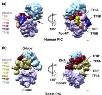
Figure 11 : Comparison of PIC structure in yeast and human ... 47
Figure 12 : Structure and compaction of the chromatin ... 49
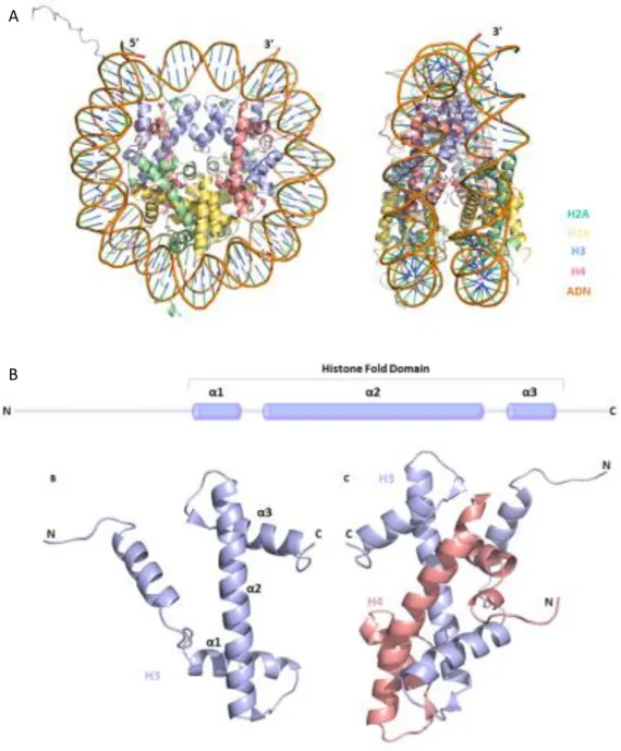
Figure 13 : Structure of the nucleosome ... 50
Figure 14 : Histone modifications are marker of the transcriptional state genes. ... 53
Figure 15 : Cross-talk between H2B ubiquitilation and methylation of histone 3. ... 54
Figure 16 : Summary of Tafs present in the different species and their nomenclatures ... 56
Figure 17 : Molecular architecture of TFIID ... 58
Figure 18 : Subunits compostion of yeast and human Mediator complex. ... 60
Figure 19 : Molecular organization of yeast and human Mediator complex... 60
Figure 20 : Mechanism of Gal4 activation upon galactose induction. ... 62
Figure 21 : Composition of the SAGA complex ... 65
Figure 22 : Conserved structure of SAGA homologues ... 67
Figure 23 : Comparison of Tafs position in SAGA and TFIID... 69
Figure 24 : Molecular architecture and nucleosome interaction site of NuA4... 73
Figure 25 : Structure of the DUB module revealed by x-ray crystallography ... 77
Figure 27 : Sus1 links transcription and mRNA export ... 79
Figure 28 : Molecular organization of yeast SAGA ... 81
Figure 29 : Modular organization of the SAGA complex ... 82
Figure 30 : Schematic representation of a Transmission Electron Microscope and path of the electrons thought the optical elements. ... 88
Figure 31 : Sample preparation techniques for single particle EM. ... 90
Figure 32 : Schematic representation of image analysis process. ... 96
Figure 33 : Sampling rate of viewing direction limits the resolution ... 97
Figure 34 : Euler angles convention. ... 99
Figure 35 : Principle of Random Conical Tilt method. ... 100
Figure 36 : Schematic representation of the Tandem Affinity Purification. ... 104
Figure 37: Summary of the different strains used in this study, and their correspondiong genotype. ... 106
Table 3 : Composition of the buffers used during purification ... 109
Figure 38 : Comparison of different lysis methods ... 118
Figure 39 : Optimization of conditions for purification ... 119
Figure 40 : Control of the quality of the sample by Electron Microscopy. ... 120
Figure 41 : PAGE-SDS analysis of SAGA complexes on a 4-15% acrylamide gel gradient. ... 121
Figure 42 : Peptide coverage of Spt7 and Sgf73. ... 122
Figure 43 : Mass spectrometry analysis of the purified complexes. ... 123
Figure 44 : Characteristic views of the SAGA complex ... 125
Figure 45: 3D reconstruction of the SAGA complex ... 126
Figure 46: Different conformations obtained by maximum likelihood 3D classification. ... 127
Figure 47 : Independent 3D reconstructions of the two separated lobes ... 128
Figure 48 : Size and position of domain V in the SAGA complex ... 129
Figure 49 : Difference between the complex from the different strains. ... 130
Figure 50 : Fitting of the DUB module structure in the density map ... 131
Figure 52 : Separation of a DUB-containing and a DUB-free SAGA complex ... 133 Figure 53 : Localization of the Spt8 subunit by immuno-labelling ... 134 Figure 54 : Class average images of SAGA observed by cryo-EM ... 136 Figure 55 : Control of the interaction betwwen the purifed SAGA complex and the mono-nucleosome ... 137 Figure 56 : Visualization of the complex formed between SAGA and mono-nucleosome ... 138 Figure 57 : Position of TBP-SAGA interacting regions ... 144
R
É
s
UMÉ
Chapitre 1.
I
NTRODUCTION
La première étape de l’expression de l’information génétique contenue dans les gènes est la synthèse d’une molécule d’ARN à partir de la matrice d’ADN, lors d’un procédé appelé la transcription. Chez les eucaryotes, la transcription des gènes codant pour les protéines est réalisée par l’ARN polymérase II (pol II). Lors de la phase d’initiation de la transcription, l’ARN polymérase et des facteurs généraux de la transcription (GTF) vont être recrutés afin de former un complexe de pré-initiation (PIC). Les GTFs reconnaissent certains éléments spécifiques du promoteur proximal du gène, et permettent ainsi (i) la reconnaissance du site d’initiation de la transcription, (ii) le recrutement de la pol II et son positionnement précis au niveau du site de démarrage de la transcription au sein du promoteur du gène, et (iii) l’ouverture de la double hélice d’ADN au niveau du site d’initiation, étape nécessaire au démarrage de la synthèse de la molécule d’ARN.
La machinerie transcriptionnelle doit également passer la barrière physique formée par la structure compacte de la chromatine, et qui limite l’accessibilité de la matrice d’ADN. En effet, la chromatine est composée de la répétition d’un élément de base, le nucléosome, formé d’un octamère de protéines histones autour duquel s’enroule la molécule d’ADN. Le passage de la forme condensée de la chromatine à un état plus ouvert, et donc plus accessible, nécessite l’action de plusieurs classes de protéines, dont notamment les chaperonnes d’histone et les facteurs de remodelage de la chromatine.
La formation du PIC est une étape très importante, qui nécessite l’action coordonnée d’un grand nombre de molécules. En particulier, les activateurs de la transcription reconnaissent des séquences spécifiques au sein ou en dehors du promoteur, et stimulent la transcription en favorisant la formation du PIC. Plusieurs complexes macromoléculaires, appelés co-activateurs transcriptionnels, sont recrutés par ces activateurs et font le lien avec les différents composants de la machinerie transcriptionnelle.
Les coactivateurs font le lien entre les activateurs de la transcription, qui lient certaines séquences d’ADN spécifiques à proximité du gène, et la machinerie transcriptionnelle. Notamment, les coactivateurs (i) interagissent avec les activateurs liés à leur séquence régulatrice, (ii) interagissent avec les différents composants du PIC pour faciliter leur recrutement, et (iii) peuvent lire et écrire des modifications spécifiques au niveau des histones, permettant l’altération de la structure de la chromatine.
Le complexe SAGA (Spt-Ada-Gcn5 acetyl transferase) est l’un de ces co-activateurs, conservé chez les eucaryotes au cours de l’évolution. Chez la levure
Saccharomyces cerevisiae, il a été montré que SAGA est requis pour la transcription d’environ
10% des gènes, la plupart étant impliqués dans la réponse de la cellule aux stresses environnementaux (Basehoar et al., 2004; Huisinga and Pugh, 2004). SAGA est un large complexe comportant au moins 19 sous-unités, pour un poids moléculaire total d’environ 1.8 MDa. Le complexe SAGA est impliqué dans la modification post-traductionnelle des histones dans le contexte du nucléosome, et en particulier dans l’acétylation des lysines sur l‘histone H3. L’acétylation des histones est généralement associée à la transcription active des gènes, et cette activité est portée au sein du complexe SAGA par l’histone acétyl-transférase (HAT) Gcn5, en association avec les protéines Ada2, Ada3 et Sgf29 qui forme un module d’acétylation au sein du complexe (Balasubramanian et al., 2002; Grant et al., 1997; Lee et al., 2011). De plus, une deuxième activité enzymatique impliquée dans la modification des histones a été identifié au sein du complexe SAGA. En effet, la protéine Ubp8, associée aux sous-unités Sgf73, Sus1 et Sgf11, est impliquée dans la coupure de l’ubiquitine liée à la lysine 123 de l’histone H2B (Daniel et al., 2004; Ingvarsdottir et al., 2005; Köhler et al., 2006; Lee et al., 2009). Cette étape est nécessaire au passage de la phase d’initiation à la phase d’élongation de la transcription (Wyce et al., 2007). SAGA est recruté au niveau du promoteur du gène par certains activateurs de la transcription, et ce recrutement se fait au travers de la protéine Tra1 (Bhaumik et al., 2004; Fishburn et al., 2005; Grant et al., 1998b). SAGA est nécessaire pour la formation du PIC au niveau du promoteur d’un ensemble de gènes (Bhaumik and Green, 2002), par le recrutement direct de la protéine TBP qui lie certains éléments de ces promoteurs et permet ainsi l’assemblage du PIC. Ce recrutement implique les protéines Spt3 et Spt8 du complexe SAGA qui interagissent directement avec TBP
(Laprade et al., 2007; Mohibullah and Hahn, 2008; Warfield et al., 2004). Enfin, le complexe SAGA contient un sous-ensemble de protéines Tafs (Grant et al., 1998a), partagée avec le facteur de transcription TFIID, et qui semblent jouer un rôle structural au sein du complexe. Finalement, SAGA contient également les protéines Spt20, Spt7 et Ada1, dont la délétion compromet l’intégrité du complexe et semble nécessaire pour le maintien de son architecture (Grant et al., 1997; Horiuchi et al., 1997).
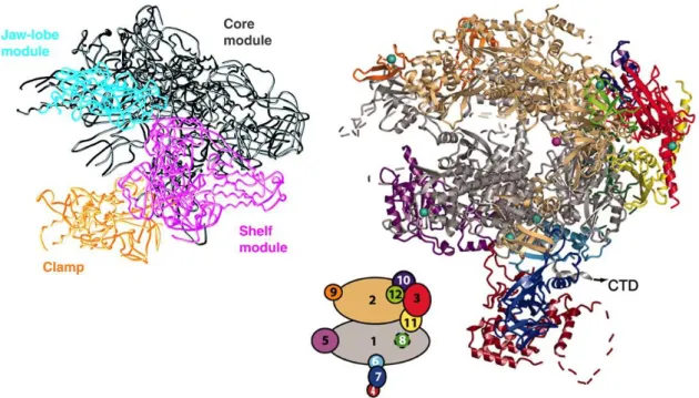
Liste des sous-unités du complex SAGA chez la levure, l’homme et la drosophile. Les sous-unités sont regroupés en fonction de leur fonction au sein du complexe.
Une forme alternative (nommée SLIK ou SALSA) du complexe SAGA a été identifiée, et se caractérise notamment par le clivage de la partie C-terminal de Spt7, et la perte de la sous-unité Spt8, et semble impliquée dans la régulation de certains gènes (Pray-Grant et al., 2002; Sterner et al., 2002)
Des études structurales et de protéomiques ont décrit l’organisation modulaire du complexe, dont les différentes sous-unités contribuant à une même fonction semblent s’associer en modules fonctionnels au sein du complexe (Lee et al., 2011; Wu et al., 2004). L’architecture moléculaire du complexe est organisée en 5 domaines, qui regroupent les différentes fonctions de SAGA. Ainsi, la fonction de liaison aux activateurs est située au sein d’un premier domaine, à l’opposé du site d’interaction avec TBP qui a été identifié au niveau d’un domaine très flexible. Les sous-unités Tafs, dont la stœchiométrie exacte n’est pas encore connue, sont regroupées au sein de deux domaines centraux, en accord avec leur rôle structural. Enfin, Gcn5 est présent au sein d’un autre domaine avec la protéine Spt7 (capable
de lier certaines lysines acétylées sur les queues d’histone), et définit une région potentielle d’interaction avec les nucléosomes.
Le complexe SAGA est organisé de manière modulaire. La structure du complexe revèle l’existance de 5 domaines, où se regoupent les différentes sous-unités se regourpent selon leurs différentes fonctions.
Le complexe SAGA est donc au centre des mécanismes d’initiation de la transcription, en intégrant différentes fonctions nécessaire à cette étape. Ainsi, le complexe SAGA fait le lien entre la machinerie transcriptionnelle et les activateurs, permet le recrutement de TBP et la formation du PIC, et modifie la chromatine par la lecture et l’écriture de modifications spécifiques sur les queues des histones. L’importance du rôle du complexe SAGA lors de la transcription est illustrée par la nécessité du bon fonctionnement de ses différentes fonctions pour le développement normal de l’embryon chez les eucaryotes supérieurs (Carre et al., 2005; Guelman et al., 2006; Weake et al., 2009; Xu et al., 2000), et par son rôle lors du développement de certains cancers (McMahon et al., 1998; Zhang et al., 2008).
Au cours de cette étude, nous nous sommes intéressés au module de déubiquitination (DUB), formé chez la levure par les protéines Sgf73, Ubp8, Sgf11 et Sus1. L’activité enzymatique du module est portée par la sous-unité Ubp8, dont l’homologue chez l’humain (USP22) a été identifié comme un marqueur de la résistance au traitement et à la formation de métastases au cours de certains cancers (Zhang et al., 2008, p. 22). De plus, l’homologue humain de Sgf73, l’ATXN7 peut présenter une extension poly-glutamine dans sa partie N-terminale, responsable de la maladie neurodégénérative SCA7 (Helmlinger et al., 2004). Afin de comprendre le rôle d’Ubp8 au sein du complexe SAGA, ainsi que l’effet de l’altération du module au cours de ces maladies, nous avons localisé le module DUB au sein du complexe. De plus, il semblerait que le module DUB puisse être détaché du reste du complexe par la
Structural role Activator binding TBP binding HAT activity Bromodomain
particule régulatrice 19S du protéasome (Lim et al., 2013), participant ainsi à la régulation fonctionnelle de SAGA. Nous avons donc analysé l’effet de la perte du module sur la structure du complexe. Enfin, le profil de transcription de certains gènes est altéré chez les modèles de souris de la maladie SCA7. Nous avons donc étudié les possibles interactions entre le module DUB et la fonction de reconnaissance du promoteur du complexe SAGA.
Pour cela, nous avons résolu la structure du complexe purifié à partir d’une souche sauvage, et de deux souches mutantes où la partie N-terminale de Sgf73 (Sgf73Δ1-104), ou la protéine entière (Sgf73Δ), a été supprimée. Les complexes ont été analysés par spectrométrie de masse et leur structure ont été comparée afin d’observer les différences dues à la perte du module.
Chapitre 2.
M
ATERIEL ET
M
ETHODES
1. Structure du complexe SAGA observé par microscopie
électronique
La plupart des processus biologiques dans la cellule nécessite l’action de protéines qui n’agissent pas de manière isolée, mais plutôt au sein de larges complexes macromoléculaires qui permettent l’action coordonnée de ces différents composants. Ces complexes sont souvent peu abondants et fragiles, et la réalisation d’étude structurale sur ces complexes s’avèrent généralement une tache compliquée.
La microscopie électronique est la méthode de choix pour l’étude structurale de ces larges assemblages macromoléculaires. En effet, la microscopie électronique permet l’observation directe de complexes de plusieurs mégadaltons, dans un état proche de celui présent dans la cellule. De plus, une étude par microscopie électronique nécessite une plus faible quantité de matériel que d’autres méthodes comme la résonnance magnétique nucléaire (RMN) ou la cristallographie aux rayons X. La microscopie électronique a le potentiel pour résoudre la structure d’un complexe biologique et de ces différentes conformations à une résolution proche de l’atome.
La visualisation du complexe en microscopie électronique est basée sur l’interaction des électrons émis par la source avec l’objet observé. Les électrons vont être déviés par les atomes qui composent le spécimen, puis focalisés par un complexe jeu de
lentilles électromagnétiques sur un détecteur qui enregistrera une série d’image du spécimen. Sur ces images, le contraste nécessaire à la bonne visualisation de l’objet est fourni par un diaphragme qui bloque les électrons fortement déviés par le spécimen (contraste d’amplitude), et par l’interférence entre les électrons non-diffusés et diffusés, ces derniers ayant subi un changement de phase due à la défocalisation du faisceau d’électron et aux aberrations des lentilles (contraste de transfert).
La préparation du spécimen se fait en plusieurs étapes. D’abord, le spécimen est adsorbé sur un film de carbone posé sur une grille de microscopie et rendu hydrophile par une décharge dans l’air. Puis, l’échantillon peut être coloré par un sel de métal lourd (tel que l’acétate d’uranyl), qui se dépose autour de l’échantillon et dévie fortement les électrons produisant ainsi un fort contraste d’amplitude. Cette technique, appelé coloration négative, a pour avantage de produire un fort contraste facilitant l’observation, mais génère des artefacts liés aux conditions salines élevées et au pH acide du colorant, ainsi qu’à la déshydratation complète du spécimen, ce qui altère la structure du complexe observé.
Une alternative à la coloration consiste à vitrifier le spécimen en le plongeant rapidement dans un bain à très basse température, tel que l’éthane liquide. Cette technique, appelée cryo-microscopie électronique, permet d’observer un spécimen complètement hydraté et proche de son état physiologique. Cependant, le contraste est ici très faible, et rend l’analyse des images particulièrement difficile.
L’irradiation du spécimen par le faisceau d’électron provoque des ruptures des liaisons moléculaires au sein du spécimen et altère sa structure. Pour limiter ces effets, plusieurs images sont enregistrées à très faible dose (nombre d’électron par unité de surface), ce qui est responsable d’un très fort bruit sur les images mais permet de répartir la dose nécessaire à la visualisions des particules sur plusieurs images. Les différentes vues du spécimen sur les différentes images seront ensuite additionnées, dans le but de réduire ce bruit.
Les images contiennent les vues (idéalement d’une même molécule) sous différents angles d’observation. Pour reconstituer un volume tridimensionnelle du spécimen, ces vues devront être combinées. Cela nécessite un long processus de traitement des images, dont le but est la détermination des angles de projections de ces différentes vues. Pour cela, les images seront regroupées en fonction de leur similarité afin de réduire le niveau de bruit présent dans les images. Ce regroupement nécessite que les images soient dans le même registre, c’est-à-dire alignées précisément les unes par rapport aux autres. Cet alignement, et
la classification des images permettant le regroupement des images similaires (c’est-à-dire correspondant à des vues proches), est un procédé itératif qui permettra d’obtenir des vues caractéristiques de l’objet présentant un rapport signal-bruit élevé. Ces vues pourront être utilisées pour reconstruire un modèle tridimensionnelle de l’objet, après l’assignation des angles de projections. De plus, la classification des différentes vues permettra de séparer les éventuelles différentes conformations du complexe.
2. Purification du complexe SAGA endogène de levure
Pour commencer une étude en microscopie électronique, il est indispensable de produire un échantillon suffisamment homogène et concentré. Le complexe SAGA est constitué de 19 sous-unités, ce qui rend la production du complexe par un système recombinant particulièrement difficile. C’est pourquoi nous avons mis en place un protocole de purification du complexe SAGA endogène chez la levure.
La méthode de TAP-tag (Puig et al., 2001) a été développé dans le but de purifier des complexes peu abondants dans des conditions douces, afin de préserver les interactions entre les différentes sous-unités du complexe. Elle est basée sur l’utilisation de deux étiquettes de purification par affinités possédant une forte affinité et une bonne spécificité.
Schéma récapitulatif de la méthode Tap-tag. Le TAP-tag se compose de deux étiquettes en tandem, l’étiquette d’afiinité protA et l’étiquette CBP, et permet la purification de la cible par étapes de purification simple en conditions natives.
Les levures sont d’abord modifiées pour exprimer le tag fusionné (généralement sur la partie C-terminal) avec une des sous-unités du complexe. Les levures sont cultivées en milieu riche YPD, puis lysées par une méthode douce. Le choix de la méthode de lyse est crucial, et doit permettre de produire un extrait cellulaire concentré où le complexe peut être purifié avec une efficacité maximum. Puis le complexe est purifié en suivant un protocole optimisé afin de permettre de préserver la stabilité du complexe. Enfin, la qualité de l’échantillon sera contrôlée par une analyse sur gel de polyacrylamide en présence de SDS (PAGE-SDS) coloré à l’argent, et par spectrométrie de masse afin de vérifier la composition protéique du complexe.
Chapitre 3.
R
ESULTATS
1. Structure du complexe SAGA par microscopie
électronique
L’analyse par microscopie électronique du complexe produit à partir de la souche sauvage a permis une description plus précise de la structure du complexe. Ainsi, les 5 domaines précédemment décrits 2 peuvent être regroupés en deux lobes. Le lobe A apparaît très homogène au sein des images et se compose du domaine I, contenant la protéine Tra1 et du domaine II. On observe une forte homologie entre le lobe A et la structure du complexe NuA4, qui contient également Tra1 (Chittuluru et al., 2011), permettant l’identification de cette sous-unité sans ambiguïté. Le lobe B contient les domaines III, IV et V, et présente une structure en forme de pince moléculaire adoptant des conformations ouvertes ou fermées.
Le complexe est organisé en 2 lobes, le lobe A étant très homogène et le lobe B montrant un haut degré de flexibilité.
Enfin, l’analyse des images semble suggérer une flexibilité dans le positionnement des deux lobes l’un par rapport à l’autre. L’extraction et l’analyse des deux lobes de manière séparée et indépendante a permis d’améliorer la définition des deux lobes, en accord avec leur flexibilité relative. Il a été ainsi possible de mettre en valeur la présence du domaine V, dans une position intermédiaire plus fortement représentée au sein du complexe sauvage, ainsi que l’apparition d’une densité supplémentaire au sein du domaine III.
L’analyse séparée des lobes A et B permet de mettre en évidence la présence du domaine III et du domaine V, formant un pince moléculaire, qui n’avait jamais été observé auparavant.
2. Localisation du module DUB et des protéines Spt7 et
Spt8
La résolution des structures des deux complexes provenant des mutants de délétion a permis d’observer une structure allongée de taille similaire à celle observée dans le cas du complexe sauvage, suggérant que l’intégrité du complexe n’a pas été affectée par la délétion. Le lobe A ne montre pas de différence majeure avec celui du complexe issu du sauvage, permettant d’exclure la présence du module au sein de ce lobe. Dans le cas du mutant Sgf73Δ1-104, le lobe B adopte toujours une forme de pince, excluant la présence du module au sein du domaine V. Cependant, une proportion accrue de conformation où ce
Lobe A Lobe B
90°
domaine n’est pas visible est observée, et cette observation est encore plus forte dans le cas du mutant Sgf73Δ. Cela semble indiquer que les propriétés dynamiques du domaine V sont affectées par le module DUB, et en particulier par la partie C-terminal de Sg73.
Présence et position du domaine V au sein des complexes purifiés à partir du souche sauvage, et de deux souches de délétion totale ou partielle de la sous-unité Sgf73.
L’analyse séparée des deux lobes, comme dans le cas du complexe issu de la souche sauvage, a permis de mettre en évidence une densité manquante au niveau du domaine III, dont la taille est compatible avec la structure cristallographique du module DUB (Köhler et al., 2010). Ceci positionne donc le module DUB à proximité du module d’acétylation des histones contenant Gcn5, et de la protéine Spt7 contenant un domaine d’interaction potentiel avec les nucléosomes, et définit ainsi un domaine au sein du complexe SAGA regroupant les fonctions de modification des histones et d’interaction avec la chromatine. Un immuno-marquage de Spt8 a également permis de mettre en évidence la présence de cette protéine dans cette région du complexe, en accord avec la présence de Spt7 et son accessibilité accrue lors de la perte du module DUB.
(A) WT
(B) Sgf73Δ1-104
(C) ΔSgf73
Position du module de déubiquitination au sein de la structure du compexe SAGA.
3. La délétion du module DUB favorise le clivage de Spt7
La caractérisation des complexes par PAGE-SDS et spectrométrie de masse (Mudpit) a permis de confirmer la perte totale du module DUB au sein des complexes obtenus à partir des deux souches mutantes. De manière surprenante, ces complexes purifiés contiennent en majorité une forme tronquée de Spt7. La protéine Spt8 est également peu présente dans ces échantillons. Ces 2 particularités sont caractéristiques de la forme alternative de SAGA, nommée SLIK ou SALSA, présentant des propriétés de liaison avec TBP différentes du complexe SAGA, et pourrait être impliquée dans la régulation de l’expression de certains gènes (Pray-Grant et al., 2002; Sterner et al., 2002). L’augmentation de l’efficacité du clivage dans le cas du mutant de délétion pourrait être liée à une augmentation de l’accessibilité de Spt7 en absence du module DUB. Cela suggère une proximité de ces sous-unités au sein du complexe.
Site actif de Ubp8 Site de liaison de l’ubiquitine
Analyse par PAGE-SDS de la composition du complexe SAGA purifié à partir de la souche sauvage (piste 1), d’une souche contenant une délétion de la partie N-terminale de Sgf73 (piste 2) et d’une souche contenant une délétion totale de Sgf73 (piste 3). La disparation des bandes marquées par une étoile (*) correspond à la perte des sous-unités du module de déubiquitination.
De plus, comme montré précédemment, la position du domaine V portant le site d’interaction avec TBP semble être différente lorsque le module DUB, et particulièrement Sgf73, est absent. Cela suggère un possible rôle inattendu pour cette protéine dans la régulation de l’activité d’activation de latranscription, mais ce résultat nécessite d’être confirmer.
4. Le module DUB n’est pas toujours présent au sein du
complexe SAGA
Il a été proposé que le module DUB pouvait être dissocié de manière fonctionnelle du complexe SAGA (Lim et al., 2013). La séparation du jeu de données basée sur la similarité des images avec un modèle du complexe comportant ou non ce module, a permis de mettre en évidence une sous-population de complexe SAGA où le module DUB est absent. De manière surprenante, il semble que la proportion de complexe ne contenant pas le module est assez élevée, et coïncide avec la proportion de complexe ayant perdu Spt8.
250 130 100 70 55 3 5 25 15 11 250 130 100 70 55 35 25 15 11 1 2 3 * * * * Spt7
La séparation du jeu de données du complexe issu de la souche sauvage selon la présence ou non du domaine III met en évidence la présence d’une large population (50% des particules observées) où le module de déubiquitination semble absent.
5. Interactions du complexe SAGA avec le nucléosome
La localisation du module de déubiquitination au sein du complexe permet de définir une possible zone d’interaction avec le nucléosome. Pour vérifier si le nucléosome se lie au complexe au travers de cette interface, nous avons reconstitué un complexe formé entre ces deux éléments et la résolution de la structure de ce complexe est en cours.
Images moyennes de classes (A) et reconstruction tri-dimensionnelle du complexe (B) formé entre SAGA et le nucléosome et montrant la présence d’une densité proche du domaine III, compatible avec la présence d’un nucléosome.
Nos résultats préliminaires indiquent que l’interaction se situe bien au niveau du domaine III, mais que la liaison semble se faire au travers d’une partie flexible du complexe et/ou du nucléosome. Il est possible que des contacts additionnels soient présents dans le contexte de la chromatine, permettant une liaison plus stable du complexe.
Chapitre 4.
C
ONCLUSIONS ET PERSPECTIVES
L’étude structurale du complexe SAGA réalisée au cours de ces travaux de thèse a permis d’obtenir une meilleure description de la structure du complexe, et de mettre en évidence l’existence d’un possible mécanisme de régulation de la fonction de reconnaissance de promoteur du complexe SAGA par une de ces sous-unités. Ainsi, la localisation du module DUB à proximité de Gcn5 permet de définir une région du complexe impliquée dans l’interaction avec la chromatine, en accord avec les études génétiques, biochimiques et fonctionnelles décrivant la structure modulaire du complexe. De plus, le complexe contient un lobe formant une pince moléculaire, dont les deux extrémités comportent une sous-unité interagissant directement avec TBP. Le module DUB, notamment la protéine Sgf73, semble impliquée dans la régulation des conformations de ce lobe, et pourrait donc permettre la régulation de la fonction de reconnaissance du promoteur de SAGA.
L’amélioration des conditions de purification du complexe a permis d’initier une étude par cryo microscopie électronique, dans le but d’obtenir des données structurales à haute résolution du complexe, nécessaire à l’étude des mécanismes de régulation du complexe. Afin de mieux comprendre comment se lie TBP avec le complexe, et quelle est la
contribution des différentes sous-unités à cette liaison et à sa régulation, il serait important d’obtenir des données structures sur des complexes fonctionnels.
Une publication scientifique reprenant ces résultats est en cours de préparation et un article de revue a été publié.
CHAPTER I :
INTRODUCTION
Chapter 1. I
NTRODUCTION
In all organisms, the genetic information of each cell is encoded in a set of deoxyribonucleic acid (DNA) molecules which composes the genome of each individual. The genes contain the information required by the cell to ensure all the biological functions and processes that will take place during its life cycle. In order to achieve this goal, this information needs to be extracted and translated in order to form active biological molecules, such as proteins or ribonucleic acid (RNA), which serves as the molecular tools of the cell. All the biological processes that lead to synthesis of these macromolecules is called gene expression.
Gene expression is a complex mechanism that comprises several steps which are tightly regulated in order to achieve basic cellular functions as well as appropriate responses to stimuli from the environment. For many years, the central dogma in molecular biology has been the conversion of the DNA into RNA, which will in turn be exported from the nuclei to the cytoplasm in order to be translated to protein.
The process of production of a RNA molecule from a DNA template, termed transcription, is a highly coordinated process mediated by RNA polymerases (RNA pols). While in prokaryote a unique polymerase ensure this process, in eukaryotes, three RNA pols (I, II, and III) were identified. RNA pol I is primarily involved in transcribing 18S and 28S ribosomal RNAs (rRNA), while RNA pol II transcribes messenger RNAs (mRNA), and RNA pol III is responsible for synthesis of cellular 5S rRNA and transfer RNAs (tRNA). The RNA pol I is localized within nucleoli, the sites for rRNA synthesis, whereas RNA pol II and III are normally present in the nucleoplasm. Transcription of protein coding genes by the RNA pol II is composed of three steps, each of them being at the center of numerous regulation processes in order to control gene expression. During initiation, the RNA pol II will be positioned on the gene promoter. Then during elongation, RNA pol II will read the coding sequence of the transcribed gene and produce a pre-mRNA which will need to be processed in several subsequent steps in order to give raise to a mature mRNA. These maturation steps are usually performed co-transcriptionally and involve the capping of the 5’ end of the mRNA, the removal of introns and the addition of a polyA tail at the 3’ end of the transcript. Eventually, in the termination step, the mRNA molecule will be released by the transcription machinery, which can resume to another cycle of transcription, and will be exported from the nucleus to the cytoplasm where the translation by the ribosomes will take place.
In eukaryote, a major obstacle for the transcription machinery is formed by the structure of the chromatin. In the nucleus, the DNA molecule is wrapped around nucleosomes, an octamer of histone proteins, in a compact structure. The RNA pol needs to gain access to the DNA template, with the help of different classes of cofactors which will alter the chromatin. These cofactors can act by adding post-translational modification on the histone proteins, thus weakening the protein-DNA interactions which bind the DNA template to the nucleosome, or by directly remodeling of the chromatin itself in an ATP-dependent manner.
1.1 - I
NITIATION OF TRANSCRIPTION BY
RNA
POL
II
1.1.1
The Promoter of the protein coding genes
To initiate transcription of the coding sequence of the gene, the transcription machinery needs first to be recruited. In this regard, it requires signals to precisely define where to start the synthesis of the RNA molecule. The promoter is the DNA region in the vicinity of a gene which integrates a set of DNA sequences (or elements) and will define the transcription start site (TSS) and regulate the frequency of the initiation event. The proximal promoter is composed of a combination of different core promoter elements (see below) in the close vicinity of the TSS which are recognized by a set of transcription factors (see section 1.1.2). These transcriptions factors will guide the polymerase to the TSS (defined as the +1 nucleotide) and allow correct initiation of the transcription. The binding of these factors to the promoter is also regulated by more distal sequence elements, termed enhancers. Enhancers are DNA sequences located upstream or downstream of the proximal promoter, in some cases far from the TSS, which can stimulate transcription upon binding of transcription factors (called activators). In yeast, these regulatory sequences are generally limited to Upstream Activation Sequences (UAS), which are located a few hundred bases upstream of the TSS.
In higher eukaryotes, seven core promoter elements have been identified, and characterized mainly in Drosophila while they are still poorly characterized in yeast (reviewed in (Thomas and Chiang, 2006) and (Juven-Gershon and Kadonaga, 2010), summarized in Figure 1). The first core promoter element that was identified is the TATA-box, an A/T-rich sequence located upstream of the TSS, in position -31 to -24 (Corden et al., 1980), with the striking exception of the yeast Saccharomyces cerevisiae (-40 to -120). The consensus sequence of the TATA-box is TATA(A/T)A(A/T)(A/G) in metazoan, and is recognized by the TATA-box Binding
Protein (TBP) to nucleate the formation of a Pre-Initiation Complex (PIC, see below). The second core promoter element is the Initiator sequence (Inr) and seems to be the most commonly occurring element (Juven-Gershon and Kadonaga, 2010; Yang et al., 2007). The Inr is a pyrimidine-rich sequence of consensus (T/C)(T/C)A+1N(T/A)(T/C)(T/C) in human and slightly
differs in Drosophila. The Inr is able to drive transcription initiation alone or in association with the TATA-box. The third core promoter element is the Downstream Promoter Element (DPE), which is located in position +28 to +34 downstream of the TSS. Its consensus sequence is (A/G)G(A/T)CGTG and it functions in synergy with the Inr, exhibiting a strict dependence on the spacing between these two elements (Kutach and Kadonaga, 2000). The DPE is conserved from Drosophila to human, but seems to be absent from yeast. Similarly to the DPE, two others core promoter elements are located downstream of the TSS: the Motif Ten Element (MTE, consensus C(G/C)A(A/G)C(G/C)(G/C)AACG(G/C)) and the Downstream Core Element (DCE). The MTE is found between +18 and +29 and works cooperatively with the Inr, whereas it can act either independently or in synergy with the DPE and the TATA-box (Lim et al., 2004). In contrast, the DCE (composed of the three motifs CTTC, CTGT, and AGC, respectively, spanning from +6 to +34) is exclusively found in the absence of the DPE. All these core promoter elements are recognition sites for proteins of the General Transcription Factor (GTF, see below part 1.1.2) TFIID. In contrast, the last two core promoter elements, namely the downstream and upstream TFIIB Recognition Elements (BREu and BREd), are recognition sites of a second GTF called TFIIB. They are located at both sides of the TATA-box, and their consensus sequences are (G/C)(G/C)(G/A)CGCC (BREu) and (G/A)T(T/G/A)(T/G)(G/T)(T/G)(T/G) (BREd). BRE elements are found in both TATA-containing and TATA-less promoters, and define the orientation of the transcription. Here again, BRE motifs seems to be largely absent in yeast promoters (Yang et al., 2007).
Figure 1 : Organization of eukaryotic promoter
The Core Promoter Elements and their respective binding partners and consensus sequence in human. From
(Thomas and Chiang, 2006)
Interestingly, none of the core promoter elements identified thus far is ubiquitous or universally required for transcription. For example, the TATA-box was originally thought to be present at the promoter of all genes but surprisingly, genome-wide studies have revealed that only a small subset, in the range of 20% in mammals and yeast (Basehoar et al., 2004), possesses this element. TATA-less promoters are often housekeeping genes which are tightly regulated, and the variability of the core promoter elements used in those promoters seems to play a critical role in this regulation. Thus, transcriptional regulation is achieved by the diversity of enhancers which regulate spatially and temporally the expression of the genes, but also by diversity in core promoter structure.
1.1.2
The Pre-Initiation Complex
On its own, none of the three eukaryotic RNA pols can initiate transcription at the TSS. The GTFs, as well as transcription factors and coactivators (see section 1.3 - Role of the Activators in transcription) are required for the specific recognition of the promoter, the
recruitment of the RNA polymerase, the interaction with regulatory factors, the unwinding of the chromatin and the recognition of the TSS. During transcription of protein coding genes by RNA pol II, several GTFs (namely TFIIA, TBP, TFIIB, TFIIF, TFIIE and TFIIH) and the pol II itself, assemble into a large PIC regrouping more than 60 different proteins. The formation of the PIC is an important and a highly regulated step of the transcription. The different GTFs present in the PIC will form multiple interactions with specific elements of the core promoter, in order to anchor the RNA pol II to the promoter DNA. In the latest steps of PIC formation, the transcription factor TFIIH will open the double-stranded DNA molecule in the close vicinity of the pol II. Then, TFIIB will locate the TSS and position the RNA pol II to the TSS, in order to start transcription. After a few rounds of abortive transcription, where short, non-productive RNA are produced, the RNA pol II pauses a few nucleotides downstream of the TSS due to the action of negative elongation factors (see section 1.1.2.7 below). This major restriction point is a key rate-limiting step in the transition from initiation to elongation, and require additional positive factors to alleviate the RNA pol II pausing and transition to the processive elongation where the RNA molecule will be fully synthetized.
Two models exist for the assembly of the PIC. In the sequential assembly pathway (Figure 2A), the different GTFs are added sequentially in a stepwise manner. TFIID is the first GTF to bind to the core promoter (at TFIID-dependent promoter), and this binding is followed by the sequential addition of TFIIA and TFIIB which stabilize the interaction of TFIID with the promoter (Buratowski et al., 1989). This allows the recruitment of the RNA pol II associated to TFIIF, and place the enzyme at the center of the PIC (Flores et al., 1991). After binding of TFIIE, TFIIH is the last factor to be recruited (Maxon et al., 1994). The RNA pol II Holoenzyme Pathway (Figure 2B) comes from the observation that PIC components, with the exception of TFIID and TFIIA, can be co-purified with the RNA pol II (Koleske and Young, 1994). In this model, TFIID will bind to the core promoter and facilitate the positioning of the RNA pol II holoenzyme to the promoter, similarly of the mechanism in the prokaryotic system. Others proteins were shown to copurify with the holoenzyme, and in particular proteins from the Mediator complex (Gustafsson et al., 1997). Mediator was identified as a protein complex which is required for bridging activator proteins to the transcription machinery, and thus belong to a second class of transcription factor, called co-activators, that will be discuss later (section 1.3.1.2).
Figure 2 : Different models for PIC assmebly. From (Thomas and Chiang, 2006)
1.1.2.1 The TATA-binding protein
In the model of the sequential assembly, TBP is the first transcription factor to bind to the promoter and nucleates formation of the PIC. In yeast, TBP is associated to two large complexes in order to stimulate transcription: (i) TFIID, which is composed of TBP and 14 TBP-associated factors (TAFs) and is responsible for expression of about 90% of the genes in yeast. Those genes are mainly housekeeping genes, which show a high basal transcription level (Basehoar et al., 2004; Huisinga and Pugh, 2004). As already mentioned, TFIID is involved in promoter recognition through the interaction of some Tafs with core promoter elements. However, TFIID also belongs to the class of the transcriptional coactivator, as it bridges the activators to the transcription machinery. The role and functions of TFIID will be discussed in section 1.3.1.1. (ii) The SAGA complex, a large coactivator complex involved in the transcription of approximately 10% of genes in yeast, which are mainly stress-induced genes and are highly regulated (Basehoar et al., 2004; Huisinga and Pugh, 2004). The SAGA complex will be the topic of section 1.4 - .
TBP is a protein of 240 amino acids in yeast (and 339 in human) highly conserved in sequence and function during evolution. The TATA-box is recognized by the C-terminal part
of TBP, and its binding to the promoter element results in the bending of the DNA by approximately 90 degrees, forming an asymmetric platform for PIC assembly. TBP structure resembles a molecular saddle, which sits on the DNA (J. L. Kim et al., 1993; Y. Kim et al., 1993; Nikolov et al., 1996). The convex face of this saddle forms an interface for interactions with transcription factors, while the concave face contains phenylalanine residues which will provoke a sharp kink of the DNA by widening the minor groove of the double helix (Figure 3). TBP is able to bind aspecifically DNA, particularly A/T rich region (Coleman and Pugh, 1995). To prevent transcription initiation at non-promoter sequence, several mechanisms of TBP-binding inhibition exist. TBP can form a homodimer through extensive contacts between the concave face of the two monomers, thus blocking the DNA-binding surface of TBP and consequently TBP function (Coleman et al., 1995; Jackson-Fisher et al., 1999; Kato et al., 1994). However, this dimerization seems dependent on TBP concentration and buffer composition, in particular for the presence of magnesium, and so far it is not clear if TBP dimerization is relevant in vivo (Vanathi et al., 2003). Similarly, TAF1 N-terminal domain (TAND) can interact with TBP to inhibit its function (Kokubo et al., 1993). The N-terminal region of the TAND (TAND1) binds the concave face of TBP, blocking protein DNA-interaction, while a second region (TAND2) interacts with the convex face and compete for TFIIA binding (see below, paragraph TFIIA) (Kokubo et al., 1998; Liu et al., 1998). An additional mechanism by which TBP function is regulated, is by the binding with the protein BTAF1 (in human) or Mot1 (in yeast), which bind to the concave face of TBP in a similar manner as TAF1, blocking TBP-promoter binding (Pereira et al., 2001). In addition, BTAF1 possesses an enzymatic activity which is able to disrupt TBP-DNA complex in an ATP (adenosine triphosphate)-dependent manner (Auble et al., 1997; Chicca et al., 1998). However, this activity was also proposed in yeast to positively acts on transcription by regulating TBP-binding to non-promoter TBP-binding site (Muldrow et al., 1999). Finally, the TBP-TATA complex can be recognized and bound by the negative cofactor 2 (NC2), which is composed of NC2α and NC2β subunits interacting via two Histone-Fold Domain (HFD) and forms a molecular clamp which block the convex surface of TBP (Goppelt and Meisterernst, 1996; Kamada et al., 2001). Here again, blocking of this surface results in the inhibition of TFIIA and TFIIB interaction with TBP, and thus prevent formation of the PIC. However, a positive role for NC2 in transcription initiation has been described, but the mechanisms involved are not fully understood (Geisberg et al., 2001)
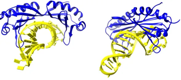
Figure 3 : Structure of yeast TBP bound to the TATA-box
Yeast TBP (in blue) sits on the minor groove of the DNA (in yellow) and provokes a strong kink of the DNA molecule. Second image is turned roughly 45° compared to the first. PDB file 1YTB (J. L. Kim et al., 1993)
Although TBP is unique in yeast, higher eukaryotes can have one or two copies of genes encoding TBP-related factors (TRF) (reviewed in (Davidson, 2003)). TRFs promotes transcription of a subset of genes in a cell-type specific manner (Kopytova et al., 2006; Martianov et al., 2002).
1.1.2.2 TFIIA
TFIIA is a heterodimer in yeast, but is composed of three proteins in human, namely TFIIAα, TFIIAβ and TFIIAγ (respectively 35 kDa, 19 kDa and 12 kDa). TFIIAα AND β derive from a single gene those product is processed by a protease cleavage to form the two subunits. TFIIA functions as a derepressor for transcription, by alleviating inhibition of TBP described before, such as TBP dimerization, binding by BTAF1/Mot1 or TAF1 in TFIID, or binding of NC2. TFIIA binds the TBP-TATA complex and makes contacts with DNA upstream of the TATA-box, in order to stabilize protein-DNA interactions. In addition, it competes for the binding of the convex face of TBP with the inhibiting TAND1 domain of TAF1 (Kokubo et al., 1998). Eventually, the TFIIA-TBP-TATA ternary complex is more resistant to BTAF1/Mot1-mediated dissociation of TBP-TATA complex. TFIIA is formed by two domains: the N-terminal domains of both subunits associate to form a domain interacting with the DNA and the N-terminal part of TBP; the C-N-terminal domains contribute to the second domain which point opposite to TBP (Geiger et al., 1996) (Figure 4)
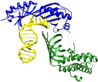
Figure 4 : Structure of TBP-DNA-TFIIA complex
Complex formed by TFIIA (in green), TBP (in blue) and the TATA-box (in yellow). TFIIA N-terminal domain is directed toward the DNA. PDB accession number 1RM1.
TFIIA can be recruited to the promoter by activators, such as the activation domain VP16 (Dion and Coulombe, 2003) and interacts with component of TFIID such as TBP, TAF1, TAF4 and TAF11 (Kraemer et al., 2001; Robinson et al., 2005; Yokomori et al., 1993). Therefore, TFIIA can stimulate transcription by stabilizing TFIID-binding to promoter in response to activator signal.
1.1.2.3 TFIIB
TFIIB plays a central role in initiation, by bridging TBP and the RNA pol II, and is essential for the recruitment of RNA pol II to the PIC. Similarly to TFIIA, but independently from its presence or absence, binding of TFIIB to the TBP-TATA complex forms a more stable ternary complex, and strengthen TBP/TFIID-DNA interaction by stabilizing the bent TBP-DNA complex and decreasing the rate of dissociation of the complex. TFIIB is composed of a single protein of 316 and 345 amino-acids in human and yeast (respectively), and its sequence is divided into 5 functional domains (Kostrewa et al., 2009).
Figure 5 : Domain composition of yeast TFIIB
From (Kostrewa et al., 2009)
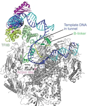
TFIIB recruits the pol II to the promoter, via direct interaction with its N-terminal domain (B-ribbon) and part of the C-terminal domain (B-core), whereas the remaining part of the B-core also binds TBP and the DNA distorted by TBP-binding (Hahn, 2004; Kostrewa et al.,
2009). Moreover, TFIIB is involved in TSS selection and opening of the promoter DNA through the B-linker domain and the B-reader domain, respectively (Kostrewa et al., 2009). Interactions of TFIIB with the DNA were mapped upstream and downstream of the TATA-box, at the BREu and BREd promoter elements, and these additional contacts help to stabilize TBP to the promoter.
A model for the role of TFIIB in transition from initiation to elongation has been proposed. In this model, DNA-bound TFIIB recruits the RNA pol II by the B-ribbon and positions the active site above the cleft of the polymerase (close complex). Then, the double-stranded DNA molecule is opened around 20 base-pairs (bp) downstream of the TATA-box, through the B-linker, and the template strand is placed inside the cleft, close to the active center of the RNA pol II (open complex). Subsequently, the template strand is scanned for an Inr motif by the B-reader. After positioning of two nucleotides in front of the Inr and formation of the first phosphodiester bound, the RNA molecule starts to grow and may interfere with the B-reader loop, leading to ejection of short RNA molecules (abortive transcription). When the RNA molecule reaches seven nucleotides, TFIIB is displaced by clashing with the transcription product and the elongation complex is formed (promoter escape) (Kostrewa et al., 2009; Sainsbury et al., 2013).
1.1.2.4 TFIIF
TFIIF is the fourth GTF to enter the PIC in the sequential assembly pathway, and plays where it plays multiple roles. It is composed of two proteins in human (RNA-pol II Associated Protein or RAP30 and RAP74) which form a heterotetramer. RAP30 is able to bind RAP74 via a N-terminal domain (amino-acids 1-98) and interact with the pol II subunit RPB5 via its central domain (amino acids 107 to 170) (Fang and Burton, 1996). Eventually, a C-terminal domain (amino-acids 164-249) is able to bind DNA in a non-specific manner. RAP74 is also made of three functional domains, involved in interactions with different partners: the N-terminal part (1-172) binds to RAP30 and the TAF1 subunit of TFIID; interactions with TFIIA subunits α and β involves two domains situated in the central part (76-136) and C-terminal part (410-444) of the protein; the C-terminal part (449-517) forms a DNA-binding domain similar to RAP30; moreover, domain spanning amino-acids 358 to 517 is involved in interactions with TFIIB. Domains involved in all these interactions are summarized in Figure 6. Similarly to many transcription factors, TFIIF can be recruited by transcriptional activators.
Figure 6 : Domains of the two subunits forming TFIIF
Position of known interaction domains are indicated. Adapted from (Thomas and Chiang, 2006)
The role and function of TFIIF in PIC are numerous: (i) TFIIF is strongly associated to RNA pol II, via interactions between RAP74 and the RPB4/RPB7 subunits, as well as RPB9, and between RAP30 and RPB5. These interactions facilitate recruitment of the RNA pol II to the promoter-bound TFIIB and TFIID (Chung et al., 2003; Rani et al., 2004); (ii) TFIIF stabilizes interaction of RNA pol II to the promoter by providing additional contacts with the DNA (Robert et al., 1998); (iii) TFIIF allows further recruitment of TFIIE and TFIIH to the PIC via direct interactions with TFIIE (Maxon et al., 1994); (iv) TFIIF, similarly to TFIIB, is located close to the active center of the RNA pol II and contribute to TSS selection driven by TFIIB (Ghazy et al., 2004); (v) TFIIF is involved in promoter escape and release of RNA pol II pauses (Zhang and Burton, 2004); (vi) TFIIF increases specificity of RNA pol II by reversing the binding of pol II to non-promoter sequence (Orphanides et al., 1996).
1.1.2.5 TFIIE
TFIIE is the next GTF to enter the PIC after TFIIF and the RNA pol II, and is essential for transition from initiation to elongation. TFIIE is composed of two subunits, α and β, of 439 and 291 amino acids in human respectively, which form a heterotetramer. This heterotetramer is also found in yeast. The N-terminal domain of TFIIEα is involved in interaction with TFIIEβ
1 139 RAP74 2 172 158 214 TAF1 TFIIEβ RAP30 358 517 TFIIB 1 517 RAP30 1 249 1 98
RAP74 107 170 RNA pol II 175 243 DNA 27 152 TFIIB 449 517 DNA 76 136 TFIIA 410 444 TFIIA 363 444 RNA pol II