HAL Id: hal-01506235
https://hal.archives-ouvertes.fr/hal-01506235
Submitted on 29 May 2020
HAL is a multi-disciplinary open access
archive for the deposit and dissemination of
sci-entific research documents, whether they are
pub-lished or not. The documents may come from
teaching and research institutions in France or
abroad, or from public or private research centers.
L’archive ouverte pluridisciplinaire HAL, est
destinée au dépôt et à la diffusion de documents
scientifiques de niveau recherche, publiés ou non,
émanant des établissements d’enseignement et de
recherche français ou étrangers, des laboratoires
publics ou privés.
Distributed under a Creative Commons Attribution| 4.0 International License
Morphological and virulence variation among isolates of
Mycosphaerella pinodes the causal agent of pea leaf
blight
Benali Setti, Mohamed Bencheikh, Jamel Henni, Claire Neema
To cite this version:
Benali Setti, Mohamed Bencheikh, Jamel Henni, Claire Neema. Morphological and virulence variation
among isolates of Mycosphaerella pinodes the causal agent of pea leaf blight. African Journal of
Agricultural Research, Academic Journals, 2011, 6 (5), pp.1067 - 1075. �hal-01506235�
ISSN 1991-637X ©2011 Academic Journals
Full Length Research Paper
Morphological and virulence variation among isolates
of
Mycosphaerella pinodes
the causal agent of pea leaf
blight
Benali Setti
1*, Mohamed Bencheikh
1, Jamel Henni
2and Claire Neema
31Institut de Biologie, Université de Chlef, BP151, 02000- Algérie. 2Institut des Sciences, Université d’Es Senia, Oran, Algérie.
3UMR de Pathologie Végétale, INRA/INA-PG/Université Paris VI, 16 rue Claude Bernard, 75231, France.
Accepted 20 October, 2010
Mycosphaerella blight caused by Mycosphaerella pinodes (Berk. and Blox.) Vestergr. is an important disease, causing severe damage in peas. Variability of 20 Algerian isolates of M. pinodes
representative of four agro climatic regions were investigated on the basis of cultural, morphological and pathogenicity. Culture and morphology showed variations in colony color, radial growth pattern and production of pycnidia and pycnidiospores. Significant differences (P < 0.05) in both pycnidia and pycnidiospores size among isolates were observed. Hence, the size of pycnidia and pycnidiospores of M. pinodes varied from 145 × 143 µm to 280 × 265 µm and from 11.5 × 2.3 µm to 22.5 × 6.3 µm respectively. Using the factor analysis, this revealed that the first principal component (pc) was more related to the growth and sporulation aspect, hence, the colony growth and both the pycnidia and pycnidiospore density were more related to the first pc, while the second pc contributed for the pycnidiospores size. The isolates were also evaluated for their pathogenicity on seven cultivars in controlled conditions. Cluster analysis based on disease rating on a scale of 1 to 5, indicated higher similarity coefficient. In addition, using Euclidian distances method, the clusters were subdivided at 70% of similarity in seven pathotype groups (PG). The two first pathotypes grouped the most isolates (70%), representing isolates from the four agro climatic regions. However, the members of same group were different in their cultural and morphological characteristics. A detailed study to investigate molecular and genetic basis of diversity is suggested.
Key words: Mycosphaerella blight, morphometry, cluster analysis. INTRODUCTION
Blight caused by Mycosphaerella pinodes is one of the most devastating diseases of pea that causes yield losses of over 50% in some years (Wallen, 1965; Bretag, 1989; Xue et al., 1997; Tivoli and Banniza, 2007) and may cause total failure to the crop under epidemic conditions.
A number of research studies have been undertaken dealing with different aspects of the disease worldwide in order to understand and manage the disease. Different parameters have been explored including
*Corresponding author. E-mail: setiben@yahoo.fr.
pathogenecity, life cycle, and disease cycle, epidemiology, breeding for resistance as well as cultural and chemical control of pea blight (Bretag, 1989; Xue et al., 1997; Onfroy et al., 2007; Setti et al., 2008, 2009).
Differences in cultural characteristics and pathogenecity among populations of this pathogen have been described (Clulow et al., 1992; Xue et al., 1997; Wroth, 1999; Zhang et al., 2004). On the other hand, Setti et al. (2009) observed different susceptibility of several cultivars with different Algerian populations of M. pinodes that revealed differences in aggressiveness.
This variation in M. pinodes is likely to enhance by the presence of the teleomorph stage under field conditions. In fact, sexual recombination, and somatic hybridization
1068 Afr. J. Agric. Res.
with or without subsequent nuclear fusion and recombination provide new pathotypes within populations.
Although blight can be controlled by the use of disease free seeds, destruction of plant debris, and fungicide treatments, under epidemic conditions, these approaches are not feasible (Tivoli et al., 1996; Tivoli and Banniza, 2007). Therefore, the best method of controlling this disease is through use of resistant cultivars. However, the development of resistant cultivars is a complex phenomenon because of the nature of the pathogen and the breakdown of the varietal resistance (Quershi and Alam, 1984; Onfroy et al., 2007).
It is in this context, the present study was thus initiated on the morphological, cultural and pathogenic variability among and within the populations of M. pinodes by using multivariate analysis which was considered the most appropriate technique for comparison (Basandrai et al., 2005).
MATERIALS AND METHODS
Cultural and morphological variation of M. pinodes
Twenty isolates of M. pinodes, obtained from four agro climatic regions in western regions of Algeria and collected during 2001 to 2005 were observed for their morphological and cultural variations. The four regions were coded Pg1, Pg2, Pg3 and Pg4.
5 mm diameter disc from actively growing cultures were placed in the center of a 90 mm diameter Petri plates containing Mathur medium (Onfroy et al., 1999). Inoculated plates were incubated at 21°C and observed for colony color of the colony, diameter (mm), number of pycnidia. For quantification of pycnidia, 1 cm² was cut at a distance of 1 cm from the center of a well sporulating culture on Mathur medium 10 days after incubation. The disc was observed under magnifying lens to count the number of pycnidia. For pycnidiospores quantification, similar disc was macerated in minimal volume of water, diluted to 10 ml and the number of spores was measured using a haemocytometer slide. For the measurements of both the length and diameter of both pycnidia, and pycnidiospores, 50 of each isolate were examined. Both the pycnidia and pycnidiospores were placed in a sterile slide, covered with a sterile cover slip, and stained with cotton blue in lactophenol solution. The measurements of each of three prepared slides (replications) were determined under a compound microscope (Motic, B1, LM-Scope, Austria) at × 40 magnification with the aid of an ocular micrometer. The ocular micrometer calibrated against a fixed and known micrometer stage (2 mm in length). The ocular micrometer is divided into 50 units, each unit equal to 4.5 m at × 40.
Pathogenic variation of M. pinodes isolates.
Plant material
The pea cvs Onward and cv Merveille de Kelvedon (MK), Douce de Provence (DP), Akel, Rondo, Grillevert, and Lucy are cultivars cultivated in most parts of western Algeria. Seeds of these cultivars were sown in 20 cm diameter pots containing an unsterilized soil/compost mixture. Ten seeds were planted per pot and seedlings were thinned to five. The plants were maintained in a growth chamber. Three replicates were used for each
combination.
Fungal material
The 20 isolates of M. pinodes, obtained from four agro climatic regions already tested for their cultural and morphological aspects were used in this study.
Inoculum production
Strains were raised on Potato Dextrose Agar (PDA) for 10 days at 21°C. Conidia from 10 days old cultures were collected by adding 10 ml of sterile distilled water to dislodge the spores. The spore suspension was filtered through two layers of cheesecloth to remove the mycelium and agar fragments. The concentration of spores was determined using a haemocytometer slide. The suspension was diluted with sterile distilled water to obtain a final concentration of 3.5 × 10⁶ conidia ml-1.
Inoculation
Plants of 15 days were inoculated by spraying to runoff with the spore suspension, using a spray atomizer with an adjustable nozzle to form a high density of fine droplets on the aerial parts of the plants. Control plants were sprayed with sterile distilled water. The plants were covered for 48 h with transparent polyethylene bags immediately after inoculation and sprayed inside the bags with distilled water to facilitate infection. After incubation, the plants were uncovered, and kept in an uncontrolled glasshouse at temperatures from 15 to 25°C.
Disease assessment
M. pinodes infection on the leaves was recorded 21 days after inoculation using a 0 to 5 disease scale according to Tivoli et al. (1996), where 0 = no lesion; 1 = a few scattered flecks; 2 = numerous flecks; 3 = 10 to 15% leaf area necrotic and presence of flecks; 4 = 50% of leaf area covered by lesions; 5 = 75 to 100% of leaf area dehydrated or necrotic. To determine the incubation period (IP) and the latent period (LP), plants were inspected daily for up to 20 days.
Data analysis
Growth rate and measurements of size and density of both pycnidia and pycnidiospores were analyzed statistically. Equality of variance was first determined using F tests. Multiple data sets were analyzed by analysis of variance. Where appropriate, means are supplemented by standard deviations in parenthesis.
The variability between isolates was examined with factor analysis. For the pathogenic variation of isolates, analysis of variance was assessed for both isolates and cultivars. Means of cultivars was performed using Tukey’s honestly significant difference (HSD) test.
A similarity matrix was constructed using Euclidian distance method. The resulting similarity data was used to construct a dendrogram. All statistics analysis were performed using statistics software SPSS (Version 8.0)
RESULTS
among the 20 isolates of M. pinodes on the basis of morphological characters and pathological test (Table 1). In cases of cultural traits and color type of the isolates, great difference was observed. These varied from cream, gray, to totally dark color. The colony of isolates HU11, SHU5, AR4, AR11 and SAR24 was light brown. Dark brown and black colony was observed in the isolates SHU8, SHU13, AR9, HU10, SHU7 and AR10. On the other hand, the isolates SAR13 and SAR21 had developed colony with gray appearance, whereas the creamy aspect was noted only for the isolate HU16 and HU19 (Figure 1).
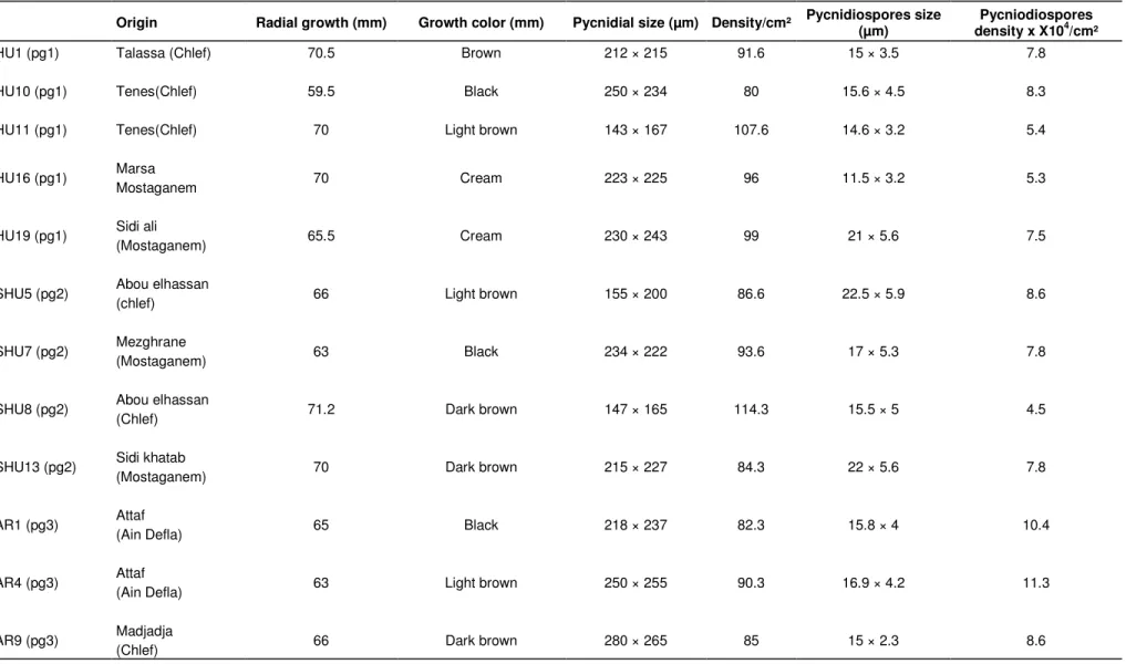
Data recorded after 10 days of incubation revealed substantial differences for linear growth among the isolates of M. pinodes. The colony diameter of the 20 isolates on Mathur medium ranged from 59.5 to 71.2 mm. The maximum colony diameter was exhibited by isolates SHU8, HU16 and SAR21 with 71.2, 70.5, and 70 mm respectively. The least growth of 59.5 was shown by the isolate HU10. Furthermore, data on morphological characteristics of the aforementioned 20 isolates of M. pinodes in respect of pycnidial formation, size of pycnidia and pycnidiospores revealed that the production of pycnidia and pycnidiospores among the isolates varied significantly. Most of the isolates produced a large number of pycnidia (> 90 pycnidia/cm²). The abundant formation of pycnidia was respectively observed on isolates SHU8 and HU11 with 113 and 112 pycnidia/cm². While the least pycnidial formation was obtained on isolate HU16 and SAR13 with 78 and 79 pycndia/cm² respectively.
The isolates also displayed significant differences in pycnidiospores density. Isolate AR4 and AR1 produced significantly more pycnidiospores than any other isolates. Generally, the number of pycnidiospores ranged from 2.3 × 10⁵ to 11.3 × 105 cm-². The mean pycnidiospore density was of 6.90 × 105 cm-² (sd = 2.29).
On the other hand, the data on size of pycnidia among isolates varied significantly. The maximum size of pycnidia was obtained from isolate AR9 and SAR13 with 280 × 265 µm and 250 × 256 µm respectively. While the least pycnidial size obtained was with isolate HU11 and SAR13 which had the dimension of 143 × 167 µm and 145 × 143 µm respectively.
Similarly, the size of pycnidiospores varied on different isolates. The average length of pycnidiospores was 18.55 (sd = 4.13). The maximum length was observed for the isolate SHU5 and SHU13 with 22.5 and 21 µm respectively. The width size also varied among isolates, this ranged from 2.3 to 6.30 µm with a mean of 4.45 µm (sd = 1.06) (Table 1). Furthermore, for accurate comparison between these isolates two multivariate analysis were used namely the principal component analysis on the basis of the six cultural and morphological characters. The principal component analysis showed that only two principal axis gave
eigenvalues greater than 1 (Table 2). While the other axis all had eigenvalues less than 1. Hence, the first two principal components were considered important and contribute the most in the distribution of variation existing among the isolates. The component 1 had an eigenvalue of 2.153, accounted for 43.04% of the overall variance in the data set (Table 2). Component 2 had an eigenvalue of 1.459 and accounted for 29.17% of the total variance. Hence, the two principal components contributed for 72.24% of the total variability (Table 2).
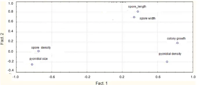
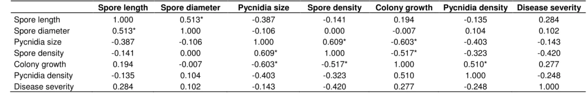
The first pc was more related to the growth and sporulation aspect, hence the colony growth and both the pycnidia and pycnidiospore densities were more related to the first pc, while the second pc contributed for the pycnidiospores size (Figure 2). On the other hand, positive correlation between the morphological and cultural characteristics was observed. Hence, the maximum correlation was noted by spore density and pycnidia size. A negative correlation was also observed between pycnidia size and colony growth and between spore density and colony growth (Table 3).
Isolate pathogenecity
There were significant differences (P < 0.001) in disease severity between isolates from different geographic areas. Variation in the distribution of the mean DS for the 20 isolates of M. pinodes across the seven cultivars was exhibited in a continuous manner. In addition, the hierarchical cluster analysis using Euclidian distances for DS was used to classify the isolates of M. pinodes. The clusters were subdivided at 70% of similarity in seven pathotype groups (PG). The two first pathotypes grouped the most isolates (60%), hence, the first PG was constituted of four isolates representing two agroclimatical regions, while the second PG was the most important numerically and it is represented by 50% of the total isolates. These isolates represented different agroclimatical regions (Figure 3). The dendrogram also indicated that isolates collected from the same location were similar to those from widely dispersed sites, or from different cultivars. Moreover, mean comparison with the t test of DS revealed no significant differences between population groups (P < 0.05). Isolates from the same area were always different from each other and had different disease indices. On the other hand, the cultivar reactions varied significantly between each other (P < 0.05). All cultivars showed symptoms involving lesions on leaves and stems and even in severe cases resulted in seedling mortality.
Finally, no positive correlation between the morphological and cultural characters and pathogenecity was observed (Table 3). The disease index of pea cultivars varied from 2.80 to 3.72, with a mean of 3.27 and a standard deviation of 1.12’ MK’ and
1070 Afr. J. Agric. Res.
Table 1. Morphological and cultural characteristics of isolates of Mycosphaerellapinodes representing four agroclimatic regions in western Algeria.
Origin Radial growth (mm) Growth color (mm) Pycnidial size (µm) Density/cm² Pycnidiospores size (µm) density x X10Pycniodiospores 4/cm²
HU1 (pg1) Talassa (Chlef) 70.5 Brown 212 × 215 91.6 15 × 3.5 7.8
HU10 (pg1) Tenes(Chlef) 59.5 Black 250 × 234 80 15.6 × 4.5 8.3
HU11 (pg1) Tenes(Chlef) 70 Light brown 143 × 167 107.6 14.6 × 3.2 5.4
HU16 (pg1) Marsa 70 Cream 223 × 225 96 11.5 × 3.2 5.3
Mostaganem
HU19 (pg1) Sidi ali 65.5 Cream 230 × 243 99 21 × 5.6 7.5
(Mostaganem)
SHU5 (pg2) Abou elhassan 66 Light brown 155 × 200 86.6 22.5 × 5.9 8.6
(chlef)
SHU7 (pg2) Mezghrane (Mostaganem) 63 Black 234 × 222 93.6 17 × 5.3 7.8
SHU8 (pg2) Abou elhassan 71.2 Dark brown 147 × 165 114.3 15.5 × 5 4.5
(Chlef)
SHU13 (pg2) Sidi khatab 70 Dark brown 215 × 227 84.3 22 × 5.6 7.8
(Mostaganem)
AR1 (pg3) Attaf 65 Black 218 × 237 82.3 15.8 × 4 10.4
(Ain Defla)
AR4 (pg3) Attaf (Ain Defla) 63 Light brown 250 × 255 90.3 16.9 × 4.2 11.3
Table 1. Contd.
AR11 (pg3) Madjadja 69.3 Lightly brown 168 × 205 94.3 18.6 × 5.4 6.7
(Chlef)
SAR2 (pg4) Madjadja 69.8 Brown 233 × 242 103.3 20 × 5.5 6.7
(Chlef)
SAR6 (pg4) Mohamedia 69.5 Dark brown 216 × 223 94.6 18.6 × 5.5 10.4
(Mascara)
SAR10 (pg4) Warizan(Rhilizane) 69 Brown 166 × 158 88.3 21.5 × 5 3.4
SAR13 (pg4) Dahmouni 63 Gray 250 × 256 87 14.6 × 4.5 5.9
(Tiaret)
SAR16 (pg4) Dahmouni 69 Dark brown 145 × 143 99 20.3 × 3.2 5.4
(Tiaret)
SAR21 (pg4) Lardjem 70 Gray 167 × 146 85.6 20.8 × 2.8 4.3
(Tissemsilt)
SAR24 (pg4) Mascara(Mascara) 69.6 Light brown 145 × 159 90.3 21 × 6.3 3.3
‘Rondo’, on partially resistant cultivars, the DS index was 2.82 and 2.85 respectively. The most susceptible cultivars were ‘Onward’, ‘Lucy’ and ‘DP’, with a disease index greater than 3.63. Mean comparison of the disease index of the seven cultivars revealed significant differences.
DISCUSSION
The study of the variability among the populations of
M. pinodes for their pathogenecity, their morpho-logical and cultural characteristics is crucial for program and strategy of breeding in order to evolve genotype with durable disease resistance.
This study reveals that M. pinodes is composed of several biotypes with marked differences in their morphological and cultural characteristics. Such variability in M. pinodes has already been reported in different countries (Barve, 2003; Peever et al., 2004; Tivoli and Banniza, 2007). Xue et al. (1997), Zhang et al. (2004), Tivoli and Banniza (2007) recorded
differences in growth rates among different isolates obtained from different regions. The growth rate in our study showed variation among isolates and this ranged from 59.5 mm to 71.2 mm with a mean of 67.96 mm (sd = 4.29).
Furthermore, the isolates tested revealed important variation in sporulation that ranged from 2.3 × 10⁵ to
11.3 × 105 cm-² with a mean of 6.90 × 105 cm-² (sd = 2.29). In fact, both the growth rate and sporulation were used by Grewal (1984) for explaining the aggressiveness and virulences of isolates. He
1072 Afr. J. Agric. Res.
Figure 1. Cultural variation in the colony of different Mycosphaerella pinodes isolates on Mathur medium at 21°C.
Table 2. Principal components for morphological and culturals traits of 20 isolates of Mycosphaerella pinodes. F1 F2 Eigenvalues 2.153 1.459 Proportion of variance 43.065 29.172 Cumulative variance 43.065 72.237 Eigenvectors pycnidiospores lengh 0.1485 0.8959 pycnidiospores width 0.0046 0.8068 Pycnidia size -0.6210 -0.2503 Colony growth 0.8360 0.1443 Pycnidia density 0.7076 -0.1954 Pycnidiospores density -0.7758 -0.0285
reported that relatively fast growing and less sporulating isolates were less aggressive while slow growing and abundantly sporulating isolates, more aggressive. However, no such correlation was noted in our study. Differences in cultural appearance among isolates from different regions have also been observed. This aspect varied from light brown to completely dark colony.
Based on pycnidia and pycnidiospores dimensions, several workers recorded variation in size of pycnidia and pycnidiospores among different isolates of the fungus (Clulow et al., 1991, 1992; Corbière et al., 1994; Peever et al., 2004; Tivoli and Banniza, 2007). In fact,
the size of pycnidia and pycnidiospores is a character which has a taxonomic importance (Agrios, 2004).
In our study, the size of pycnidia ranged from 145 × 143 µm to 280 × 265 µm, and the pycnidiospores varied from 11.5 × 2.3 µm to 22.5 × 6.3 µm. The variance analysis of both the size of pycnidia and pycnidiospores had significant differences between isolates. Similar variations have been reported in others species of Ascochyta namely A. pisi (Jameli et al., 2005), A. lentis (Kaiser et al., 1993). The observations on A. rabiei have revealed variations among isolates obtained from different countries (Haware, 1987; Nene and Reddy, 1987;
1.0 0.8 0.6 0.4 0.2 0.0 -0.2 -0.4 -1.0 -0.6 -0.2 0.2 0.6 1.0
Figure 2. Scattered diagram for two factors in 20 isolates of Mycosphaerella pinodes from four agro climatic regions in western Algeria.
Iqbal et al., 2004).
These variations could be the result of different genetic exchange occurring in population as sexual recombination, hybridization with or without subsequent nuclear fusion and parasexual cycle. In fact, M. pinodes is a teleomorph of A. pinodes. This pathogen forms its pseudothercia on the sensescent stipules during the second part of cropping season (Barve et al., 2003). These sexual fruiting structures permit the fungi to overwinter and are considered to play an important role in generating pathogen variability (Tivoli and Banniza, 2007; Ali et al., 2009). In fact, with the presence of sexual reproduction, new combination of genes arises into the field, from one growing season to the other.
In the multivariate analysis, separation between the populations groups of M. pinodes examined was not evident. This showed that neither PCA nor the hierarchical classification (HCA) were able to distinguish between isolates according to their origin. Therefore, we conclude that there were no consistent morphological or cultural differences between M. pinodes populations groups.
The isolates tested in the present study showed variation in pathogenicity among a collection of 20 isolates of M. pinodes against 7 commercial cultivars with different levels of resistance ranging from susceptible to partially resistant.
The disease rating of each isolate of M. pinodes towards the cultivars exhibited continuous variability. All symptoms involving both leaves and stems initially produce small lesions in the form of numerous flecks. Leaves with many lesions wither before the lesions become large, especially on the lower portion of the plants. The most aggressive isolates were from different population group namely SAR21, SHU8 and HU16,
whereas, the less aggressive isolates were AR11, SAR10 and HU10.
The hierarchical cluster analysis using Euclidian distances were subdivided at 70% of similarity in seven pathotypes (Aggressiveness groups) two of which were the most important numerically and they grouped more than 70% of the total isolates. Inconsistent clustering pattern of isolates obtained from the same origin may be attributed towards frequent exchange of breeding materials. Several reasons have been suggested, such as the increase of pea growing area and the introduction of new cultivars that contribute to extend the diversity of the pathogen population. The mode of reproduction of M. pinodes also contributes in extending the variability (Crino et al., 1985; Hussain and Barz, 1997). Kaiser (1992) and Ali et al. (2009) also suggested that the sexual stage can generate new recombinants with varying aggressiveness spectrum.
Moreover, it is likely that morphological and cultural variations can provide only the preliminary variation in M. pinodes isolates, since these variations did not correlate with the geographical origin and pathogenic variations. On the other hand, in previous study, using more isolates showed that these cultivars had different levels of quantitative resistance (Setti et al., 2009). The mean comparison test of the DS showed that the seven cultivars fell into three groups (P < 0.0001) going from susceptible to partially resistant. In fact, the studies on pea’s resistance to M. pinodes have shown the absence of specific resistance (Nasir and Hope, 1991; Clulow et al., 1992). Recently, many authors described the observed resistance in peas cultivars as partial (Onfroy et al., 1999; Wroth and Khan, 1999; Wang et al., 2000, Fondevilla et al., 2005). In fact, the partial resistance results in the slow down of disease progress and
1074 Afr. J. Agric. Res.
Table 3. Pearson linear correlation coefficient between the six morphological and cultural characters and the disease severity.
Spore length Spore diameter Pycnidia size Spore density Colony growth Pycnidia density Disease severity
Spore length 1.000 0.513* -0.387 -0.141 0.194 -0.135 0.284 Spore diameter 0.513* 1.000 -0.106 0.000 -0.007 0.104 0.102 Pycnidia size -0.387 -0.106 1.000 0.609* -0.603* -0.403 -0.143 Spore density -0.141 0.000 0.609* 1.000 -0.517* -0.323 -0.420 Colony growth 0.194 -0.007 -0.603* -0.517* 1.000 0.510* 0.277 Pycnidia density -0.135 0.104 -0.403 -0.323 0.510 1.000 -0.248 Disease severity 0.284 0.102 -0.143 -0.420 0.277 -0.248 1.000 *:P values < 0.05.
Figure 3. Dendrogram showing clustering of the pathogenicity of Mycosphaerella pinodes on seven cultivars.
or reduction in the pathogen multiplication (Parlevliet, 1979).
Finally, this study showed that none of the morphometrics characters or the origin of the isolate could be correlated with pathogenic variability. The use of any characters to
distinguish aggressiveness between unknown isolates of the fungus requires much attention and verification.
Biochemical and molecular approaches may be helpful for further study to confirm the association and correlation in this respect.
Furthermore, this study indicated that M. pinodes
isolates collected from Algeria were composed of various aggressiveness groups. Such results are useful for choosing pathotypes representing populations of the pathogen rather than individual in screening for utilization in breeding
programme.
REFERENCES
Ali SR, Iqbal SHM, Iqbal U, Ghafoor A, Akram A (2009). Pathogenic diversity in Ascochyta rabiei (Pass)Lib of chickpea. Pakistan J. Bot., 41(1): 413-419.
Agrios GN (2004). Plant pathology (5th Ed.). Elsevier Academic Press. Barve MP, Arie T, Slimath SS, Muehlbauer FJ, Peever TL (2003).
Cloning and characterization of the mating type (MAT) locus from
Ascochyta rabiei (teleomorph: Didymella rabiei) and a MAT phylogeny of legume-associated Ascochyta spp. Fungal Genet. Biol., 39: 151-167.
Basandrai AK, Pande S, Krishna Kishore G, Crouch JH, Basandrai D (2005). Cultural, Morphological and Pathological Variation in Indian Isolates of Ascochyta rabiei, the Chickpea Blight Pathogen. Plant Pathol. J., 21(3): 207-213.
Bretag TW (1989). Resistance of pea cultivars to Ascochyta blight caused by Mycosphaerella pinodes, Phoma medicagenis and
Ascochyta pisi. Ann. Appl. Biol., 114: 157-159.
Crino PA, Porto-Puglia A, Saccordo F (1985). Reaction of chickpea lines to Ascochyta rabiei in winter sowing in Italy. Int. Chickpea Newslett., 12: 27-29.
Clulow SA, Lewis BG, Parker ML, Matthews P (1991). Infection of pea epicotyls by Mycosphaerella pinodes. Mycol. Res., 95: 817-820. Clulow SA, Lewis BG, Matthews P (1992). Expression of resistance to
Mycosphaerella pinodes in Pisum sativum. Plant Pathol., 41: 362-369.
Corbière R, Gelie B, Molinero V, Spire D, Agarwal VK (1994). Investigations on seedborne nature of Mycosphaerella pinodes in pea seeds. Seed Res., 22: 26-30.
Fondevella S, Avila CM, Cubero JI, Rubiales D (2005). Response to
Mycosphaerella pinodes in a germplasm collection of Pisum spp. Plant Breed.,124: 313-315.
Grewal JS (1984). Evidence of physiologic races in Ascochyta rabiei of chickpea. In: Saxena M.C. and K.B. Singh. K.B. (eds.), Proceeding Workshop on Ascochyta Blight and Winter Sowing of Chickpea. ICARDA, 4–7 May, 1981, Syria, pp. 55-65.
Hussain S, Barz W (1997). Isoenzym polymorphism in Ascochyta rabiei isolates from Pakistan Pakistan J. Bot., 29: 207-216. Haware MP (1987). Pathogenic variability in Ascochyta rabiei.
ICARDA Food Legume Improvement Program. Annu. Rep., pp. 129-130.
Iqbal SM, Ghafoor A, Ayub N, Ahmad Z (2004). Pathogenic diversity in
Ascochyta rabiei isolates collected from Pakistan. Pakistan J. Bot., 36: 427-39.
Jamali AR, Iqbal SM, Rauf CA, Akram A (2005). Studies on the pathogenic variability in Ascochyta pisi. Int. J. Agri. Biol.,7: 272-774. Kaiser WJ (1992). Fungi associated with the seeds of commercial lentils
from the US Pacific Northwest. Plant Dis.,76: 605-610.
Kaiser WJ, Hannan RM, Rogers JD (1994). Factors affecting growth and sporulation of Ascochyta fabae f.sp. lentis. Plant Dis., 78: 374-379.
Kaiser WJ, Hellier BC (1993). The teleomorph of Ascochyta fabae f.sp
lentis on lentil straw. Phytopathol., pp. 82-692.
Nasir M, Hoppe HH (1991). Studies on pathotype differentiation within
Mycosphaerella pinodes (Berk&Blox) Vesterger, a component of the Ascochyta-disease-complex of peas (Pisum sativum L.). Zeit. Pflanzenkrank. Pflanschutz, 98: 619-627.
Nene YL, Reddy MV (1987). Chickpea diseases and their control. In: The Chickpea, ed. by M. C. Saxena and K. B. Singh. CAB International. Oxon, UK, pp. 233-270.
Onfroy C, Tivoli B, Corbière R, Bouznad Z (1999). Cultural, molecular, and pathogenic variability of Mycosphaerella pinodes and Phoma medicagenis var. pinodella isolates in dried pea (Pisum sativum) in France. Plant Pathol., 48: 218-229.
Onfroy C, Baranger A, Tivoli B (2007). Biotic factors affecting the expression of partial resistance in pea to ascochyta blight in a detached stipule assay. Eur. J. Plant. Pathol., 119: 13-27.
Parlevliet JE (1979). Components of resistance that reduce the rate of epidemic development. An. Rev. Phytopathol., 17: 203–222.
Peever TL, Salimath SS, Su G, Kaiser WJ, Muehlbauer FJ (2004). Historical and contemporary multilocus population structure of
Ascochyta rabiei (teleomorph: Didymella rabiei) in the Pacific Northwest of the United States. Mol. Ecol., 13: 291-309.
Qureshi SH, Alam SS (1984). Pathogenic behavior of Ascochyta rabiei
isolates on different cultivars of chickpea in Pakistan. Int. chickpea Newsltr.,10: 29-31.
Setti B, Bencheikh M, Henni J, Neema C (2008). Effect of pea cultivar, pathogen isolate, inoculums concentration and leaf wetness duration on Ascochyta blight caused by Mycosphaerella pinodes.
Phytopathol. Med., 47(3):214-222.
Setti B, Bencheikh M, Henni J, Neema C (2009). Comparative aggressiveness of Mycosphaerellapinodes on peas from different regions in western Algeria. Phytopathol. Med.,48(2): 195-204. Tivoli B, Beasse C, Lemarchand E, Masson E (1996). Effect of
Asochyta blight (Mycosphaerella pinodes) on yield components of single pea (Pisum sativum) plants under field conditions. Ann. Appl. Biol., 129: 207-216.
Tivoli B, Banniza S (2007). Comparaison of the epidemiology of ascochyta blights on grain legumes. Eur. J. Plant Pathol., 119: 59-76. Wallen VR (1965). Field evaluation of the importance of the
Ascochyta complex on peas. Can. J. Plant Sci., 45: 27-33.
Wang H, Hwang SF, Chang KF, Turnbull GD, Howard RJ (2000). Characterization of Ascochyta isolates, and susceptibility of pea cultivars to the Ascochyta disease complex in Alberta. Plant Pathol., 49: 540-545.
Wroth JM (1999). Evidence suggests that Mycosphaerella pinodes
infection of Pisum sativum is inherited as a quantitative trait. Euphytica, 107: 193-204.
Wroth JM, Khan TN (1999). Differential responses of field pea (Pisum sativum L.) to ascochyta blight (Mycosphaerella pinodes): Rating disease in the field. Aus. J. Agric. Res., 50: 601-615.
Xue AG, Warkentin TD, Kenaschuk EO (1997). Effect of timings of inoculation with Mycosphaerella pinodes on yield and seed infection of field pea. Can. J. Plant Sci., 77: 685-689.
Zhang JX, Fernando WGD, Xue AG (2004). Temporal and spatial dynamics of Mycosphaerella blight (Mycosphaerella pinodes) in field pea. Can. J. Plant Pathol., 26: 522-525.