HAL Id: hal-01837483
https://hal.archives-ouvertes.fr/hal-01837483
Submitted on 25 May 2020
HAL is a multi-disciplinary open access
archive for the deposit and dissemination of sci-entific research documents, whether they are pub-lished or not. The documents may come from teaching and research institutions in France or abroad, or from public or private research centers.
L’archive ouverte pluridisciplinaire HAL, est destinée au dépôt et à la diffusion de documents scientifiques de niveau recherche, publiés ou non, émanant des établissements d’enseignement et de recherche français ou étrangers, des laboratoires publics ou privés.
Eleostearic Phospholipids as Probes to Evaluate
Antioxidants Efficiency against Liposomes Oxidation
Erwann Durand, André Delavault, Claire Bourlieu, Jérôme Lecomte, Bruno
Barea, Maria-Cruz Figueroa Espinoza, Eric A. Decker, Françoise Michel
Salaun, Gilles Kergourlay, Pierre Villeneuve
To cite this version:
Erwann Durand, André Delavault, Claire Bourlieu, Jérôme Lecomte, Bruno Barea, et al.. Eleostearic Phospholipids as Probes to Evaluate Antioxidants Efficiency against Liposomes Oxidation. Chem-istry and Physics of Lipids, Elsevier, 2017, 209, pp.19-28. �10.1016/j.chemphyslip.2017.10.006�. �hal-01837483�
Version postprint
Accepted Manuscript
Title: Eleostearic Phospholipids as Probes to Evaluate Antioxidants Efficiency against Liposomes Oxidation. Authors: Erwann Durand, Andr´e Delavault, Claire Bourlieu, J´erˆome Lecomte, Bruno Bar´ea, Maria Cruz Figueroa
Espinoza, Eric A. Decker, Franc¸oise Michel Salaun, Gilles Kergourlay, Pierre Villeneuve
PII: S0009-3084(17)30226-8
DOI: https://doi.org/10.1016/j.chemphyslip.2017.10.006
Reference: CPL 4597
To appear in: Chemistry and Physics of Lipids Received date: 22-8-2017
Revised date: 21-9-2017
Accepted date: 19-10-2017
Please cite this article as: Durand, Erwann, Delavault, Andr´e, Bourlieu, Claire, Lecomte, J´erˆome, Bar´ea, Bruno, Espinoza, Maria Cruz Figueroa, Decker, Eric A., Salaun, Franc¸oise Michel, Kergourlay, Gilles, Villeneuve, Pierre, Eleostearic Phospholipids as Probes to Evaluate Antioxidants Efficiency against Liposomes Oxidation.Chemistry and Physics of Lipids https://doi.org/10.1016/j.chemphyslip.2017.10.006
This is a PDF file of an unedited manuscript that has been accepted for publication. As a service to our customers we are providing this early version of the manuscript. The manuscript will undergo copyediting, typesetting, and review of the resulting proof before it is published in its final form. Please note that during the production process errors may be discovered which could affect the content, and all legal disclaimers that apply to the journal pertain.
Version postprint
Eleostearic Phospholipids as Probes to Evaluate Antioxidants
Efficiency against Liposomes Oxidation.
Erwann Durand1*,André Delavault1, Claire Bourlieu2, Jérôme Lecomte1, Bruno Baréa1, Maria Cruz Figueroa Espinoza3, Eric A. Decker4, Françoise Michel Salaun5, Gilles Kergourlay5, Pierre Villeneuve1
1 CIRAD, UMR IATE, Montpellier F-34398, France 2 INRA, UMR IATE, Montpellier F-34060, France
3 Montpellier SupAgro, UMR IATE, Montpellier F-34060, France 4 Food Chem. Dept., U. Mass., Amherst, MA, USA
5 Diana Pet Food, ZA du Gohélis, 56250 ELVEN, France
*Corresponding author:
Dr. Erwann DURAND, CIRAD, UMR IATE, Montpellier
F-34398, France
E-mail address: erwann.durand@cirad.fr
Tel: +33 (0)4 99 61 20 30
Version postprint
Graphical abstract
Highlights
A new oxidation high-throughput assay based on the UV properties of a phospholipid probe is proposed.
Two site of oxidation (membrane or aqueous phase) were developed.
The new method, tested with several antioxidants, is rapid and reproducible.
Aqueous or membrane-induced oxidations provide further information on the antioxidants efficacy.
ABSTRACT:
Regardless of the applications: therapeutic vehicle or membrane model to mimic complex
biological systems; it is of a great importance to develop simplified, reproducible and rapid
model assays allowing for a relevant assessment of the liposomal membrane oxidation and
therefore antioxidant activity of selected molecules. Here, we describe a new and
high-throughput assay that we called “Vesicle Conjugated Autoxidizable Triene (VesiCAT)”. It is
based on specific UV absorbance spectral properties of a new phospholipid probe, synthesized
with natural conjugated eleostearic acid extracted from Tung oil. The VesiCAT assay has
been developed with two different radical generators (2,2’-azobis(2-amidinopropane)
Version postprint
constant flux of oxidant species, either in membrane or in aqueous phase. This method
appears very efficient in assessing the effect of various pure antioxidant molecules in their
ability to preserve liposomes from oxidative degradation. In addition, the AAPH- and
AMVN-induced oxidations offer the possibility of extracting different but complementary
information with respect to the antioxidants efficacy.
KEYWORDS: Eleostearic Phospholipid, Antioxidants, Vesicles, Liposomes, Lipid
oxidation.
INTRODUCTION
Liposomes consist in spherical phospholipids vesicles which stabilize an aqueous core from
the external medium 1. Over the past decades, several commercial applications of liposomes have been developed to deliver bioactive molecules or therapeutic agents into their target zone
of activity within organisms (e.g., cells, tissues) while improving clinical efficacy and limiting
side effect linked to non-target delivery 2. Modulation of liposome structure is easy since both the alkyl chains or polar heads of phospholipids can be tailored to modify vesicle size, charge
but also membrane physical state or fluidity 3. Liposomes present different physical structures that depend on their chemical composition and their method of preparation 4. Multilamellar vesicles (MLV) contain several bilayers surrounding each other whereas unilamellar
liposomes are made of a single bilayer. These latter can be distinguished as small unilamellar
vesicles (SUV; diameter <100 nm) or large unilamellar vesicles (LUV; diameter >100 nm).
Multi-vesicular vesicles (MVV) correspond to smaller vesicles trapped into a large vesicle.
In the context of their use as bioactive delivery vehicles, liposomes exhibit drawbacks owing
to their relative poor physical or chemical stability 5. For example, depending on environmental conditions (e.g., temperature, ionic strength, pH), they may undergo
Version postprint
aggregation phenomena 6. Similarly, liposomes are prone to chemical degradation mainly via oxidation of their fatty acid constitutive moieties. Despite the relative simplicity of liposomal
lipids, oxidation mechanisms remain complex because they depend on the oxidation inducer,
the antioxidant’s partitioning and activity, but also on both the composition and physical
properties of the liposomes 7. For example, the size and number of layers in the liposome impact its stability 3. Apparently, LUV maintain their structural integrity when exposed to reactive oxygen species (ROS) generated outside the bilayer (in aqueous media). LUV are
more sensitive to oxidation than MLV and they avoid the problem of partial accessibility of
the oxidation inducer to external lipid bilayer as in MLV 7. The oxidation of liposomes can be limited using the same strategies that are employed for the protection of classical oils (made
of triacylglycerols). Such strategies involve the limitation of high temperature, light or oxygen
exposure, as well as the use of exogenous antioxidants that can act by various mechanisms
(e.g., metal chelators or radical scavengers) 8.
In addition to their therapeutic use, liposomes have been used extensively as models for in
vitro lipid oxidation studies 9. The involvement of oxidants in several pathological disorders,
including cancer, diabetes, cardiovascular diseases, chronic inflammatory disease,
postischaemic organ injury, neurodegenerative disorders, and xenobiotic/drug toxicity has
been widely documented 10–12. No matter how oxidation is involved in tissue injury in human disease (origin of the disease or simply produced during the development of the damage), the
mimetic bilayer structure of liposome represents an interesting tool to investigate the
antioxidant potential of a compound, as well as drug-membrane interaction.
In a complex system where liposomes are involved, either in food, cosmetic or
pharmaceutical formulations, the efficiency of these antioxidants would be governed, not only
by their chemical reactivity, but also by their interactions with other components and their
Version postprint
ones expressing the best capacity to locate in the close vicinity, or more specifically, in the
phospholipid membrane of the liposome. In that context, relevant methods are needed to
evaluate the efficiency of antioxidants for the protection of liposomes against oxidation.
Typically, the majority of existing methods are based on the measurement of oxidation rate in
the absence or presence of the tested antioxidant in experimental protocols where
phospholipids oxidation is generally induced by radical initiator or metal, and oxidation is
measured by quantifying lipids primary and/or secondary oxidation compounds. Many other
methods use fluorescent probes to evaluate the extent of lipid peroxidation in liposomal
systems. For example, Kuypers et al. (1987) 13 demonstrated the advantages of using cis-parinaric acid (9-cis,11-trans,13-trans,15-cis-octadecatetraenoic acid) as fluorescent probe.
This fatty acid containing four conjugated double bonds exhibits high fluorescence properties
(λex/em: 320/432 nm), as well as high sensitivity to free radical attacks. The key point is that
the fluorescence of cis-parinaric acid is irreversibly lost during its oxidation, so many tests
have been developed using this substrate 14. For example, Osaka et al. (1997) 15 assessed the ability of amphotericin B to overcome peroxidation of cis-parinaric acid complexed in
liposomes using lipophilic AMVN (2,2'-azobis (2,4-dimethylvaleronitrile)) azo initiator.
Cis-parinaric acid as probe is indeed attractive due to the fact that it can be anchored in
membranes which have led to a good detection sensitivity of oxidative processes in these
highly organized structures. However this probe has some drawbacks: it is air sensitive and
photolabile and undergoes photodimerization under illumination, which can result in loss of
fluorescence and overestimation of the extent of lipid peroxidation 16. Moreover, the results can be biased if the interfering molecule absorbs all or part of the excitation and/or emission
photons. Also, the fluorescence and absorption measurements of the polyene is particularly
Version postprint
unclear how this probe in its free fatty acid form could affect antioxidant effect due to its
undefined membrane anchoring or because it may alter physical properties of membranes.
C11-BODIPY581/591 (4,4-difluoro-5-(4-phenyl-1,3-butadienyl)-4-bora-3a,4a-diaza-s-indacene-3-undecanoic acid), initially developed by Naguib (1998) 18, is another fluorescent probe that, once incorporated in liposomes, is extensively used as oxidizable substrate to evaluate lipid
membrane peroxidation. This fluorescent fatty acid analog, which the BODIPY core is
connected to a phenyl moiety via a conjugated diene, displays bright red fluorescence. This
substrate is highly oxidizable by peroxyl radicals (ROO.) and its oxidation leads to gradual
extinction of the fluorescent signal. However, Huang et al (2002) 19 showed that the C11-BODIPY581/591 probe could undergo photobleaching and lose 30% of its fluorescence in the absence of AMVN. This suggests that, like cis-parinaric acid, C11-BODIPY581/591 is photosensitive and should thus be taken into account. More recently, other probes corresponding to BODIPY conjugates of α-tocopherol were also used to evaluate the activity
of antioxidants in liposomes 20,21. The mechanism of these probes relies on the reaction of the phenolic moiety of the probe with ROO. resulting in enhanced fluorescence in comparison
with classical BODIPY probes. Although all the probes mentioned above are widely used to
assess antioxidant in liposomal systems, one can question the relevance of the obtained results
since these probes are artificial molecules that are not encountered in living systems or
formulations where liposomes are involved. In addition, most of the synthetic probes used to
evaluate the antioxidant capacity (e.g., BODIPY and its derivatives, HDAF
(hexadecanoylaminofluorescein)22, DPH-PA (diphenylhexatriene propionic acid)23) have different lipophilic and charge properties, as a result different membrane affinity, mobility,
location, and penetration depth, and therefore skew the antioxidant interpretation and
Version postprint
the evaluation of antioxidant potency in liposomal media where the used probe would
correspond to real phospholipids instead of artificial probes.
In 2008, our group has developed a new method to evaluate antioxidants capacity in
oil-in-water emulsion, using Tung oil as oxidizable substrate which is particularly rich in
trieleostearin 24. Due to its conjugated triene, eleostearic acid exhibits a unique UV absorbance spectrum that is very convenient to estimate its oxidation rate by UV
spectrophometry equipment (micro-plate reader). This method (CAT assay) is now used to
assess the potential of natural antioxidants or plant extracts 25,26, phenolipids 27–29, synthetic antioxidants 30 or essential oils 31. More recently, this method was also adapted to the use of a lipophilic azo initiator, namely AMVN to compare the behavior of hydrophilic and lipophilic
antioxidants 32.
Herein, we propose a further version of this eleostearic-based emulsion assay, adapted to
vesicle suspension. For this, an eleostearic phospholipid probe was synthesized, and its
concentration in artificial membrane suspension was fine tuned in order to visualize its natural
absorbance using microplate reader. Then, the conditions were established with the aim at
promptly and efficiently probe the membrane oxidation by simply following the eleostearic
phospholipid absorbance decay. Finally, this new method called “Vesicle Conjugated Autoxidizable Triene (VesiCAT)”, was tested with addition of diverse antioxidants. Either in
aqueous or membrane region, linear and reproducible responses over the concentration range
were observed, with respect to liposomes induced peroxidation and antioxidant efficacy.
Version postprint
MATERIAL AND METHODS
Chemicals
Tung oil from Aleurites fordii seeds (Tung oil, average MW=872 g/mol), phosphate buffer
solution pH 7.2 (PBS), PPyr32 (4-pyrrolidinopyridine) and DCC (N,
N′-dicyclohexylcarbodiimide) and all solvents (HPLC or analytical grade) were purchased from
Sigma-Aldrich (Saint Quentin, France). 1,2-dipalmitoyl-sn-glycero-3-phosphocholine (DPPC)
and 1,2-dilauroyl-sn-glycero-3-phosphocholine (DLPC) were purchased from Avanti
(Alabama, USA). sn-Glycero-3-phosphocholine (GPC) was purchased from Larodan (Solna,
Sweden). Trolox (97%) was obtained from Acros Organic (Geel, Belgium).
2,2’-azobis(2-amidinopropane) dihydrochloride (AAPH) and 2,2'-azobis (2,4-dimethylvaleronitrile)
(AMVN) were obtained from Wako Chemical (Neuss, Germany). Studied antioxidants: gallic
acid (99%), chlorogenic acid (99%), quercetin dihydrate (99%) and rosmarinic acid (96%)
were all purchased from Sigma-Aldrich (USA).
Extraction and purification of α-eleostearic acid from Tung Oil.
Eleostearic acid was isolated from Tung oil using a method adapted from 33–35. Tung oil (34 g) and potassium hydroxide (40.5 g) were dissolved in absolute ethanol (250 mL) in a 500 mL
round-bottom flask under an argon atmosphere in the dark. The reaction mixture was stirred
and refluxed for 3 h. The mixture was cooled to room temperature, to which 200 mL of
distilled water was added, and the aqueous phase was washed three times with hexane (3 ×
150 mL). The aqueous phase was then acidified (pH 2) with sulfuric acid solution (50%
Version postprint
ether solution was dried over anhydrous sodium sulfate and evaporated to dryness under
reduced pressure at 15°C in the dark (Buchi Rotavapor ® R-210, Germany). The resulting
eleostearic acid was purified by recrystallization. The crude powder obtained after evaporation was dissolved in acetone at room temperature and recrystallized twice at −20 °C.
After vacuum filtration, a white powder of pure eleostearic acid (8.4 g) was obtained and
dried under vacuum for further esterification. The product was identified by 1D NMR (1H
and 13C). 1H NMR (300 MHz, CDCl3): δ 9.244 (s, br, 1H), 6.36−6.24 (dd, 1H), 6.12−5.97 (q, 2H), 5.96−5.85 (dd, 1H), 5.68−5.56 (dt, 1H), 5.36−5.25 (dt, 1H), 2.30 (t, 2H), 2.16−2.01 (m,
4H), 1.60 (q, 2H), 1.41−1.20 (m, 12H), 0.83 (t, 3H) ppm. 13C NMR (75 MHz, CDCl3): δ 179.66 (COOH), 135.1−125.42 (5 CH), 33.8−22.1 (10 CH2), 13.9 (1 CH3) ppm.
Synthesis of 1,2-α-Eleostearoyl-sn-glycero-3-phosphocholine (DEPC) adapted from 36:
To a solution of sn-Glycero-3-phosphocholine (GPC) (0.100 g, 0.4 mmol), pure α-eleostearic
acid (0.556 g, 2 mmol) was added, in alcohol-free and anhydrous CHCl3 (6 mL), under stirring and inert atmosphere. A solution of freshly recrystallized PPyr32 (4-pyrrolidinopyridine, 0.296 g, 2 mmol) and DCC (N, N′-dicyclohexylcarbodiimid, 0.388 g, 2
mmol), in 2 mL of CHCl3, was then added dropwise. After 24 h at 20°C, the reaction mixture was filtered on a Sartorius mini-sart filter syringe (0,45 µm), concentrated and re-diluted in
6mL of ethanol/water (9:1 v/v). Then, the product was purified, by HPLC semi-preparative
chromatography (Thermo Ultimate 3000, Thermo Fisher Scientific, France) equipped with a
fraction collector (AFC-3000), and a Hypersil Gold C18 column (5 μm, 175 Å, 21.2 × 150
mm; Thermo Fisher Scientific, France). Flow rate and detection set at 20 mL/min and 273
nm, respectively. Eluent gradient was a binary mixture of MeOH/H2O, starting with 50% MeOH from 0 to 15 min, then 100% MeOH from 15 to 40 min). Fractions containing the
Version postprint
product were collected and this latter was identified by 1D NMR (1H and 13C) and 2D NMR
(1H-1H and 1H-13C). 135 mg of pure compound was obtained (45% yield). 1H NMR (300 MHz CDCl3): δ 0.87 (t, 6H, J = 6.9, C18-CH3, C36-CH3), 1.21−1.43 (m, 24H, C(4−7, 16, 17, 22−25,34, 35)−CH2), 1.47-1.63 (m, 4H, C(3, 21)−CH2), 1.95−2.19 (m,8H, C(8, 15, 26, 33)−CH2), 2.22−2.29 (m, 4H C(2, 20)−CH2), 3.32 (s, 9H, N-(CH3)3), 3.68-3.83 (m, 2H, CH2−N), 3.84−3.99 (m, 2H, CH2 sn-3), 4.03-4.11 (m, 1H, CHsn-1), 4.22-4.42 (m, 3H, PO−CH2, CH sn-1), 5.12−5.25 (m, 1H,CH sn-2) 5.31−5.40 (m, 2H C(9,27)−CH), 5.56−5.72 (m, 2H, C14, C32-CH), 5.92−6.17 (m,6H, C10, C11, C13, C28, C29, C31-CH), 6.30−6.38 (m, 2H,C12, C30-CH). 13C NMR (100 MHz CDCl3): δ 14.1 (C18,36), 22.4 (C17, 35), 25.1 (C15, 33), 28.0 (C8, 26), 29.25,29.32, 29.41, 29.51, and 29.85 (C 4−7, 22−25), 31.7(C16,34), 32.7 (C3, 21), 34.3 and 34.4 (C2,20), 54.7 (N(CH3)3),59.5 (C−N, JC−P = 5.1), 63.1 (C sn-1), 63.7 (PO-C,JC−P = 5.1), 66.6 (C sn-3, JC−P = 6.6), 70.7 (Csn-2, JC−P = 7.3), 126.0 (C12, 30), 128.9 (C10, 28), 130.7 (C13, 31), 131.9 (C9,27), 133.0 (C11, 29), 135.4 (C14, 32), 173.3 and 173.6 (C1, 19).
Liposome oxidation assays with AAPH.
Preparation of liposomes started by combining of 8 mg of DPPC (Tc = 42°C) or DLPC (Tc
(phase transition temperature) = -1°C) and 0.8 mg DEPC (800µL from DEPC stock solution
at 1 mg.mL-1 in anhydrous CHCl
3) in CHCl3. The mixture was put in a round bottom flask and the solvent was then slowly removed, in the dark, at room temperature by rotary
evaporator to form a thin lipid layer at the bottom of the flask. The lipid film was kept under
vacuum for 2 h to remove the solvent traces. The phospholipids were resuspended for 30 min
with 10 mL of phosphate buffer (PBS, pH=7.2) at a temperature above the Tc (50°C for
Version postprint
35 KHz, Transonic T 425/H, Elsloo, The Netherlands) and put back in the incubator for
another 30 min of stirring and returned to the bath sonicator for an additional 15 min. The
suspension was subsequently subjected to 10 cycles of extrusion in an Avanti mini-extruder
apparatus (Alabama, USA) with 100-nm polycarbonate filters. After extrusion, the 10 mL of
liposomes were diluted with 10 mL of PBS. All liposome samples were freshly prepared the
day of the experiments. Particle size distribution was assessed with a nanoparticle size
analyzer (Nicomp N3000 DLS System, Port Richey, USA). The Dynamic Light Scattering
analyses showed LUV to be monodisperse, with mean diameter = 133.8 ± 20.6 nm.
A total of 240 μL of these suspensions was transferred to a 96-wells microplate (Greiner,
Frickenhausen, Germany). For assays, the liposomes were treated with 0.5 to 4 µM of
antioxidant (30 µL from stock solution in PBS were added to the wells) and incubated with a
control temperature at 34.5 ± 0.5°C (microplate reader temperature setting at 40°C). Finally, a
total of 30 μL of a solution of AAPH in PBS (20mM), prepared immediately before reading,
was added to wells to induce oxidation. The progress of reactions was immediately monitored
by recording the decrease in absorbance at 273 nm. Measurements were performed every 2
min for 7.5 h, with a 5 s agitation before each measure, using an Infinite M1000 microplate
reader (TECAN, Gröedig, Austria) equipped with Magellan software. All measurements were
performed in triplicate and reported as the average ± standard deviation (SD).
Liposome oxidation assays with AMVN.
Preparation of liposomes started by dissolution of 16 mg of DLPC (Tc = -1°C) and 0.8 mg
DEPC (800 μL from DEPC stock solution at 1mg.mL in anhydrous CHCl3), and 1.5 mg of AMVN prepared in 2 mL of CHCL3, put in a round bottom flask and the solvent was then slowly removed at room temperature by rotary evaporator, to form a thin lipid layer at the
Version postprint
bottom of the flask. The lipid film was kept under vacuum for 2 h to remove the solvent
traces. The phospholipids were resuspended with 10 mL of PBS at 20°C (30 min in the
incubator). This suspension was sonicated in a bath sonicator (5 min) and put back in the
incubator for another 30 min of stirring and returned to the bath sonicator for an additional 15
min. The suspension was subsequently subjected to 10 cycles of extrusion in an Avanti
mini-extruder apparatus (Alabama, USA) with 100-nm polycarbonate filters. After extrusion, the
10 mL of liposomes were diluted with 10 mL of PBS. All liposome samples were freshly
prepared the day of the experiments. Particle size distribution was assessed with a
nanoparticle size analyzer (Nicomp N3000 DLS System, Port Richey, USA). The Dynamic
Light Scattering analyses showed LUV to be monodisperse, with mean diameter = 132.3 ±
11.3 nm.
A total of 240 μL of these suspensions was transferred to a 96-wells microplate. For assays,
the liposomes were treated with 1 to 8 µM of antioxidant (60 µL from stock solution in PBS
were added to the wells). To induce oxidation, the temperature in the microplate wells was
increased to 43 ± 0.5 °C and the progress of reactions was immediately monitored by
recording the decrease in absorbance at 273 nm. Measurements were performed every 15 min
for 7.5 h using an Infinite M1000 microplate reader equipped with Magellan software. An
orbital agitation with a control temperature at 43 ± 0.5 °C in the microplate wells (temperature
setting 47°C, equipment) before each measure was performed using a Grant Bio PHMP
thermoshaker for microplates. All measurements were performed in triplicate and reported as
the average ± SD.
Version postprint
To normalize data, the raw absorbance signal was transformed in relative absorbance
according to the Equation 1
Relative absorbance = Abst / Abs0 (1)
Where Abst and Abs0 are absorbances measured at times t and 0 min, respectively. It is worth
mentioning that if the measurement is not rapid enough after initiating the oxidation, the Abs0
for the blank (without antioxidant) may be lower than the sample containing the antioxidant.
In this case, to normalize Abs0, the experimental Abs0 of blank can be artificially replaced
with the Abs0 of samples in Equation (1). The area under curve (AUC) corresponding to
relative absorbance decay was then calculated as follows
AUC = 1 + Abst1/Abs0 + Abs2/Abs0 … + Abs299/Abs0 … + Abs300/Abs0 (2)
The net protection area provided by an antioxidant sample was then calculated using the
difference between the AUC in the presence of an antioxidant sample (AUCSample) and the AUC of the blank (AUCControl), the latter consisting of the same mixture without antioxidant. Trolox was used as a calibrator for antioxidant capacity measurements. Thus, the antioxidant
capacity of a sample relative to Trolox (VesiCAT value) is given as:
VesiCAT value = [(AUCSample - AUCControl) / (AUCTrolox – AUCControl)] x [(moles of
Trolox/moles of sample)] (3)
A VesiCAT value was calculated for both AAPH- and AMVN-induced oxidation. Regarding
the AMVN-induced oxidation, AUC80 corresponding to the area under the curve obtained after 80% of DEPC oxidation was calculated. The net protection area provided by the
antioxidants and the VesiCAT values were calculated in the same way using equations (2) and
Version postprint
RESULTS AND DISCUSSION
1: Description of the methodology
Method development for evaluation of the antioxidant properties of molecules or extracts is of
a great challenge. Indeed, it is very important for both industrial and academic prospects, to
implement relevant, rapid, easy, and reliable assay. Here, we describe a new and
high-throughput assay that we called “Vesicle Conjugated Autoxidizable Triene (VesiCAT)”,
based on the spectral properties of conjugated fatty acid naturally present in Tung oil. The
strategy consisted of synthesizing a phospholipid containing eleostearic acid (an
octadecatrienoic acid with a conjugated triene).
For this, a two-steps procedure (Figure. 1) was applied to the synthesis of a
phosphatidylcholine having two eleostearic alkyl chains: first, the extraction and purification
by recrystallization of the eleostearic acid from Tung oil35, and, second, phospholipid synthesis through nucleophilic substitution using sn-glycero-3-phosphocholine36. The so-obtained di-eleostearic phospholipid (DEPC) was then used as new marker to assess oxidation
once incorporated into model membrane systems (liposomes). Indeed, the conjugated trienes
bound onto the phosphocholine moiety exhibit very high oxidative sensitivity and strong
absorption in the ultraviolet domain characterized by a signal having three peaks at 263, 273,
and 283 nm (Figure. 2). Under oxidizing conditions induced by a hydrophilic peroxyl radical
generator (AAPH) at 40°C in phosphate buffer solution (pH 7.2), we showed that oxidative
degradation of such liposomes enriched in DEPC probe, could be kinetically followed by
measurement of the absorbance decrease at 273 nm (Figure 2). Concomitantly, an increase in
absorbance at around 230 nm was also observed due to oxidative degradation of the
Version postprint
We also studied the natural oxidation of the DEPC in a liposomal system in the absence of
azo initiator (data not shown). However, we observed that oxidation kinetics were much
slower (~days) than artificially induced oxidation (~hours), which appears to be incompatible
with high-throughput purpose. That is why the use of azo initiators was chosen. Indeed,
despite their artificiality, they are easy to use, enabling a constant, fast, and
temperature-controlled rate of peroxidation. Two different radical generators (namely AAPH (hydrophilic)
and AMVN (lipophilic)) were used with the aim at comparing water-soluble and interface
membrane radical initiation. We first adjusted the main parameters (e.g., oxidizing conditions,
liposome preparation, DEPC concentration), and then validated the method by using some
phenolic antioxidants.
2. VesiCAT assay development and validation with antioxidants
VesiCAT with water radical initiation (AAPH)
The method was developed with the simplest liposomal system, made with non-oxidizable
phospholipids having different alkyl chain lengths, namely DLPC and DPPC. For this, two
different large unilamellar vesicles, prepared with a blend of DEPC/DLPC and DEPC/DPPC,
were made to evaluate how membrane structure can affect the kinetics of oxidation and
antioxidant response. Liposomes suspensions were oxidized at 34.5 ± 0.5 °C (liquid
temperature in wells) with a constant flux of radical initiators generated in the aqueous phase
by thermo degradation of AAPH. For this, the AAPH concentration was fixed at 2 mM, which
Version postprint
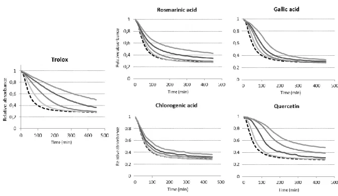
Dotted lines Figures. 3 and 4 show the results of DEPC oxidation experiments carried out in
DLPC or DPPC model membranes, respectively. A mass ratio of 1:10 DEPC to other
phospholipids allowed for the best condition to observe a linear absorbance related to its
concentration. The results indicated that the DEPC oxidation kinetic in DLPC or DPPC
membranes is almost the same, with 50% of the DEPC being oxidized after 34.5 ± 4.9 min
and 32 ± 5.1 min, in DLPC and DPPC respectively. Overall, the VesiCAT assay validation
consisted of verifying whether the addition of an antioxidant compound resulted in a delay in
oxidation of the model liposomes, which could be spectrophotometrically evaluated by
monitoring the decay in absorbance at 273 nm. First, Trolox (water-soluble analog of
α-tocopherol) was used as a reference standard similarly to what we did previously when
developing the CAT assay21. Its addition at different concentrations (from 0.5 to 8 µM) in liposome suspensions before initiating oxidation with AAPH led to an expected, and a
desired, progressive delay in the absorbance decay (Figures. 3 and 4).
Many strategies could be used to measure and represent the antioxidant capacity such as: the
area under the curve (AUC), the reaction rate with free radicals, the inhibition time, and the
antioxidant concentration necessary to achieve 50% inhibition (IC50). A good method should
be able to differentiate the antioxidants with different reaction kinetics. Measurement of the
AUC seems more suitable when the objective is to compare antioxidant capacity of various
molecules/extracts, independently of the antioxidative mechanism (i.e., retarder or chain
breaker). Indeed, for methods using a fixed time or inhibition degree as endpoint, the time or
Version postprint
different inhibition degrees may provide different antioxidant values (even change the
ranking) because any activity of the reaction after the fixed point is totally overlooked 38. However, for methods utilizing AUC, there is a clear starting point and a clear endpoint, and
its calculation exploits both inhibition time and degree of oxidation, thus reflecting the
different reaction kinetics. From those reasons, we believe that assays using AUC provide
global information, whereas other approaches may give more specific data. However, kinetic
parameters such as lag phase duration and initial rate can be also evaluated to get more insight
on the oxidation mechanisms.
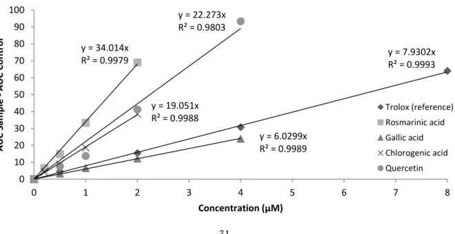
In the AAPH-induced oxidations, the net protection AUC of the reference (AUCTrolox – AUCControl) versus concentration, allowed for a perfect linear relationship (R2 ≥ 0.997), with values equivalent to 9.8-fold ± 0.4 the Trolox concentration in DEPC/DLPC (Figure. 3) and
8.3-fold ± 0.5 the Trolox concentration in DEPC/DPPC membranes (Figure. 4). In addition,
various antioxidants at different concentrations were tested in the different membrane
composition (Figures. 3 and 4). These model antioxidants were chosen according to their
occurrence in the literature dealing with antioxidant behaviors and also on the fact that they
cover different chemical structures from simple phenolic compounds to more complex
molecules. Through calculation of the AUC, good linear relationships (R2 ≥ 0.98) were established between net protection areas (AUCAntioxidants–AUCControl) and antioxidant concentrations for all phenolics tested, both in DLPC (Figure. 5) and DPPC (Figure. 6)
vesicles.
Thus, regardless of the nature of the antioxidants and membrane composition, the oxidation of
Version postprint
the leading coefficient of the linear function represented by the net protection versus
antioxidant concentration, the better the antioxidant capacity is. Thus, rosmarinic acid showed
the best antioxidant ability over the rest of the molecules. The results are not very surprising
because this compound has been already attested to be an efficient antioxidant whether in
formulated lipid dispersions or homogenous systems 24,39. Its strong antioxidant capacity is often correlated with its two catechol functions. On the contrary, gallic acid presented the
lowest efficiency, whereas chlorogenic acid and quercetin exhibited very similar protection
effect. Here, the antioxidant capacity is the result of the scavenging/reducing free radicals
properties of molecules, but also of their capacity to alter the reactive oxygen species
propagation from the fatty acids within the membranes. It is worth noting that the main
purpose of this article is not to compare and discuss about the antioxidant effects, but rather to
describe the new method. Nevertheless, it is necessary to recognize that the method exposes
predictable antioxidant ranking for such experiments with rosmarinic acid > quercetin ~
chlorogenic acid > gallic acid, in accordance with theoretical expectations, because of the
number and position of the hydroxyl groups as well as the degree of conjugation of the whole
molecule 40,41.
VesiCAT with membrane radical initiation (AMVN)
The same approach was performed using liposoluble AMVN as azo initiator. AMVN is a
synthetic azo-compound that dissociates to form C-centered free radicals and then peroxyl
radicals by oxygen reaction, in the hydrophobic phospholipid bilayer. Unlike water-based
initiators, AMVN must be added before vesicles formation. For that reason, the VesiCAT
method was described with DLPC membranes, where the lower liquid-crystalline phase
transition temperature than DPPC allowed preparing of LUV at low temperatures (20° C)
Version postprint
suspensions were oxidized at 43 ± 0.5 °C (liquid temperature in wells) with a constant flux of
radical initiators generated in the membrane bilayer by thermo degradation of AMVN. Dotted
lines Figure. 7 show the result of DEPC oxidation carried out with a mass ratio DEPC to
DLPC of 1:20, suitable to observe a linear absorbance related to concentration. In these
conditions, 50% of the probe was oxidized after 48.5 ± 4.9 min.
As previously for AAPH induced oxidation, Trolox was used as a reference standard, and its
addition at different concentrations (from 0.5 to 5 µM) in the LUV suspension before starting
oxidation by increasing temperature, led to the same observation with a progressive delay in
the absorbance decay (Figure. 7). However, in the oxidation experiments carried out with
AMVN, the kinetic behavior, especially the very slow return to baseline, was different
making the calculation of the total AUC more difficult. This is possibly the result of more
complex reactivity between antioxidants and oxidant species, with a higher order reaction rate
than the ones obtained in the AAPH assays. Thus, for AMVN-induced oxidation, the AUC80 corresponding to the area under the curve obtained after 80% of DEPC oxidation was
calculated. In this condition, the net protection AUC80 of the reference (AUC80_Trolox – AUC80_Control) versus concentration, allowed for a perfect linear relationship (R2 ≥ 0.998), with values equivalent to 3.4-fold ± 0.6 the Trolox concentration in DEPC/DLPC membranes
(Figure. 7). From a kinetic standpoint, Trolox exhibited different behaviors in both oxidation
assays. It essentially showed inhibition of probe degradation in the AMVN-induced oxidation,
with an insignificant oxidation lag phase. Conversely, in AAPH-induced oxidation, Trolox
exhibited an oxidation lag phase proportionate to its concentration. Very similar observation
has been made by Panya and co-workers while comparing oxidation after lipid or aqueous
phase induction in Tung oil in water emulsion 32. The same antioxidants were tested at different concentrations to prevent DEPC oxidative degradation by means of AMVN-induced
Version postprint
oxidation (Figure. 7). Through calculation of the AUC80, good linear relationships (R2 ≥ 0.98) were established between net areas at 80% of DEPC oxidation (AUC80_Antioxidant– AUC80_Control) and antioxidant concentrations (Figure. 8). Unlike the AAPH-induced oxidation, Trolox appeared to be the best antioxidant. Contrariwise, chlorogenic acid showed
the poorest antioxidant efficacy, whereas it was one of the best ones with AAPH assays.
VesiCAT assays: comparison of the results
It appears that the order of effectiveness of the AAPH-induced oxidation assays is gallic acid
< Trolox < quercetin ~ chlorogenic acid < rosmarinic acid, whereas it is chlorogenic acid <
gallic acid < quercetin < rosmarinic acid < Trolox for AMVN-induced oxidations. When the
equation (3) provided in material and methods section is applied, rosmarinic acid exhibited
the highest VesiCAT value in all assays, followed by quercetin, whereas gallic acid showed
very low values (Table. 1). As already mentioned, chlorogenic acid showed very opposite
trends with respect to antioxidant capacity. It is an efficient antioxidant with aqueous
generated oxidants, but poor antioxidant while oxidant species are generated in membrane
domain. This is the perfect example that the effectiveness of antioxidants could be related to
the type of inducer and the region where oxidant species are initiated and spread. Beside the
membrane composition effect, the antioxidant capacity of molecules may result not only on
their chemical interaction with other molecules involved in the oxidation pathway, but also on
their location, concentration, distribution into the different regions (e.g., interfacial lipid
membranes, water), or even penetration depth in membrane 42. In addition, another fundamental facet to consider is the mobility of antioxidants in such assays monitored over
Version postprint
interfacial lipid membrane to reach its equilibrium distribution between interface and water
phases. However, depending on the nature of antioxidant, this kinetic process (and
equilibrium constant) may be significantly different and sometimes not fully resolved within
the lifetime of the experiment.
A striking example to understand how the distribution affects the results is the important
difference that may be observed in antioxidant responses in liposome assays, if antioxidants
are blended with phospholipid before vesicles formation or added after. Indeed, it is different
if the antioxidant has to move from dispersed liposome (core or interface) to dispersion water
phase, or the reverse. Thus, the combination between mobility and equilibrium constant
distribution has to be considered. In addition, one should keep in mind that all these
phenomena explained with the parent antioxidant molecules, are also true with the daughter
antioxidant molecules (oxidized antioxidants), introducing another complexity in the
antioxidant interpretation due to the different chemico-physical properties of the
newly-formed molecule. Overall, the VesiCAT method, developed with the two main different sites
of oxidation (membrane and aqueous) provides consistent results and additional details. In
this VesiCAT method, AMVN assays seem to provide more information about the molecule
distribution and mobility toward the membrane region, whereas AAPH experiments would
give more information about scavenging/reducing free radicals ability. In the light of the
aforementioned statement, one may say that among the tested antioxidants, rosmarinic acid is
the best one to scavenge/reduce free radical (VesiCAT values (TE) > 4, in the AAPH assays);
chlorogenic acid and quercetin have intermediate efficiencies (VesiCAT values (TE) ~ 2.5, in
the AAPH assays); whereas gallic acid has the lowest value (VesiCAT values (TE) < 1, in the
AAPH assays). In addition, Trolox seems to have the best mobility and/or distribution in
membrane, since it showed a low antioxidant efficacy in the AAPH assays, but the highest net
Version postprint
scavenger/reducer of free radicals (AAPH assays), is very poor in the AMVN assay. One
reason could be a slow mobility combined, or not, with a low distribution in the interfacial
domain.
CONCLUSION
This study describes a new absorbance probe-based method (VesiCAT) for measuring
liposome oxidation. The di-eleostearic phospholipid (DEPC) probe was synthesized through a
two-steps procedure, and the experimental conditions were finely tuned to observe and
measure its oxidation in liposomal systems. This is very unique system, and the big advantage
over the other artificial probes is that the probe corresponds to real phospholipids. Indeed, the
current probes used to evaluate the antioxidant capacity have different lipophilic and charge
properties, and when blending with membrane, they could alter physical properties. In
addition, their membrane affinity, mobility, location, penetration depth, and orientation are
uncertain and would likely be different to a fatty acid esterified on a phospholipid. All these
characteristics are essential while measuring antioxidant activity and therefore, imprecisions
could misinterpret the antioxidant responses.
Considering that the region of oxidation may induce different oxidation pathways and
therefore antioxidant responses, two inducers, with opposed solubility, were implemented to
initiate oxidation. Results, carried out on several antioxidants, showed that the assays are
reproducible and efficient, and pointed out that the AAPH- and AMVN-induced oxidations
provide further information on the antioxidants efficacy. Moreover, methods were developed
with liposomes made with DLPC or DPPC, but the new DEPC probe could also be combined
with more complex membrane composition or different types of vesicles.
Version postprint
REFERENCES
(1) Bangham, A. D.; Standish, M. M.; Watkins, J. C. Diffusion of univalent ions across the
lamellae of swollen phospholipids. J. Mol. Biol. 1965, 13 (1), 238–252.
(2) Fan, Y., Zhang, Q. Development of liposomal formulations: From concept to clinical
investigations. Asian J. Pharm. Sci. 2013, 8 (2), 81–87.
(3) Akbarzadeh, A.; Rezaei-Sadabady, R.; Davaran, S.; Joo, S. W.; Zarghami, N.;
Hanifehpour, Y.; Samiei, M.; Kouhi, M.; Nejati-Koshki, K. Liposome: classification,
preparation, and applications. Nanoscale Res. Lett. 2013, 8 (1), 102.
(4) McClements, D. J. Encapsulation, protection, and release of hydrophilic active
components: Potential and limitations of colloidal delivery systems. Adv. Colloid
Interface Sci. 2015, 219, 27–53.
(5) Maherani B, Arab-Tehrany E, MozafariMR, Gaiani C, L. M. Liposomes: a review of
manufacturing techniques and targeting strategies. Curr. Nanosci. 2011, 211 (7), 436–
452.
(6) Taylor, T. M.; Weiss, J.; Davidson, P. M.; Bruce, B. D. Liposomal Nanocapsules in
Food Science and Agriculture. Crit. Rev. Food Sci. Nutr. 2005, 45 (7–8), 587–605.
(7) Schnitzer, E.; Pinchuk, I.; Lichtenberg, D. Peroxidation of liposomal lipids. Eur.
Biophys. J. 2007, 36 (4–5), 499–515.
(8) Waraho, T.; McClements, D. J.; Decker, E. A. Mechanisms of lipid oxidation in food
dispersions. Trends Food Sci. Technol. 2011, 22 (1), 3–13.
(9) Reis, S., Lucio, M., Segundo, M., & Lima, J. L. F. C. Liposomes: Methods and
Version postprint
V., Ed.; 2010; pp 167–188.
(10) Ziech, D.; Franco, R.; Georgakilas, A. G.; Georgakila, S.; Malamou-Mitsi, V.;
Schoneveld, O.; Pappa, A.; Panayiotidis, M. I. The role of reactive oxygen species and
oxidative stress in environmental carcinogenesis and biomarker development. Chem.
Biol. Interact. 2010, 188 (2), 334–339.
(11) Jellinger, K. A. Basic mechanisms of neurodegeneration: a critical update. J. Cell. Mol.
Med. 2010, 14 (3), 457–487.
(12) Bergamini, C. M.; Gambetti, S.; Dondi, A.; Cervellati, C. Oxygen, reactive oxygen
species and tissue damage. Curr. Pharm. Des. 2004, 10 (14), 1611–1626.
(13) Kuypers, F. A.; Berg, J. J. M. van den; Schalkwijk, C.; Roelofsen, B.; Op den Kamp, J.
A. F. Parinaric acid as a sensitive fluorescent probe for the determination of lipid
peroxidation. Biochim. Biophys. Acta - Lipids Lipid Metab. 1987, 921 (2), 266–274.
(14) Tsuchiya M, Kagan VE, Freisleben H-J, Manabe M, P. L. Antioxidant activity of
α-tocopherol, β-carotene, and ubiquinol in membranes: cis-parinaric acid-incorporated
liposomes. Methods Enzym. 1994, 234, 371–383.
(15) Osaka K, Ritov VB, Bernardo JF, Branch RA, K. V. Amphotericin B protects
cis-parinaric acid against peroxyl radicalinduced oxidation: amphotericin B as an
antioxidant. Antimicrob Agents Chemother. 1997, 41, 743–747.
(16) Morgan C. G, Hudson B, Wolber P. K. Photochemical dimerization of parinaric acid in
lipid bilayers. Proc. Natl. Acad. Sci. 1980, 77, 26–30.
(17) Sklar, L. A; Hudson, B. S.; Simoni, R. D. Conjugated polyene fatty acids as fluorescent
Version postprint
828.
(18) Naguib, Y. M. A. A Fluorometric Method for Measurement of Peroxyl Radical
Scavenging Activities of Lipophilic Antioxidants. Anal. Biochem. 1998, 265 (2), 290–
298.
(19) Huang, D.; Ou, B.; Hampsch-Woodill, M.; Flanagan, J. A.; Deemer, E. K.
Development and validation of oxygen radical absorbance capacity assay for lipophilic
antioxidants using randomly methylated beta-cyclodextrin as the solubility enhancer. J.
Agric. Food Chem. 2002, 50 (7), 1815–1821.
(20) Krumova, K.; Friedland, S.; Cosa, G. How Lipid Unsaturation, Peroxyl Radical
Partitioning, and Chromanol Lipophilic Tail Affect the Antioxidant Activity of
α-Tocopherol: Direct Visualization via High-Throughput Fluorescence Studies
Conducted with Fluorogenic α-Tocopherol Analogues. J. Am. Chem. Soc. 2012, 134
(24), 10102–10113.
(21) Li, B.; Harjani, J. R.; Cormier, N. S.; Madarati, H.; Atkinson, J.; Cosa, G.; Pratt, D. A.
Besting Vitamin E: Sidechain Substitution is Key to the Reactivity of Naphthyridinol
Antioxidants in Lipid Bilayers. J. Am. Chem. Soc. 2013, 135 (4), 1394–1405.
(22) Santos, F.; Teixeira, L.; Lúcio, M.; Ferreira, H.; Gaspar, D.; Lima, J. L. F. C.; Reis, S.
Interactions of sulindac and its metabolites with phospholipid membranes: An
explanation for the peroxidation protective effect of the bioactive metabolite. Free
Radic. Res. 2008, 42 (7), 639–650.
(23) Lúcio, M.; Nunes, C.; Gaspar, D.; Ferreira, H.; Lima, J. L. F. C.; Reis, S. Antioxidant
activity of vitamin E and Trolox: Understanding of the factors that govern lipid
Version postprint
(24) Laguerre, M.; López-Giraldo, L. J.; Lecomte, J.; Baréa, B.; Cambon, E.; Tchobo, P. F.;
Barouh, N.; Villeneuve, P. Conjugated autoxidizable triene (CAT) assay: a novel
spectrophotometric method for determination of antioxidant capacity using
triacylglycerol as ultraviolet probe. Anal. Biochem. 2008, 380 (2), 282–290.
(25) Durand, E.; Lecomte, J.; Upasani, R.; Chabi, B.; Bayrasy, C.; Baréa, B.; Jublanc, E.;
Clarke, M. J.; Moore, D. J.; Crowther, J.; Wrutniak-Cabello, C; Villeneuve, P.
Evaluation of the ROS Inhibiting Activity and Mitochondrial Targeting of Phenolic
Compounds in Fibroblast Cells Model System and Enhancement of Efficiency by
Natural Deep Eutectic Solvent (NADES) Formulation. Pharm. Res. 2017.
(26) Laguerre, M.; Lόpez Giraldo, L. J.; Piombo, G.; Figueroa-Espinoza, M. C.; Pina, M.;
Benaissa, M.; Combe, A.; Rossignol Castera, A.; Lecomte, J.; Villeneuve, P.
Characterization of Olive-Leaf Phenolics by ESI-MS and Evaluation of their
Antioxidant Capacities by the CAT Assay. J. Am. Oil Chem. Soc. 2009, 86 (12), 1215–
1225.
(27) Grajeda-Iglesias, C.; Salas, E.; Barouh, N.; Baréa, B.; Panya, A.; Figueroa-Espinoza,
M. C. Antioxidant activity of protocatechuates evaluated by DPPH, ORAC, and CAT
methods. Food Chem. 2016, 194, 749-757.
(28) Sørensen, A. M.; Durand, E.; Laguerre, M.; Bayrasy C.; Lecomte J.; Villeneuve P.;
Jacobsen C. Antioxidant Properties and E ffi cacies of Synthesized Alkyl Ca ff eates,
Ferulates, and Coumarates. J. Agric. Food Chem. 2014, 62 (52), 12553–12562.
(29) Laguerre, M.; López Giraldo, L. J.; Lecomte, J.; Figueroa-Espinoza, M.-C.; Baréa, B.;
Weiss, J.; Decker, E. A.; Villeneuve, P. Relationship between hydrophobicity and antioxidant ability of “phenolipids” in emulsion: a parabolic effect of the chain length
Version postprint
(30) Zago, E.; Durand, E.; Barouh, N.; Lecomte, J.; Villeneuve, P.; Aouf, C. Synthesis of
Lipophilic Antioxidants by a Lipase-B-Catalyzed Addition of Peracids to the Double
Bond of 4-Vinyl-2-methoxyphenol. J. Agric. Food Chem. 2015, 63 (41), 9069–9075.
(31) Tchobo, F. P.; Alitonou, G. A.; Soumanou, M. M.; Barea, B.; Bayrasy, C.; Laguerre,
M.; Lecomte, J.; Villeneuve, P.; Souhounhloue, K. C. D. Chemical Composition and
Ability of Essential Oils from Six Aromatic Plants to Counteract Lipid Oxidation in
Emulsions. J. Am. Oil Chem. Soc. 2014, 91 (3), 471–479.
(32) Panya, A.; Temthawee, W.; Phonsatta, N.; Charoensuk, D.; Deetae, P.; Visessanguan,
W.; Decker, E. A. Apolar Radical Initiated Conjugated Autoxidizable Triene
(ApoCAT) Assay: Effects of Oxidant Locations on Antioxidant Capacities and
Interactions. J. Agric. Food Chem. 2015, 63 (34), 7546-7555.
(33) Nicolet, B. H. C18 Fatty Acids. I. the Non-Identity of Eleostearic Acid Tetrabromide
From Tung Oil With Ordinary Linolic Acid Tetrabromide.1. J. Am. Chem. Soc. 1921,
43 (4), 938–940.
(34) O’Connor, R. T.; Heinzelman, D. C.; McKinney, R. S.; Pack, F.; C. The
spectrophotometric determination of the alpha and beta isomers of eleostearic acid in
tung oil. J. Am. Oil Chem. Soc. 1947, 24 (6), 212–216.
(35) Durand, E.; Zhao, Y.; Coupland, J. N.; Elias, R. J. Assessing Interactions between
Lipophilic and Hydrophilic Antioxidants in Food Emulsions. J. Agric. Food Chem.
2015, 63 (49), 10655–10661.
(36) El Alaoui, M.; Noiriel, A.; Soulère, L.; Grand, L.; Queneau, Y.; Abousalham, A.
Development of a High-Throughput Assay for Measuring Phospholipase A Activity
Version postprint
Microtiter Plates. Anal. Chem. 2014, 86 (21), 10576–10583.
(37) Brauer R.W., Steadman L. T. The autoxidation of β-eleostearic acid: the application of
the spectrophotometer to the study of the course and the kinetics of the reaction. J. Am.
Chem. Soc. 1944, 66, 563–569.
(38) Prior, R. L.; Wu, X.; Schaich, K. Standardized methods for the determination of
antioxidant capacity and phenolics in foods and dietary supplements. J. Agric. Food
Chem. 2005, 53 (10), 4290–4302.
(39) Soobrattee, M. A.; Neergheen, V. S.; Luximon-Ramma, A.; Aruoma, O. I.; Bahorun, T.
Phenolics as potential antioxidant therapeutic agents: Mechanism and actions. Mutat.
Res. Mol. Mech. Mutagen. 2005, 579 (1–2), 200–213.
(40) Apak, R.; Özyürek, M.; Güçlü, K.; Çapanoğlu, E. Antioxidant Activity/Capacity
Measurement. 1. Classification, Physicochemical Principles, Mechanisms, and Electron
Transfer (ET)-Based Assays. J. Agric. Food Chem. 2016, 64 (5), 997-1027.
(41) Rice-Evans, C. A.; Miller, N. J.; Paganga, G. Structure-antioxidant activity
relationships of flavonoids and phenolic acids. Free Radic. Biol. Med. 1996, 20 (7),
933–956.
(42) Durand, E.; Jacob, R. F.; Sherratt, S.; Lecomte, J.; Baréa, B.; Villeneuve, P.; Mason, R.
P. The nonlinear effect of alkyl chain length in the membrane interactions of
phenolipids: Evidence by X-ray diffraction analysis. Eur. J. Lipid Sci. Technol. 2017,
Version postprint
Figure. 1: Synthesis pathway of the 1,2-α-Eleostearoyl-sn-glycero-3-phosphocholine (DEPC) probe.
Figure. 2: UV signal of DEPC probe in DLPC membrane (1:10, w:w), and monitoring of its oxidation
induced by 1 mM of AAPH at 40°C. ~230 nm Conjugated diene formation 273 nm Conjugated triene degradation KOH/Ethanol 3 h, reflux Tung oil ~ 80 % of trieleostearin O O O O O P O- O O N+ C H3 CH3 CH3 CH3 CH3 OH O CH3 PPyr32/DCC/CHCl3 24 h, 20°C Eleostearic acid 1,2-α-Eleostearoyl-sn-glycero-3-phosphocholine (DEPC) Phospholipid probe with specific UV absorbance
Version postprint
Figure 3: Kinetics of relative absorbance decay in the absence and presence of various amounts of
antioxidants. The liposome suspension was made with DEPC/DLPC (1:10, w:w ratio) in PBS, pH 7.2, at 34.5 ± 0.5 °C and contained AAPH (2 mM) without (---) and with (___) an antioxidant: Trolox (0.5
to 8 μM); or gallic acid (0.5 to 4 μM); or rosmarinic acid (0.2 to 2 μM); or chlorogenic acid (0.2 to 2 μM); or quercetin (0.5 to 4 μM).
Version postprint
Figure 4: Kinetics of relative absorbance decay in the absence and presence of various amounts of
antioxidants. The liposome suspension was made with DEPC/DLPC (1:10, w:w ratio) in PBS, pH 7.2, at 34.5 ± 0.5 °C and contained AAPH (2 mM) without (---) and with (___) an antioxidant: Trolox (0.5
to 8 μM); or gallic acid (0.5 to 4 μM); or rosmarinic acid (0.2 to 2 μM); or chlorogenic acid (0.2 to 2 μM); or quercetin (0.5 to 4 μM).
Figure 5: Net protection AUC (AUCSample – AUCControl) versus concentration for five different
antioxidants (including Trolox as reference), in VesiCAT-AAPH assays made with DEPC/DLPC (1:10, w:w ratio), in PBS, pH 7.2, at 34.5 ± 0.5 °C. y = 9.4927x R² = 0.9977 y = 40.098x R² = 0.9992 y = 7.613x R² = 0.9978 y = 23.949x R² = 0.9995 y = 25.642x R² = 0.9970 0 20 40 60 80 100 120 0 1 2 3 4 5 6 7 8 A UC Sam p le -A UC Co n tr o l Concentration (µM) Trolox (reference) Rosmarinic acid Gallic acid Chlorogenic acid Quercetin y = 7.9302x R² = 0.9993 y = 34.014x R² = 0.9979 y = 6.0299x R² = 0.9989 y = 19.051x R² = 0.9988 y = 22.273x R² = 0.9803 0 10 20 30 40 50 60 70 80 90 100 0 1 2 3 4 5 6 7 8 A UC Sam p le -A UC Co n tr o l Concentration (µM) Trolox (reference) Rosmarinic acid Gallic acid Chlorogenic acid Quercetin
Version postprint
Figure 6: Net protection AUC (AUCSample – AUCControl) versus concentration for five different
antioxidants (including Trolox as reference), in VesiCAT-AAPH assays made with DEPC/DPPC (1:10, w:w ratio), in PBS, pH 7.2, at 34.5 ± 0.5 °C.
\
Figure 7: Kinetics of relative absorbance decay in the absence and presence of various amounts of
antioxidants. The liposome suspension was made with DEPC/DLPC (1:20, w/w) in PBS, pH 7.2, at 43 ± 0.5 °C and contained AMVN (0.3 mM) without (- - -) and with (___) an antioxidant: Trolox (0.5 to 5
μM); or gallic acid (1 to 20 μM); or rosmarinic acid (0.5 to 6 μM); or chlorogenic acid (1 to 20 μM); or quercetin (1 to 9 μM).
Version postprint
Figure 8: Net protection AUC (AUCSample – AUCBlank) versus concentration for five different
antioxidants (including Trolox as reference), in VesiCAT-AMVN assays made with DEPC/DLPC (1:20, w:w), in PBS, pH 7.2, at 43 ± 0.5 °C. y = 3.8397x R² = 0.9924 y = 2.0839x R² = 0.9920 y = 0.332x R² = 0.9827 y = 0.2307x R² = 0.9909 y = 1.8602x R² = 0.9932 0 2 4 6 8 10 12 14 16 18 20 0 2 4 6 8 10 12 14 16 18 20 A UC Sam p le -A UC Co n tr o l Concentration (µM) Trolox (reference) Rosmarinic acid Gallic acid Chlorogenic acid Quercetin
Version postprint
Table 1: Comparison of the antioxidant values obtained with the VesiCAT assays, expressed as
Trolox equivalents (TE) of four pure phenolic compounds. All measurements were performed in triplicate VesiCAT assays and reported as the average ± standard deviation (SD).
VesiCAT Values (TE)
Assays Rosmarinic acid Gallic acid Chlorogenic acid Quercetin
AAPH DEPC/DLPC 4.13 ± 0.13 0.93 ± 0.22 2.62 ± 0.26 2.63 ± 0.58
DEPC/DPPC 4.27 ± 0.02 0.78 ± 0.04 2.41 ± 0.01 2.92 ± 0.16