HAL Id: hal-00275721
https://hal.archives-ouvertes.fr/hal-00275721
Submitted on 25 Apr 2008HAL is a multi-disciplinary open access archive for the deposit and dissemination of sci-entific research documents, whether they are pub-lished or not. The documents may come from teaching and research institutions in France or abroad, or from public or private research centers.
L’archive ouverte pluridisciplinaire HAL, est destinée au dépôt et à la diffusion de documents scientifiques de niveau recherche, publiés ou non, émanant des établissements d’enseignement et de recherche français ou étrangers, des laboratoires publics ou privés.
Crystal structure of Thermus thermophilus tRNA
m1A58 methyltransferase and biophysical
characterization of its interaction with tRNA.
Pierre Barraud, Beatrice Golinelli-Pimpaneau, Cédric Atmanene, Sarah
Sanglier, Alain van Dorsselaer, Louis Droogmans, Frédéric Dardel, Carine
Tisné
To cite this version:
Pierre Barraud, Beatrice Golinelli-Pimpaneau, Cédric Atmanene, Sarah Sanglier, Alain van Dorsselaer, et al.. Crystal structure of Thermus thermophilus tRNA m1A58 methyltransferase and biophysical characterization of its interaction with tRNA.. Journal of Molecular Biology, Elsevier, 2008, 377 (2), pp.535-50. �10.1016/j.jmb.2008.01.041�. �hal-00275721�
Crystal structure of Thermus thermophilus tRNA m
1A
58
methyltransferase
and biophysical characterization of its interaction with tRNA.
Pierre Barraud1, Béatrice Golinelli-Pimpaneau2
, Cédric Atmanene3
, Sarah Sanglier3
, Alain Van Dorsselaer3
, Louis Droogmans4
, Frédéric Dardel1
& Carine Tisné1*
1
Laboratoire de Cristallographie et RMN biologiques, Université Paris-Descartes, CNRS UMR 8015, 4 avenue de l’Observatoire, 75006 Paris, France
2
Laboratoire d’Enzymologie et Biochimie Structurales, CNRS Bâtiment 34, 1 avenue de la Terrasse, 91190 Gif-sur-Yvette, France
3
Laboratoire de Spectrométrie de Masse Bio-Organique, IPHC-DSA, ULP, CNRS UMR7178, 25 rue Becquerel, 67087 Strasbourg, France
4
Laboratoire de Microbiologie, Institut de Recherches Microbiologiques Wiame, Université Libre de Bruxelles, avenue E. Gryson 1, B-1070 Bruxelles, Belgium
*Address correspondence to: Dr. Carine Tisné, phone: +33 1 53 73 15 72, fax: +33 1 53 73 99 25, e-mail: carine.tisne@univ-paris5.fr
Short running title : Biophysical characterization of the interaction of thTrmI with tRNA The abbreviations used are: AdoHcy, homocysteine; AdoMet, S-adenosyl-L-methionine; ESI-MS, Electrospray ionization mass spectrometry; MTases, methyltransferases; TrmI, m1
A58 tRNA methyltransferase; thTrmI, Thermus thermophilus
Abstract
Methyltransferases (MTases) from the TrmI family catalyse the S-adenosyl-L-methionine (AdoMet)-dependent N1-methylation of tRNA adenosine 58. The crystal structure of Thermus
thermophilus m1
A58 tRNA MTase (thTrmI) in complex with S-adenosyl-L-homocysteine was
determined at 1.7 Å resolution. This structure is closely related to that of Mycobacterium
tuberculosis TrmI (mycoTrmI), and their comparison enabled us to enlighten two grooves in
the TrmI structure that are large enough and electrostatically compatible to accommodate one tRNA per face of TrmI tetramer. We have then conducted a biophysical study based on electrospray ionization mass spectrometry, site-directed mutagenesis and molecular docking. First, we confirmed the tetrameric oligomerisation state of TrmI and we showed that this protein remains tetrameric upon tRNA binding with formation of complexes involving one to two molecules of tRNA per TrmI tetramer. Secondly, three key residues for the methylation reaction were identified, the universally conserved D170, and two conserved aromatic residues Y78 and Y194. We then used molecular docking to position a N9-methyladenine in
the active site of TrmI. The N9-methyladenine snugly fits in the catalytic cleft where the side
chain of D170 acts as a bidentate ligand binding the amino moiety of AdoMet and the exocyclic amino group of the adenosine. Y194 interacts with the N9-methyladenine ring
whereas Y78 can stabilize the sugar ring. From our results, we propose that the conserved residues that form the catalytic cavity (D170, Y78 and Y194) are essential to fashion an optimized shape of the catalytic pocket.
Keywords: TrmI, m1A58 methyltransferase, X-ray structure, noncovalent mass spectrometry, Protein-RNA interactions
Introduction
Functional tRNAs carry a number of chemically modified nucleosides that are formed enzymatically after transcription, during the tRNA maturation process. To date, more than 90 different modifications have been identified in tRNAs from various organisms.1
Among these post-transcriptional nucleoside modifications, N1-methyladenosine (m
1
A) is found at a highly conserved A58 position in the TΨC loop of many tRNAs in the three domains of life
(Bacteria, Archaea and Eukarya). This modification occurs infrequently in bacteria and, for instance, is absent in E. coli tRNAs. On the contrary, it is common in the tRNAs of most eukaryotes and archaea, and this modification seems to be essential for some organisms. Actually, in the yeast Saccharomyces cerevisiae, mutants defective in N1-methylation of A58
are non-viable.2
Likewise, in Thermus thermophilus, gene disruption studies have shown that m1
A58 modification is required for growth of this bacterium at high temperatures. 3
S-Adenosyl-L-methionine (AdoMet) dependent methyltransferases (MTases) are involved in a wide variety of biological processes involving methylation of nucleic acids, proteins, phospholipids and small molecules using the ubiquitous methyl donor AdoMet. Recently, two essential genes of S. cerevisiae, GCD10 and GCD14 (renamed TRM6 and TRM61), were identified to encode the two types of subunits of the yeast m1
A58 MTase. 4
The recombinant enzyme behaves as an α2β2 heterotetramer, Trm61p being responsible for AdoMet binding
and presumably catalysis of the methyl transfer reaction,2
whereas both types of subunits are essential for tRNA-binding.5
Indeed, the purified recombinant Trm61p subunit, which binds AdoMet, cannot bind tRNA in vitro in the absence of Trm6p.2
Interestingly, Trm61p was found to be closely related to a group of prokaryotic proteins which share not only the same AdoMet-binding site but also other highly conserved motifs. Therefore, the corresponding prokaryotic proteins were presumed to act as m1
A58 MTases. 6
This hypothesis was demonstrated experimentally for the bacterial orthologs in Thermus thermophilus and
Mycobacterium tuberculosis and for the archaeal ortholog in Pyrococcus abyssi.3,7-9
These proteins are α4 homotetramers of Trm61p-like proteins, hereafter called TrmI. Interestingly,
orthologs of the Trm6p protein could only be found in eukaryotes. It was shown that Trm6p, despite the absence of the characteristic MTases motifs, is structurally and evolutionary related to Trm61p suggesting that the eukaryotic m1
A58 MTases evolved by gene duplication
and speciation to form a heteromultimeric protein, whereas their prokaryotic orthologs remained homomultimers.6
Despite the low level of sequence similarity between the various families of AdoMet-MTases, most of them contain a structurally highly conserved catalytic AdoMet-binding domain organized in a Rossman-like fold (For a review see 10
). Only one crystal structure of an m1
A58 tRNA MTase has been reported to date, that of the M. tuberculosis Rv2118c protein
(mycoTrmI) in complex with S-adenosyl-L-methionine.7
C-terminal domain is very similar to that of other AdoMet-dependent MTases whereas the N-terminal domain which is mainly composed of β-sheets is not found in other MTases of known structure.
In the present work, we have first solved the crystal structure of the m1
A58 tRNA MTase
from T. thermophilus (thTrmI) at 1.7 Å resolution in complex with S-Adenosyl-L-homocysteine (AdoHcy), the product after methyl transfer. We then focused our attention on the tRNA-binding properties of thTrmI using several biophysical techniques, including electrospray ionization mass spectrometry (ESI-MS), site-directed mutagenesis, steady-state kinetic assays, fluorescence spectroscopy and molecular docking. ESI-MS has demonstrated its particular suitability for the investigation of noncovalent complexes.11-13
More particularly, so-called noncovalent ESI-MS has been extensively used to characterize supramolecular assemblies involving oligonucleotides in interaction with drugs and proteins (For reviews see
14-17
). As protein:RNA systems are partly driven by electrostatic-based interactions which are strongly enhanced in the gas phase, ESI-MS is well suited for their characterization.18,19
Indeed, numerous studies of protein-RNA complexes have revealed a strong agreement between mass spectrometric gas-phase measurements and results obtained by solution phase techniques.20-24
In our work, we used noncovalent ESI-MS: i) to unambiguously assess the oligomerisation state of thTrmI ; ii) to monitor the thTrmI oligomerisation state upon tRNA binding and iii) to determine the stoichiometry of the thTrmI-tRNA complexes. In addition, a mutagenesis study was performed to identify residues potentially crucial for the methylation reaction. We thus showed that the universally conserved D170 residue, together with two conserved aromatic residues, Y78 and Y194, which line the catalytic pocket are key residues for the enzymatic catalysis. Actually, D170A and Y78A variants are severely altered in their catalytic efficiency. Finally, we have used molecular docking to position a N9-methyladenine
Results and Discussion
Structure determination of T. thermophilus TrmI
Production and purification of T. thermophilus TrmI (thTrmI) have been described by Droogmans and coworkers.3
However, the recombinant His6-tagged protein exhibited a low
solubility (< 0.5 mg/mL) unless high salt concentrations were added (0.2 M imidazole-HCl and 0.5 M KCl). This was a serious issue for crystallization assays, in which precipitating solutions must be added to concentrated protein. In order to improve solubility, the N-terminal His6-tag of thTrmI was cleaved with thrombin. Indeed, after His6-tag removal,
thTrmI became highly soluble (> 10 mg/mL), even at moderate ionic strengths (150 mM).
This type of behaviour has already been reported for some His-tagged proteins.25
We then carried out crystallization assays on the complex between thTrmI, AdoHcy and E. coli tRNAi
Met
. The latter tRNA was selected because it is devoid of modification on A58 and is a
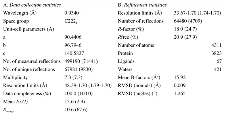
substrate of the T. thermophilus enzyme (see paragraph on steady-state kynetic assays). Although the tRNA was present in all crystallization conditions, crystals only contained the free thTrmI protein in complex with AdoHcy. Table 1 summarizes the data collection and refinement statistics. The structure of T. thermophilus TrmI protein was then solved by molecular replacement with the structure of m1
A58 tRNA MTase from M. tuberculosis TrmI
(PDB entry code 1I9G)7
using PHASER26
(Z score = 23.7). Actually, thTrmI shares 39% identical and 13% similar residues with mycoTrmI. The model was then refined to 1.7 Å, to a final Rfree factor of 20.9% (Table 1). The last four residues at the N-terminus were too poorly
ordered to be included, and therefore the final model consisted of 251 residues per monomer. The asymmetric unit contained a dimer of thTrmI. However, the symmetry-related subunits interact extensively leading to a tetrameric organization of thTrmI (Figure 1(b)).
Overall structure of thTrmI
The thTrmI monomer is composed of two distinct domains connected by an extended linker containing a short α-helix (helix 2, Figure 1(a)). The large C-terminal domain (residues 63-255) adopts a typical class I MTase fold which consists of a central seven-stranded β sheet (β1 to β6 and β8 – Figure 1(a)) flanked by α helices on both sides (αA to αE – Figure 1(a)).
10
The first five strands of the β sheet (β1 to β5 on Figure 1(a)) are parallel whereas the last two
(β6 to β8) are antiparallel. The C-terminal domain contains the AdoMet binding site and the
catalytic pocket. The smaller N-terminal domain (residues 5-62) is largely a β structure composed of six β-strands (βA to βF Figure 1(a)) and one small α helix (α1).
The thTrmI tetramer is organised as a dimer of tight dimers. Two monomers interact via their extended β6 strands which form a cross-subunit, antiparallel β-sheet (Figure 1(b), pale
and dark blue). This dimer is further stabilized by ionic interactions between helices α2 and
β6 strands of the other dimer to form a central anti-parallel β-sheet structure. The tetramer
interaction is limited to this β-sheet region and stabilized by both ionic and hydrophobic interactions, mainly a salt bridge between E220 and R224, hydrophobic interactions between W226 and F245, and between H234 and F237 from one dimer to H242 from the other dimer. The salt bridge and the hydrophobic interactions between W226 and F245 have been also observed on the mycoTrmI structure7
and involve conserved residues.
In the tight dimer found in the asymmetric unit, the two cofactor-binding sites are occupied by AdoHcy molecules in identical conformations, similar to that of AdoMet in the
M. tuberculosis TrmI structure. The binding cleft is formed by residues belonging to
conserved motifs, named I, II, III, IV, (Figures 1(c), 1(d) and 7(a)).27
The adenine ring of the AdoHcy moiety is bound by motifs II, III, and IV via water-mediated hydrogen bonds between the exocyclic amino group and residues E155 and E173, and between the N7 position
of the adenine and the backbone amide of E173 (Figure 1(c)). Adenine binding is also reinforced by hydrophobic contacts between its aromatic ring and residues L171, V177, K153, L154, Y124, A126 and A100. The ribose moiety of AdoHcy is bound by motif II, essentially by hydrogen bonds involving the 2’ and 3’ hydroxyl groups and residues E125 and H130. The amino-acid part of the cofactor is mainly bound by motif I. Interestingly, an aspartate side chain that could be involved in the methylation reaction, the carboxylate chain of D170 from motif IV, participates to a direct polar interaction with the ammonium group of AdoHcy. The GXGXGG pattern of motif I binds to the amine and the carboxylate through a tight network of water-mediated hydrogen bonds involving the backbone (Figure 1(c)). Comparison with mycoTrmI structure and tRNA recogition
Globally, the overall structure of thTrmI closely resembles that of mycoTrmI. Superimposition of T. thermophilus and M. tuberculosis TrmI structures gives a root mean square deviation of 4.47 Å for Cα, the largest variation occurring in the β-strands (β6, β7, β8)
that protrude out of the structure. When comparing the active sites more accurately, that of
mycoTrmI is narrower by about 1 Å than that of thTrmI. Moreover, the protruding R249 in mycoTrmI makes like a supplementary step at the exit of the pocket that does not exist in thTrmI as R249 is replaced by a shorter side chain residue (V240). As a consequence, the
accessible surface of Y78 is twice larger in thTrmI than that of Y84 in mycoTrmI. All these features seem to make the methyl donor less easily accessible in mycoTrmI. Overall, in both structures, the adenine should enter deeply in the binding cleft to have access to the methyl donor therefore implicating a major deformation of the phosphodiester backbone around A58.
It is also highly interesting to compare electrostatic potentials of their molecular surfaces (Figure 2). These calculations uncover that both proteins have two linked grooves of positive electrostatic potentials indicated by large blue surfaces on Figure 2 that are large enough to accommodate double-helical RNA. The first groove that covers the β-sheet region forming
the tetrameric architecture is less positively charged in thTrmI than in mycoTrmI; whereas the second groove that encompasses the N-terminal domain of one TrmI subunit and that looks like an open hand is more positively charged in thTrmI than in mycoTrmI. Several residues with basic side chains are conserved among the TrmI protein family, namely K13 (R in some sequences), R15 (K), R72, R127, R217 and R229 (K) together with the histidines involved in the formation of the tetramer H234 and H242. R15 and R217 are only conserved in bacterial TrmI proteins. These grooves have the dimensions to accommodate the acceptor arm (groove 1, dimensions: 25 Å large and 45 Å long) and the anticodon one (groove 2, dimensions: 25 Å large and roughly 30 Å long) without steric clashes, enabling us to position the adenosine 58 near the catalytic pocket (manual docking, data not shown). The grey line in Figure 2(a) indicates a possible position of the helix axis of tRNA. Numerous clashes occur within the T-arm that undoubtedly undergoes huge conformational changes that probably involves the entire T-arm and not only a simple flip of the adenosine 58. These changes are difficult to predict and, for this reason, we could not pursue this docking. In conclusion, the conservation of two grooves of positively charged surfaces supports the idea that TrmI binds tRNA as a tetramer and that two molecules of tRNA can interact simultaneously with the TrmI tetramer. The fact that, in eukaryotes, the enzyme has evolved from an α4 homotetramer to an α2β2
heterotetramer is compatible with a two tRNA per TrmI tetramer stoichiometry, one tRNA being expected to interact with an αβ subsystem.
thTrmI is a tetramer and binds up to two tRNA molecules as a tetramer. Close inspection of the crystal structures of thTrmI and mycoTrmI7
revealed that the tetramers are formed by two pairs of extensively interacting subunits stabilized by relatively small numbers of contacts between the two dimers. Given the architecture of TrmI, it was of interest to investigate the oligomerization state of thTrmI alone and upon tRNA binding, and to determine the binding stoichiometry of the thTrmI-tRNA complexes. Previously reported gel filtration experiments have indicated that both mycoTrmI and thTrmI should form tetramer in solution.3,9
However, in the case of thTrmI, due to an inherent limited precision of this technique, it is difficult to unambiguously state whether the protein is tetrameric or pentameric.3
In this study, we used ESI-MS, which has been proven to be a valuable technique for the determination of oligomerization states of noncovalent assemblies28-31
to first confirm the oligomerization state of thTrmI. Figure 3(a) presents the ESI mass spectrum obtained for thTrmI under non-denaturing conditions and carefully controlled operating conditions (see Materials and Methods): a single ion distribution is observed in the m/z 4000-5000 range. With a measured molecular weight (MW) of 115450 ± 5 Da, this distribution can be assigned to the +23 to +30 charge states of a thTrmI tetramer (theoretical MW = 115453 Da). Thus, ESI-MS allowed us to unambiguously assess the tetrameric nature of the protein in agreement with our crystallographic results.
The tRNA binding stoichiometry was first investigated by gel retardation assay. As shown on Figure 4, the free tRNA band progressively disappears upon addition of increasing amounts of thTrmI evidencing thus the existence of thTrmI-tRNA interactions. Moreover, the free tRNA band completely disappears at 1:2 thTrmI:tRNA ratio, substantiating the formation of complexes involving two molar equivalents of thTrmI monomer per tRNA. To further confirm gel retardation experiments, we also used the potentialities of noncovalent ESI-MS to investigate the oligomerization state of thTrmI upon tRNA binding and to determine the tRNA binding stoichiometry. Titration experiments, monitored by noncovalent ESI-MS, involving a fixed concentration of thTrmI and increasing amounts of tRNAi
Met
, revealed the presence of three ion distributions (Figure 3(b-d)). The first one in the m/z 4000-5000 region with a molecular mass of 115450 ± 5 Da is related to the thTrmI tetramer. For the second ion distribution in the m/z 5000-5900 region, the molecular mass of 140347 ± 6 Da corresponds to the (thTrmI)4:(tRNA)1 complex. Finally, a third ion distribution within the m/z 5900-6800
range with a molecular mass of 165455 ± 10 Da refers to the (thTrmI)4:(tRNA)2 complex.
Even a ten-fold molar excess of tRNA per thTrmI tetramer does not lead to the detection of complexes with higher tRNA binding stoichiometries. Interestingly, upon increase of the tRNA concentration from 1.5 to 10 molar equivalents per thTrmI tetramer, the relative abundances of (thTrmI)4:(tRNA)1 and (thTrmI)4:(tRNA)2 complexes statistically increase,
which is in favour of non-cooperative tRNA binding system and further suggests the presence of two independent tRNA binding sites.32
The same experiments were also carried out in presence of AdoHcy (data not shown) and no effect either on the binding stoichiometry of the complex or on the binding affinity of the tRNA for thTrmI was observed.
As ESI-MS detects species in the gas phase of the mass spectrometer, control experiments are always needed to ensure that mass spectra faithfully reflect the behaviour in solution.33
Thus, since electrostatic interactions are sensitive to the ionic strength of the medium,34,35
experiments were carried out at different ammonium bicarbonate concentrations (Figure 5). Decreasing the ammonium bicarbonate concentration from 200 mM to 100 mM displaces the equilibrium towards the formation of (thTrmI)4:(tRNA)1 and (TrmI)4:(tRNA)2 complexes
(Figure 5(a-b)). However, the signal intensities were dramatically lower when 100 mM ammonium bicarbonate was used rather than 200 mM buffer, mainly because of protein precipitation. Therefore, no ESI-MS analysis could be performed at buffer concentrations lower than 100 mM. While both (thTrmI)4:(tRNA)1 and (thTrmI)4:(tRNA)2 complexes are
favoured at low ionic strengths, the use of higher salt concentrations destabilizes these assemblies (Figure 5(c-d)), leading even to a complete dissociation of thTrmI:tRNA complexes into thTrmI tetramer at 1250 mM ammonium bicarbonate (Figure 5(d)). The fact that mass spectra do reflect expected changes induced by modification of the solution conditions is a further evidence that allows us to definitely rule out the possibility that the TrmI-tRNA complexes result from an artefact of the technique. Moreover, as described in
other publications,22,36
an additional ESI-MS control experiment was performed in strictly identical experimental and operating conditions with a non-substrate RNA as a negative control. The absence of any thTrmI-control RNA complex on ESI mass spectra even in presence of a five-fold molar excess of control RNA (Figure 6) indicates that the gas phase detection of the TrmI-tRNA complexes arises from a specific recognition in solution and not from any gas phase artefact.
In conclusion, noncovalent ESI-MS results clearly support that thTrmI remains tetrameric upon tRNA binding and that thTrmI binds up to two molecules of tRNA.
The D170A and Y78A variants of thTrmI are severely altered in their catalytic efficiency.
On the basis of multiple sequence alignments of the TrmI family members (Figure 7(a)) and on the comparison of the crystallographic structures of TrmI from M. tuberculosis7
and T.
thermophilus, we chose to mutate three conserved residues, D170, Y194 and Y78 in order to
study their involvement in adenosine binding and/or in catalysis. Figure 7(b) presents the
thTrmI residues that are conserved or semi-conserved in the catalytic pocket, except the
conserved residues that bind to AdoMet since their role in the catalysis mechanism is known. Yet, the universally conserved AdoMet-binding D170 was selected as it forms the back of the catalytic pocket and may also bind the adenine 58 ring of the tRNA substrate (Figure 7(b)). Y194 and Y78 were mutated because they respectively form the left side and the floor of the adenosine-binding pocket in the 3D structure (Figure 7(b)) and are conserved as aromatic residues among m1
A58 tRNA MTases, belonging respectively to motifs V and X (Figure
7(a)). Therefore, these aromatic residues could be involved in the stabilization of the target adenine that needs to be flipped out of the tRNA structure to be methylated. P196 that is involved in making the shape of the catalytic cavity was not selected for mutation, because in
M. tuberculosis, it is naturally replaced by an alanine. Therefore, we individually mutated
Y78, D170 and Y194 to alanines. We expressed and purified the variant proteins as described in Materials and Methods section. We then determined the kinetic and RNA-binding parameters for wild-type and variant proteins (Table 2).
First, we investigated whether the mutations altered tRNA binding by the enzyme. Interaction with tRNA induces a quenching of the intrinsic tryptophan fluorescence of the protein of about 40 %. We therefore used fluorimetric titrations to determine the apparent dissociation constant between thTrmI variants and tRNAi
Met
(Table 2). All mutants showed quite unchanged dissociation constants (Kd about 15 ± 3 nM), thereby indicating that they
retained a native folding and similar RNA binding ability compared to the wild-type enzyme. We then analysed their catalytic properties by determining their kinetic parameters kcat and KM
for both AdoMet and tRNA substrates. The overall results are summarized in Table 2. Interestingly, all mutants retain some catalytic activity, but are altered to various extents,
essentially in their catalytic turnover kcat. The D170A mutant retains some activity, but its kcat
value is severely reduced, by a factor of about 300. D170A also shows an increased KM for
AdoMet substrate in agreement with the crystal structure. It seems therefore to be the most important residue for the catalysis of the reaction. The Y78A mutant leads to a roughly 20-fold decrease of the catalytic turn-over constant. Its binding constant for the methyl donor is unchanged compared to the wild-type protein. The Y194A mutant shows a smaller decrease of kcat, by a factor of 3 (Table 2). The KM value for AdoMet is increased suggesting that this
residue is involved in cofactor binding. Yet, in the crystal structure, it is not involved in a direct interaction with AdoHcy in contrast to D170. The mutation of Y194 to an alanine probably slightly destabilizes the catalytic pocket or indirectly weakens AdoMet binding. In conclusion, D170A is the variant that is most largely altered in its catalytic efficiency followed by Y78A and then Y194A. And, therefore, D170, Y78 and Y194 are key residues in the catalytic mechanism.
The substrate adenine snugly fits in the active site pocket of thTrmI.
To investigate the positioning of the target A58 into the catalytic pocket of T. thermophilus
and M. tuberculosis TrmI, we decided to locate a N9-methyladenine in the catalytic pocket of
these proteins by molecular docking. We docked a N9-methyladenine and not an adenosine or
an AMP molecule since it is impossible to predict the conformation of the sugar of this flipped-out nucleotide. Moreover Y78 that makes the exit of the catalytic pocket and which may interact with the ribose of the adenosine 58 can also flip to prevent from steric clashes with the A58 ribose. As the molecules of AdoHcy present in the active sites of thTrmI have
identical conformations to the AdoMet co-factor present in mycoTrmI, we modelled an AdoMet molecule in the catalytic pocket of thTrmI by superimposing the AdoMet from
mycoTrmI with AdoHcy in thTrmI. We used the program HADDOCK37
that can take into account mutagenesis results by introducing data as Ambiguous Interaction Restraints (AIRs) to drive the docking process. An AIR is defined as an ambiguous distance between all residues shown to be involved in the interaction. Ambiguous intermolecular distance restraints were applied between D170, Y194, Y78 and the AdoMet cofactor on the one hand, and the N9-methyladenine, on the other hand. A distance restraint between the N1 atom of N9
-methyladenine and the methyl of AdoMet was also added. The model resulting from the docking with thTrmI is presented in Figure 8(a) and shows that the N9-methyladenine fits
snugly in the cavity where methylation takes place. The same result was obtained for
mycoTrmI (data not shown). Moreover, the methyl group introduced to substitute the ribose
is pointing above Y78 (Figure 8(b)) enabling us to add a ribose without steric hindrance. Therefore, Y78 is likely to strongly interact with the ribose of the adenosine 58 probably participating to stabilization of the flipped-out conformation. The distance between the N9
between the N9-methyladenine N1 and the AdoMet sulphur atom of 4.1 Å. This is in keeping
with distances observed in co-crystal structures of other endocyclic purine N-MTases (distances between 3.6 Å and 4.5 Å).38
The docked model of the N9-methyladenine ring
suggests a binding mode involving essentially Van der Waals interactions with Y194, and a polar contact between the exocyclic amino group of the purine and the carboxylate of D170 (Figure 8(b)). Therefore, the side chain of D170 could act as a bidentate ligand binding the amino terminal moiety of AdoMet and the N6 exocyclic amino group of the adenine ring
substrate. Thus, the sulphur-methyl bond of AdoMet is almost coplanar with the N9
-methyladenine ring and is pointing to the same direction as the N1 lone-pair orbital, which
can be of significant importance for the chemical mechanism. We thus propose that D170 could minimize the transition state energy by positioning both the AdoMet and the target base in an optimal orientation for the methylation reaction. D170 could also deprotonate the N6
group to activate the N1 lone-pair orbital in order to attack the methyl group of AdoMet and
to generate the exocyclic imino tautomer of N1-methyladenosine as the initial product. The
nucleophilic attack leads to the m1
A basic form which has to be subsequently protonated to give the m1
A cationic form, stable at physiological pH (pKa = 9.3).39
As the deprotonation of N6 of m
1
A is easier than the deprotonation of N6 of the non-methylated adenine ring (pKa =
16.7),40
it seems more reasonable that the deprotonation and the nucleophilic attack occur in the same elementary step. The model perfectly situates D170 to serve that role, which is most definitely consistent with the kinetic results.
The chemical mechanism of adenine N1 methylation of RNA had not yet been investigated
in contrast to the N1 methylation of guanine. 41
In the case of m1
G RNA methyltransferase (TrmD),41
an aspartate residue was shown to be involved in the deprotonation step of N1 of
guanine. The two processes are likely to be different, as under physiological conditions, the N1 of guanine is protonated but that of adenine is not. DNA methyltransferases have been
extensively studied, but the m1
A modification is not encountered in DNA. The methylation of the N6 position of adenine has been investigated in DNA (for structural studies see
42-44
) and more recently in rRNA.45,46
Interestingly, the main catalytic residue in m6
A RNA or DNA MTases has been clearly identified by mutagenesis studies to be the D/N/S residue of the conserved pattern (D/N/S)PP(Y/F/W) which belongs to MTase motif IV (for reviews on DNA MTases see 47,48
). In known structures of m6
A DNA MTases (for example M•TaqI, PDB code 2ADM) or m6
A RNA MTases (ErmC’, PDB code 1QAN) in complex with cofactor (AdoMet or AdoHcy), the position of the D/N/S residue of motif IV relative to the cofactor is closely similar to that of D170 residue in the T. thermophilus TrmI crystal structure. Additionally, two other residues have been identified to be important in catalysis in m6
A DNA methyltransferases. These residues are aromatic and stabilize the flipped-out target base and/or act as cation-π catalysts. The first one is the aromatic Y/F/W residue of motif IV, which is proposed to stabilize the cationic transition state by cation-π interaction.49,50
second one is the aromatic residue Y/F/W of motif VIII, which stabilizes the flipped base outside the DNA helix.49,50
These aromatic residues are conserved in motifs IV and VIII in m1
A tRNA MTases (Figure 7(a), symbol o), but they are not part of the catalytic pocket in the TrmI structure. Strikingly, two other aromatic residues, i.e. Y78 and Y194, are located in the active site cavity of thTrmI (Figure 7(b)) at relative positions with respect to the AdoMet cofactor similar to those of the aromatic residues listed above for DNA MTases. These residues are also conserved as aromatic residues among m1
A58 tRNA MTases and belong
respectively to motifs X and V (Figure 7(a)). The noticeable structural similarity between the catalytic pocket of m1
A tRNA MTases and m6
A DNA MTases suggests a similar role in catalysis for these residues. Y78 is thus the most probable candidate for the stabilization of the flipped-out adenosine given its position in the catalytic pocket and the dramatic decrease of kcat of the Y78A mutant. Then, Y194 could act as a cation-π catalyst to stabilize a cationic
transition state. These roles are also in agreement with our docking results.
Coordinates and structure factors for T. thermophilus TrmI protein in complex with AdoHcy have been deposited at the Protein Data Bank with accession code 2PWY.
Materials and Methods
Expression and purification of thTrmI for structural studies
Recombinant T. thermophilus TrmI protein (255 residues) was overexpressed and purified as previously described.3
The N-terminal His6-tag was removed by thrombin cleavage
(25 units of thrombin per mg of protein) performed overnight at ambient temperature. The
thTrmI protein was then further purified by gel filtration on a HiLoad 26/60 Superdex 200
prepgrade chromatography column (Amersham Biosciences) equilibrated in 50 mM Tris-HCl buffer (pH 8.0), 100 mM KCl. Fractions containing pure recombinant protein were pooled, concentrated to 8-10 mg/mL using Amicon Ultra (Millipore) and stored at 4 °C.
Crystallization and crystal structure determination
Crystallization was performed at 19 °C using the sitting drop vapour diffusion method. Protein samples were prepared at 3 mg/mL in 50 mM Tris-HCl buffer (pH 8.0) containing 100 mM KCl, 2 mM AdoHcy and 1 equivalent of tRNAi
Met
. Drops of 1 µL were prepared by using a Cybi-Disk robot system that mixes equal volume of protein and reservoir solutions. Reservoir volumes of 100 µL were used. Crystals of thTrmI were obtained in 2 M ammonium sulphate, isopropanol 5 % (v/v) (condition number 5 of Hampton Research Crystal Screen II kit). The crystals were harvested, soaked in a cryoprotectant solution (2.2 M ammonium sulphate, isopropanol 5 % (v/v), glycerol 20 % (v/v)) and flash-frozen in liquid nitrogen before data collection. Diffraction data were collected at beam line ID14-1 of the European Synchrotron Radiation Facility (ESRF, Grenoble, France).
All crystallographic calculations were performed using the CCP4 suite version 651
as implemented in the graphical user interface.52
X-ray diffraction data were processed using MOSFLM53
and scaled with SCALA.54
The structure of thTrmI was solved by molecular replacement using the program PHASER26
and the structure of m1
A58 tRNA MTase from M.
tuberculosis (PDB entry code 1I9G) as a model. In the model, non-conserved residues were
truncated to alanine. Model building of thTrmI was first performed with ARP/wARP55
using the warpNtrace automated procedure. Restrained refinements of the structure were performed with the program REFMAC5.56
Model and maps visualisations for manual reconstruction were performed with the program COOT.57
Solvent molecules were automatically added using ARP waters module implemented in REFMAC5. In the last stages of refinement, TLS parameters were refined58
using one group for each domain of the protein, i.e. the large C-terminal domain and the small N-C-terminal domain.
We computed the electrostatic surface potential using APBS Tools, an interface for performing APBS electrostatics calculation59
Expression and purification of tRNAi Met
The E. coli tRNAi Met
was overexpressed from plasmid pBSTtRNAf Met
in E. coli JM101TR using a protocol derived from that of Meinnel and Blanquet.60
Briefly, after phenol extraction of RNAs from bacteria, total tRNA was separated by gel filtration on a HiLoad 26/60 Superdex 75 prepgrade chromatography column (Amersham Biosciences) equilibrated in 25 mM Tris-HCl pH 7.0. The overexpressed tRNAi
Met
was then separated from other tRNAs by an anion exchange step (Resource Q column) equilibrated in 25 mM Tris-HCl pH 7.0. tRNAs were eluted using a 350 mM to 550 mM NaCl gradient in the same buffer. The fractions containing the purified tRNAi
Met
were pooled together, dialysed against 50 mM Tris-HCl, 100 mM KCl, pH 8.0, concentrated using Amicon Ultra (Millipore) and stored at -20 °C.
Electrospray ionization mass spectrometry
Noncovalent mass spectrometry experiments were performed on an electrospray time-of-flight mass spectrometer (LCT, Waters). Samples were continuously infused into the mass spectrometer with a syringe pump (Harvard Apparatus) at a flow rate of 6 µl/min. Mass spectra were acquired in the positive ion mode on the mass range 2000-8000 m/z after calibration with the multiply charged ions produced by horse heart myoglobin diluted to 2 µM in a 1:1 water/acetonitrile mixture (v/v) acidified with 1% (v/v) formic acid. Deconvoluted mass spectra were obtained using the Transform algorithm of MassLynx 4.0 software. Instrumental parameters were optimized to get the best compromise between ion desolvatation, ion transmission and preservation of noncovalent complexes during the ionization and desorption processes. This optimization concerned especially the pressure in the interface (Pi) and the accelerating voltage (Vc) which were set to 6.1 mbar and 120 V, respectively.
Purity and homogeneity of thTrmI were checked in denaturing conditions by diluting the protein to 5 µM in a 1:1 water/acetonitrile mixture acidified with 1% (v/v) formic acid. A good agreement was found between the measured molecular mass (28863.0 ± 0.3 Da) and the mass calculated from the amino acid sequence (28863.3 Da). For experiments performed in non-denaturing conditions, different ammonium bicarbonate concentrations ranging from 100 mM to 1250 mM were tested. A concentration of 200 mM ammonium bicarbonate was chosen as the best compromise to provide good quality ESI mass-spectra without extensive
thTrmI:tRNA complexes dissociation (which takes place at high ionic strengths) or protein
aggregation (occurring at low ionic strengths). Thus, this buffer concentration was used to study both thTrmI oligomerization state and its tRNA-binding properties. All experiments were carried out at 20 µM thTrmI (tetramer concentration) whereas E. coli tRNAi
Met
concentration (molecular weight MW = 24888 Da) was increased up to 200 µM for titration experiments. To study the influence of AdoHcy on the thTrmI-tRNA complexation equilibrium, thTrmI and AdoHcy, both at 20 µM (tetramer concentration), were incubated in
the ammonium bicarbonate buffer in presence or in absence of tRNA (30 µM). Finally, a non-substrate control RNA of 59 nucleotides (MW = 19255 Da) was also used in a five-fold molar excess relative to the thTrmI tetramer concentration in order to investigate the specificity of the thTrmI-tRNA interactions. All solutions were incubated for 15 minutes at room temperature before analysis under non-denaturing conditions.
Non-denaturing agarose gel electrophoresis
Samples were prepared by mixing constant amount of E. coli tRNAi Met
(4 µg) with increasing amounts of thTrmI protein (from 0 to 1 molar equivalent of thTrmI tetramer) in loading buffer (Tris-HCl pH 8.0 50 mM, KCl 100 mM, glycerol 18 % (v/v)). Samples were kept at 4 °C for 12 hours and loaded on a 2 % (w/v) agarose gel. Migration was performed for one hour in Tris-Acetate buffer pH 8.0 at 4 °C under an electric field of 100 V. Bands were visualized by UV shadowing.
Site-directed Mutagenesis
Alanine site-directed mutagenesis was carried out using the Quickchange kit and protocol (Stratagene). All oligonucleotides for mutagenesis were designed to have appropriate pairing stability (fusion temperature near 80 °C) and were between 27 to 32 nt long. All mutant genes were fully sequenced and contained only the desired substitutions. Expression and purification of wild-type and mutant thTrmI proteins for kinetic studies
The wild type and mutant proteins were overexpressed and purified as for structural studies except that the thrombin cleavage was omitted. Protein concentrations were evaluated with the Bradford method. The proteins were then diluted in 50 mM Tris-HCl pH 8.0 to a final concentration of 0.25 mg/mL and stored at 4 °C.
Fluorescence titrations of thTrmI by tRNAi Met
Fluorescence measurements were performed at 20 °C on a JASCO spectrofluorimeter. Excitation and emission wavelengths were 280 nm and 343 nm respectively. The excitation and emission bandwidths were 5 nm and 10 nm respectively.
Fluorescence titrations experiments were performed by adding increasing concentrations of nucleic acid to a fixed amount of thTrmI protein (15 nM) in 50 mM Tris-HCl, 200 mM KCl, 0.5 % (w/v) PEG 8000, pH 8.0. Fluorescence intensities were corrected for dilution and were fitted using Eq. (1) assuming two equivalent tRNA binding sites per thTrmI tetramer (see Mass Spectrometry Results).
(1) I = I0" I0" I# 2nNt Kd+ Lt+ nNt"
(
Kd + Lt+ nNt)
2 " 4LtnNt $ % & ' ( )where I0, Fluorescence intensity without tRNA; I, fluorescence intensity at a given
concentration of tRNA; I∞, fluorescence intensity at the plateau; n, number of tRNA binding
sites on the protein; Nt, total concentration of protein; Lt, total concentration of tRNA.
Confidence limits on the parameters were estimated by Monte-Carlo sampling.61
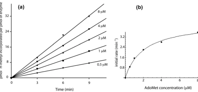
Steady-state kinetic assay [Methyl-3
H]-AdoMet (555 GBq/mmol, 20.5 MBq/mL, MP Biomedicals) was mixed with non-radioactive AdoMet (Sigma) to achieve a specific radioactivity of about 500 cpm/pmol (16.7 Bq/pmol). The methylation kinetic assays were performed in 50 mM Tris-HCl pH 8.0 at 60 °C. The tRNA and [methyl-3
H]-AdoMet were thermally equilibrated in 1.25 x concentrated reaction buffer (96 µL) at 60 °C for 5 min, and the reaction was started by adding the enzyme (24 µL). Aliquots (25 µL) were removed after different incubation times and transferred into 800 µL of 5 % (w/v) trichloroacetic acid (TCA) at 0 °C for 30 min in order to quench the reaction and to precipitate the tRNA. The precipitates were collected by filtration through GF/C filters (Whatman). The filters were washed with cold 5 % TCA, dried, and the radioactivity was measured by liquid scintillation counting for 2 min, resulting in a counting error below 4 %. Data were corrected by substracting the background radioactivity determined from a control without enzyme.
For the determination of AdoMet KM and kcat, the reaction mixtures contained MTase (wild
type, 25 nM ; Y194A, 50 nM ; Y78A, 100 nM ; D170A, 500 nM), tRNAi Met
(20 µM) and 3
H-AdoMet (wild type, 0.5-8 µM ; Y194A, 0.5-8 µM ; Y78A, 0.5-8 µM ; D170A, 5-50 µM). For tRNAi
Met
KM and kcat, the reaction mixtures contained MTase (wild type, 25 nM ; Y194A, 50
nM ; Y78A, 100 nM ; D170A, 500 nM), tRNAi Met
(wild type, 0.5-8 µM ; Y194A, 0.5-8 µM ; Y78A, 0.25-4 µM ; D170A, 0.5-8 µM) and 3
H-AdoMet (wild type, 20 µM ; Y194A, 50 µM ; Y78A, 20 µM ; D170A, 50 µM). Initial rates (vi) for each substrate concentration (tRNAi
Met
) were determined from the slope of linear fits of time course data points (wild type, 9 min ; Y194A, 20 min ; Y78A, 40 min ; D170A, 40 min). Enzyme parameters were obtained by non-linear least square fitting using Eq. (2) of Michaelis-Menten kinetics (wild type and mutant Y194A) or Eq. (3) in cases where enzyme concentration was not negligible compared to substrate concentration (mutant Y78A and D170A). kcat values were calculated for two
catalytic sites per TrmI tetramer. Confidence limits on the parameters were estimated by Monte-Carlo sampling using the MC-Fit program.61
(2) ! vi= VmaxS0 KM + S0 (3) ! vi= Vmax 2E0 KM + S0+ E0" (KM + S0+ E0) 2 " 4S0E0
(
)
Molecular Docking
Docking of the N9-methyladenine ring into the active site of TrmI was performed using
HADDOCK 1.337
(http://www.nmr.chem.uu.nl/haddock/) that makes use of CNS62
as structure calculation software. HADDOCK allows to deal with ambiguous constraints, i.e. restraints can be applied between residues and not only between atoms. The protein structure used for the docking was the tetrameric T. thermophilus TrmI crystal structure in which the AdoHcy ligand was replaced with the AdoMet methyl donor. A 2 Å distance was used to define ambiguous restraints (AIRs) that were applied between the adenine and a set of neighbours composed of AdoMet cofactor and thTrmI residues identified by mutagenesis (i.e. Y78, D170 and Y194). One unambiguous restraint set to 2 Å was applied between the atoms that react in the methyl transfer reaction, i.e. the N1 position of the N9-methyladenine ring and
the Cε atom of the AdoMet ligand. During the rigid body energy minimisation, 2000 structures were calculated and the 1000 best solutions based on the intermolecular energy were used for the semi-flexible refinement. The docking converged to a single solution. The same protocol was also applied to the M. tuberculosis TrmI.
REFERENCES
1. Rozenski, J., Crain, P. F. & McCloskey, J. A. (1999). The RNA Modification Database: 1999 update. Nucl. Acids Res. 27, 196-197.
2. Anderson, J., Phan, L. & Hinnebusch, A. G. (2000). The Gcd10p/Gcd14p complex is the essential two-subunit tRNA(1-methyladenosine) methyltransferase of
Saccharomyces cerevisiae. Proc. Natl. Acad. Sci. USA 97, 5173-5178.
3. Droogmans, L., Roovers, M., Bujnicki, J. M., Tricot, C., Hartsch, T., Stalon, V. & Grosjean, H. (2003). Cloning and characterization of tRNA (m1A58)
methyltransferase (TrmI) from Thermus thermophilus HB27, a protein required for cell growth at extreme temperatures. Nucl. Acids Res. 31, 2148-2156.
4. Anderson, J., Phan, L., Cuesta, R., Carlson, B. A., Pak, M., Asano, K., Bjork, G. R., Tamame, M. & Hinnebusch, A. G. (1998). The essential Gcd10p-Gcd14p nuclear complex is required for 1-methyladenosine modification and maturation of initiator methionyl-tRNA. Genes Dev. 12, 3650-3662.
5. Ozanick, S. G., Bujnicki, J. M., Sem, D. S. & Anderson, J. T. (2007). Conserved amino acids in each subunit of the heteroligomeric tRNA m1A58 Mtase from Saccharomyces cerevisiae contribute to tRNA binding. Nucl. Acids Res. 35, 6808-6819.
6. Bujnicki, J. M. (2001). In silico analysis of the tRNA:m1A58 methyltransferase family: homology-based fold prediction and identification of new members from Eubacteria and Archaea. FEBS Letters 507, 123-127.
7. Gupta, A., Kumar, P. H., Dineshkumar, T. K., Varshney, U. & Subramanya, H. S. (2001). Crystal structure of Rv2118c: an AdoMet-dependent methyltransferase from Mycobacterium tuberculosis H37Rv. J. Mol. Biol. 312, 381-391.
8. Roovers, M., Wouters, J., Bujnicki, J. M., Tricot, C., Stalon, V., Grosjean, H. & Droogmans, L. (2004). A primordial RNA modification enzyme: the case of tRNA (m1A) methyltransferase. Nucl. Acids Res. 32, 465-476.
9. Varshney, U., Ramesh, V., Madabushi, A., Gaur, R., Subramanya, H. S. &
RajBhandary, U. L. (2004). Mycobacterium tuberculosis Rv2118c codes for a single-component homotetrameric m1A58 tRNA methyltransferase. Nucl. Acids Res. 32, 1018-1027.
10. Schubert, H. L., Blumenthal, R. M. & Cheng, X. (2003). Many paths to
methyltransfer: a chronicle of convergence. Trends Biochem. Sci. 28, 329-335.
11. Loo, J. A. (1997). Studying noncovalent protein complexes by electrospray ionization mass spectrometry. Mass Spectrom. Rev. 16, 1-23.
12. Loo, J. A. (2000). Electrospray ionization mass spectrometry: a technology for studying noncovalent macromolecular complexes. Int. J. Mass Spectrom. 200, 175-186.
13. Heck, A. J. & Van Den Heuvel, R. H. (2004). Investigation of intact protein complexes by mass spectrometry. Mass Spectrom. Rev. 23, 368-389.
14. Veenstra, T. D. (1999). Electrospray ionization mass spectrometry: a promising new technique in the study of protein/DNA noncovalent complexes. Biochem. Biophys.
Res. Commun. 257, 1-5.
15. Beck, J. L., Colgrave, M. L., Ralph, S. F. & Sheil, M. M. (2001). Electrospray ionization mass spectrometry of oligonucleotide complexes with drugs, metals, and proteins. Mass Spectrom. Rev. 20, 61-87.
16. Hofstadler, S. A. & Griffey, R. H. (2001). Analysis of noncovalent complexes of DNA and RNA by mass spectrometry. Chem. Rev. 101, 377-390.
17. Rusconi, F., Guillonneau, F. & Praseuth, D. (2002). Contributions of mass
spectrometry in the study of nucleic acid-binding proteins and of nucleic acid-protein interactions. Mass Spectrom. Rev. 21, 305-348.
18. Robinson, C. V., Chung, E. W., Kragelund, B. B., Knudsen, J., Aplin, R. T., Poulsen, F. M. & Dobson, C. M. (1996). Probing the nature of noncovalent interactions by mass spectrometry. A study of protein-CoA ligand binding and assembly. J. Am.
Chem. Soc. 118, 8646-8653.
19. Rogniaux, H., Van Dorsselaer, A., Barth, P., Biellmann, J. F., Barbanton, J., van Zandt, M., Chevrier, B., Howard, E., Mitschler, A., Potier, N., Urzhumtseva, L., Moras, D. & Podjarny, A. (1999). Binding of aldose reductase inhibitors: Correlation of crystallographic and mass spectrometric studies. J. Am. Soc. Mass Spectrom. 10, 635-647.
20. Mei, H. Y., Mack, D. P., Galan, A. A., Halim, N. S., Heldsinger, A., Loo, J. A., Moreland, D. W., Sannes-Lowery, K. A., Sharmeen, L., Truong, H. N. & Czarnik, A. W. (1997). Discovery of selective, small-molecule inhibitors of RNA complexes--I. The Tat protein/TAR RNA complexes required for HIV-1 transcription. Bioorg. Med.
Chem. 5, 1173-1184.
21. Liu, C., Tolic, L. P., Hofstadler, S. A., Harms, A. C., Smith, R. D., Kang, C. & Sinha, N. (1998). Probing RegA/RNA interactions using electrospray ionization-fourier transform ion cyclotron resonance-mass spectrometry. Anal. Biochem. 262, 67-76. 22. Potier, N., Donald, L. J., Chernushevich, I., Ayed, A., Ens, W., Arrowsmith, C. H.,
Standing, K. G. & Duckworth, H. W. (1998). Study of a noncovalent trp repressor: DNA operator complex by electrospray ionization time-of-flight mass spectrometry.
Protein Sci. 7, 1388-1395.
23. Hagan, N. & Fabris, D. (2003). Direct mass spectrometric determination of the
stoichiometry and binding affinity of the complexes between nucleocapsid protein and RNA stem-loop hairpins of the HIV-1 Psi-recognition element. Biochemistry 42, 10736-10745.
24. Kilic, T., Sanglier, S., Van Dorsselaer, A. & Suck, D. (2006). Oligomerization behavior of the archaeal Sm2-type protein from Archaeoglobus fulgidus. Protein Sci. 15, 2310-2317.
25. Hamilton, S., Odili, J., Pacifico, M. D., Wilson, G. D. & Kupsch, J. M. (2003). Effect of imidazole on the solubility of a His-tagged antibody fragment. Hybridoma and
Hybridomics 22, 347-355.
26. McCoy, A. J., Grosse-Kunstleve, R. W., Storoni, L. C. & Read, R. J. (2005).
Likelihood-enhanced fast translation functions. Acta Crystallog. sect. D 61, 458-464. 27. Fauman, E. B., Blumenthal, R. M. & Cheng, X. (1999). Structure and evolution of
AdoMet-dependent methyltransferases. In S-adenosylmethionine-dependent
methyltransferases: structures and functions (Cheng, X. & Blumenthal, R. M., eds.),
pp. 1-38. World Scientific Publishing.
28. Rostom, A. A. & Robinson, C. V. (1999). Detection of the intact GroEL chaperonin assembly by mass spectrometry. J. Am. Chem. Soc. 121, 4718-4719.
29. van Berkel, W. J., van den Heuvel, R. H., Versluis, C. & Heck, A. J. (2000). Detection of intact megaDalton protein assemblies of vanillyl-alcohol oxidase by mass spectrometry. Protein Sci. 9, 435-439.
30. Sanglier, S., Ramstrom, H., Haiech, J., Leize, E. & Van Dorsselaer, A. (2002). Electrospray ionization mass spectrometry analysis revealed a similar to 310 kDa noncovalent hexamer of HPr kinase/phosphatase from Bacillus subtilis. Int. J. Mass
31. Sanglier, S., Leize, E., Van Dorsselaer, A. & Zal, F. (2003). Comparative ESI-MS study of approximately 2.2 MDa native hemocyanins from deep-sea and shore crabs: from protein oligomeric state to biotope. J. Am. Soc. Mass Spectrom. 14, 419-429. 32. Rogniaux, H., Sanglier, S., Strupat, K., Azza, S., Roitel, O., Ball, V., Tritsch, D.,
Branlant, G. & Van Dorsselaer, A. (2001). Mass spectrometry as a novel approach to probe cooperativity in multimeric enzymatic systems. Anal. Biochem. 291, 48-61. 33. Smith, R. D. & Light-Wahl, K. J. (1993). The observation of non-covalent
interactions in solution by electrospray ionization mass spectrometry: Promise, pitfalls and prognosis. Biol. Mass Spectrom. 22, 493-501.
34. Kapur, A., Beck, J. L., Brown, S. E., Dixon, N. E. & Sheil, M. M. (2002). Use of electrospray ionization mass spectrometry to study binding interactions between a replication terminator protein and DNA. Protein Sci. 11, 147-157.
35. Gupta, R., Hamdan, S. M., Dixon, N. E., Sheil, M. M. & Beck, J. L. (2004). Application of electrospray ionization mass spectrometry to study the hydrophobic interaction between the epsilon and theta subunits of DNA polymerase III. Protein
Sci. 13, 2878-2887.
36. Cassiday, L. A., Lebruska, L. L., Benson, L. M., Naylor, S., Owen, W. G. & Maher, L. J., 3rd. (2002). Binding stoichiometry of an RNA aptamer and its transcription factor target. Anal. Biochem. 306, 290-297.
37. Dominguez, C., Boelens, R. & Bonvin, A. M. (2003). HADDOCK: a protein-protein docking approach based on biochemical or biophysical information. J. Am. Chem.
Soc. 125, 1731-1737.
38. McCarthy, A. A. & McCarthy, J. G. (2007). The structure of two
N-methyltransferases from the caffeine biosynthetic pathway. Plant Physiol. 144, 879-889.
39. Kettani, A., Gueron, M. & Leroy, J. L. (1997). Amino proton exchange processes in mononucleosides. J. Am. Chem. Soc. 119, 1108-1115.
40. Stewart, R. & Harris, M. G. (1977). Amino Group Acidity in Nucleotide Bases. Can.
J. Chem. 55, 3807-3814.
41. Elkins, P. A., Watts, J. M., Zalacain, M., van Thiel, A., Vitazka, P. R., Redlak, M., Andraos-Selim, C., Rastinejad, F. & Holmes, W. M. (2003). Insights into catalysis by a knotted TrmD tRNA methyltransferase. J. Mol. Biol. 333, 931-949.
42. Labahn, J., Granzin, J., Schluckebier, G., Robinson, D. P., Jack, W. E., Schildkraut, I. & Saenger, W. (1994). Three-dimensional structure of the adenine-specific DNA methyltransferase M.Taq I in complex with the cofactor S-adenosylmethionine. Proc.
Natl. Acad. Sci. USA 91, 10957-10961.
43. Tran, P. H., Korszun, Z. R., Cerritelli, S., Springhorn, S. S. & Lacks, S. A. (1998). Crystal structure of the DpnM DNA adenine methyltransferase from the DpnII restriction system of streptococcus pneumoniae bound to S-adenosylmethionine.
Structure 6, 1563-1575.
44. Goedecke, K., Pignot, M., Goody, R. S., Scheidig, A. J. & Weinhold, E. (2001). Structure of the N6-adenine DNA methyltransferase M.TaqI in complex with DNA and a cofactor analog. Nat. Struct. Biol. 8, 121-125.
45. Schluckebier, G., Zhong, P., Stewart, K. D., Kavanaugh, T. J. & Abad-Zapatero, C. (1999). The 2.2 A structure of the rRNA methyltransferase ErmC' and its complexes with cofactor and cofactor analogs: implications for the reaction mechanism. J. Mol.
Biol. 289, 277-291.
46. Maravic, G., Feder, M., Pongor, S., Flogel, M. & Bujnicki, J. M. (2003). Mutational analysis defines the roles of conserved amino acid residues in the predicted catalytic pocket of the rRNA:m6A methyltransferase ErmC'. J. Mol. Biol. 332, 99-109.
47. Bheemanaik, S., Reddy, Y. V. & Rao, D. N. (2006). Structure, function and mechanism of exocyclic DNA methyltransferases. Biochem. J. 399, 177-190. 48. Jeltsch, A. (2002). Beyond Watson and Crick: DNA methylation and molecular
enzymology of DNA methyltransferases. Chembiochem 3, 274-293.
49. Pues, H., Bleimling, N., Holz, B., Wolcke, J. & Weinhold, E. (1999). Functional roles of the conserved aromatic amino acid residues at position 108 (motif IV) and position 196 (motif VIII) in base flipping and catalysis by the N6-adenine DNA
methyltransferase from Thermus aquaticus. Biochemistry 38, 1426-1434.
50. Roth, M., Helm-Kruse, S., Friedrich, T. & Jeltsch, A. (1998). Functional roles of conserved amino acid residues in DNA methyltransferases investigated by site-directed mutagenesis of the EcoRV adenine-N6-methyltransferase. J. Biol. Chem. 273, 17333-17342.
51. Collaborative Computational Project Number 4. (1994). The CCP4 suite: programs for protein crystallography. Acta Crystallog. sect. D 50, 760-763.
52. Potterton, E., Briggs, P., Turkenburg, M. & Dodson, E. (2003). A graphical user interface to the CCP4 program suite. Acta Crystallog. sect. D 59, 1131-1137.
53. Leslie, A. G. W. (1992). Recent changes to the MOSFLM package for processing film
and image plate data. Joint CCP4+ ESF-EAMCB Newsletter on Protein
Crystallography (Science and Engineering Research Council, D. L., Ed.), Daresbury Laboratory, Warrington, UK.
54. Evans, P. R. (1993). Proceedings of the CCP4 Study Weekend: Data collection and
processing. Science and Engineering Research Council, Daresbury Laboratory,
Warrington, United Kingdom.
55. Perrakis, A., Morris, R. & Lamzin, V. S. (1999). Automated protein model building combined with iterative structure refinement. Nat. Struct. Biol. 6, 458-463.
56. Murshudov, G. N., Vagin, A. A. & Dodson, E. J. (1997). Refinement of
macromolecular structures by the maximum-likelihood method. Acta Crystallog. sect.
D 53, 240-255.
57. Emsley, P. & Cowtan, K. (2004). Coot: model-building tools for molecular graphics.
Acta Crystallog. sect. D 60, 2126-2132.
58. Winn, M. D., Isupov, M. N. & Murshudov, G. N. (2001). Use of TLS parameters to model anisotropic displacements in macromolecular refinement. Acta Crystallog. sect.
D 57, 122-133.
59. Baker, N. A., Sept, D., Joseph, S., Holst, M. J. & McCammon, J. A. (2001). Electrostatics of nanosystems: application to microtubules and the ribosome. Proc.
Natl. Acad. Sci. USA 98, 10037-10041.
60. Meinnel, T. & Blanquet, S. (1995). Maturation of pre-tRNA(fMet) by Escherichia coli RNase P is specified by a guanosine of the 5'-flanking sequence. J. Biol. Chem. 270, 15908-15914.
61. Dardel, F. (1994). MC-Fit: using Monte-Carlo methods to get accurate confidence limits on enzyme parameters. Comput. Appl. Biosci. 10, 273-275.
62. Brunger, A. T., Adams, P. D., Clore, G. M., DeLano, W. L., Gros, P., Grosse-Kunstleve, R. W., Jiang, J. S., Kuszewski, J., Nilges, M., Pannu, N. S., Read, R. J., Rice, L. M., Simonson, T. & Warren, G. L. (1998). Crystallography & NMR system: A new software suite for macromolecular structure determination. Acta Crystallog.
sect. D 54, 905-921.
63. Schluckebier, G., O'Gara, M., Saenger, W. & Cheng, X. (1995). Universal catalytic domain structure of AdoMet-dependent methyltransferases. J. Mol. Biol. 247, 16-20.
ACKNOWLEGMENTS
The authors would like to thank Franck Brachet for his skill in the set up of the pipetting robot. We acknowledge Dr Thibaut Crépin for his kind help in the synchrotron data collection on beamline ID14-1 at ESRF (Grenoble). We thank Nathalie Ulryck for help in site-directed mutagenesis. Pierre Barraud and Cédric Atmanene are supported by a studentship from the “Ministère de la recherche”. We thank the CNRS for financial support. Louis Droogmans was supported by grants from the FRFC (Fonds pour la Recherche Fondamentale Collective) and IISN (Institut Interuniversitaire des Sciences Nucléaires).
FIGURE LEGENDS
Figure 1: Crystal Structure of T. thermophilus TrmI.
(a) Overall structure of thTrmI monomer. The secondary structure elements are labelled following the nomenclature defined in Schluckebier et al.63
The cofactor AdoHcy is shown in blue sticks. (b) Organization of thTrmI as a tetramer. Each monomer is drawn with a different colour. The two tight dimers are respectively in red and yellow, and in pale and dark blue. The tetramerization interface involved strands β6 and β7.
(c) The AdoHcy binding site. The ligand (blue) and the enzyme residues (gray) are shown as sticks. Water molecules are drawn as blue spheres. Polar contacts are represented with dashed lines. Strictly conserved residue among m1
A58 tRNA MTases are labelled with bold letters.
Characteristic motifs from MTases are highlighted. (d) Density map (2Fo-Fc at 1σ) around the cofactor. The AdoHcy ligand (blue) and the enzyme residues (green) are shown as sticks. Water molecules are represented as red spheres.
Figure 2: Electrostatic surface potential of (a) T. thermophilus TrmI and (b) M. tuberculosis TrmI. Blue indicates positive charge and red negative one with the maximum color saturation corresponding to -3 kT (red) and +3 kT (blue). The figure was prepared using the APBS59
PyMOL plugin.
Figure 3: Noncovalent ESI-MS analyses of thTrmI and thTrmI-tRNA assemblies. ESI mass spectra obtained under non-denaturing conditions (200 mM ammonium bicarbonate buffer pH 8.0; Vc = 120 V; Pi = 6.1 mbar) for (a) thTrmI tetramer (20 µM) alone and in presence of (b) 30 µM, (c) 100 µM and (d) 200 µM of E. coli tRNAi
Met
. Inserts correspond to deconvoluted spectra showing molecular weights on the x-scale. T (MW = 115450 ± 5 Da) corresponds to (thTrmI)4, T+1tRNA (MW = 140347 ± 6 Da) corresponds to
(thTrmI)4:(tRNA)1 complex and T+2tRNA (MW = 165455 ± 10 Da) corresponds to
(thTrmI)4:(tRNA)2 complex. (*) correspond to tRNA signals.
Figure 4: Non-denaturing agarose gel electrophoresis Constant amount of E. coli tRNAi
Met
(4 µg) with increasing amounts of thTrmI protein (from 0 to 1 molar equivalent of thTrmI tetramer). Bands were visualized by UV shadowing.
Figure 5: Influence of the buffer ionic strength on thTrmI-tRNA complexes stabilities. ESI mass spectra obtained under non-denaturing conditions (ammonium bicarbonate pH 8.0; Vc = 120 V; Pi = 6.1 mbar) for thTrmI tetramer (20 µM) in presence of 100 µM of E. coli tRNAi
Met
. Ammonium bicarbonate concentration was set to (a) 100 mM, (b) 200 mM, (c) 400 mM and (d) 1250 mM. Inserts correspond to deconvoluted spectra showing molecular
weights on the x-scale. T corresponds to (thTrmI)4, T+1tRNA corresponds to
(thTrmI)4 :(tRNA)1 complex and T+2tRNA corresponds to (thTrmI)4:(tRNA)2 complex. (*)
correspond to tRNA signals.
Figure 6: Specificity of thTrmI-tRNA complexes formation. ESI mass spectra obtained under non-denaturing conditions (200 mM ammonium bicarbonate buffer pH 8.0; Vc = 120 V; Pi = 6.1 mbar) for thTrmI tetramer (20 µM) in presence of (a) 100 µM E. coli tRNAi
Met
(b) 100 µM non substrate control RNA. Inserts correspond to deconvoluted spectra showing molecular weights on the x-scale. T corresponds to (thTrmI)4, T+1tRNA corresponds to
(thTrmI)4 :(tRNA)1 complex and T+2tRNA corresponds to (thTrmI)4:(tRNA)2 complex. (*)
corresponds to RNA signal.
Figure 7: Sequence alignments of TrmI proteins and conserved residues in thTrmI catalytic pocket.
(a) Part of sequence alignment of T. thermophilus, M. tuberculosis, P. abyssi, M. musculus,
H. sapiens and S. cerevisiae m1
A58 tRNA MTases. Residue numbers are those of T.
thermophilus sequence. Highly conserved residues are shown on a dark blue background and
residues with a similar physico-chemical character are on a pale blue background. The secondary structure of the T. thermophilus TrmI is shown below the sequences as arrows for β-strands and cylinder for α-helices. The names of the secondary structure elements refer to Figure 1(a). The sequence motifs typical of the AdoMet dependent MTase family are also indicated. Residues marked with a star (*) (i.e. Y78, D170 and Y194) are involved in the catalytic pocket formation and are those studied by mutagenesis in the present work. Residues marked with a circle (o) (i.e. W175 and F245) are conserved aromatic residues in motifs IV and VIII, belonging to the catalytic pocket in m6
A DNA MTases. (b) Conserved and semi-conserved residues around the catalytic pocket of T. thermophilus TrmI. The strictly conserved residues among the m1
A58 tRNA MTase family are shown as sticks on the T.
thermophilus structure except the conserved residues that bind AdoMet. AdoMet was
modeled from AdoHcy by adding a methyl group on the sulphur atom, the methyl is positionned by the sulfonium chirality. Y78, Y194 and D170 are indicated as red sticks. Figure 8: Modeling a N9-methyladenine in the catalytic pocket of thTrmI
(a) Average docked structure of the N9-methyladenine ring inside the catalytic pocket of
thTrmI. The enzyme surface is coloured according to the electrostatic surface potential.
Positive potential is shown in blue and negative potential in red. The AdoMet ligand and the docked N9-methyladenine ring are shown as sticks (carbon atoms in light blue, nitrogen
atoms in dark blue, oxygen atoms in red, sulphur atom in yellow and hydrogen atoms in white). The Van der Waals surface of the N9-methyladenine ring is shown as white dots.
(b) Distances between the average structure of the docked N9-methyladenine ring and key
moiety of the enzyme. The enzyme is shown in gray with the cartoon representation. The AdoMet ligand, the docked N9-methyladenine ring and residues D170, Y78 and Y194 are