HAL Id: tel-02003364
https://tel.archives-ouvertes.fr/tel-02003364
Submitted on 1 Feb 2019
HAL is a multi-disciplinary open access
archive for the deposit and dissemination of sci-entific research documents, whether they are pub-lished or not. The documents may come from teaching and research institutions in France or abroad, or from public or private research centers.
L’archive ouverte pluridisciplinaire HAL, est destinée au dépôt et à la diffusion de documents scientifiques de niveau recherche, publiés ou non, émanant des établissements d’enseignement et de recherche français ou étrangers, des laboratoires publics ou privés.
and human mitochondria
Mariia Baleva
To cite this version:
Mariia Baleva. Study of the mechanisms of tRNA targeting into yeast and human mitochondria. Cellular Biology. Université de Strasbourg; Université Lomonossov (Moscou), 2016. English. �NNT : 2016STRAJ096�. �tel-02003364�
Université de Moscou
2016
École doctorale des sciences de la vie et de la santé
THÈSE
présentée pour l’obtention du grade de
DOCTEUR DE L’UNIVERSITÉ DE STRASBOURG
Discipline : Sciences du vivant
Domaine : Aspects moléculaires et cellulaires de la biologie
par
BALEVA MARIIA
ÉTUDE DES MÉCHANISMES D’ADRESSAGE D’ARN DE TRANSFERT
DANS LES MITOCHONDRIES DE LEVURE ET HUMAINES
Soutenue le 16 Decembre 2016 devant la commission d’examen:
M.S. JOHANSEN
M.M. PATRUSHEV
M.M. BLAISE
A-M. DUCHENE
M.I. TARASSOV
A.P. KAMENSKI
M.B. MASQUIDA
Rapporteur externe
Rapporteur externe
Rapporteur externe
Examinateur
Examinateur
Co-directeur de thèse
Directeur de thèse
UMR N°7156 UdS-CNRS
«Génétique Moléculaire, Génomique et Microbiologie»
TABLE OF CONTENTS
ABBREVIATIONS ... 1 INTRODUCTON ... 3 Origin and general structure of mitochondria ... 3 Protein Import Pathways into Mitochondria ... 3 Targeting and sorting signals of mitochondrial precursor proteins ... 5 The TOM complex as the entry platform ... 7 Multifunctional inner membrane translocase TIM23 ... 8 TIM22 complex - a specialized translocase for metabolite carriers ... 10 The MIA pathway: intermembrane space import and assembly ... 10 The SAM complex – a platform for β-barrel proteins biogenesis ... 11 The MIM machinery ... 12 RNA Import into Mitochondria ... 12 tRNA import into protozoan mitochondria ... 15 tRNA import into plant mitochondria ... 19 tRNA import into yeast mitochondria ... 20 Enolase in other life processes ... 23 Mitochondrial RNA import in mammals ... 26 THESIS PROJECT AND OBJECTIVES ... 30 RESULTS ... 31 A moonlighting human protein is involved in mitochondrial import of tRNA ... 31 PUBLICATION I ... 33 Human enolase overexpression or down-regulation does not affect tRK1 mitochondrial import in vivo. ... 34 Probing tRK1 and tRK2 conformations ... 36 Factors beyond enolase 2 and mitochondrial lysyl-tRNA synthetase precursor are required for tRNA import in yeast mitochondria ... 42 The analysis of protein content of Eno2p-containing samples ... 63Analysis of Eno2p structure by SAXS and X-ray crystallography ... 72 Analysis of tRK1-protein complexes purified from yeast ... 75 DISCUSSION ... 80 CONCLUSIONS AND PERSPECTIVES ... 88 MATERIAL AND METHODS ... 89 REFERENCES ... 114 RÉSUMÉ DE THÈSE ... 128 SUMMERY ... 135
1
ABBREVIATIONS
aaRS ADP APS ATP BSA DMEM DMSO DNA DTT EDTA EMSA FRET HEPES HdV HMH IMS IPTG ITC Kd kDa KRS LB mtDNA MIA MIM MISS MMP aminoacyl-tRNA synthetase adenosine diphosphate ammonium persulfate adenosine triphosphate bovine serum albumin dulbecco modified Eagle's medium dimethyl sulfoxide deoxyribonucleic acid dithiothreitol ethylenediaminetetraacetic acid electrophoretic mobility shift assay fluorescence resonance energy transfer 4-(2-hydroxyethyl)-1-piperazineethanesulfonic acid hepatitis delta virus ribozyme hammerhead ribozyme intermembrane space isopropyl β-D-1-thiogalactopyranoside isothermal titration microcalorimetry dissociation constant kilo Dalton cytosolic lysyl-tRNA synthetase lysogeny broth mitochondrial DNA mitochondrial intermembrane space import and assembly machinery of the inner membrane mitochondrial intermembrane space signal mitochondrial processing peptidase2 MIP MTS MW NADH OD PAM PBS PIPES PNPase preMSK preKARS PAGE RIC PCR pI SAM SDS siRNA SSC TAB TBE TEMED TOM Tris tRNA UV VDAC UPS Δψ mitochondrial intermediate peptidase mitochondrial targeting sequence molecular weight nicotinamide adenine dinucleotide reduced optical density presequence translocase associated motor phosphate buffered saline piperazine-N,N'-bis(ethanesulfonic acid) polynucleotide phosphorylase precurcor of yeast mitochondral lysyl-tRNA synthetase precurcor of human mitochondral lysyl-tRNA synthetase polyacrylamide gel electrophoresis RNA import complex polymerase chain reaction isoelectric point sorting and assembly machinery sodium dodecylsulfate small interferring RNA saline sodium citrate buffer tubulin antisense-binding protein tris-borate-EDTA buffer tetramethylethylenediamine translocase of the outer membrane (hydroxymethyl)aminomethane transfer RNA ultra violet voltage dependent anion channel ubiquitin-proteasome system membrane potential
3
INTRODUCTON
Origin and general structure of mitochondria
More than 1.5 billion years ago an α-proteobacteria–like ancestor gave the origin of mitochondria (Dyall et al, 2004; Gray, 2012). Moreover, mitochondria still have a set of features similar to modern prokaryotes. Thus, as well as plasma membrane of prokaryotes the mitochondrial membrane contains electron transport proteins, mitochondria also possess a circular genome and specific genetic code, mitochondrial transcription is coupled to translation (Shadel, 2004), organella division and replication occur independently of host cell division. In addition they are surrounded by a double-membrane. The outer membrane contains diverse pore-forming proteins (porins) and translocases (Campo et al, 2016) that provide transport of different molecules ranging from ions and small uncharged molecules to proteins. The inner membrane is permeable only to oxygen, carbon dioxide, and water, while the other molecules reach the mitochondrial inner space by the means of specific membrane transport proteins. The inner mitochondrial membrane forms numerous invaginations called cristae, which greatly increase the total surface of the inner membrane. This increased surface confers to the inner membrane very effective transport capacities to the entire matrix volume. Cristae contain complexes of the electron transport chain and the ATP synthase. The inner membrane surrounds a mitochondrial matrix where the citric acid cycle and the other biosynthetic reactions take place. Membranes are separated by a thin gap (~20 nm) forming the intermembrane space. (Kühlbrandt, 2015)Protein Import Pathways into Mitochondria
Mitochondria possess their own genome, which reflects their bacterial origin as well as divergent evolution. Mitochondrial DNA shows a great deal of variation in size, physical form, coding capacity, modes of organization and expression (Gray, 2012). The transition from autonomous endosymbionts to mitochondria results from genome reduction through the loss of the part of endosymbiotic genes or their transposition to the nucleus (Allen, 2003). To explain the retention of a set of genes as well as of mechanisms of4 genome maintenance in mitochondria, several hypotheses were suggested. Thus, mitochondrial genome encodes eight highly hydrophobic membrane proteins, which are believed retained to avoid mistargeting. According to an alternative hypothesis, mtDNA keeps genes which expression can be regulated depending on the redox state of gene products (Allen, 2015). Furthermore, the non-canonical mitochondrial genetic code can make a barrier to expression of mitochondrial protein-coding genes in nucleus (Allen, 2003). Figure 1. Pathways sort mitochondrial precursor proteins into the mitochondrial subcompartments. Precursors of multi-spanning outer membrane proteins are recognized by Tom70 and transferred to the MIM machinery, which mediates their integration and assembly into the membrane. The TOM complex forms the general entry gate for 90% of mitochondrial precursors Thereafter, sorting pathways branch off. With the help of small TIM chaperones, β-barrel precursors are directly transferred to the SAM complex, which mediates their integration into the outer membrane (OM). Precursors with a cleavable presequence are transferred to the TIM23 complex, which mediates either lateral release into the inner membrane (IM) or transport into the mitochondrial matrix. Precursor translocation via the TIM23 complex into the matrix additionally requires the ATP-consuming action of the PAM module. The presequence is removed upon import by the mitochondrial processing peptidase. The small TIM chaperones guide precursors of multi-spanning carrier proteins to the TIM22 complex, which facilitates their insertion into the inner membrane. The membrane potential across the inner membrane (Δψ) drives TIM23 and TIM22 dependent import. Mia40 sorts cysteine-rich precursors into the intermembrane space and mediates their oxidative folding. Erv1 shuttles the electrons from Mia40 to cytochrome c of the respiratory chain (adapted from (Böttinger et al, 2015). IMS – the intermembrane space. Large-scale proteomic analysis and computational studies allow to identify ∼1000 distinct proteins in fungi mitochondria and ∼1500 - in animal mitochondria (Fukasawa et al, 2015). However the majority (∼99%) of mitochondrial proteins have a nuclear origin
5 (Chacinska et al, 2009). They are synthesized in cytosol and imported to one of the four mitochondrial compartments by translocator complexes located in the mitochondrial membranes (Figure 1) (Bolender et al, 2008; Campo et al, 2016; Rao et al, 2012; Wenz et al, 2015).
Targeting and sorting signals of mitochondrial precursor proteins
The precursors of mitochondrial proteins are firstly synthesized in the cytosol and then targeted to mitochondria. To reach the correct mitochondrial subcompartment, they possess cleavable or not-cleavable sequences, which mark the precise sorting pathway and determine the final location of imported proteins. While the nature of these signals is not uniform (Chacinska et al, 2009), they have rather common features (Figure 2). Figure 2. Targeting and Sorting Signals of Mitochondrial Precursor Proteins. Three groups of targeting signals can be identified (A) presequences located at the N-terminus, (B) multiple internal signals and (C) redox-regulated signals that provide transient covalent interaction with the corresponding import receptor (adapted from Chacinska et al., 2009). Most matrix-targeting proteins contain a 10-50 amino acid cleavable pre-sequence located at the N-terminal extremity. Presequences are rich in positively charged, hydrophobic,6 and hydroxylated amino acids, and tend to form amphiphilic α-helices. Both positive charges and amphipathic property of the presequence are important for its recognition by receptors of TOM-complex and following translocation (Abe et al, 2000). Upon translocation pre-sequences are usually cleaved by different peptidases such as the mitochondrial processing peptidase (MMP) (Taylor et al, 2001), mitochondrial intermediate peptidase (MIP) or others (Quiros et al, 2015). A few matrix proteins, as chaperonin 10, are synthesized with an amino-terminal presequence that remains a permanent part of the protein (Ryan et al, 1994). A hydrophobic stretch following the N-terminal presequence serves as additional sorting signal. It arrests the translocation of protein through the mitochondria inner membrane with following lateral release into the membrane (Glick et al, 1992). A subset of mitochondrial proteins contains targeting signal at various positions within a mature protein, which are not cleaved upon translocation. Thus, the outer membrane β-barrel proteins bear the C-terminal targeting signal formed by the last β-strand which anchors the protein molecule in the membrane (Kutik et al, 2008). For the anchoring of Tom70 and Tom20 on the surface of mitochondria both N-terminal transmembrane domain and its positively charged C-flanking region are important (Kanaji et al, 2000). The outer membrane proteins of the α-helical type contain targeting sequences represented by the α-helical transmembrane segment often flanked by positively charged residues and located at the amino (signal anchor sequence) or at the carboxy terminus (tail anchor) as well as in the middle of the proteins (Chacinska et al, 2009). Targeting signals of inner membrane proteins as metabolite carriers are more heterogeneous. Usually they consist of three to six domains, each of about 10 amino acid residues, scattered across the molecule. Cooperation of domains is required for recognition by import receptors and for efficient translocation of hydrophobic preproteins (Wiedemann et al, 2001). Some mitochondrial inter-membrane space proteins contain non-cleaved signal (MISS) that include a conserved cysteine motif involved in transient disulfed-bonded intermediate formation with a receptor (Milenkovic et al, 2007; Milenkovic et al, 2009; Mordas and Tokatlidis, 2015).
7
The TOM complex as the entry platform
TOM complex is the general entry gate for more than 90% of all mitochondrial proteins that transfers them to distinct translocators (Campo et al, 2016). In contrast to known membrane protein complexes, which structures have been defined, the TOM complex consists of both α-helical and β-barrel integral membrane proteins. Recently, the architecture of this complex was defined (Shiota et al, 2015). Tom20 is a general import receptor that cooperates with Tom22 to form a presequence receptor, cis site (Shiota et al, 2011; Yamano et al, 2008). While Tom20 interacts with the hydrophobic side of the presequence, Tom22 recognizes its hydrophylic region (Schulz et al, 2015). The proteins with internal targeting signals are preferably recognized by Tom70 (Brix et al, 1997; Endo and Kohda, 2002). Being recognized, precursor proteins cross the outer membrane through the β-barrel Tom40 channel (Gessmann et al, 2011), which possesses two different translocation pathways for different types of precursors. Cross-linking experiments revealed the acidic path within the Tom40 pore, while carrier proteins follow the path formed by hydrophobic residues (Gornicka et al, 2014; Shiota et al, 2015). Then the presequence is recognized by the so-called trans site of the TOM40 complex formed by the intermembrane space domain (IMS) regions of Tom40, Tom22, and Tom7 (Shiota et al., 2011). The increase in affinity toward the trans side provides the driving force for precursor translocation across the membrane (Schulz et al, 2015). It was demonstrated that the amino-terminal segment of Tom40 crosses the β-barrel from the cytosolic side to the intermembrane space possibly to block the pore or participate in translocation of the precursor proteins throughout the barrel (Shiota et al, 2015). It was found that TOM complex presents two isoforms: the mature trimeric form dynamically exchanges with dimeric Tom22-less isoform providing the platform for reassembling and regulating protein transport (Shiota et al, 2015). TOM complex subunits could be phosphorylated: the phosphorylation of Tom40 precursor reduces import, whereas the phosphorylation of Tom6 precursor stimulates import and membrane integration (Rao et al, 2012; Schulz et al, 2015). In addition, the small subunits Tom5, Tom6, and Tom7 are involved in the assembly and stability of the TOM complex (Sherman et al, 2005). The deletion of Tom6 destabilizes the8 TOM complex, while the interaction of TOM subunits is stabilized in the absence of Tom7 (Becker et al, 2011b).
Multifunctional inner membrane translocase TIM23
The TIM23 complex is one of the most complicated protein translocase in the cell presenting a highly dynamic structure. TIM23 contains the core proteins Tim23, Tim17, and Tim50, as well as Mgr2 and Tim21 as accessory subunits and the motor-associated proteins Pam 17, Tim16 (Pam16), Tim14 (Pam18), Tim44, mtHsp70, which are responsible for mediating the ATP-driven translocation across the inner membrane, and Mge1. The TIM23 complex is a dynamic structure allowing translocation of proteins (Figure 3) with presequences into the mitochondrial matrix, as well as Mgr2/Tim17-dependent insertion of proteins bearing a stop transfer signal in the N-terminal region (Schulz et al, 2015). Tim23 is the central subunit of the complex. The C-terminal and membrane bound domain of Tim23 form a channel while the unstructured IMS domain of Tim23 (Tim23IMS)provides various interactions with different complex subunits, Tom22IMS and presequences
(Alder et al, 2008; Waegemann et al, 2015). While Tim23 represents the minimal pore, the native translocase channel is built by Tim23/Tim17 oligomers. It was demonstrated that Tim17 is involved in substrate-induced voltage gating of the translocase channel, recruits with Pam17 and Pam18 of the PAM complex and participates in the lateral release of
precursors into the inner membrane (Alder et al, 2008; Waegemann et al, 2015). The lateral release of precursors is also under control of Mgr2, the lateral gatekeeper (Ieva et al, 2014), which is, in addition, involved in Tim21 recruitment and cycling of Pam18 (Waegemann et al, 2015). Tim50 is the presequence receptor at the inner membrane (Schulz et al, 2011). Tim50 interacts with Tim23 via their IMS domains promoting the closed state of the channel in the absence of substrates, therefore maintaining the permeability barrier across the inner membrane (Meinecke, 2006).
9
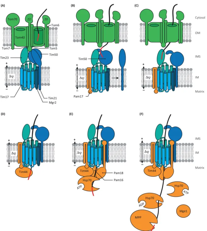
Figure 3. The presequence pathway of mitochondrial protein import (adapted from (Schulz et al, 2015). (A) TOM and TIM complexes are kept in close proximity by interactions of Tom22IMS with Tim50, Tim23 and Tim21. (B) Tim50 recognizes the presequence on the trans side of the TOM complex triggering Tim21 dissociation from the translocase and association of Pam17 with the complex. (С) Thereafter, presequence is passed to Tim23; (D) enters the channel and crosses the inner membrane driven by Δψ. On the matrix side, Tim44 binds the presequence. (E) Interaction of the presequence with Tim44, as well as assembling of the import motor, recruits Hsp70 in the ATP-bound state. (F) Pam18 stimulates Hsp70 to hydrolysis ATP and Hsp70 dissociation of Hsp70-ADP tightly bound to the substrate. Mge1 stimulates the precursor and Hsp70 dissociation by releasing of ADP. Thereafter, the pre-sequence can be removed by the mitochondrial processing peptidase and then processed by the intermediate cleaving peptidase 55 (Icp55) or the mitochondrial intermediate peptidase Oct1. OM – the outer membrane, IM – the inner membrane, IMS – inter-membrane space (Schulz et al, 2015).
10
The TIM23 complex cooperates with the TOM complex forming a path for effective translocation of ≈ 70% of proteins targeted into mitochondria (Waegemann et al, 2015). Complexes are kept in close proximity by interactions of Tom22IMS with Tim50 and Tim23 as
well as Tim21 (Figure 3A), which also participates in the substrate-directed switching of the TIM23 complex between matrix translocation and inner membrane sorting modes (Mokranjac et al, 2005).
TIM22 complex - a specialized translocase for metabolite carriers
A specialized TIM22 translocase is required for the biogenesis of a class of multispanning inner membrane proteins, including metabolite carriers or some translocase subunits like Tim17, Tim22, and Tim23. Upon translocation into the intermembrane space, this type of precursor is taken by small soluble Tim proteins arranged in subcomplexes, and is transferred to the TIM22 complex. The TIM22 translocase consists of four membrane subunits – Tim22, Tim18, Tim54, and Sdh3, and a peripheral Tim12 subunit (Campo et al, 2016). Large C-terminal domain of Tim54 exposed to the intermembrane space serves as a platform for docking of small Tim complexes bound with precursors. The protein-conducting channel of TIM22 consists of two pores formed by Tim22 (Rehling, 2003). The combined action of membrane potential and carrier precursor is responsible for the coordinated opening and closing of the TIM22 translocase (Peixoto et al, 2007). However, the mechanism of releasing of carrier precursors from the TIM22 complex into the inner membrane is still unknown.The MIA pathway: intermembrane space import and assembly
The biogenesis of IMS proteins with internal non-cleavable mitochondrial IMS-targeting/sorting signals (MISS) relies on Mia40, a redox-regulated IMS receptor that introduces disulfide bonds via several electron-transfer reactions (Chacinska et al, 2004). Another component of the MIA pathway is the thiol oxidase Erv1, which function is to recycle Mia40 by accepting electron from reduced Mia40 and transfering it to cytochrome c or molecular oxygen. A scheme of MIA pathway is represented on figure 4 (Mordas and Tokatlidis, 2015).11 The import of IMS-targeted proteins does not require the inner membrane potential or matrix ATP hydrolysis. This is the only mitochondrial import pathway that results in a covalent modification of the imported precursors (Chacinska et al, 2004). Figure 4. The mechanism of the MIA pathway (adapted from Mordas and Tokatlidis, 2015). The reduced and unfolded cytosolic precursor of IMS-protein containing MISS enters the mitochondria through the TOM complex following involvement into the MIA pathway. The reduced precursor donates the electron to the redox active cysteine-proline-cysteine (CPC) motif of Mia40. Erv1 reoxidizes Mia40 and transfere the electron to O2 directly or through the cytochrome c (Cyt c) and cytochrome c oxidase. Ccp - cytochrome c peroxidase. ETC – the electron transport chain
The SAM complex – a platform for β-barrel proteins biogenesis
β-barrel proteins of the outer membrane of mitochondria fulfill the protein and metabolite transport into mitochondria. Precursors of β-barrel proteins are imported via the TOM complex and directed by small Tim chaperons to the SAM (sorting and assembly machinery) complex, which mediates folding and integration of the precursors into the outer membrane (Wiedemann et al, 2003). The SAM complex consists of Sam50 (Tob55) (Kozjak et al, 2003), as central component of the complex, and two peripheral Sam35 and Sam37 subunits, which are largely exposed to the cytosol (Klein et al, 2012). Sam50 contains a soluble conserved polypeptide-transport associated domain (POTRA) and a transmembrane β-barrel channel (Stroud et al, 2011). The POTRA domain promotes a release of precursors from the SAM complex. Sam35 (Tob38) functions as a receptor that specifically recognizes the targeting12 signal of β-barrel proteins (Waizenegger et al, 2004). Sam37 (Mas37) interacts with Tom22 on the cytosolic side of the mitochondrial outer membrane and thus contributes TOM–SAM supercomplex formation, as well as promotes SAM stability and precursor maturation (Wenz et al, 2015).
The MIM machinery
The MIM (Machinery of the Inner Membrane) complex is crucial for the biogenesis of multispanning outer membrane proteins. Precursors of these proteins are recognized by the import receptor Tom70, but not its partner Tom20, and transfered to the MIM machinery, which mediates the insertion (Papić et al, 2011). The MIM complex is formed by multiple copies of Mim1 and Mim2 (Becker et al, 2011a; Dimmer et al, 2012). Mim1 associates with the SAM complex and is involved in the assembly pathway of β-barrel protein Tom40 (Becker et al, 2008). In addition, Mim1 stimulates the biogenesis of Tom20 and Tom70, which are anchored into the membrane via a single N-terminal α-helix (Becker et al, 2008). All these precursor proteins are delivered to the MIM machinery from the cytosolic side of the outer membrane. The insertion of precursors of other single-spanning outer membrane proteins from the cytosol involves TOM and SAM subunits or may occur independently. An example of the MIM complex cooperation with protein import machineries of both membrane is demonstrated for Om45 . The precursor of Om45 is recognized by the receptors Tom20 and Tom22, translocated across the outer membrane by the means of the pre-sequence protein import machine but assembled into the outer membrane via the MIM machinery.RNA Import into Mitochondria
Apart from several protein genes, mitochondrial DNA contains a set of RNA genes including rRNAs and tRNAs. Except in the cases of land plants, a subset of green, red and brown algae, as well as protozoan Reclinomonas, most examined mitochondrial genomes lack a 5S rRNA gene (Adams and Palmer, 2003).13 The number of mitochondria encoded tRNAs varies widely among eukaryotes from the absence of any tRNAs in apicoplexa or trypanosomatids (Pino et al, 2010; Tan et al, 2002) to self-sufficient in translation set of 22 to 27 tRNAs (Adams and Palmer, 2003). The absence of tRNA genes is rescued by mitochondrial import of nuclear-encoded tRNAs from cytosol. Sequence analysis of mitochondrial genomes suggests the existence of a mitochondrial tRNA import mechanism in a vast majority of species belonging to six eukaryotic supergroups (Figure 5). This analysis predicts the number of mitochondria tRNA genes and compares it with codons that are used by a corresponding mitochondrial translation systems. Hence, the import of cytosolic tRNA is suggested in case of absence of the relevant tRNAs genes in mtDNA. Nevertheless, yeast possess a complete set of tRNAs sufficient for mitochondrial translation but still require the import of nuclear-encoded tRNALys (Tarassov and Entelis,
1992). Such tRNAs candidates for «redundant» import cannot be identified by sequence analysis. Moreover, mitochondria encoded tRNAs can have non-canonical secondary structures, which depart significantly from canonical tRNA structures, that prevents their recognition using bioinformatic algorithms (Dörner et al, 2001). There is no doubt that mitochondrial tRNA import appeared early in evolution (Lithgow and Schneider, 2010; Schneider and Maréchal-Drouard, 2000). There are lots of examples (especially in Opisthokonta) when one species possess a complete set of mitochondrial tRNAs while its close relative species lost all tRNAs. This argues in favour of the hypothesis of a polyphyletic origin of mitochondrial tRNA import: it was invented many times in different clades of the phylogenetic tree (Lithgow and Schneider, 2010; Schneider and Maréchal-Drouard, 2000).
14
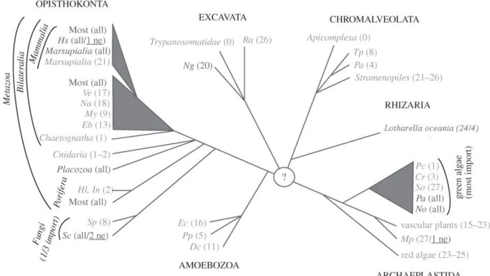
Figure 5. Occurrence and distributionof mitochondrial tRNA import (adapted from Lithgow and Schneider, 2010). Unrooted phylogenetic tree of the six eukaryotic supergroups (indicated in capitals). Branching order reflects the phylogenetic relationship of taxons but branch length is not to scale. Organisms having a complete set of mitochondrial tRNA genes are shown in dark grey. Organisms lack a variable number of apparently essential mitochondrial tRNA genes are shown in light grey. The numbers of tRNA genes encoded in the different mitochondrial genomes are indicated. «All» indicates that the mitochondrial-encoded tRNA gene set is complete and «0» indicates complete absence of mitochondrial tRNA genes. If organisms retain a single mitochondrial tRNA gene only it is always the tRNAMet. The minimal number of tRNAs required for mitochondrial translation is, depending on the wobble rules and the genetic code variations, between 20–22. Even in organisms having more than 22 mitochondrial tRNA genes, the set of mitochondrial-encoded tRNA is often not complete and import of cytosolic tRNAs is required. In most of these cases, it is the tRNAThr that is imported. In a few systems, import of a cytosolic tRNA that has the same decoding capacity as a still existing mitochondrial-encoded tRNA gene has been shown experimentally (shown underlined). Acronyms for species: Hs, Homo sapiens; Ve, Vanhornia eucnemidarum; Na, Neomaskellia andropogonis; My, Mizuhopecten yessoensis; Eb, Epiperipatus biolleyi; Hl, Hypospongia lachne; In, Igornella notabilis; Sp, Spizellomyces punctatus; Sc, Saccharomyces cerevisiae; Ng, Naegleria gruberi; Ra, Reclinomonas americana; Tp, Tetrahymena pyriformis; Pa, Paramecium aurelia; Pc, Polytomella capuana; Cr, Chlamydomonas reinhardtii; Sc, Scenedesmus obliquus; Pa, Pseudendoclonium akinetum; No, Nephroselmis olivacea; Mp, Marchantia polymorpha; Ec, Entamoeba castelanii; Pp, Physarum polycephalum; Dc, Dictyostelium citrinum. (Lithgow and Schneider, 2010). The mitochondrial RNA import is a complex process which can be subdivided into two main stages – targeting a subpart of specific RNA pool to the surface of mitochondria followed by their translocation into organelles. Years of close scrutiny in a variety of organisms belonging to different phylogenetic branches suggest at least two distinct mechanisms of this process. The tRNA import in yeast, T. brucei and mammals seems to rely on protein factors while plants and Leishmania utilize factor-independent mechanism to deliver tRNAs into
15 organelles. The mitochondrial RNA import process is still largely not understood. The difficulty of studying mitochondrial RNA import stems from the lack of reliability of experimental in vitro system, which can satisfactorily reflect the in vivo situation. The better understood in vitro RNA import process of trypanosomatids does not rely on soluble factors to provide selectivity, which is crucial in vivo (Bouzaidi-Tiali et al, 2007; Tan et al, 2002). In in vitro import experiments with different cytosolic tRNAs into isolated Solanum tuberosum mitochondria, the S. tuberosum cytosol-specific tRNAGlyGCC was imported into isolated
mitochondria with the same efficiency as normally imported tRNAGlyUCC (Delage et al, 2003a; Salinas et al, 2006), so this situation does not correspond to that in vivo.
tRNA import into protozoan mitochondria
The mitochondrial genomes of trypanosomatids such as T. brucei and Leishmania spp. are devoid of tRNA genes (Schneider, 2011). So all mitochondrial tRNAs are imported from the cytosol, except tRNAMeti and tRNASec that have exclusively cytosolic localization. The extent
of mitochondrial localization of different nuclear-encoded tRNAs ranges from 1 to 7.5% (Tan et al, 2002). The mechanisms of regulation of those distributions have not been elucidated yet. It is widely accepted that selectivity is based on the presence of tRNA import determinants, which favor the interaction with protein factors, and anti-determinants retaining non-importable tRNAs in the cytosol. Based on the discovery of import selectivity in different organisms, one can conclude that there is no universal import signaling. The signal for mitochondrial targeting can be represented as specific sequence or structural motifs spread over the tRNA molecules. In Tetrahymena, the anticodon of tRNAGln apparently acts as an import determinant (Rusconi and Cech, 1996). Thus, Tetrahymena contains three tRNAGln isoacceptors, two cytosol-specific ones, which decode codons UAA and UAG, while the third tRNAGlnUUG is partly imported into mitochondria. Nucleotide substitutions in the UUG
anticodon uniquely abolished import while substitution of a single anticodon nucleotide (UUA → UUG) confered import of a normally non-imported glutamine tRNA. All these facts
demonstrate that the anticodon UUG is both necessary and sufficient for tRNA import (Rusconi and Cech, 1996).
16
The comparison of tRNAMeti and the tRNAMete revealed that the pair U51:A63 in the T-stem is the main anti-determinant of mitochondria localization in T. brucei. It inhibits the import of tRNAMeti and prevents its interaction with the cytosolic elongation factor eEF1a (Crausaz Esseiva et al, 2004). Such a mechanism is also inherent in tRNASec where the U8:U66
base pair also acts as anti-determinant and retains tRNASec in the cytosol by preventing
binding with eEF1a (Bouzaidi-Tiali et al, 2007).
Chemical modifications may also affect the mitochondrial import process. The study of Leishmania tarentolae tRNAGluUUC and tRNAGlnUUG demonstrated that cytosol-specific
thiolation of tRNAs at wobble position of anticodon served as the anti-determinant for import of those RNAs (Kaneko et al, 2003). This mechanism is not general and does not seem to take place in T. brucei, a close relative of Leishmania (Bruske et al, 2009). The down regulation of TbNfs, the master desulfurase responsible for both Fe-S cluster formation and tRNA thiolation, led to reduction of the overall thiolation level but did not alter the distribution of T.
brucei tRNAs (Paris et al, 2009).
In Leishmania tropica two types of import sequences were discernible depending on their localization: type I (D-arm motif YAGAGY of several imported tRNAs and D-arm motif shared by tRNAIle and tRNAValCAC as well as anticodon arm of tRNAArgACG) and type II (CUG3-4U
located in V-T region of tRNAIle and other tRNAs) (Bhattacharyya et al, 2002). These types
demonstrated different import abilities: type I RNAs are efficiently transferred through the inner membrane and are inhibited by type II whereas type II RNAs have poor inner membrane transfer efficiencies and are stimulated by type I (Bhattacharyya et al, 2002). Thus, regulation of tRNA pools inside mitochondria is provided by cooperative or antagonistical interaction of tRNAs with different receptors on the inner mitochondrial membrane in a so-called «ping-pong» import regulation model (Bhattacharyya et al, 2003).
Some tRNAs may have several determinants; for example, L. tarentolae tRNAIle contains
both the D domain and the V-T region motifs. D-arm exchange between the mainly cytosolic tRNAGln and the efficiently imported tRNAIle leads to increased localization of tRNAGln in
mitochondria, but does not prevent the mitochondrial localization of the chimeric tRNAIle
17 It was observed that the determinants for tRNA cytosolic localization coincide with anti-determinants for eEF1a interaction (Bouzaidi-Tiali et al, 2007; Crausaz Esseiva et al, 2004). In addition, the depletion of eEF1a in T. brucei reduced import of newly synthesized tRNAs. All this indicates that eEF1a plays an important role in tRNA import process. eEF1a discriminates imported tRNAs, then hands them over toward the outer membrane where they interact with a specific receptor. The receptor is not able to discriminate between cytosol-specific tRNAs and imported tRNAs, which is consistent with the loss of specificity in in vitro tRNA import system lacking eEF1a (Bouzaidi-Tiali et al, 2007). To explain why a translation factor eEF1a helps a subpopulation of aminoacylated tRNAs to escape cytosolic translation and targets them to the mitochondria, the existence of two functionaly different eEF1a populations was suggested (Bouzaidi-Tiali et al, 2007). A precise mechanism of the tRNA translocation across the mitochondrial double membrane in trypanosomatids is unknown but it depends on ATP hydrolysis. In T. brucei tRNA mitochondrial import shows a connection with the mitochondrial protein import like in S. cerevisiae and in plants but, unlike plants, does not require VDAC (Pusnik et al, 2009). TbTim17 and TbmHsp70 are directly involved in tRNA import in vivo as deletion of these proteins leads to inhibition of tRNA import. However, possible reason for the tRNA import inhibition could be a lack of import of additional unknown tRNA import factor (Tschopp et al, 2011). It was suggested that TbTim17 and TbmHsp70 are physically associated with other proteins including Tb11.01.4590 and Tb09.v1.0420 to form the tRNA translocon of T. brucei (Seidman et al, 2012). In L. tropica, prior to translocation, tRNAs interact with the outer mitochondrial membrane receptors one of witch is TAB (tubulin antisense-binding protein) (Adhya et al, 1997; Bhattacharyya et al, 2002). The experiments with mutant forms of the D-arm of tRNATyr allowed suggesting the presence of distinct receptors in the outer and the inner membranes (Bhattacharyya et al, 2000). Northing is known about a pathway for tRNA translocation through the double-mitochondrial membrane. However, it was suggested that a unique channel found in L. tropica provides a transport of tRNAs through the inner mitochondrial membrane (Figure 6B).
18 Figure 6. Mitochondrial tRNA-import translocation pathways of S. cerevisiae, L. tropica and S. tuberosum (adapted from Salinas et al., 2008). A) In S. cerevisiae, the import of the nuclear-encoded tRNALysCUU (blue ribbon) relies on its non-canonical interaction with its carrier protein preMSK1p (red). The receptor Tom20 (turquoise) (but not Tom70; blue) of the TOM complex and Tim44 (turquoise) of the TIM complex are essential for tRNA import. B) In Leishmania tropica, tRNAs are first bound to outer mitochondrial membrane (OM) receptors one of which is TAB (orange). Next, tRNAs are translocated through the outer membrane via an unknown pathway. The translocation of tRNAs across the the inner membrane (IM) might occur through RIC complex. Two receptors, RIC1 and RIC8A, bind with tRNAs, which then are translocated through the pore constituted by RIC6 and RIC9. C) In Solanum tuberosum, tRNA import depend on two major components of the TOM complex, TOM20 (turquoise) and TOM40 (turquoise), which fix tRNAs at the mitochondrila surface. The voltage-dependent anion channel (VDAC; orange) creates a pore for tRNAs translocation through the outer mitochondrial membrane. A pathway of the inner membrane translocation remains unknown. This is the 640 kDa tRNA import complex (RIC) located on the inner mitochondrial membrane. The system reconstructed with RIC in phospholipid vesicles demonstrated characteristic features of the Leishmania RNA import including the allosteric regulation between two different subsets of tRNAs (type I and type II) (Chatterjee et al, 2006). Native RIC contains 11 different subunits, 6 of which are essential and sufficient for import activity. The knockdown of these six subunits led both to inhibition of mitochondrial tRNA import and mitochondrial translation. RIC1 corresponds to the α subunit of the F1-ATP synthase of the
complex V and provides energy for import whereas RIC8A is the subunit 6b of complex III of the respiratory chain. As well as RIC1, RIC8A appeared as a receptor allosterically interacting with distinct tRNA subsets (Bhattacharyya et al, 2003; Mukherjee et al, 2007). Three other
19 components, RIC5, RIC6, and RIC9, are also parts of respiratory complexes. RIC9 transfers tRNA from receptors to the import pore. A voltage-gated minimal translocation pore of RIC is constituted by RIC6, RIC9, and RIC4A (Koley and Adhya, 2013). However, the published data raise several questions concerning the existence and functioning of RIC (Salinas et al, 2008). To date, the RIC complex has been identified only in L. tropica and it was demonstrated that mitochondrial tRNA import in T. brucei does not depend on Rieske protein, which is one component of RIC (Paris et al, 2009).
tRNA import into plant mitochondria
In plants, the number and identitiy of imported tRNAs vary greatly even in closely related species. Nevertheless, the existence of exclusively cytosolic tRNAs and distinct extent of mitochondrial localization for different tRNAs point to a selection step in mitochondrial import. Different nucleotides or domains including the anticodon, the D-arm and T-domain in tRNAVal (Delage et al, 2003b; Laforest et al, 2005), position 70 in tRNAAla (Dietrich et al, 1996)were revealed as import determinants. Absence of common import sequence signature led to a hypothesis that a conformation of the tRNA and not particular nucleotides may be specifically recognized during import (Laforest et al, 2005). Mutagenetic studies demonstrated a certain level of correlation between import and recognition by a cognate aminoacyl-tRNA synthetase indicating its involvement into mitochondrial RNA import in plants (Delage et al, 2003a). Although it is suggested that the aminoacyl-tRNA synthetase is implicated into tRNA mitochondrial import in plants but is not sufficient for that process. For example, in S. tuberosum two cytosolic tRNAGly with UCC and CCC anticodons are selectively
imported into mitochondria, and tRNAGlyGCC is retained in cytosol while all isoacceptors can be recognized by the glycyl-tRNA synthetase (Delage et al, 2003a). As an explanation, it was suggested that the fate of a cytosolic tRNA depends on the interaction with either cytosolic aaRS, which addresses it to cytosolic translation, or precursor form of mitochondrial aaRS that directs it into mitochondria (Duchêne et al, 2011; Duchene et al, 2005). tRNA import into plant mitochondria requires both a membrane potential and ATP. Whereas in vitro import of tRNAs can be achieved independently from additonal cytosolic factors, the selectivity of this system is limited (Salinas et al, 2006). Two components of the
20 plant TOM complex (Tom20 and Tom40) are required for ATP-dependent tRNA-binding at the mitochondrial surface in vivo (Salinas et al, 2006; Salinas et al, 2014). Thereafter, tRNAs are translocated across the membrane through a pore formed by VDAC (Figure 6C). An angagement of VDAC is confirmed by inhibition experiments of tRNA import into isolated mitochondria by VDAC antibodies and ruthenium red (Salinas et al, 2006).
tRNA import into yeast mitochondria
The mitochondrial import of Saccharomyces cerevisiae tRNALysCUU (tRK1) represents a highly selective system. A fraction lower than 5% of the cytosolic tRK1 was shown to
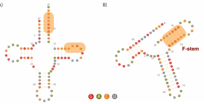
associate with mitochondria, whereas the other isoacceptor tRNALysUUU (tRK2) is restricted to cytosol (Tarassov and Entelis, 1992). Previously, it was suggested that the anticodon of tRK1 (especially the wobble position C34 of the anticodon) and the acceptor stem contain determinants for its import selectivity (Entelis et al, 1998). Thereafter, it was assumed that a tRNA secondary structure rearrangement provides the major contribution to import selectivity (Figure 7). Only molecules comprising a folded TΨC hairpin, and which are able to adopt a particular F-hairpin immediately followed by the 3′-end (Figure 7B) proceed to the import pathway (Kolesnikova et al, 2010). Figure 7. Secondary structures of tRK1. (A) classical clover-leaf structure and (B) F-form (Baleva et al, 2015). Regions of tRK1 involved in F-stem formation as well as F-stem are in color box.
21 Transfer RNAs are involved in cytosolic translation and recycled during protein synthesis. To be targeted into mitochondria nuclear-encoded tRNAs must escape the translation machinery cycle possibly due to interaction with protein factors (Figure 8A). In S. cerevisiae, the imported tRK1 is firstly charged by the cytosolic lysyl-tRNA synthetase (KRS) (Tarassov et al, 1995b), and then is specifically recognized by Eno2p - one of the two isoforms of the glycolytic enzyme enolase (Entelis et al, 2006). It was suggested that Eno2p induces conformational changes in the aminoacceptor stem of tRNA molecules and discriminates imported molecules. The tRNA-Eno2p-containing complex is then transfered toward the mitochondrial surface, where the tRNA is handed to the precursor of mitochondrial lysyl-tRNA synthetase (preMSK1p) (Tarassov et al, 1995b), which is synthesized mainly in the vicinity of mitochondria (Entelis et al, 2006). In complex with preMSK1p, tRK1 adopts an “intermediate” conformation, which thereafter facilitates re-folding of tRK1 into the classic L-shape structure (Kolesnikova et al, 2010). Enolase integrates into a glycolytic multiprotein complex that is associated with the mitochondrial outer membrane, whereas a tRK1-preMSK1p complex co-imports into mitochondria (Entelis et al, 2006). In S. cerevisiae, tRK1 is co-imported with preMSK1p. While the translocation of preproteins through the mitochondrial membrane requires it to be unfolded, it is not clear how tRNA remains bound to the carrier protein. It is possible that while in complex with tRK1 during the import, preMSK1p adopts a conformation allowing interaction with tRNAs (Entelis et al, 1998). This agrees with the experimentally demonstrated targeting of branched polypeptides or chimeric proteins fused with single- or double stranded oligonucleotides into mitochondria (Brokx et al, 2002). In addition, the experiments with a nicked tRK1 transcript were in favor of import of folded tRK1 molecule as well as importance of the tRNA L-shape structure (Entelis et al, 1998). The tRK1 translocation into mitochondria requires ATP and the membrane potential as well as the intact protein machinery. The investigation of tRK1 import in yeast strains carrying deletions of different receptors of TOM and TIM translocases revealed the importance of Tom20 and Tim44 (Figure 6A) (Tarassov et al, 1995a). The deletion of those proteins abolished tRK1 import while the absence of Tom70 did not show any effect (Tarassov et al, 1995a).
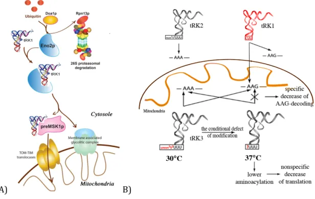
22 A) B) Figure 8. Mitochondrial tRK1 import in S. cerevisiae, its possible regulation, and implication in the adaptation. A) Aminoacylated tRK1 can be addressed either towards the cytosolic translation or towards the mitochondria. This choice can be determined by interaction of the RNA with either cytosolic retention factors or mitochondrial import factors and regulated by the ubiquitin-proteasome system (UPS). Two proteins were involved in this mechanism. Doa1p participates in specific ubiquitination of import factors as Eno2p or factors involved in tRK1 retention in cytosol while Rpn13p recognizes ubiquitinated proteins and degraded them by the proteasome (Brandina et al., 2007). Eno2p selects imported tRK1 molecules and deliver them to newly synthesised preMSK1p molecules at the mitochondrial surface. Eno2p participates to a large membrane associated glycolytic complex whereas tRK1 is delivered into mitochondrial matrix in complex with preMSK1p possibly via protein import translocases (Entelis et al., 2006) B) Adaptation mechanism involving tRK1 import (adapted from Kamenski et al., 2007). Mitochondrial tRK3 can decode both AAA and AAG codons in mitochondrial mRNAs at normal conditions (30°)C, while at non-permisive conditions (at 37°C) loose the ability for efficient decoding of AAG due to hypomodified U34 in the wobble position of tRK3. Thus, imported tRK1 can cure the deficiency by decoding the AAG codons.
In S. cerevisiae, cytosolic tRNALysCUU (tRK1) is involved in organellar translation. Under normal conditions tRNALysCUU import seems redundant since there is mitochondria-specific tRNALysUUU (tRK3) that can decode AAA and AAG codons, both found in mitochondrial open reading frames. Yet, elevated temperature leads to modification defect of the 2-thio group of the U34 from the tRK3 anticodon. The absence this modification may prevent translation of AAG codons, while the imported tRK1 cures this deficiency (Figure 8B) (Kamenski et al, 2007). Since tRK1 role inside the mitochondria is conditional, tRK1 import can be also regulated conditionally. Thus, in S. cerevisiae two proteins, Rpn13p and Doa1p, which are the components of the ubiquitin-proteasome system (UPS), were identified as possible tRNA
23 mitochondrial import regulators (Brandina et al, 2007). Depletion either Rpn13p or Doa1p increased efficiency of tRK1 import. It was proposed that Doa1p participates in specific ubiquitination of import factors as Eno2p or others not yet identified, while Rpn13p recognizes them and proceeds to proteasome degradation (Figure 8A). Moreover, other proteins, which promote retention of the tRNA in the cytosol, can be substrates of UPS-degradation (Brandina et al, 2007) and regulation of their level can also influence tRK1 mitochondrial import efficiency.
It was published that nuclear-encoded tRNAGlnCUG and tRNAGlnUUG were imported into yeast mitochondria (Rinehart et al, 2005) as well as into rat and human mitochondria (Rubio et al, 2008). Unlike tRK1 import, the import of tRNAGlnCUG and tRNAGlnUUG in vitro does not require aminoacylation or any cytosolic factors. The tRNAGlnCUA was able to suppress amber mutation of mitochondrial gene in yeast, which indicates its participation in mitochondrial translation (Rinehart et al, 2005). Nevertheless, the biological functions of in vivo import of those tRNAs are not clear. It was shown that mitochondria use the transamidation pathway to generate mitochondrial Gln-tRNAGln that is used only by the mitochondrial translation apparatus to decode both CAG and CAA glutamine codons (Araiso et al, 2014; Frechin et al, 2009).
Enolase in other life processes
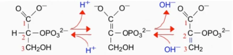
Studies of RNA mitochondrial import have reported that the enzyme enolase could be a major player in this process (Entelis et al, 2006). That was confirmed by numerous tests (genetic and biochemical). Enolase is a very abundant protein in cell that somehow constraining cells to adapt many functions to its presence. Hence, with this idea in mind, it is interesting to describe in the present sub-part the numerous and very diverse functions in which enolase is involved, from bacterial infections to cancer response. Enolase (or 2-phospho-D-glycerate hydrolase) is a highly conserved protein found in nearly all organisms both aerobic or anaerobic. Enolase is a metaloenzyme (Poyner et al, 2001) that is responsible for the reversible catalysis of the conversion of 2-phosphoglycerate to phosphoenolpyruvate (Figure 9) in glycolysis and gluconeogenesis.24 Figure 9. The scheme of the reaction catalyzed by enolase showing the removed hydroxyl group on carbon 3 and the resulting formation of the enol moiety. Many organisms have different enolase isoforms, the expression of which vary according to the pathophysiological conditions of cells, metabolic demands, or developmental stage of the organism (McAlister and Holland, 1982; Tanaka et al, 1985; Barbieri et al, 1990). In mammals, like in all vertebrates, enolase occurs as three isoenzymes: α-enolase is found in a variety of tissues including liver, β-enolase is muscle specific and γ-enolase is found in neuron and neuroendocrine tissues (Royds et al, 1982). Expression of different enolase isoforms changes during embryonic development for example β-enolase is expressed in differentiated skeletal muscles, while in the embryonic primary fibers and myotubes enolase is presented mainly by -isoform (Barbieri et al, 1990). Yeast have two isoforms of enolase. The expression of yeast enolase isozymes is dependent on the carbon source, glucose, or a nonglycolytic substrate, while their kinetic properties are very similar (McAlister and Holland, 1982). Recent studies have reported a lot of other functions for enolase in addition to its innate glycolytic function (Pancholi, 2001; Diaz-Ramos et al, 2012). Thus, enolase was identified as τ-crystallin in fish, reptiles, birds and lamprey eye lens. In lens, enolase is present as monomer and shows significantly low enzymatic activity (Wistow et al, 1988). The alternative translation of the α-enolase mRNA results in a 37 kDa protein which lacks the first 96 amino acid residues - c-myc promoter-binding protein 1 (MBP-1). Unlike enolase, which has cytosolic localization, MBP-1 is found in the nucleus. MBP-1 binds the c-myc P2 promoter and down-regulates expression of the c-myc proto-oncogen, which plays an important role in the regulation of cell growth and differentiation (Subramanian and Miller, 2000).
25
In Saccharomyces cerevesiae, enolase was identified as a heat-shock protein (HSP48) involved in thermal tolerance and growth control (Iida and Yahara, 1985).
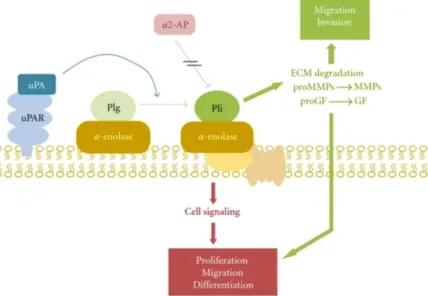
Enolase was found as a strong plasminogen-binding receptor on the surface of different cells such as hematopoetic cells, epithelial cells, neuronal cells, carcinoma cells and also on Gram-positive cocci, Candida and parasites. α-enolase interacts with plasminogen and induces activation of plasminogen by plasminogen activation system (PAs) and protects plasmin from inhibition by α2-antiplasmin (Figure 10). (Diaz-Ramos et al, 2012) Figure 10. Scheme of α-enolase/plasminogen interaction on the cell surface. α-Enolase enhances plasminogen activation on the cell surface, concentrates plasmin proteolytic activity on the pericellular area and protects plasmin from its inhibitor α2-antiplasmin. Once activated, plasmin can degrade most of the components of the extracellular matrix, directly or indirectly by activating metalloproteases. It is also capable to activate prohormones of progrowing factors. Abbreviations: Plg, plasminogen; Pli, plasmin, α2-AP, α2-antiplasmin; uPA, urokinase-type plasminogen activator, uPAR, urokinase-type plasminogen activator; ECM, extracellular matrix; MMPs, metalloproteases; GF, growing factors. (Diaz-Ramos et al, 2012) Plasminogen binding ability of cell surface expressed enolases results in the involvement of enolase in initiating some disease processes, tumor invasion and metastasis through activation of a serine-protease involved in extracellular matrix degradation. Yet, enolase participates in remodelling of actin cytoskeleton by induction of actin polymerisation and distribution and regulation of the PhoA cyclin-dependent kinase (Hafner et al, 2012). In addition, enolase interacts with actin and tubulin and could contribute to myogenesis (Keller et al, 2007). Similarly, interaction of enolase with the cytoskeleton may be closely related to the invasiveness of cancer cells (Trojanowicz et al, 2009). Another major contribution of
26 enolase to tumor progression is its requirement for maintaining the Warburg effect (aerobic glycolysis) in cancer cells. Enolase knockdown rescues oxidative phosphorylation and impairs the tumor growth (Capello et al, 2016). Enolase also acts as pro-survival factor supporting cancer cells adaptation to different stresses including hypoxia, chemo- and radiotherapy (Yan et al, 2011). Taken together, these finding strongly suggest that enolase could be used as a promising therapeutic target for cancer diagnosis and anti-cancer therapy (Jung et al, 2013; Benjamin et al, 2016)
Mitochondrial RNA import in mammals
Mammalian mitochondria encode 13 proteins, 2 rRNAs and a set of 22 tRNAs required for reading all codons (Florentz et al, 2003). Since mammalian mitochondria possess a full minimum set of tRNAs for efficient translation, there is no reason to suggest the existence of mitochondrial tRNA import. Nevertheless, the analysis of human mitochondrial transcriptome demonstrated more diverse population of mitochondrial RNAs than previously suggested (Mercer et al, 2011), together with the enrichment of several nuclear RNAs including several tRNAs. In addition, it is not known yet whether nuclear-encoded mammalian tRNALys can beimported into mitochondria while tRNALys is absent in the marsupial mitochondrial genome
(Dörner et al, 2001). Moreover, the sequence analysis of the only nuclear-encoded tRNA which is associated with highly pure mitochondria, shows the enrichment of tRNALys (Dörner
et al, 2001). Demonstration of yeast tRK1-targeting into isolated human mitochondria in the presence of yeast import factors has unraveled the existence of a cryptic mechanism for tRNA import (Kolesnikova et al, 2004). This mechanism presents similarities with that of yeast (Gowher et al, 2013). Thus, preKARS2, the precursor of human mitochondrial lysyl-tRNA synthetase, is implicated both in vitro and in vivo in tRNA import in human mitochondria (Gowher et al, 2013). Moreover, yeast or rabbit enolase facilitated tRNA-preKARS2 interaction in vitro and further import of this complex into isolated human mitochondria.
Artificial RNA minimal import substrates bearing two hairpins characteristic of the tRK1 F- 27 form were also efficiently imported into human mitochondria in vivo and in vitro (Gowher et al, 2013). Import of RNA components of RNase MRP and RNase P was also suggested. RNase MRP is a site-specific endoribonuclease that is suggested to be involved in the processing of mitochondrial RNAs and also in mitochondrial DNA replication (Stohl and Clayton, 1992). Mammalian RNase P participates in the processing and removal of tRNAs separating the regions coding for oxydative phosphorylation (OXPHOS) protein subunits (Doersen et al, 1985; Klemm et al, 2016). The mitochondrial genome of S. cerevisiae encodes the RNase P RNA but lots of species do not possess a certain gene in their mtDNA. The first evidence for import of both RNaseP RNA and RNase MRP RNA came when their processing activity was recovered from isolated human mitochondria pretreated with micrococcal nuclease (Doersen et al, 1985). Thereafter, two RNAs were identified to be identical in sequence to nuclear H1 RNA and MRP RNA by Nothern blot hybridization (Puranam and Attardi, 2001). Nothing is known about the mechanisms that address these RNAs to mitochondria. However, a highly conserved 3′→5′ exoribonuclease, PNPASE, seems to be implicated in the import of RNase P and MRP RNA components (Wang et al, 2010). It was suggested that a 20 ribonucleotide stem-loop of both RNAs interact with PNPASE in a manner that triggers only import rather than processing. Moreover, a fusion of this structure with non-imported RNA allowed import of these constructions (Wang et al, 2010; Wang et al, 2012). However, the mitochondrial import of RNA component of RNase P was questioned after reporting that human mitochondrial RNase P is proteinaceous and consists of three MRPP subunits (for PROtein-only RNase P or PROteinaceous RNase P) (Holzmann et al, 2008; Klemm et al, 2016). PROPR was also found in plant mitochondria and chloroplasts. PROPR proteins were able to functionally replace RNA-containing RNase P in complementation experiments in Escherichia coli (Gobert et al, 2010) and Saccharomyces cerevisiae (Weber et al, 2014). Nevertheless, at present, it is assumed a possibility for a co-existence of both types of RNase P within mitochondria (Wang et al, 2010). 5S rRNA is an integral part of the large ribosomal subunit of all cytosolic ribosomes, but mitochondria of only land plants, algaes and few protists encode its gene (Adams and Palmer,
28 2003). It was demonstrated that cytosolic 5S is imported into mitochondria representing an example of a highly selective targeting system. 5S RNA is rather short (120 nt) with highly conserved secondary and tertiary structures (Smirnov et al, 2008b). It adopts a three-domain Y-shaped organisation (Figure 11). The α-domain is formed by helix I, domains β and γ have a heterogenous structure that includes both helical and loop regions. Two regions of 5S rRNA, the proximal part of helix I containing the conserved G7-U112 pair and the site associated with the loop E-helix IV region of domain γ, were discribed as determinants of mitochondrial localization. Destruction or destabilisation of these parts significantly decreased the import efficiency, whereas disruption of both sites led to loss of import (Smirnov et al, 2008a). Figure 11. The secondary structure of human 5S rRNA (adapted from Smirnov et al., 2011). TFIIIA is involved in the export of newly synthesized 5SrRNA from the nucleus. The following fate of 5S rRNA then depends on interaction with protein factors. Ribosomal protein L5 reimports 5S rRNA molecules into nucleus while the mitochondrial ribosomal protein L18 (preMRP-L18) and rhodanese target them to mitochondria. Two proteins were identified as targeting factors delivering 5S rRNA. 5S rRNA escapes from the “back to the nucleus” pathway due to interaction with a precursor of mitochondrial ribosomal protein L18 (preMRP-L18), which changes the RNA conformation making it unrecognizable by ribosomal protein L5. Then, 5S rRNA is handed to rhodanese, a mitochondrial thiosulfate sulfurtransferase, and transported to the mitochondrial surface. The translocation of 5S rRNA across the mitochondrial membrane requires ATP, an electrochemical membrane potential, and an intact protein import apparatus (Smirnov et al, 2010; Smirnov et al, 2008a) suggested by the observation that blocking protein import also inhibits 5S rRNA mitochondrial import.
29 In summary, RNA mitochondrial import does not reveale a unique universal mechanism but some features seem to be common. They are a requirement of ATP, protein factors providing the selectivity, and involvement of protein import machinery at least it receptor part. Mechanisms governing tRNA targeting into mitochondria remain poorly understood as well as protein factors involved in this process either have not identified or their functioning is not clear enough.
30
THESIS PROJECT AND OBJECTIVES
The main objective of the present work was to decrypt the mechanisms of transfer RNA targeting into yeast and human mitochondria. This work focused mainly on the initial targeting step of tRK1 import into mitochondria both in human cells and yeast as well as on the role of glycolytic enzyme Eno2p in this process.Functional studies of the mitochondrial import in yeast have suggested that a second isoform of the glycolytic enzyme enolase is an essential part of the tRK1 mitochondrial targeting process (Entelis et al., 2006). Together with another protein factor, preMSK1p, this enzyme represents a minimum system sufficient to provide the mitochondrial import of tRK1 in vitro. There are data indicating the interaction between Eno2p and tRK1 as well as a chaperone activity of Eno2p in the complex with tRK1 (Entelis et al., 2006; Kolesnikova et al., 2010). Nevertheless, the nature of this interaction is still unkown. We also hypothesized that human cells use a similar mechanism for induced tRK1 import implicating human enolase as well. This hypothesis was based on previous studies indicating the engagement of orthologous proteins for the tRNA mitochondrial import in human cells. In order to reach the stated objective, we: Study whether human enolases are involved in tRK1 import into human mitochondria in vitro and in vivo; Study differences of conformational features of tRK1 and tRK2 in solution; Study the process of targeting of tRK1 into yeast mitochondria;
Study the structures of tRK1-protein complexes involved in tRK1 import into yeast mitochondria.
31
RESULTS
A moonlighting human protein is involved in mitochondrial import of tRNA
It was previously demonstrated in our laboratory that yeast tRNALysCUU as well as some of its synthetic transcripts can be targeted into human mitochondria indicating the existence of a cryptic mechanism for tRNA mitochondrial import (Entelis et al, 2001; Gowher et al, 2013). Moreover, this mechanism presents a certain level of similarity with the mitochondrial targeting of tRK1 in yeast. In vitro studies revealed that the human precursor of mitochondrial lysyl-tRNA synthetase (preKARS2) and also rabbit muscle enolase are both necessary and sufficient to direct import of tRK1 into mitochondria (Gowher et al, 2013). It could then be hypothesized that the human enolase was implicated in the tRNA mitochondrial import pathway in human cells by playing a role similar to that of Eno2p in this process in yeast. In human, as in all vertebrates, enolase is represented by three isoenzymes: α-enolase (Enolase 1) is found in a variety of tissues, while two others are tissue specific. β-enolase (Enolase 3) is mainly found in muscles and γ-enolase (Enolase 2) is only present in neurons and neuroendocrine tissues. All isoforms are very conserved and show similar kinetic properties (Díaz-Ramos et al, 2012). We studied the capacity of human enolases to participate in the tRK1 mitochondrial import process by applying various in vitro approaches. These results and their discussion are presented in publication 1 (See below). Each of three isoforms of human enolase was overexpressed and His6-tag-purified in E. coli. We studied the capacity of human enolases to participate in tRK1 import process by means of electrophoretic mobility shift assay (EMSA) and in vitro import approach. All isoforms revealed comparable abilities to direct import of synthetic tRK1 into isolated human mitochondria in the presence of recombinant preKARS2. EMSA is commonly used method in the characterization of protein-nucleic acid interactions based on the differential migration of complexes and free nucleic acid during native gel electrophoresis. According to results of EMSA, recombinant human enolases possess affinities to labeled synthetic transcript of yeast tRK1. The apparent Kd for this interaction is 2.0 ± 0.5
32 μM, which is close to 2.5 ± 0.2 μM apparent Kd of tRK1 and yeast Eno2p interaction (Entelis et al, 2006). Human enolases as well as yeast Eno2p showed lower affinity to tRK1 transcript than preKARS2 (or preMSK1p in yeast)
,
so in the presence of preKARS2, all tRK1 was shifted to forming the complex tRK1-preKARS2. In addition, human enolases facilitated the tRK1-preKARS2 complex formation resulting in decreasing the apparent Kd by 10 fold from 300 nM to less than 20 nM. The same trend was observed for the yeast recombinant Eno2p. Yeast Eno2p decreases the apparent Kd of tRK1-preMSK1p from 180 nM to 40 ± 10 nM. Summing up the results of these experiments, we suggested that human enolases seemed to participate in tRNA mitochondrial import in human cells fulfilling the same targeting functions as Eno2p in yeast cells. The data also support the idea that human cells possess a cryptic tRNA import mechanism that can be activated under specific conditions. .33
PUBLICATION I
A Moonlighting Human Protein Is Involved in
Mitochondrial Import of tRNA
International Journal of
Molecular Sciences
ISSN 1422-0067
www.mdpi.com/journal/ijms Article
A Moonlighting Human Protein Is Involved in Mitochondrial
Import of tRNA
Maria Baleva 1,2,†, Ali Gowher 1,†,‡, Piotr Kamenski 2, Ivan Tarassov 1, Nina Entelis 1
and Benoît Masquida 1,*
1 Department of Molecular and Cellular Genetics, UMR 7156 Génétique Moléculaire,
Génomique, Microbiologie (GMGM), CNRS—Université de Strasbourg, 67084 Strasbourg, France; E-Mails: [email protected] (M.B.); [email protected] (A.G.); [email protected] (I.T.); [email protected] (N.E.)
2 Department of Molecular Biology, Biology Faculty of Moscow State University,
119992 Moscow, Russia; E-Mail: [email protected]
† These authors contributed equally to this work.
‡ Present address: Genome Institute of Singapore, #02-01 Genome, Singapore 138672.
* Author to whom correspondence should be addressed; E-Mail: [email protected];
Fax: +33-3-6885-1365. Academic Editor: Michael Ibba
Received: 28 January 2015 / Accepted: 15 April 2015 / Published: 24 April 2015
Abstract: In yeast Saccharomyces cerevisiae, ~3% of the lysine transfer RNA acceptor 1
(tRK1) pool is imported into mitochondria while the second isoacceptor, tRK2, fully remains in the cytosol. The mitochondrial function of tRK1 is suggested to boost mitochondrial translation under stress conditions. Strikingly, yeast tRK1 can also be imported into human mitochondria in vivo, and can thus be potentially used as a vector to address RNAs with therapeutic anti-replicative capacity into mitochondria of sick cells. Better understanding of the targeting mechanism in yeast and human is thus critical. Mitochondrial import of tRK1 in yeast proceeds first through a drastic conformational rearrangement of tRK1 induced by enolase 2, which carries this freight to the mitochondrial pre-lysyl-tRNA synthetase (preMSK). The latter may cross the mitochondrial membranes to reach the matrix where imported tRK1 could be used by the mitochondrial translation apparatus. This work focuses on the characterization of the complex that tRK1 forms with
human enolases and their role on the interaction between tRK1 and human pre-lysyl-tRNA synthetase (preKARS2).
Keywords: human mitochondria; tRNA targeting; enolase
1. Introduction
Mitochondria are the centres of critical cellular processes, such as oxidative phosphorylation, apoptosis, fatty acids, amino acids and Fe–S cluster metabolisms. Despite this central role, mitochondria present genomes only encoding a fistful of proteins and RNAs dedicated to oxidative phosphorylation or mitochondrial translation. The regulation of mitochondrial activities thus relies on nuclear-encoded factors, which need to be imported in a concerted way in order to tune mitochondrial activity with cytosolic conditions [1,2]. The process of mitochondrial import of cytosolic proteins is far better understood than the process of RNA import, although mitochondrial RNA import has been described in fungi, protozoa, plants and mammals [3].
Regarding tRNAs, the existence of mitochondrial genomes encoding few or no tRNAs in certain organisms such as cniderians or protozoa, respectively, certainly indicates that tRNAs import is necessary for translation [4,5]. However, tRNAs can also be imported in mitochondria of organisms with a full set of mitochondrial DNA-encoded tRNAs. In the yeast Saccharomyces cerevisiae, tRNALys
acceptor 1 (tRK1) is imported despite the presence of a mitochondrial encoded tRNALys (tRK3) [6].
In this case, a ~3% fraction of the tRK1 cytosolic pool is constitutively routed towards mitochondria. The import of tRK1 does not confer any obvious advantage to the cell per se, except when position 34 of tRK3 becomes hypomodified at non-permissive temperature (37 °C), which creates a dependence of mitochondrial translation upon tRK1 import [7].
Yeast tRK1 can also be imported in human mitochondria in vitro [8] and in vivo [9], despite that tRNALys mitochondrial import has not been demonstrated so far in mammals. At present, the reasons for
maintaining this cryptic mechanism in human cells are unknown. The capacity of human cells to import tRK1 into mitochondria points to the possibility to use tRK1 as a vector to target foreign RNA into the mitochondrial matrix. This strategy has been demonstrated experimentally by achieving replacement of the non-functional tRNALys in the case of the MERRF syndrome [9], or by the inhibition of the
replication of mitochondrial genomic copies harbouring deletions or mutations [10–12]. These studies suggest that RNA import mechanisms in yeast and human mitochondria should be related, and the factors involved in tRK1 import in human mitochondria are expected to share the functional characteristics necessary for RNA import in yeast mitochondria [13]. Better understanding of the targeting mechanism in yeast and human cells is critical for optimisation of potential therapeutic approaches.
During the last decade, significant efforts have resulted in identifying the factors responsible for tRK1 mitochondrial targeting in yeast [14–16]. According to this mechanism, tRK1 is handled by the glycolytic enzyme enolase in the first place, and further targeted to the mitochondrial membrane where it forms a complex with the precursor of the mitochondrial lysyl-tRNA synthetase (preMSK) (Figure 1A). The RNA determinants, which confer tRK1 import selectivity versus tRK2 have been analyzed [17,18] and show that the CUU anticodon and nucleotides from the acceptor arm are critical.