HAL Id: tel-01737469
https://tel.archives-ouvertes.fr/tel-01737469
Submitted on 19 Mar 2018HAL is a multi-disciplinary open access archive for the deposit and dissemination of sci-entific research documents, whether they are pub-lished or not. The documents may come from teaching and research institutions in France or abroad, or from public or private research centers.
L’archive ouverte pluridisciplinaire HAL, est destinée au dépôt et à la diffusion de documents scientifiques de niveau recherche, publiés ou non, émanant des établissements d’enseignement et de recherche français ou étrangers, des laboratoires publics ou privés.
Role of neutrophils and leukotrienes in atherosclerotic
plaque destabilisation : implication of endotoxemia
Marie-Anne Mawhin
To cite this version:
Marie-Anne Mawhin. Role of neutrophils and leukotrienes in atherosclerotic plaque destabilisation : implication of endotoxemia. Quantitative Methods [q-bio.QM]. Université de Strasbourg, 2017. En-glish. �NNT : 2017STRAJ034�. �tel-01737469�
UNIVERSITÉ DE STRASBOURG
École doctorale des
Sciences de la Vie et de la Santé ED 414
INSERM U1148, LVTS
Laboratory for Vascular Translational Science
CNRS UMR 7104
THÈSE
présentée par :Marie-Anne MAWHIN
soutenue le : 03 juillet 2017pour obtenir le grade de :
Docteur de l’université de Strasbourg
Discipline/ Spécialité: Biologie des systèmes
Role of neutrophils and leukotrienes in
atherosclerotic plaque destabilisation
-Implication of endotoxemia-
THÈSE dirigée par :
Dr FABRE Jean-Etienne DR, INSERM U1148 LVTS, Hôpital Bichat Pr MARK Manuel DR, IGMB, Université de Strasbourg
RAPPORTEURS :
Dr WITKO-SARSAT Véronique DR, INSERM U1016, Institut Cochin Dr HO-TIN-HOÉ Benoît CR, INSERM U1148 LVTS, Hôpital Bichat
AUTRES MEMBRES DU JURY :
Remerciements
Je tiens tout d’abord à adresser mes remerciements aux membres du jury qui ont gracieusement accepté d’évaluer mes travaux de thèse. Au Dr Véronique Witko-Sarsat et au Dr Benoît Ho-Tin-Hoé, pour leur relecture attentive de mon manuscrit et leurs corrections pertinentes. Au Pr Jean-Luc Davideau pour sa participation à ce jury de thèse.
Un grand merci au Pr Manuel Mark, pour avoir examiné ces travaux avec attention et pour son rôle de directeur strasbourgeois.
Je souhaite remercier à Jean-Etienne Fabre de m’avoir laissé autant de liberté et d’indépendance dans la gestion de ce travail, de m’avoir fait confiance toutes ces années, et de m’avoir permis de devenir une chercheuse autonome.
Je remercie le Dr Didier Letourneur, directeur de l’U1148 m’a donnée la possibilité de réaliser ma thèse dans de bonnes conditions.
Un immense merci à Peggy Tilly qui m’a formé dans toute son expertise au tout début de ma thèse, à Thomas Lavaux pour nos conversations, à Camille Jost pour avoir été une première stagiaire au top, aux membres du service microscopie de l’IGMBC sans qui mes analyses aurait été d’autant plus difficiles et du service histologie qui m’ont appris ‘à couper’.
Je souhaiterais adresser mes remerciements sincères à toutes les personnes qui ont contribué à l’avancement de mes travaux. A Gaïa et Miao, mon équipe de choc, pour toute votre aide, tous ces moments joyeux, et vos ‘treats’ culinaires. Vous avez trop la classe ! A Xavier Norel pour ces idées précieuses sur mes données, pour sa disponibilité, pour tous les réactifs ‘anciens’ qui marche finalement bien et pour m’avoir permis d’améliorer mes connaissances sur les médiateurs lipidiques. A Jean-Baptiste Michel pour m’avoir donné la possibilité de travailler sur les échantillons humains de la biobanque et à Catherine pour m’avoir consacré du temps et pour avoir arroser mes plantes ;) . A l’équipe du 4ème, sans qui mes expériences de cytométrie n’aurait pas été possible. Merci Alexia de
m’avoir formé et aidé dans de nombreuses occasions et un grand merci à Marie, Tony et Pina d’avoir consacré de leur temps pour résoudre cette manip’ difficile. Merci à Francesco pour nos petites expériences des jours fériés, pour son partage de connaissances et ses dons réguliers. Merci à Marion pour son aide précieuse en microscopie. A Lilliane ‘Lilli’ qui m’a tellement aidé pour mes colorations bizarres, qui m’a toujours bien conseillé pour mes animaux ‘particuliers’ et pour avoir toujours été là quand je ne savais ni comment faire ou ni comment trouver quoique ce soit ! Merci pour ta sympathie et ton soutien. A Véronique Ollivier et son coloc’ de bureau qui ont toujours été présents lorsque des
mes nombreuses interrogations et pour leur expertise dans les neutrophiles et la microscopie. A Magnus Bäck pour sa contribution cruciale sur les leucotriènes.
Je tiens à remercier à Martine Jandrot-Perrus et Marie-Christine qui m’ont accordé du temps et guidé dans plusieurs occasions.
Je souhaite également remercier Corinne pour son assistance, pour tous les ‘tricks’ administratifs et je te dirais bientôt où en sont les champignons.
À tous ceux sans qui ces années de thèse n’aurait pas été les mêmes au laboratoire, un grand merci à :
Kristell, ma petite bretonne, amie et voisine de bureau et de maison, tu as toujours été là pour moi et notre complicité m’a soutenue tout au long de cette thèse. Merci pour toutes ses conversations et ses petits verres dans les bars du quartier.
Kamel ‘bouquet’, aka charles m. (j’espère que tu pourras bientôt me pourchasser dans les couloirs pour ça !), nous avons tellement partagé autant d’un point de vue scientifique qu’amical, merci pour tous ces rires.
Angèle, sans qui cette fin de thèse n’aurait pas été la même, j’espère que bientôt tu verras la vie en rose. Sophie, ‘swiff’, pour ces moments de bonne humeur et d’écoute, je suis contente que tu es trouvée ta voie.
Varouna, ‘minnie coloc’ de pièce à chirurgie, pour tous ces moments, ces apéroquais et ces concerts de folie.
Adrian, mon dentiste favori, pour tous ses conseils, ses supers débats et sa gentillesse - même si tu n’as pas aérer mon bureau ;)
Sandy, pour ton soutien, ta sympathie et ton réalisme et tous ces petites pauses dans le bureau des libanais.
Karen, pour ta joie de vivre et ton sourire et Nicolas pour tes bonnes blagues et ta bonne humeur. Sébastien, dit maurice, pour tous les bons petits moments passés.
Nicolas dit de Bobigny (ça sonnerait presque classe) pour ton soutien, tous nos échanges et tous ces moments.
Je souhaite également remercier Chabba et Gulsev pour ces moments échangés particulièrement lors de notre rébellion contre les francfortois. Soumaya et Audrey les voisines les plus rigolotes ! Maya pour nos conversations de doctorantes nocturnals et Soraya pour les pauses clopes du soir. Redouane pour le
dolicrâne et autres folies. Mansour pour sa vitalité. Dévy pour son accueil. Richard sans qui mon premier séminaire aurait été bien solitaire.
Je tiens aussi à remercier toute l’équipe du LVTS qui m’a accueilli si chaleureusement au cours de ma thèse, Marie-Paule, Cédric, Laurence, Mathilde, Marie-Sylvie, Stéphane, Yacine, Benjamin, Christine, Jamila, Asma, Marie-Nathalie, Charlotte, Sonia, Youmna, Pétra, Yara, Agathe, Pauline, Anaïs, PAG, Ali, Leila, Jessica, Emanuele, Aurélie, Louise, Ziad, Harry, Clément, Mélodie, Sandrine, Lamia, Louis, Kévin, Thang, Bo, Marisol, Alexandre, Rachida, Guillaume, Jean-phi et tous ceux que j’oublie…
Pour finir, je pense aux personnes extérieures qui m’ont énormément soutenu au cours de cette thèse: ma famille, en particulier mes parents, ma sœur Barbara, Christine et Grand-maman. Merci à Nawel, Marion, Eva, Cédric, Marlène, Pauline et Zoé, sans vous ça n’aurait pas été pareil. Merci à Paul, pour tout, without out you this would not have been possible.
Table of contents
List of figures ... 5 List of tables ... 6 Abbreviations ... 7 Introduction ... 9 Atherosclerosis ... 10I. From healthy to atherosclerotic vessels ... 10
A/ Structure of arteries ... 10
B/ Generalities on atherosclerosis ... 11
1. Definition ... 11
2. Prevalence and risk factors ... 11
C/ The formation of atheroma or atherogenesis ... 12
1. Lipoprotein accumulation and inflammation ... 12
2. Necrotic core and fibrous cap formation ... 13
II. Atherosclerotic plaques: from stability to vulnerability and rupture ... 15
A/ The concept of vulnerable plaques ... 15
B/ Mechanisms of plaque destabilisation ... 16
1. Cellular death ... 16
2. Accumulation of necrotic cells ... 16
3. Degradation of the fibrous cap and proteolytic activity ... 16
4. Intraplaque haemorrhages ... 17
5. Calcification and cholesterol crystals... 18
C/ Resolution versus chronicity of inflammation ... 18
D/ From vulnerable plaque to cardiovascular events ... 18
1. Mechanisms of plaque rupture ... 18
2. Plaque erosion ... 19
3. Healing of plaques ... 19
E/ Murine model of atherosclerosis ... 20
III. The role of infectious diseases in atherosclerosis ... 21
A/ Potential implications of bacterial and viral infections ... 21
1. Clinical arguments ... 21
2. Animal studies ... 22
B/ Possible mechanisms ... 23
2. Indirect impact of pathogens on plaques ... 23
C/ Endotoxemia and atherosclerosis ... 24
1. TLRs signalling in atherosclerosis ... 25
2. Endotoxemic models in atherosclerotic mice ... 25
3. Association between endotoxemia and atherosclerosis in humans ... 26
Neutrophils ... 27
I. The life of neutrophils ... 27
A/ The granulopoiesis ... 27
1. Production ... 27
2. Release and retention ... 27
3. Regulation of neutrophil production ... 27
4. Granule formation ... 28
B/ Trafficking of neutrophils ... 29
1. Circulation, margination and lifespan ... 29
2. Recruitment of neutrophils into tissues ... 30
II. Immune functions of neutrophils ... 31
A/ Defence systems ... 31
1. Phagocytosis ... 32
2. Reactive oxygen species ... 32
3. Primary granule proteins ... 32
4. Secondary and tertiary granule proteins ... 32
5. NETosis ... 33
B/ Immune cell crosstalk ... 34
C/ Resolution and apoptosis ... 35
III. Emerging roles of neutrophils in atherosclerosis ... 36
A/ Perturbation of neutrophil homeostasis in atherosclerosis ... 36
1. Hyperlipidaemia and neutrophilia ... 36
2. Systemic increase and activation of neutrophils in human atherosclerosis... 36
3. Other factors linked to atherosclerosis can affect neutrophil homeostasis ... 37
B/ The identification of neutrophils in plaques ... 37
1. In human vulnerable plaques ... 37
2. Identification of neutrophils in murine plaques ... 37
C/ Role of neutrophils in atherogenesis ... 38
1. Implication of neutrophils in murine atherogenesis ... 38
2. Potential mechanisms ... 39
1. Murine plaque vulnerability and neutrophils ... 40
2. Neutrophils are linked to human plaque destabilisation ... 41
3. Potential mechanisms of neutrophil-mediated plaque vulnerability ... 42
E/ Recruitment of neutrophils to plaques ... 43
1. Lesion entry ... 43
2. Pathways involved in neutrophil recruitment to plaques ... 44
Leukotrienes ... 47
I. Biosynthesis of leukotrienes ... 47
A/ Reaction pathway ... 47
B/ Cellular production of LTB4 ... 47
C/ Control and regulation of leukotriene production ... 48
D/ Alternative pathways: the SPMs and the 5-oxo-ETE ... 49
II. Functions of leukotrienes ... 51
A/ Leukotriene receptors ... 52
B/ Responses of immune cells to leukotrienes ... 52
1. Neutrophils ... 52
2. Monocytes/macrophages ... 54
3. Other immune cells ... 54
C/ Actions of leukotrienes on other cells ... 55
1. Smooth muscle cells... 55
2. Endothelial cells (ECs) ... 55
III. Leukotrienes in atherosclerosis ... 55
A/ Leukotriene biosynthesis in atherosclerosis ... 55
B/ Involvement of leukotrienes in cellular responses in atherosclerosis ... 56
1. Immune cell recruitment and activation ... 56
2. Endothelial responses ... 57
3. Smooth muscle cells... 57
C/ Animal models ... 58
1. 5LO targeting ... 59
2. FLAP targeting ... 59
3. BLT1 and BLT2 targeting ... 60
4. Targeting of other leukotriene pathways... 60
5. Limitation of mouse models ... 60
D/ Genetic associations in humans ... 61
1. Early studies ... 61
IV. Role of leukotrienes in other pathologies, with an emphasis on infections ... 62
Aims of the thesis ... 63
Results ... 64
Discussion and perspectives ... 66
I. Production of leukotriene B4 in atherosclerotic plaques ... 67
Basal production of LTB4 ... 67
Stimulation of LTB4 production ... 68
Cellular production in plaques ... 69
II. The specific recruitment of neutrophils by leukotriene B4 in plaques ... 69
The increased neutrophil content is a consequence of a recruitment ... 69
Means by which neutrophils could enter plaques ... 70
Potential implications of other chemoattractants ... 70
Sequential attraction leukocytes in plaques ... 71
Activation of other plaque cells by LTB4 and LPS ... 72
III. Effects of endotoxemia, leukotriene and neutrophils on plaque vulnerability and rupture .. 73
Neutrophil activation in plaques ... 73
MMP activity and collagen degradation ... 75
Necrotic core and SMC apoptosis ... 76
Endotoxemia and plaque rupture ... 76
IV. Perspectives ... 77
Beyond the deleterious role of neutrophils, the resolution ... 77
Targeting LTB4 in infectious contexts in patients at risk of cardiovascular diseases ... 78
V. Conclusion ... 79
French summary ... 80
I. Introduction ... 81
A/ Le développement de l’athérosclérose ... 81
B/ La déstabilisation et la rupture de la plaque d’athérosclérose ... 81
C/ L’infection et l’athérosclérose ... 82
D/ Les neutrophiles dans l’athérosclérose ... 83
E/ Le leucotriène B4 dans l’athérosclérose ... 84
II. Objectifs de la thèse et résultats ... 85
A/ Objectifs ... 85
B/ Approches expérimentales et résultats ... 85
III. Conclusions et perspectives ... 86
Bibliographic references ... 87
List of figures
Figure 1. Structure of an artery. ... 10
Figure 2. Photograph and representative diagram of a human coronary artery showing adaptive intimal thickening of arteries. ... 12
Figure 3. Photograph and representative diagram of a human intimal xanthoma. ... 13
Figure 4. Representative diagram and photograph showing the pathological intimal thickening of a human coronary artery. ... 14
Figure 5. Representative diagrams and photographs of a human fibrocalcific plaque and a fibroatheroma. ... 14
Figure 6. Microphotographs of histological sections and macroscopical views of disrupted, vulnerable (TCFA), and stable plaques with different degrees of stenosis. ... 15
Figure 7. Microphotographs and magnifications of an eroded plaque, a ruptured plaque and a healed plaque. ... 20
Figure 8 Potential mechanisms of plaque growth and destabilisation mediated by bacteria. ... 24
Figure 9. Regulation of granulopoiesis. ... 28
Figure 10. Neutrophil granules types, stage of formation and contents. ... 29
Figure 11. The neutrophil recruitment cascade. ... 31
Figure 12. Electronic photographs and mechanisms of NETosis. ... 34
Figure 13. Interactions of between neutrophils and the immune system. ... 35
Figure 14. Immunostaining showing the colocalisation of neutrophils with their enzymes in human culprit plaques and murine aortic root lesions. ... 38
Figure 15. Roles of neutrophils in atherogenesis. ... 40
Figure 16. Optical projection tomography fluorescence intensity maps showing murine plaque disruption during LPS-induced lung injury. ... 41
Figure 17. Cholesterol crystals induce the release of NETs which prime macrophages. ... 43
Figure 18. Possible mechanisms of arterial neutrophil recruitment. ... 44
Figure 19. Cellular biosynthesis of leukotrienes. ... 49
Figure 20. The lipid mediator class switch. ... 50
Figure 21. LTB4 is required for the clustering of neutrophils at sites of infection and injury. ... 53
Figure 22. Colocalisation of 5LO, FLAP and LTA4H with macrophages in human and murine atherosclerotic plaques. ... 56
Figure 23. Roles of leukotrienes and other lipid mediators in the regulation of plaque inflammation. 58 Figure 24. The crosstalk between cytokines and leukotrienes. ... 68
Figure 25. The sequential recruitment and functions of neutrophils and monocytes... 72
List of tables
Table 1. Main MMPs involved in atherosclerotic plaque destabilisation. ... 17
Table 2. Involvement of neutrophil recruitment in murine models of atherosclerosis. ... 46
Table 3. Principal sources, targets, and functions of leukotrienes. ... 51
Abbreviations
12LO: 12-lipoxygenase 15LO: 15-lipoxygenase 5LO: 5-lipoxygenase
A
AA: arachidonic acid APOE: apolipoprotein E APOAI: apolipoprotein AI
B
BLT: leukotriene B4 receptor 1
BMT: bone marrow transplantation
C
CB: endocannabinoid receptor CCL: chemokine (C-C motif) ligand CCR: chemokine (C-C motif) receptor CMV: cytomegalovirus
cPLA2: cytosolic phospholipase A2 CR: complement receptor
CRP: C-reactive proteint
CXCL: Chemokine (C-X-C motif) ligand CXCR: Chemokine (C-C motif) ligand cysLT: cysteinyl leukotriene
D
DAMP: damage-associated motif pattern DC: dendritic cells
DC-SIGN: dendritic cell-specific intercellular adhesion molecule-3 grabbing non-integrin
E
EC: endothelial cell ECM: extracellular matrix
ERK: extracellular signal–regulated kinases
F
FAAH: fatty-acid amide hydrolase FLAP: 5LO-activating protein FPR: formyl-peptide receptor
G
G-CSF: granulocyte-colony stimulating factor GROα: growth-related oncogene-α
H
HFD: high-fat diet HSP: heat-shock protein
I
ICAMs: Intercellular adhesion molecule IL: interleukin
K
KLF: krüppel-like factor
L
LDL: low-density lipoprotein
LDLR: low-density lipoprotein receptor LPS: lipopolysaccharide
LT: leukotriene LTA4: leukotriene A4
LTA4H: leukotriene A4 hydrolase
LTB4: leukotriene B4
M
MAPK: mitogen-activated protein kinase MCP: monocyte chemoattractant protein MIP: macrophage inflammatory protein MPO: myeloperoxidase
N
NADPH: nicotinamide adenine dinucleotide phosphate
NAMPT: nicotinamide phosphoribosyltransferase NF-κB: nuclear factor kappa B NO: nitric oxide
O
oxLDL: oxidised low-density lipoprotein
P
PAD: peptidyl arginine deiminase
PAMP: pathogen-associated motif pattern PG: prostaglandin
PGP: proline-glycine-proline peptide PI3K: phosphoinositide 3-kinase PKC: protein kinase C
PMN: polymorphonuclear neutrophil PNCA: proliferating cell nuclear antigen PRR: pattern-recognition receptors
R
ROS: reactive oxygen species
S
SAA: serum amyloid A SMC: smooth muscle cells
SPM: specialised pro-resolving lipid mediator
T
TGF: transforming growth factor TNF: tumour necrosis factor TYK: tyrosine kinase
V
VLDL: very low-density lipoprotein
W
Atherosclerosis
I. From healthy to atherosclerotic vessels
A/ Structure of arteries
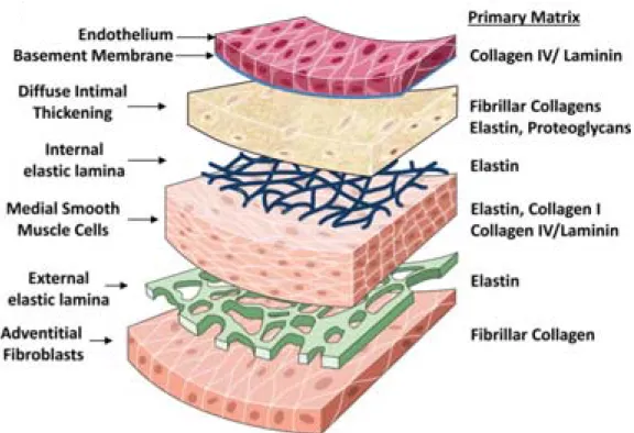
Since William Harvey’s description of blood circulation, the cardiovascular system is commonly acknowledged as an organ system that allows for blood to circulate and deliver nutrients, metabolites and oxygen to cells [1]. Arteries are composed of three main layers: the tunica intima, the tunica media and the tunica adventitia (Figure 1).
Figure 1. Structure of an artery. Illustration showing the composition and disposition of the three
layers that form arteries (adapted from Yurdagul A Jr, 2016).
The inner layer, the intima, is formed by a monolayer of endothelial cells (ECs) overlaying a basal lamina rich in type IV collagen. A small number of vascular smooth muscles cells (SMCs) are observed in the intima and are thought to contribute to the production of extracellular matrix (ECM) proteins that constitute the basal membrane.
The middle layer, the media, is made up of interposed elastic laminae and SMC layers that ensure the vessel contractility. The media is rich in type I and III collagen fibres that have important tensile strength. The intima and the media are separated by the internal elastic lamina while the external elastic lamina delimits the interface between the media and the adventitia.
The aforementioned layer of loose connective tissue mainly contains fibroblasts, perivascular nerves, lymphatic vessels, and microvessels named vasa vasorum.
B/ Generalities on atherosclerosis
1. Definition
During the XVI and XVII centuries, renaissance anatomists, the most famous of whom Leonardo Da Vinci, described the degeneration of arteries with advancing age, already hinting at the progressive aspect of atherosclerosis [1]. Lobstein coined the term arteriosclerosis or atherosclerosis in 1829, derived from the Greek ‘athere’ (gruel) and ‘skleros’ (hardening).
Atherosclerosis develops in the intimal layer of large- and medium-sized arteries, such as carotid arteries, aortas and coronary arteries [2]. The development of atherosclerosis involves complex interactions between blood-derived elements, for instance monocytes or lipoproteins, and vascular wall components, such as SMCs. Lesions start as a lipid deposition in arterial walls. This lipid build-up leads to inflammation within the intima [3] and evolves into atheroma plaques. As such, plaques are mainly asymptomatic. However, they can thicken, leading to a reduction of the arterial lumen, or stenosis. Plaque stenosis alone can sometimes obstruct the blood flow although it is rarely fatal, as downstream tissues are weakly ischaemic [4]. Conversely, acute cardiovascular events are principally caused by the formation of thrombus over the plaque or atherothrombosis. Plaques weaken, rupture and release their content into the bloodstream. Atherosclerotic plaques contain thrombogenic elements capable of activating platelets, leading to atherothrombosis. Thrombi can directly occlude the blood flow or embolise and block smaller vessels. This induces the ischaemia of downstream organs and necrosis of tissues, which can be fatal.
2. Prevalence and risk factors
Atherosclerosis accounts for the majority of cardiovascular diseases, the first cause of death worldwide, with an estimated 15 million deaths in 2015 [5]. Atherosclerosis manifests in multiple forms depending on the affected organ. For instance, ruptured plaques can provoke myocardial infarction, ischemic stroke and acute limb ischaemia.
The main causal factor identified in the initiation of atherosclerosis is low-density lipoprotein (LDL) [6]. These proteins carry cholesterol, are elevated in the blood of hypercholesteraemic patients and can easily accumulate in the vessel walls. Several risk factors for atherosclerosis have been identified and include diet, smoking, genetics, hypertension, hypercholesterolemia, hyperlipidaemia, hyperglycaemia, and diabetes. This multiplicity of risk factors highlights the multifactorial facet of atherosclerosis. Infections are also considered to be risk factor for atherosclerosis [7], [8] and will be discussed in more details in a subsequent part.
However, despite the development of preventive strategies and the use of tension and lipid-lowering drugs such as statins, atherosclerotic plaque rupture is still an important concern and for this reason it is crucial to better clarify the aetiology of plaque destabilisation.
C/ The formation of atheroma or atherogenesis
1. Lipoprotein accumulation and inflammation
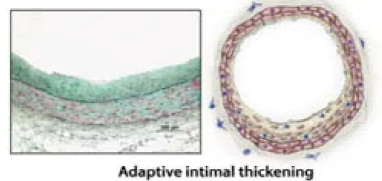
Atherosclerosis develops predominantly in areas of low shear stress within arteries, e.g. bifurcations [9]. In these areas, the intima is thickened, probably as an adaptation to resident mechanical constraints [4]. This stage of plaque formation is named ‘adaptive intimal thickening’ (Figure 2).
Figure 2. Photograph and representative diagram of a human coronary artery showing adaptive intimal thickening of arteries. The intima is enriched in smooth muscle cells and matrix fibres in
areas of low shear stress (adapted from Bentzon et al., 2014).
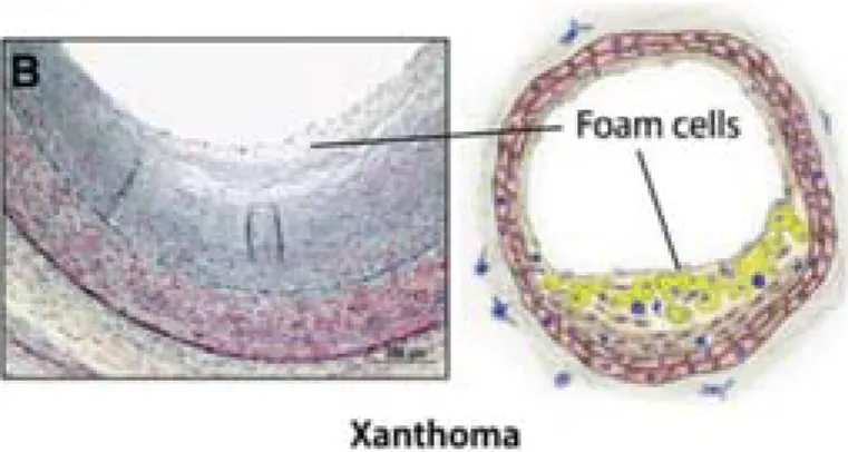
In these sites of predilection, LDLs accumulate in the intimal layer, where they oxidise and aggregate. The oxidation of LDLs is thought to rely on the activity of myeloperoxidase (MPO), lipoxygenases, and reactive oxygen species (ROS). The scavenging of modified LDLs (oxLDLs) by resident macrophages and notably SMCs that reside in the intima results in the accumulation of lipids inside the cells, turning them into ‘foam’ cells [10]. These processes involve cholesterol efflux and autophagy [11]. OxLDLs stimulate the polarisation of macrophages to a pro-inflammatory phenotype that secretes cytokines and produces reactive oxygen species, which subsequently promote the further retention and oxidation of LDLs in the intima [12].
Figure 3. Photograph and representative diagram of a human intimal xanthoma. This lesion displays
an accumulation of foam cells derived from recruited macrophages and SMCs (adapted from Bentzon, 2014).
Modified LDLs are recognised as danger signals and induce a response to injury in ECs, SMCs, resident macrophages and dendritic cells, stimulating the secretion of chemoattractants and the expression of adhesion molecules. This will lead to the attraction innate immune cells, in particular monocytes, through the interaction of classical monocytic chemokines, such as, CCL2 (monocyte chemotactic protein, MCP-1), CCL5 (RANTES) and CX3CL1 (fractalkine), with their receptors CCR2, CCR5 and CX3CR1 [13]. Recruited monocytes differentiate into specialised phagocytes, i.e. macrophages and dendritic cells, which can further ingest oxLDLs. T cells are also recruited during the initial stages of atherosclerosis in response to cytokines or chemokines secreted by macrophages and ECs.
2. Necrotic core and fibrous cap formation
The ongoing intimal inflammation stimulates the mobilisation of medial SMCs, which migrate to the intima, proliferate and lose their contractile function to acquire a synthetic and phagocytic phenotype. Interestingly, SMC proliferation has been shown to rely on leukotrienes [14]. SMCs then start to produce ECM proteins, such as collagen or elastin. This phenotypic change also leads to the increased retention and oxidation of LDLs, promoting foam cell formation. This type of lesion (Type III-IV) is characterised by a pathological intimal thickening (Figure 4).
Figure 4. Representative diagram and photograph showing the pathological intimal thickening of a human coronary artery. This type of lesion exhibits increased extracellular lipid accumulation in the
absence of a defined necrotic core (adapted from Bentzon et al., 2014).
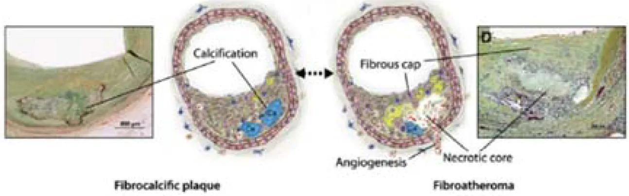
Foam cell accumulation induces to the apparition of a necrotic and lipidic core composed of apoptotic phagocytes and extracellular lipids in the form of cholesterol esters and crystals [15]. Many factors present in plaques can induce the apoptosis of foam cells and SMCs. In physiological conditions, apoptotic cells are cleared by phagocytes, however in plaques phagocytes exhibit defective efferocytosis, or clearance of apoptotic remnants, which contributes to the necrotic core growth [16]. Progressively, a fibrous cap composed of a matrix of type I and type III collagens mainly produced by intimal SMCs encapsulates this necrotic area [17]. Besides type I and III collagens, fibrous caps contain elastin and other ECM fibres. These lesions (Type V) are characterised as fibroatheroma or fibrocalcific plaques if they display calcifications (Figure 5).
Figure 5. Representative diagrams and photographs of a human fibrocalcific plaque and a
fibroatheroma. Advanced lesions comprise two types: fibrocalcific plaques (left) distinguished by the
presence of calcified necrotic cores surrounded with tissue, and fibroatheroma (right) characterised by the presence of a necrotic core, a fibrous cap, and neovessels (adapted from Bentzon et al., 2014).
II. Atherosclerotic plaques: from stability to vulnerability and rupture
A/ The concept of vulnerable plaques
Stable plaques are generally rich in SMCs that are predominantly localised in a thick fibrous cap. Necrotic cores are rather small or absent in stable lesions. The collagen and elastin present in the fibrous cap maintain the integrity of plaques and limit the extrusion of necrotic core materials into the bloodstream. The balance between SMC proliferation and apoptosis and between the synthesis and enzymatic degradation of ECM fibres determines the thickness of fibrous caps [18].
The presence of a thin cap and large necrotic core is the hallmark of vulnerable plaques [4]. The large necrotic core is associated with a high leukocyte infiltration and the thinned cap with a decrease in SMC and ECM content. These vulnerable plaques can also exhibit haemorrhages.
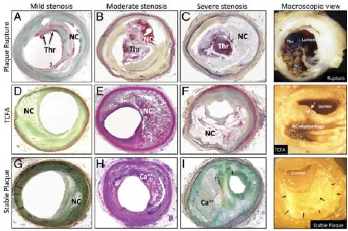
This type of lesion (Type VI) is named thin-cap fibroatheroma (TCFA) and are considered to be at high-risk of rupture (Figure 6).
Figure 6. Microphotographs of histological sections and macroscopical views of disrupted, vulnerable (TCFA), and stable plaques with different degrees of stenosis. (A to C and top right)
Ruptured plaques with thrombi (Thr) overlying disrupted fibrous caps (red arrowhead on macroscopic view) and in contact with necrotic cores (NC). (D to F and middle right) Thin cap fibroatheromas (TCFA) showing large necrotic cores (NC) and thinned fibrous caps (white arrows on macroscopic view). (G to I and bottom right) Stable plaques rich in fibrous tissue, with relatively small necrotic cores
B/ Mechanisms of plaque destabilisation
1. Cellular death
As aforesaid, SMCs are crucial for plaque stability as they maintaining the fibrous cap thick; therefore SMC apoptosis is an important marker of plaque vulnerability [19]. Indeed, the disappearance of SMCs results in reduced synthesis of matrix fibres and thus thinner caps. Recruited inflammatory leukocytes secrete proteases that induce a loss of SMC-matrix interaction by degrading matrix fibres, resulting in their cellular death by anoikis. Moreover, the ingestion of oxLDLs by SMCs and macrophages engenders endoplasmic reticulum stress and hence promotes apoptosis.
SMC and macrophage apoptosis leads to the necrotic core expansion and the exacerbation of plaque inflammation [20]. Cathepsins released from activated neutrophils are implicated in macrophage death. In addition, activated CD8+ lymphocytes and macrophages release tumour necrosis factor (TNF) which
is pro-apoptotic [21]. Mast cells can also promote apoptosis through the activation of Toll-like receptor (TLR) 4 by their enzymes [22].
2. Accumulation of necrotic cells
Clearance of apoptotic bodies by efferocytosis promotes the resolution of inflammatory processes and avoids the accumulation of apoptotic cells. If not cleared, apoptotic cells start to enter secondary necrosis. ‘Find-me’ and ‘eat-me’ signals plays a key role in the ingestion of apoptotic cells. However, in plaques, oxLDLs compete with find-me signals as they bind the same scavenger receptors on phagocytes [23]. Moreover, the overabundance of ‘eat-me’ signals in plaques hampers efferocytosis by overloading scavenger receptors. Finally, several proteases could also cleave these receptors. Altogether, necrotic cells accumulate in plaques, which lead to the expansion of the necrotic core
3. Degradation of the fibrous cap and proteolytic activity
Leukocyte infiltration is known to contribute to inflammatory reactions and proteolysis that destabilise plaque. Indeed, leukocytes, ECs and SMCs release metalloproteases (MMPs) that can digest the fibrous cap. Macrophages and neutrophils can be directly activated by inflammatory components in plaques and are a rich source of MMPs in plaques [24]. In addition, once activated, T cells stimulate macrophages to release matrix proteases. In early stages of atherosclerosis, MMPs are thought to be more protective as they facilitate the migration of SMCs from the media to the intima and thus the formation of a fibrous cap [20], [25]. Conversely in later stages, increased MMP release leads to the digestion of the ECM proteins in the fibrous cap, and therefore weakens plaques. For example, the constitutively-expressed MMP-2, which cleaves type IV collagen, is involved in SMC migration, while MMP-8 is involved in fibrous cap thinning through type I collagen degradation [26].
In addition to MMPs, cathepsins and serine proteases such as the neutrophil elastase digest elastin, and thus thin the fibrous cap. Indeed, the fibrous cap is mainly composed of collagen type I, but also contains elastin and proteoglycans. Expression of collagen type VIII has also been shown in aortic SMC from atherosclerotic mice but not wild-type mice [27].
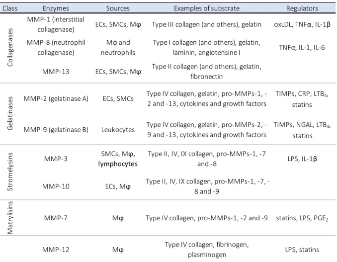
MMPs have other roles beside proteolytic degradation, and contribute to the ongoing inflammation as they cleave cytokines, pro-MMPs (the secreted inactive form of MMPs), growth factors and receptors. Table 1 recapitulates the activities and sources of the main MMPs involved in plaque weakening.
Table 1. Main MMPs involved in atherosclerotic plaque destabilisation.
4. Intraplaque haemorrhages
As plaques grow, their supply of crucial oxygen and metabolites is lessened. To palliate this nutrient deprivation and ischaemia, neovessels grow into the intima. These neovessels, i.e. vasa
vasorum, originate mainly from the adventitia, but can also emerge from the luminal side. They provide
an entrance for monocytes and immune cells in plaques. Moreover, this neo-angiogenesis gives rise to a fragile and often leaky microvasculature, which promotes the extravasation of red blood cells [15]. Haemoglobin from erythrocytes is highly cytotoxic and thus promotes apoptosis of resident plaque cells
Class Enzymes Sources Examples of substrate Regulators
MMP‐1 (interstitial
collagenase) ECs, SMCs, Mϕ Type III collagen (and others), gelatin oxLDL, TNFα, IL‐1β
MMP‐8 (neutrophil collagenase) Mϕ and neutrophils Type I collagen (and others), gelatin, laminin, angiotensine I TNFα, IL‐1, IL‐6 MMP‐13 ECs, SMCs, Mϕ Type II collagen (and others), gelatin, fibronectin
MMP‐2 (gelatinase A) ECs, SMCs Type IV collagen, gelatin, pro‐MMPs‐1, ‐
2 and ‐13, cytokines and growth factors
TIMPs, CRP, LTB4,
statins
MMP‐9 (gelatinase B) Leukocytes Type IV collagen, gelatin, pro‐MMPs‐2, ‐
9 and ‐13, cytokines and growth factors TIMPs, NGAL, LTB4, statins MMP‐3 SMCs, Mϕ, lymphocytes Type II, IV, IX collagen, pro‐MMPs‐1, ‐7 and ‐8 LPS, IL‐1β MMP‐10 ECs, Mϕ Type II, IV, IX collagen, pro‐MMPs‐1, ‐7, ‐ 8 and ‐9 Ma tr yl is in s MMP‐7 Mϕ Type IV collagen, pro‐MMPs‐1, ‐2 and ‐9 statins, LPS, PGE2 MMP‐12 Mϕ Type IV collagen, fibrinogen, plasminogen LPS, statins Co llage n as es Gela ti n as es St ro m el ys in s Mϕ = macrophages; TIMPs = Tissue Inhibitors of MMPs; CRP = C‐Reative Protein
[28]. These intraplaque haemorrhages are frequent in advanced plaques and have been shown to result in the accumulation of protease-rich neutrophils in plaques [29]. Therefore by conveying neutrophils, intraplaque haemorrhages can also contribute to plaque weakening.
5. Calcification and cholesterol crystals
Apoptotic, necrotic and degraded matrix materials trap calcium granules and deposits. When expanding, this trapped calcium results in the calcification of plaques. The genesis of these calcification is however not fully understood. Another type of crystals found in plaques derive from cholesterol, which can be synthesised by SMCs in response to collagen type I [30]. These cholesterol crystals can exhibit several forms, including nodules, plaques and spikes [31]. While the role of calcification, especially the nodular type, in plaque destabilisation is still debated, cholesterol crystals are found perforating the fibrous cap at sites of rupture.
C/ Resolution versus chronicity of inflammation
The evolution of plaques is a slow process that occurs over years in the life of an individual. Lesions are thought to be able to regress or to remain stable. Several processes occurring in plaques display a duality that counterbalance the inflammation to avoid a swift exacerbation. The endoplasmic reticulum stress generated by the ingestion of oxLDLs activates a kinase involved in macrophage survival and autophagy, and thus reduces the excessive apoptosis [32]. The pro-inflammatory activation of plaque cells by inflammatory lymphocytes can be countervailed by regulatory lymphocytes [33]. The prostaglandin PGE2 expressed in plaques promotes macrophage activation, but also increases the
production of anti-inflammatory IL-10 [34]. Macrophages themselves differentiate into either a pro- or anti-inflammatory phenotype and can egress from lesions [12]. Moreover, lipoxygenases, such as 12/15-lipoxygenases oxidise LDLs, while they also produce pro-resolutive lipoxins and resolvins [35]. The balance between these inflammatory and resolutive signals determines the progression and ultimately the fate of the lesion.
D/ From vulnerable plaque to cardiovascular events
1. Mechanisms of plaque rupture
For a plaque to rupture, it must be structurally weak to be prone to tearing in the fibrous cap that exposes the thrombotic material to the bloodstream. In ruptured plaque specimens obtained from autopsy, thinned fibrous caps are consistently evidenced and are particularly localised in the shoulders of eccentric plaques, i.e. plaques that form an arc in the blood vessel [4]. As previously mentioned, fibrous cap thinning involves both the loss of SMCs, thus the reduction of ECM fibres synthesis, and
the digestion of matrix fibres by enzymatic activities [19]. Concurrently, the infiltration of leukocytes such as monocytes and neutrophils results in their subsequent activation and MMP, cathepsin, and plasminogen activator releases [36], accelerating both the thinning of the fibrous cap and enhancing the thrombogenicity of plaques.
The underlying causes of plaque rupture remain undefined. Physical or psychological stress, infection, and temperature variation have been evoked as triggers of cardiovascular events. Circadian rhythm may also play a part, as myocardial infarctions are more frequent in the morning [37], [38]. Mechanistically, the activation of the sympathetic nervous system is the main pathway evoked, as it leads to increased heart rate and blood pressure. Vasoconstriction, vasodilatation and wall shear stress could thus participate in plaque rupture [18], [39].
Observational findings have shown that only a minor part of ruptures results in symptoms. Therefore, it is possible that plaques with limited thrombogenicity also rupture, but only induce a minor thrombus formation and heal silently.
2. Plaque erosion
Atherothrombosis also forms on ‘eroded’ plaques, i.e. with an absence of endothelium over an intact fibrous cap [40]. These plaques are more fibrous and rich in SMCs (Figure 7). Local shear stress combined with oxidative stress promotes endothelial dysfunction and death. The subsequent endothelial denudation activates platelets causing them to form a thrombus.
3. Healing of plaques
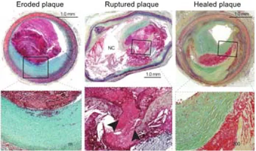
Observations of human specimen have revealed the presence of thrombi incorporated into plaques and layers of dense type I collagen and loose type III collagen [41]. This suggests that plaques rupture silently and heal by rebuilding layers of tissue (Figure 7). This fibrotic remodelling could also favour the stenosing of plaques.
Figure 7. Microphotographs and magnifications of an eroded plaque, a ruptured plaque and a healed plaque. (Left) Plaque erosion is characterised by a thrombus overlying an intact and thick fibrous cap.
(Middle) A ruptured plaque showing an occlusive thrombus in direct contact with the thrombogenic necrotic core (NC); arrowheads indicate the disruption of the thinned cap. (Right) A thrombus has been incorporated into the plaque which displays distinct layers of collagen, representative of a healed plaque (adapted from Patri et al., 2013)
E/ Murine model of atherosclerosis
Mice do not develop spontaneous atherosclerosis diseases, unlike pigs, non-human primates and surprisingly, birds [42]. Mice are atheroresistant, but some backgrounds are more susceptible to atherogenesis than others when fed a high fat diet (HFD), for example the C57BL/6 background. The most frequent murine models of atherosclerosis are genetically knock-out Apoe-/- and Ldlr-/- mice [43].
In both models, hyperlipidaemia results from a defective cholesterol trafficking. Apolipoprotein E (APOE) is the ligand of the low-density lipoprotein receptor (LDLR) and other receptors on hepatocytes necessary for lipid uptake of chylomicrons and very low-density lipoprotein remnants. Apoe-/- mice are
hyperlipidaemic under a chow diet and develop plaques early. Plaque progression can be accelerated by a HFD, leading to plaques rich in macrophages and lipids. Ldlr-/- mice do not have such an increased
lipidemia, as other receptors than LDLR can uptake lipoproteins. These mice need to be fed a HFD in order to observe the early apparition of lesions. To palliate this disadvantage, Ldlr-/- mice have been
crossed with other mouse lines such as Apoe-/- mice or mice knock-in for human ApoB100 transgene.
Interestingly, after HFD feeding, Apoe-/-Ldlr-/- mice show significant coronary lesions and suffer from
myocardial infarction, in particular in stress conditions [44]
In terms of atherosclerosis modelling, aged chow-fed Apoe-/- mice show lesions that are
morphologically closer to human lesions. After 20 weeks, lesions are fibrous, rich in SMCs and ECM. In older mice, intraplaque haemorrhages have been evidenced, suggesting that these plaques could
spontaneously become vulnerable. However, APOE is expressed on bone marrow-derived cells and therefore bone transplantation from mice expressing APOE is not possible, as it will interfere with atherogenesis. Moreover, Apoe-/- mice exhibit an increased leucocytosis and cognitive dysfunction,
underlining the multiple roles of APOE.
III. The role of infectious diseases in atherosclerosis
The notion of the implication of bacterial infections in atherosclerotic disease stems from clinical observations. As early as in the 19th century, physicians were noticing a connection between the
incidence of stroke and infections [45]. Thereafter, a large body of evidence has emerged from both clinical and experimental studies, linking atherosclerosis with different pathogens [46]–[49]. These include bacteria such as Chlamydia pneumoniae (C.pneumoniae), Porphyromonas gingivalis (P.gingivalis), Helicobacter pylori (H.pylori), but also viruses, such as influenza or cytomegalovirus (CMV). Nevertheless, the role of infection in the pathogenesis of atherosclerosis and associated disorders remains elusive, undoubtedly because of the complexity of this possible contribution.
A/ Potential implications of bacterial and viral infections
1. Clinical arguments
Identification of pathogens in plaques: C. pneumoniae, a bacteria responsible for low-grade respiratory infections, is one of the first pathogens identified in human atherosclerotic vessels. Microscopic observations by immunohistochemistry or electronic microscopy and PCR identification of this pathogen were later confirmed by the isolation of viable organisms from human plaques [50]. C.
pneumonia was found in cells from human atherosclerotic vessels, but not from healthy tissues, but its
presence was not correlated to plaque instability. Genomic material from several commensal bacteria and viruses have been further identified in human plaques. For example, a large variety of dental germs have been detected by PCR or immunocytochemistry in human plaques [51].
Association with cardiovascular events: In the early 2000s, a large cohort study involving more than 40.000 patients confirmed previous smaller-scale studies on the association between acute infection and myocardial infarction or stroke [52]. Numerous seroepidemiological studies demonstrated associations between bacterial or viral seropositivity and cardiovascular outcomes [49], [53]–[56]. However these associations are weakened by conflicting results, showing how heterogeneities in studied parameters or in study designs could lead to opposing results. Moreover, the lack of suitable and reliable techniques for the identification of pathogens could also underlie this variability.
Antibiotherapy trials: Following these studies, several clinical trials were implemented using antibiotherapy targeting C. pneumoniae but showed no benefits on cardiovascular mortality. These studies are however partially inconclusive, as C.pneumoniae is an intracellular pathogen and hence result in a persistent infection, in which the pathogen resides in cells for long periods without proliferating and escapes antibiotic treatment. Furthermore, the timing and duration of antibiotic treatments may have been either too late or too short in the course of the infectious process. Moreover, prolonged antibiotherapy leads to an alteration of the lipid metabolism and atherogenesis. Finally, other studies have suggested that cumulative exposures to a large number of pathogens is responsible for the increased risk of cardiovascular events.
The concept of infectious burden: Early studies already showed that the higher the number of pathogens identified, the higher the prevalence of coronary artery disease [57]. In the Northern Manhattan prospective study, the risk of stroke [58] and the thickness of carotid plaques [59] were found to increase in patients seropositive for at least five pathogens, and correlated positively although non-significantly with seropositivity for only one pathogen. Therefore, as different pathogens are identified in plaques, it is possible that the cumulative actions of different bacteria and viruses renders plaques vulnerable, as opposed to the single action of one specific pathogen.
2. Animal studies
In the 70s, an American group showed that following infection with the avian herpes virus, chickens develop atherosclerotic-like lesions in arteries [60]. Since then, numerous models in mice, rabbits and rats have clarified the impact of infection on atherosclerosis.
Repeated injections of C.pneumoniae increases lesion size in Apoe-/-mice, Ldlr-/- mice, and
cholesterol-fed rabbits, but the antibiotic treatments of these animals does not provide consistent data. In advanced atherosclerosis, infections results in increased MMP activity and reduction of fibrous cap thickness but do not increase lesion size.
Another well-studied group of bacteria in atherosclerosis are dental germs. Oral or systemic deliverance of P.gingivalis increases systemic inflammation [61], [62] and accelerates atherosclerosis in Apoe-/- mice and rabbits [63]. These increases are also associated with an augmentation of
inflammatory cells, lipids, and cytokines within plaques. In several studies, genomic material from
P.gingivalis has been detected in aortic plaques in murine models of periodontitis. Other dental
pathogens accelerate atherosclerosis, in particular during polymicrobial dental infection, which implies a role for the infectious burden in atherogenesis [64].
The impact of H.pylori and Mycoplasma pneumoniae on atherogenesis is still controversial because of conflicting data.
Viral infections are potential accelerators of atherosclerosis, as infections of mice with CMV or influenza virus enhance T cell and macrophage infiltration and lesion size.
B/ Possible mechanisms
1. Direct action of pathogens on plaques
Bacteria and viruses could directly act on plaques or infect macrophages, SMCs or ECs, resulting in pro-inflammatory responses and expression of adhesion molecules [65] (Figure 8). For example, CMV infection triggers the expression of enzymes from the 5LO pathway in SMCs [66] and could hence favour pro-inflammatory responses. Bacterial infections also promote LDL oxidation and foam cells formation [67].
2. Indirect impact of pathogens on plaques
Molecular mimicry: PAMPs or Pathogen-Associated Molecular Patterns expressed at the surface of bacteria present structures that resemble host proteins (Figure 8). For example, heat shock proteins (HSPs) found at the surface of C.pneumoniae, H.pylori or P.gingivalis, are similar to human HSPs [68]. The cross-reactivity between these HSPs can trigger a humoral response directed against both pathogenic and human HSPs, initiating an auto-immune response against plaque cells.
Systemic inflammatory responses: The presence of infectious agents at distant non-vascular sites such as the teeth or the lungs induces an augmentation of circulating cytokines and acute phase proteins which may reach the vascular site of plaques. Circulating levels of IL-6 and C-reactive protein (CRP) are for example elevated in patients with periodontal disease [68], [69] and in murine models of periodontal diseases [61]. Moreover, circulating bacterial or viral products increase the activation of blood leukocytes, which directly affect plaques, as discussed in more detail in the following section.
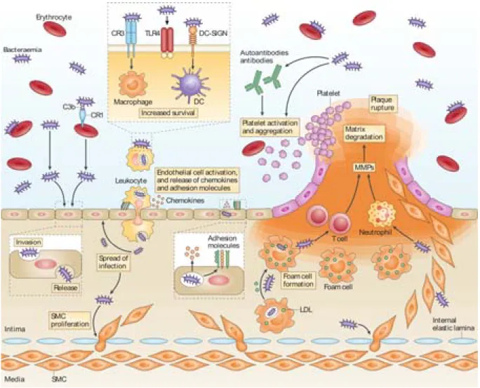
Figure 8 Potential mechanisms of plaque growth and destabilisation mediated by bacteria. Bacteria
directly invade leukocytes and ECs and increase systemic inflammation through TLR4 signalling, leading to the upregulation of adhesion molecules, the secretion of chemokines and the subsequent transmigration of leukocytes. Pathogens enter safely within leukocytes by the activation of complement receptor-3 (CR3) and dendritic cell-specific ICAM-3 grabbing non-integrin (DC-SIGN). The presence of bacteria in the intima induces the proliferation and migration of smooth muscles cells and promotes the uptake of oxLDLs by macrophages. Bacteria, along with inflammatory signals, induce the secretion of MMPs and consequently degrade the fibrous cap. Bacterial signals also enhance platelet aggregation by a direct action and by the production of pro-thrombotic autoantibodies (adapted from. Hajishengallis, 2014).
C/ Endotoxemia and atherosclerosis
When gram-negative bacteria are dividing or dying, their cellular wall breaks down, leading to the release of their surface components, such as endotoxins or lipopolysaccharides (LPS). LPS is composed of polysaccharides bound to a hydrophobic moiety called lipid A, which has different immuno-potencies depending on the species of bacteria. When hydrophobic LPS enters the blood flow, it is
carried by proteins such as LPS-binding proteins (LBP) or APOE. These proteins either activate target cells or lead to hepatic clearance [70]. LPS is known to induce pro-inflammatory responses and cell death through pathways involving TLR, CD14, MYD88 and NF-κB. Various conditions can lead to the translocation of LPS into the circulation, for example intestinal leakage [71].
1. TLRs signalling in atherosclerosis
LPS binds preferentially to TLR4, however LPS originating from P.gingivalis has been shown to exert its effect through both TLR4 [72] and TLR2 [73]. Genetic deletions of TLR2, TLR4 or the adaptor protein Myd88 in Apoe-/- mice decrease macrophage infiltration and diminished atherosclerotic
lesion size [74]. Accordingly, human polymorphisms in the TLR4 or TLR2 genes promote or reduce susceptibility to cardiovascular events [75].
Other ligands of TLR4/CD14 complex and TLR2 include saturated fatty acids, which are also known to promote LPS translocation and metabolic diseases [76], [77]. Moreover, macrophagic TLR4 binds oxLDL [78] and its activation by LPS and LDL also upregulates chemokine expression in macrophages [79].
2. Endotoxemic models in atherosclerotic mice
Systemic administration of LPS: Lehr et al. first showed that repeated intravenous injections of LPS in hypercholesteraemic rabbits results in accelerated atherogenesis [80]. More recently, repeated intraperitoneal LPS injections for 10 weeks in Apoe-/- mice was shown to increase plaque size, oxidation
of LDLs and natural killer cell infiltration. Apoe-/- mice lacking APOCI are protected from LPS-induced
atherosclerosis and their plaques contain less T cells and macrophages, emphasizing the role played by lipoproteins in the regulation of LPS-induced inflammatory responses [81]. Similarly, APOAI protects mice from accelerated atherogenesis and the increased infiltration of CD68-positive macrophages [82]. Moreover, in a model of accelerated atherosclerosis induced by collar placement, chronic LPS exposure in HFD-fed Apoe-/- mice promotes macrophage infiltration and vulnerability, in particular in stressful
conditions [83]. Finally, in mice injected with subclinical/super-low doses of LPS for two months, monocytes polarise towards an inflammatory phenotype, impairing the resolution of subsequent inflammatory responses in plaques [84].
LPS-induced lung inflammation: A recent study pointed out the role of neutrophils in plaque vulnerability following LPS exposure in a model of acute lung inflammation induced by intratracheal LPS instillation in HFD-fed Apoe-/- mice [85]. In this model of plaque destabilisation occurs relatively
fast, i.e. 24 hours after LPS administration, which is consistent with the rapid reactivity of neutrophils known to be fast effector in inflammatory processes.
LPS from dental germs: In Apoe-/- mice, chronic infusion of LPS from P.gingivalis activates the
COX-2 pathway in plaque macrophages and also accelerates atherogenesis [67], [86]. This is in agreement with the increased TLR expression in atherosclerotic plaques in murine models of periodontal disease [73] and the induction of inflammatory responses in ECs by LPS from P.gingivalis [62], [65].
Metabolic endotoxemia: Mice fed a HFD develop what is called metabolic endotoxemia, which has been implicated in metabolic disorders such as diabetes or obesity [87]. This translocation of LPS due to increased intestinal permeability is dependent on modifications of the gut microbiota [88]. Administration of Akkermansia muciniphila in HFD-fed Apoe-/- mice restores the integrity of the
intestinal barrier, prevents metabolic endotoxemia and reduces the atherosclerotic burden [89].
3. Association between endotoxemia and atherosclerosis in humans
Human blood vessels respond to LPS by expressing and producing inflammatory signals: chemokines (IL-8, CCL2…), cytokines, ROS, PAF, and adhesion molecules [8].
Plasma levels of endotoxin and cardiovascular events: In 1999, results from the Bruneck study provided evidence that plasma levels of LPS correlates to the incidence of atherosclerosis [90]. In patients undergoing peritoneal dialysis, the elevated level of plasmatic endotoxin is associated with increased carotid intimal thickness [91]. Increased plasmatic LPS levels correlates to the risk of metabolic syndromes and the 10-year cardiovascular mortality [92]. Endotoxemia hence is related to cardiovascular events in humans.
Periodontal diseases: During human periodontal diseases, LPS released from dental germs enters the bloodstream [93]. Periodontitis-induced endotoxemia is associated with cardiovascular diseases, as shown in the FINRISK study [94].
Metabolic endotoxemia: High-fat feeding in humans potentially results in the translocation of LPS in the blood, as suggested by studies showing that high-fat meals induce endotoxemia [95]. The levels of plasmatic endotoxin are increased in patients with chronic heart failure presenting a bowel wall oedema that induces gut leakage [96] and LPS translocation [97]. The increased risk of cardiovascular events and mortality observed in endotoxemic patients with chronic kidney disease can also be linked to increased gut permeability [98]. In addition, LPS translocation occurs during long term western-type diets, which are rich in fats and is known to contribute to the onset of atherosclerosis and metabolic syndromes [99]. Metabolic endotoxemia and the gut microbiome are thus likely contributing to atherosclerosis in humans [100].
Neutrophils
I. The life of neutrophils
A/ The granulopoiesis
1. Production
Neutrophils belong to the myeloid cell line, which emerges from the differentiation of hematopoietic stem progenitor cells (HSPCs) and includes granulocytes, monocytes, dendritic cells, platelets and erythrocytes [101]. Granulocytes comprise basophils, eosinophils and neutrophils, and are identified by polylobed nuclei and the presence of cytoplasmic granules.
Granulopoiesis takes place in bone marrow niches, where HSPCs are committed under the instruction of chemokines and transcriptions factors to differentiate into mature neutrophils (Figure 9), by going sequentially through different stages: myeloblasts, promyelocytes, myelocytes, metamyelocytes and band cells.
2. Release and retention
Once mature, neutrophils can exit the bone marrow to enter the bloodstream (Figure 9). This release is antagonistically regulated by the two receptors CXCR4 and CXCR2 [102], [103]. CXCR4, highly expressed on newly formed and senescent neutrophils, binds CXCL12 produced by bone marrow ECs and osteoblasts [104]. CXCR4 retains mature neutrophils in the bone marrow and targets senescent neutrophils back to the bone marrow. Inversely, CXCR2 and its ligands, CXCL1 (GROα, growth-related oncogene-α/KC) and CXCL2 (macrophage inflammatory protein, MIP-2), both expressed by ECs, facilitate the egress of neutrophils into the circulation.
3. Regulation of neutrophil production
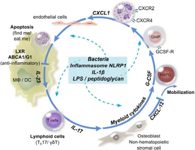
Granulocyte-colony stimulating factor (GCS-F) stimulates bone marrow neutrophil production and egress, by shifting the balance between CXCR4 and CXCR2 (Figure 9). Bacterial products, IL-1 and TNF can also promote neutrophil release into the blood circulation during ‘emergency’ granulopoiesis [105]. Conversely, the ingestion of apoptotic neutrophils by macrophages reduces IL-23 secretion through a LXR-dependent transcription [106]. IL-23 stimulates the production of IL-17 by specialised lymphocytes which will in turn promote the production of G-CSF and hence the release of neutrophils.
Figure 9. Regulation of granulopoiesis. Hematopoietic stem progenitor cells (HSPCs) differentiate
into granulocyte/macrophage progenitors (GMP), which following stimulation by G-CSF differentiate into mature neutrophils. Osteoblasts and stromal cells in the bone marrow inhibit the mobilisation of neutrophils by secreting CXCL12, the ligand of CXCR4. The egress and mobilisation of mature neutrophils is promoted by the axis CXCR2/CXCL1. The ingestion of apoptotic neutrophils by tissue macrophages (Mϕ) or dendritic cells (DC) inhibits the secretion of IL-23 via LXR-dependent
transcription. IL-23 conversely induces the release of IL-17 by specialised lymphocytes (Th17/γδT),
resulting in increased G-CSF production. Bacteria and their products or inflammatory signals, such as NLRP1 (NLR family, pyrin domain containing 1) inflammasome, tune the production of neutrophils (adapted from Wirths et al., 2014).
4. Granule formation
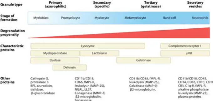
Belonging to the granulocyte family, neutrophils are rich in granules and secretory vesicles, filled with proteins necessary for their functions from recruitment to microbial defence [107]. Neutrophil granules are divided into four types and are sequentially formed and released in a reverse fashion, meaning that the first formed is the last released (Figure 10).
- Primary/azurophil granules are firstly formed and contain high amounts of MPO and serine proteases. - Secondary/specific granules contain high levels of lactoferrin and collagenases.
- Tertiary/gelatinase granules, as stated in their name, are rich in gelatinase, i.e. MMP-9.
- Secretory granules or vesicles are formed by endocytosis and are rapidly released upon chemotactic stimulation. They are an important reservoir of membrane surface receptors involved neutrophil priming and adhesion, such as CD11b/CD18, complement receptors or the co-receptor of TLR, CD14.
Figure 10. Neutrophil granules types, stage of formation and contents. Four types of granules are
formed during granulopoiesis and contain various factors necessary for neutrophil functions, such as antimicrobial proteins, receptors, adhesion molecules, and proteases. The first granules formed are the last released following neutrophil activation (adapted from Amulic et al., 2012).
B/ Trafficking of neutrophils
1. Circulation, margination and lifespan
In humans, neutrophils represent about 50 to 70% of the circulating leukocytes, but only about 10 to 25% in mice. However, under inflammatory stimulation, mice show a significant neutrophil mobilisation. In physiological conditions, the number of circulating neutrophils fluctuates throughout the day due to the circadian control of HSPCs [108].
Beside the bone marrow, neutrophils are found in steady state in the lungs, the spleen and the liver [109], [110]. This pool of neutrophils marginated along the organ blood vessels is thought to constitute reservoirs for rapid deployment during acute processes.
Neutrophils are considered as short-lived cells with an estimated lifespan of 3 to 16 hours in the circulation [111]. In the absence of inflammatory signals, neutrophils enter spontaneous apoptosis, however during inflammatory processes their lifespan is prolonged, mainly through the inhibition of apoptosis pathways [34], [112], [113].
2. Recruitment of neutrophils into tissues
Following an injury or invasion of microorganisms, tissue-resident sentinel leukocytes and ECs, expressing pattern-recognition receptors (PRRs), recognise damage-associated molecular patterns (DAMPs) or pathogen-associated molecular patterns (PAMPs). Consequently, activated sentinel cells secrete cytokines and lipid mediators. In response to these signals, ECs start to express leukocyte adhesion molecules [114] that promotes the recruitment of neutrophils. Sentinel cells also attract neutrophils to affected tissues by releasing chemokines and lipids mediators. The cascade of recruitment and transmigration of neutrophils into tissues is well-described [115] and comprises several steps (Figure 11):
1. Tethering and rolling: A weak binding between P-selectin and E-selectin, expressed on the surface of ECs, and PSGL-1 (P-selectin glycoprotein ligand 1), expressed on neutrophils, leads to the rolling of neutrophils on surface of the endothelium.
2. Firm adhesion: Rolling neutrophils are primed by cytokines, chemokines and/or DAMPs/PAMPs present at the endothelial surface. This priming leads to conformational changes that increase the affinity of surface integrins, CD11a/CD18 (LFA-1) and CD11b/CD18 (Mac-1), and the release of intracellular integrin stores. When integrins bind intracellular adhesion molecules (ICAMs) on ECs, neutrophils arrest firmly on the endothelium.
3. Diapedesis: Once adherent, neutrophils start to crawl onto the endothelium to search for an appropriate site of entry into tissues. To extravasate, neutrophils need to cross the endothelium and the basement membrane. This transmigration is mainly paracellular but can also occur transcellularly.
As an alternative to rolling, neutrophils can form ‘slings’ and ‘tethers’ enriched in CD11a/CD18 to ensure their arrest in the high shear forces occurring in large arteries [115] (Figure 11). Moreover, platelets could contribute to the endothelial adhesion of neutrophils, notably through the secretion of chemokines, for example CCL5 [116].
Interestingly, neutrophils undergo reverse transmigration in certain pathological conditions, such as ischemia-reperfusion [117]. In such condition, the reverse transmigration of neutrophils is dependent on the degradation of junctional adhesion molecules by neutrophil elastase released upon LTB4
stimulation [118]. This process could prevent neutrophils from residing in tissue when not needed to fight infections.
Figure 11. The neutrophil recruitment cascade. (a) In response to an inflammatory stimuli, ECs
express adhesion molecules which allow the tethering of rolling neutrophils. Neutrophils then crawl and adhere to the endothelium. After firm arrest, neutrophils start diapedesis. (b) Alternatively, neutrophils can form tethers and slings to anchor themselves to the endothelium and overcome high shear stress (adapted from Kolaczkowska et al., 2013).
II. Immune functions of neutrophils
Neutrophils are the first line of defence in bacterial infection. Once in tissues, they navigate towards the inflammed site following a hierarchy of chemotactic molecules which includes LTB4, IL-8
(CXCL8), CXCL1, CXCL2, CCL3, CCL5, C5a, and fMLP [119], [120]. On the inflammatory site, neutrophils activate to perform their defensive functions.
A/ Defence systems
Neutrophils present various mechanisms involved in microbial killing and immune functions, from phagocytosis to granule release [105], [115], [121]–[123], which will be briefly described herein.
1. Phagocytosis
Similarly to macrophages, neutrophils have a high phagocytic capacity for pathogens and cellular debris after recognition by PRRs, Fc receptors or complement receptors. Once in the cell, pathogens are destroyed in a specialised compartment, the phagosome that becomes functional upon its fusion with granules and the assembly of NADPH oxidase.
2. Reactive oxygen species
The respiratory burst occurs following neutrophil activation and results in the generation of Reactive Oxygen Species (ROS). On the plasma or phagosomal membrane, nicotinamide adenine dinucleotide phosphate (NADPH) oxidase is assembled into a complex that reduces molecular oxygen to superoxide ions (•O
2-). Those ions can further be converted into various species including hydrogen
peroxide (H2O2) by dismutation or peroxynitrite (ONOO−) after reaction with NO. ROS can contribute
to the reversible regulation of several enzymes, such as metalloproteases.
3. Primary granule proteins
Myeloperoxidase (MPO) has an important oxidant activity. During the respiratory burst, MPO reacts with hydrogen peroxide to form hypochlorous acid (HOCl). It can also chlorinate tyrosine residues in proteins, such as the HDL, ApoAI, and form reactive nitrogen intermediates [124]. MPO can either be released into the phagosome or into the extracellular milieu.
Defensins are cationic antimicrobial peptides that can form pores in bacterial membranes and promote monocyte and T cell chemotaxis.
Another antimicrobial protein localised in primary granules is the bactericidal/permeability-increasing protein (BPI) of which the primary function is to bind LPS.
Finally, primary granules contain the three main serine proteases of neutrophils: proteinase-3, cathepsin G, and elastase. These enzymes digest various ECM components, including type IV collagen and elastin. They can also promote the activation of ECs, macrophages and lymphocytes and the attraction of monocytes and T cells.
4. Secondary and tertiary granule proteins
Secondary granules contain several antimicrobial peptides, such as the neutrophil gelatinase-associated lipocalin (NGAL) which plays an important role in iron-depleting immune strategies. Other peptides include LL37 (CRAMP), involved in the chemotaxis of neutrophils, T cells and monocytes, and metal-chelating proteins: lactoferrin which sequestrates iron and calprotectin (S100A8/S100A9) which chelates zinc and manganese.