HAL Id: hal-02640118
https://hal.inrae.fr/hal-02640118
Submitted on 28 May 2020
HAL is a multi-disciplinary open access
archive for the deposit and dissemination of sci-entific research documents, whether they are pub-lished or not. The documents may come from teaching and research institutions in France or abroad, or from public or private research centers.
L’archive ouverte pluridisciplinaire HAL, est destinée au dépôt et à la diffusion de documents scientifiques de niveau recherche, publiés ou non, émanant des établissements d’enseignement et de recherche français ou étrangers, des laboratoires publics ou privés.
Copyright
Bisphenol A exposure disrupts neurotransmitters
through modulation of transaminase activity in the
brain of rodents
Daniel Zalko, Ana Soto, Cécile Canlet, Marie Tremblay-Franco, Fabien
Jourdan, Nicolas J. Cabaton
To cite this version:
Daniel Zalko, Ana Soto, Cécile Canlet, Marie Tremblay-Franco, Fabien Jourdan, et al.. Bisphenol A exposure disrupts neurotransmitters through modulation of transaminase activity in the brain of rodents. Endocrinology, Endocrine Society, 2016, 157 (5), pp.1736-1739. �10.1210/en.2016-1207�. �hal-02640118�
Bisphenol A Exposure Disrupts Neurotransmitters
Through Modulation of Transaminase Activity in the
Brain of Rodents
Daniel Zalko, Ana M. Soto, Cecile Canlet, Marie Tremblay-Franco, Fabien Jourdan, and Nicolas J. Cabaton
Toxalim (D.Z., C.C., M.T.-F., F.J., N.J.C.), Université de Toulouse, INRA (Institut National de la Recherche Agronomique), 31027, Toulouse, France; and Department of Integrative Physiology and Pathobiology (A.M.S.), Tufts University School of Medicine, Boston, Massachusetts 02111
A
n increasing body of literature suggests that peri-natal exposure to low doses of bisphenol A (BPA) has lasting effects on brain development and/or behav-ior in rodents (1, 2). Concerns about the occurrence of similar effects in humans are based on the parallel in-crease of neurobehavioral conditions in humans that appear in rodents exposed to BPA (3), and on epidemi-ological studies (4). In their recent article, Franssen et al (5) demonstrated opposite dose-dependent effects of BPA on the neuroendocrine maturation of female rats ex-posed during early life from postnatal day (PND)1 toPND15. In animals exposed to a 0.025-g BPA/kg bw
(body weight)/d dose, neuroendocrine maturation related to puberty was found to be delayed, with opposite effects observed in animals exposed to a 5000-g BPA/kg bw/d
dose. Modulation of inhibitory ␥-aminobutiric acid
(GABA)ergic neurotransmission was found to be associ-ated with the delayed maturation of GnRH secretion
through increased GABAergic tone for the 0.025-g
BPA/kg bw/d dose; opposite effects were observed for the higher dose.
These new findings prompted us to reexamine in more detail our results obtained for male mice exposed to very low doses of BPA from gestational day 8 to PND16 (maternal exposure to 0-, 0.025-, 0.25-, or
25-g BPA/kg bw/d) (6). In this 2013 study, we used
untargeted 1H-nuclear magnetic resonance (NMR)
metabolomics to explore the metabolites present in a set of tissues, including male brains at PND21. All brain metabolites were included in a partial least
square-dis-criminant analysis model, which demonstrated ex-tremely significant differences between the 4 groups. GABA was one of the major metabolites responsible for intergroup differences. To better understand the mech-anisms by which BPA disrupts brain development, we reanalyzed our raw data from 2013 with the purpose of 1) challenging the hypotheses by Franssen et al, and 2) seeking additional hypotheses that could explain the disruption of GABAergic pathways. We addressed the former by means of a 2 by 2 group comparison and the latter by analyzing the genome-scale metabolic
net-ISSN Print 0013-7227 ISSN Online 1945-7170 Printed in USA
Copyright © 2016 by the Endocrine Society Received March 29, 2016. Accepted March 29, 2016. For related article see page 1740
Abbreviations: BPA, bisphenol A; bw, body weight; GABA,␥-aminobutiric acid; NMR, nuclear magnetic resonance; PND, postnatal day.
Figure 1. Relative intensity of GABA in the brain of PND21 mice
perinatally exposed to 0-, 0.025-, 0.25-, or 25-g BPA/kg bw/d (based on1H-NMR semiquantitative spectral data from Cabaton et al [6]).
C o u n t e r p o i n t
work based on our 20131H-NMR brain metabolomics data, and by extracting the most prominent pathways (eg, subnetworks) modulated by BPA exposure.
In the first place, our results clearly support the con-clusions by Franssen et al about GABA being a key
de-terminant of BPA effects, despite species, sex, and exposure scenario differences (perinatal in our study, strictly postnatal in the Franssen et al work). In our study, GABA was significantly increased in
whole-brain extracts from the 0.025-g
BPA/kg low-dose group compared with controls (vehicle). No compar-isons could be carried out for the 5000-g/kg dose, not addressed in Cabaton et al (6). However, untar-geted metabolomics already high-lighted a decrease of GABA at the highest dose tested in Cabaton et al,
namely 25-g BPA/kg compared
with controls (Figure 1). In other words, the lower GABAergic tone observed by Franssen et al at 5000-g BPA/kg in specific brain re-gions was already obvious at 25-g BPA/kg in a perinatal scenario when using NMR-based metabolomics on whole-brain extracts.
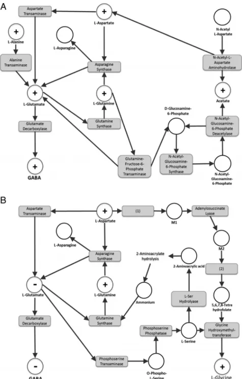
In a second step, based on our raw data from brain extracts, metabolic networks analysis was performed for the lowest and highest BPA doses used in Cabaton et al. The discrimi-nant metabolites were examined us-ing MetExplore, the open access webserver we developed for map-ping, visualization, and mining of
omics data (www.metexplore.fr)
(7). The subnetworks of interest
re-lating to 0.025-g BPA/kg bw/d
animals (vs controls) and 25-g
BPA/kg bw/d animals (vs controls) are displayed in Figure 2, A and B, respectively.
L-glutamate is the primary sub-strate for GABA synthesis, which is carried out by brain glutamate de-carboxylases. Both L-glutamate and GABA were found to change in a similar way: increased in
0.025-g BPA/kg exposed mice
and decreased in 25-g BPA/kg ex-posed mice. For this reason, and because there is only 1 report so far (8) suggesting regulation of the glutamate decarboxylase activity by BPA (at 40g/kg, in rats), it is unlikely that this enzymatic pathway would play a
Figure 2. Metabolic subnetwork extractions obtained using the webserver MetExplore
(www.metexplore.fr) showing the metabolic pathways modulated by BPA perinatal exposure in PND21 mice: (A) 0.025-g/kg bw/d vs control and (B) 25-g/kg bw/d vs control. The “⫹” dots represent the identified metabolites that were increased, the “⫺” dots represent the identified metabolites that were decreased, and the white dots represent the metabolites predicted to be involved in the corresponding biochemical pathway. The gray boxes are the enzymes catalyzing the reactions linking substrates and products. Metabolites: M1, (S)-2-[carboxamido]succinate; M2, 5-amino-1-(5-phospho-D-ribosyl)imidazole-4-carboxamide. Enzymes: (1), phospho-ribosyl-amino-imidazole-succino-carboxamide synthase; (2), phospho-ribosyl-amino-imidazole-succino-carboxamide formyltransferase.
major role in the observed alterations of GABA levels. Rather, we clearly favor the hypothesis that the con-centration of L-glutamate itself is the determining pa-rameter in all of these studies.
Network analysis based on the Cabaton et al study provides further support for BPA-driven GABA modula-tion by highlighting the active pathways of L-glutamate consumption and production.
For L-glutamate consumption, an enhanced deple-tion of L-glutamate ultimately leading to the producdeple-tion of the final metabolite Glycine was demonstrated for the highest BPA dose of 25-g BPA/kg. Glycine was signif-icantly increased in the brain of these animals (Figure 2B). This observation could be explained by the mod-ulation of 2 metabolic pathways, the most direct one being the production of L-serine. No existing data cur-rently documents alteration of this specific pathway by BPA. However, up-regulation of the phosphoserine transaminase gene by estrogen was previously shown in vitro in female rat trigeminal ganglia, consistent with an enhanced consumption of L-glutamate (9). This path-way may participate in lowering glutamate (and con-sequently GABA) levels via increased phosphoserine transaminase activity.
For L-glutamate production, for 0.025-g BPA/kg
exposed animals (Figure 2A), the study by Cabaton et al demonstrated that at least 2 direct substrates of L-glu-tamate, namely the metabolites L-alanine (through the L-alanine transaminase pathway) and L-aspartate (through the aspartate transaminase pathway) were sig-nificantly higher in BPA exposed animals than in con-trols. Also, the latter metabolite is unequivocally involved in the regulation of L-glutamate (and
conse-quently, of GABA) in the high-dose group (25-g
BPA/kg bw/d) (Figure 2B).
All these features strongly suggest that the modula-tion of transaminases involved in the synthesis and con-sumption of L-glutamate are key factors involved in the effects of BPA on neuroendocrine maturation high-lighted by Franssen et al. Although data supporting this hypothesis based on experimental studies involving xeno-estrogens are lacking, it should be stressed that estradiol and ageing were previously found to decrease transaminase activity in rat brains (10), which is con-sistent with Franssen et al’s conclusions for their high-dose BPA group, eg, an accelerated maturation of rats through diminished inhibitory GABAergic neurotrans-mission. It is hypothesized on the basis of network modeling that an opposite situation may take place for lower doses of BPA. BPA undoubtedly impacts brain
development, and it is likely that part of its effects occur at the level of brain transaminases.
Our data together with those of Franssen et al suggest that the pathways of biosynthesis, action and metabolism of excitatory and inhibitory amino acids may be a funda-mental component of BPA disruption of neurodevelop-ment. Finally, given that the brain is a heterogeneous or-gan, it is counterintuitive that BPA could sufficiently affect the content of the metabolites implicated in the synthesis of neurotransmitters such as GABA and glutamate, so that the observed effect would remain significant even in ex-tracts of the whole organ. This suggests that BPA modu-lates transaminase activity in a generic way, as suggested for estrogens (10).
Acknowledgments
We thank the editorial contributions by Cheryl Schaeberle. Address all correspondence and requests for reprints to: Dr Daniel Zalko, Toxalim, Université de Toulouse, INRA, 180 Che-min de Tournefeuille, 31027 Toulouse, France. E-mail: daniel.zalko@toulouse.inra.fr.
This work was supported in part by the National Institute of Environmental Health Sciences Award R01ES08314 and by the PhenoMeNal project, European Commission, Horizon 2020 Programme, Grant 654241. The content is solely the responsi-bility of the authors and does not necessarily represent the official views of the National Institute of Environmental Health Sciences or the National Institutes of Health.
Disclosure Summary: The authors have nothing to disclose.
References
1. Rebuli ME, Patisaul HB. Assessment of sex specific endocrine dis-rupting effects in the prenatal and pre-pubertal rodent brain. J
Ste-roid Biochem Mol Biol. In press.
2. Rubin BS, Lenkowski JR, Schaeberle CM, Vandenberg LN,
Ron-sheim PM, Soto AM. Evidence of altered brain sexual
differen-tiation in mice exposed perinatally to low, environmentally rel-evant levels of bisphenol A. Endocrinology. 2006;147:3681– 3691.
3. vom Saal FS, Akingbemi BT, Belcher SM, et al. Chapel Hill bi-sphenol A expert panel consensus statement: integration of mech-anisms, effects in animals and potential to impact human health at current levels of exposure. Reprod Toxicol. 2007;24: 131–138.
4. Mustieles V, Pérez-Lobato R, Olea N, Fernández MF. Bisphenol A: human exposure and neurobehavior. Neurotoxicology. 2015;49: 174 –184.
5. Franssen D, Gerard A, Hennuy B, Donneau AF, Bourguignon JP,
Parent AS. Delayed neuroendocrine sexual maturation in female rats
neurotransmission and opposing effects of a high dose.
Endocrinol-ogy. 2016;1740 –1750.
6. Cabaton NJ, Canlet C, Wadia PR, et al. Effects of low doses of bisphenol A on the metabolome of perinatally exposed CD-1 mice.
Environ Health Perspect. 2013;121:586 –593.
7. Cottret L, Wildridge D, Vinson F, et al. MetExplore: a web server to link metabolomic experiments and genome-scale metabolic net-works. Nucleic Acids Res. 2010;38:W132–W137.
8. Zhou R, Chen F, Chang F, Bai Y, Chen L. Persistent overexpression of DNA methyltransferase 1 attenuating GABAergic inhibition in
basolateral amygdala accounts for anxiety in rat offspring exposed perinatally to low-dose bisphenol A. J Psychiatr Res. 2013;47:1535– 1544.
9. Puri V, Puri S, Svojanovsky SR, et al. Effects of oestrogen on tri-geminal ganglia in culture: implications for hormonal effects on migraine. Cephalalgia. 2006;26:33– 42.
10. Moorthy K, Sharma D, Basir SF, Baquer NZ. Administration of estradiol and progesterone modulate the activities of antioxidant enzyme and aminotransferases in naturally menopausal rats. Exp
![Figure 1. Relative intensity of GABA in the brain of PND21 mice perinatally exposed to 0-, 0.025-, 0.25-, or 25- g BPA/kg bw/d (based on 1 H-NMR semiquantitative spectral data from Cabaton et al [6]).](https://thumb-eu.123doks.com/thumbv2/123doknet/14388376.507758/2.877.462.816.629.923/figure-relative-intensity-perinatally-exposed-semiquantitative-spectral-cabaton.webp)