Characterizing Corticostriatal Circuit Function During
Performance of Habitual Action Sequences
by
Nune Martiros
B. S., Massachusetts Institute of Technology 2008
MASSACHUSETTS INSTITUTE OF TECHNOLOGY
JUL 2
0
2016
LIBRARIES
ARCHNES
SUBMITTED TO THE DEPARTMENT OF BRAIN AND COGNITIVE SCIENCES IN PARTIAL FULFILLMENT OF THE REQUIREMENTS FOR THE DEGREE OF:
DOCTOR OF PHILOSOPHY AT THE
MASSACHUSETTS INSTITUTE OF TECHNOLOGY MAY 2016
2
21k s e
I CL.
2016 Massachusetts Institute of Technology. All rights reserved
Signature of Author ...
Certified by ...
Signature redacted
Department of Brain and Cognitive SciencesApril 9, 2016
Signature redacted
It /
Ann M. Graybiel
Institut or and Professor in Brain and Cognitive Sciences
Thesis Supervisor
Accepted by ...
Signature redacted
Matthew A. Wilson
She an Fairchild Professor of Neuroscience and Picower Scholar
Director of Graduate Education for Brain and Cognitive Science
MITLibraries
DISCLAIMER NOTICE
Due to the condition of the original material, there are unavoidable
flaws in this reproduction. We have made every effort possible to
provide you with the best copy available.
Thank you.
The images contained in this document are of the
best quality available.
Characterizing Corticostriatal Circuit Function During Performance of Habitual Action Sequences
by Nune Martiros
Submitted to the Department Of Brain and Cognitive Sciences on May, 2016 in Partial Fulfillment of the Requirements for the Degree of Doctor of Philosophy in Neuroscience.
ABSTRACT
The striatum is the largest nucleus in the basal ganglia and the recipient of dense dopamine input. Multiple cortico-basal ganglia-thalamic loops are thought to function together during the learning and performance of reinforced behaviors, with the dorsolateral circuit being particularly critical for the learning of habitual chains of action sequences. However, how this circuit works to generate such behavior is poorly understood. To explore the nature of striatal neural representations during learned action sequences, I designed a task targeted at disambiguating movement-related responses from habit representations in striatum. In combination with this task, I employed electrophysiology and optogenetics techniques to characterize task-related neuronal activity in the corticostriatal circuit. I found that, unlike in motor cortex, neurons in striatum did not respond simply to particular individual actions, but responded preferentially at the initiation and termination of learned action sequences. These experiments provide a test for the existence of a generalized striatal signal marking the start and end of units of habitual behaviors which may be produced with the contribution of striatal interneurons, providing a mechanism by which striatum can control the encoding and performance of chunked action sequences. In a separate set of experiments, I explored the effect of dopamine depletion on local field potential oscillations in the same region of striatum. My goal was to investigate the interaction between abnormal oscillations caused by dopamine depletion in Parkinson's disease and the functional task-related oscillations that normally occur in healthy striatum. Against our expectations, I found that local unilateral dopamine depletion in dorsolateral striatum did not result in changes in pre-task baseline strength of oscillations, but rather in the overexpression of the normal task-related oscillations. These studies add support to theories of striatal function and dysfunction that emphasize selective network modulation by learned behaviors.
Thesis supervisor: Ann Graybiel
TABLE OF CONTENTS
A CKNO W LEDG EM ENTS ... 6
Chapter 1: GENERAL INTRODUCTION ... 7
Striatum : inferring function from structure ... 7
Striatum: Inferring function from lesion deficits ... 10
Movement correlates in dorsolateral striatum ... 13
Correlates of habits and rewards in dorsolateral striatum ... 16
W hy is this w ork im portant?... 17
Chapter 2: Generalized striatal signal for chunked behavioral repertoires ... 18
S U M M A RY ... 18
INTRO D U CTIO N ... 19
R ESU LTS ... 2 1 Rats learn individualized stereotyped movement patterns to execute correct sequence... 21
Motor cortex, but not dorsolateral striatum task-related neuronal activity can be accounted for by individual m otor actions... 24
Dorsolateral striatum SPNfiring reflects the boundaries of the learned action sequence across a variety of different learned lever press sequences ... 25
Experience of reinforcement on behavioral sequence results in accentuation of DLS SPNfiring at task boundaries. ... 32
Task-boundary activity occurs in partial correct sequences but not in the most commonly perform ed incorrect sequence... 32
DLS activity marks beginning and end of learned sequence similarly in isolated correct trials and correct trials within high performance periods ... 34
Inhibiting cortical cell bodies has weak effects on DLS SPN and FSI firing ... 36
Motor cortex neuronal population lacks task-boundary activity seen in DLS... 39
Motor cortex terminal silencing does not affect striatal task-boundary response ... 40
Striatal narrow-waveform fast spiking interneurons may be shaping the task-boundary activity in the spiny projection neurons... 41
Two groups of striatal SPNs, both active at task-boundaries, may exert opposing influence on
m otor cortex. ... ,... 43
Performance of learned lever press sequence elicits strong activation in lateral but not medial corticostriatal circuit ... 45
DISCUSSION ... 46
Future directions... 53
SUPPLEMENTARY FIGURES ... 57
M ATERIALS AND M ETHODS ... 67
Anim als ... 67
Recording drive ... 67
Surgical procedures ... 67
Behavioral-training ... 68
Devaluation procedure ... 68
Optogenetic Inhibition of cortical term inals in-task... 69
Optogenetic inhibition of cortical cell bodies and cortical terminals out-of-task ... 69
Data acquisition... 69
Spike sorting and quality assessm ent... 70
Classifying putative cell types... 70
Analysis of neural activity ... 71
Comparison of task representations in motor cortex and striatum ... 71
Assessing cortical neuronal responses to SPN bursts ... 72
Histology ... 72
Chapter 3: Effects of dopamine depletion on LFP oscillations in striatum are task- and learning-dependent and are selectively reversed by L-DOPA ... 73
SUMMARY ... 73
INTRODUCTION ... 74
RESULTS ... 75
Dopamine Depletion Amplifies Oscillations Selectively only during Task Times in Which They Are Actively M odulated. ... 75
Effects of Dopamine Depletion on LFP Oscillations Emerge after Learning on the Associative
T-M a ze Ta sk... 7 7
L-DOPA Normalizes Power in All Oscillations with the Exception of the Low Gamma
O scilla tio n ... 79
D ISCU SSIO N ... 8 3 Dopamine Depletion Amplifies Low Frequency LFP Oscillations Only During the Performance of a W ell Learned Task... 84
L-DOPA Fails to Normalize Elevated Low Gamma Oscillations Which May Be Specifically Linked to FSI Firing in the Dopamine-Depleted Dorsolateral Striatum. ... 85
Dopamine Depletion Alters the Phase Relationships between Spiking and LFPs... 85
Dopamine Depletion in Dorsolateral Striatum Affects a Broad Range of Behaviorally Relevant LFP Oscillations in a Dynamic, Task, and Learning Dependent Manner. ... 86
SUPPLEM ENTA RY FIG U RES ... 88
M ATERIALS AND M ETHODS ... .94
Animals, Dopamine Depletion and Tetrode Implantation... 94
Behavioral Training and Data Collection... 95
N euronal Recordings . ... 96
Fast-Scan Cyclic Voltam m etry. ... 97
Behavioral D ata A nalysis. ... 97
LFP Data Analysis...98
Spike-LFP Coupling Analysis... 99
ACKNOWLEDGEMENTS
There are certain transitional moments in one's lifetime that urge one to stop and consider current circumstances from a large distance. To imagine the infinite universe around me and the speck of stardust from which I arose, and my brain arose, which in turn convinced me to study itself is one such exercise that helps me realize the scope of the incredible circumstances under which I was able to complete this chapter in life, and is both bewildering and humbling. When I started this scientific and life journey toward a PhD, I had passion in thinking about the brain and behavior but little idea of the reality of the scientific pursuit. As it turns out, being a neuroscientist is much more about supportive people, knowledgeable people, technology, and experience than I would have ever guessed. I am thankful to have this opportunity to express my gratitude, which I often find difficult to express, toward the many people who have in numerous ways helped me learn these lessons, inspired me, given me confidence in myself, and given me the courage to think differently from others. Of the many people I would like to thank, the first is my husband Martin Lemaire, with whom we have bravely faced adulthood. He has inspired me with his persistence, discipline (in which I am sorely lacking), and with his intense focus on priorities at hand. In fact, he is sitting across from the table from me at this moment, intensely focused on studying for his most difficult MIT Masters in Finance final exam tomorrow. Moreover, Martin has been an essential force in my life in his unfaltering belief in my abilities and his trust in me. Second, I would like to thank my son Remi who has taught me to be selfless, and who has brought a previously unimaginable joy into my life and my heart each day since he was born and even while he was in the womb. I am infinitely grateful to my parents Art and Ana who have always acted selflessly to take care of me, who taught me to think critically, and instilled a sense of independence in me. I would like to thank my PhD advisor Ann Graybiel for her endless support of me and for inspiring me. As one of the first women in neuroscience, she has overcome enormous obstacles, yet she is one who does not care to mention them. She is always in the present, always looking forward, never still or indifferent. Most importantly, she is one of the many people that have helped me understand that the first determinant of success is persistence and hard work. I always wondered what is was about me that made me lucky enough to catch her attention, but I am certain that I have enjoyed the pleasure of her mentorship due to this luck. I would like to thank my classmates for their comradery while we navigated becoming scientists and being in our twenties. I was lucky enough to fall into a group of wonderful people who constituted my classmates, the kinds of friends with whom I am quite sure I could easily pick up where we left off easily ten years from now. One huge force in providing perspective in our lives throughout the many years of graduate school have been our undergraduate students for whom we have served as graduate resident tutors. We are some of the few lucky people to have been friends with many of our future leaders at the ages of 18, and I am hopeful from knowing the compassion that they possess. Additionally, I am grateful for the patience of Jessica Pourian, Alexandra Burgess, Devin Ahern, and Jennifer Bustamante who spent their hours and years meticulously assembling recording drives under microscopes and training rats. I am grateful also for the guidance of my thesis committee members Matthew Wilson, Jerry Schneider, and Naoshige Uchida who were kind enough to donate their time to me, who saw value in my work, and who helped me generate ideas for new ways of looking at the data. I am incredibly obliged to very many other current and past members of our laboratory for their support and help, including Henry Hall, Jannifer Lee, Michael Riad, Min Jung Kim, Eric Burguiere, Christiane Schreiweis, Jill Crittenden, Kyle Smith, Leif Gibb, Hisham Atallah, Alexander Friedman, Mark Howe, Ledia Hernandez, Carolyn Lacey, Yasuo Kubota, Bernard Bloem, Daniel Gibson, and others.
CHAPTER 1: GENERAL INTRODUCTION
The primary area of study of this thesis work and of our laboratory are the mechanisms of function of basal ganglia circuits in the learning and performance of learned behaviors including chains of actions performed in a sequence, and decision making. While a clear set of mechanisms for striatal based learning including the involvement of striatal microcircuitry and striatal inputs and outputs is still far from reach, it is critical to make use of all levels of description to work toward such a theory of striatal function. Here, I will summarize some findings from anatomical work, lesioning work, and neuronal activity monitoring which have led to the views and
hypotheses relating to striatal function and especially of the function of the
dorsolateral/habit/sensorimotor striatum that have motivated the work in this thesis.
Striatum: inferring function from structure
The striatum is the largest nucleus in the basal ganglia and one of the most evolutionarily preserved brain structures specializing in action selection by reinforcement learning by being the recipient of the densest dopamine input in the brain and serving as an interface between many sensory and motor control brain regions (Medina and Reiner, 1995, Striedter, 2005, Stephenson-Jones et al., 2011, Schneider, 2014). While the initial sensory and motor regions providing input to striatum were subcortical, as neocortical sensory and motor regions developed they became among those providing input to striatum with almost every cortical region sending inputs to striatum (Alexander et al., 1986, Schneider, 2014). The neocortex also become one of the primary targets of basal ganglia output through the thalamus. While the basic microcircuitry of the striatum appears to be fairly homogeneous throughout the large structure, and preserved through evolution (Medina and Reiner, 1995), the input and output pathways to and from different striatal areas are very diverse. As such, the striatum appears to provide a multi-purpose microcircuitry for the integration of many inputs and the result has been several parallel circuits that have been roughly characterized as sensorimotor, associative, and limbic (Alexander et al., 1986, Voorn et al., 2004).
Cortical input to the striatum follows a complex organization. Generally, cortical input to the striatum is topographically organized such that the ventral striatum receives input from limbic corticies, the dorsomedial striatum receives input from more medial associative corticies, and the dorsolateral striatum receives input from more lateral sensorimotor corticies (McGeorge and Faull, 1989, Sesack et al., 1989, Ebrahimi et al., 1992, Brown et al., 1998, Hoffer and Alloway, 2001, Voorn et al., 2004, Wu et al., 2009). On the anterior-posterior axis, there is also a rough topographic organization with anterior corticies such as motor cortex sending inputs to the anterior striatum and posterior corticies such as visual cortex sending inputs to the posterior striatum. On a smaller scale, a different level of functional organization is prevalent; some cortical areas or layers project preferentially to striosome or matrix compartments of the striatum (Eblen and Graybiel, 1995, Trytek et al., 1996) and there is overlap in the projection zones of functionally
related cortical areas, for example overlap in projections from somatosensory and motor cortices representing the same body part (Parthasarathy et al., 1992, Brown et al., 1998). Axons to these overlapping sensory-motor projection areas terminate on discrete groups of matrix and striosome clusters of neurons called matrisomes which in turn send axons that converge in globus pallidus (Flaherty and Graybiel, 1994) presenting the possibility that they are modular processing units of the striatum (Amemori et al., 2011). Furthermore, the striatum receives two types of cortical projections, those that are collaterals of corticospinal axons arising from layer 5B pyramidal cells and projecting only ipsilaterally, and a separate set of short-range axons to striatum arising from both layer 2/3 and layer 5B neurons and projecting bilaterally (Akintunde and Buxton, 1992, Anderson et al., 2010). Individual striatal neurons receive inputs from tens of thousands of cortical neurons with each individual cortical neuron contributing few synapses (Shepherd, 2003). How this anatomical connectivity translates to functional connectivity is unknown with only a handful of studies finding rough agreement between the known topography of corticostriatal projections and the location of local field potential responses or c-Fos expression in striatum in response to cortical electrical stimulation (Parthasarathy and Graybiel, 1997, Sgambato et al., 1997, Glynn and Ahmad, 2002). However, there is some agreement between the types of known functions of these corresponding cortical and striatal areas (Bailey and Mair, 2007, Boulougouris et al., 2007, Jonkman et al., 2009), which further suggests that
parallel corticostriatal circuits are functionally important for different aspects of learning and behavior.
The organization of outputs from the striatum at a basic level is similarly homogeneous throughout the large structure with spiny projection neurons expressing D1 type dopamine receptors and substance P (direct pathway) projecting primarily to globus pallidus internal segment (or in rodents entopeduncular nucleus) and substantia nigra pars reticulata, which in turn send another set of inhibitory connections to thalamus disinhibiting it. These same neurons often have collaterals to the external segment of globus pallidus and subthalamic nucleus, the primary target structures of the indirect pathway. The spiny projection neurons expressing mostly D2 type dopamine receptors and enkephalin (indirect pathway) do not normally project to the basal ganglia output structures, but to the intermediate structures of the globus pallidus external segment and subthalamic nucleus (Parent et al., 2000).
Major theories of striatal function have been developed from the anatomical structure of these direct and indirect pathways, with the direct pathway involving two inhibitory links to the thalamus thus providing disinhibition to the thalamus and the indirect pathway having three inhibitory links thus providing inhibition to the thalamus. As such, it is thought that while direct and indirect pathway spiny projection neurons may receive similar inputs providing information about sensory stimuli, context, and motivational levels; the direct pathway can use such information to potentiate appropriate behaviors by disinhibiting thalamus while the indirect pathway can use it to inhibit behaviors (Albin et al., 1989). However, in recent years many aspects of this model have been challenged and the model has been revised as will be discussed in a later section. One additional clue about basal ganglia function from its structure comes from the reduction in size and apparent funneling down of information from the input stage (striatum) to the output stages (globus pallidus internal segment/entopeduncular nucleus and substantia nigra pars reticulata) (Oorschot, 1996). Together, the vast diversity of inputs to the striatum, the funneling down of this information through the basal ganglia structures, and the potential for opposite influences of the direct and indirect pathways on behavior, as well as the highly dense dopamine input to the striatum indicate that the striatum is likely to play an important role in
learning and action selection based on sensory/contextual information and a history. of reinforcement (Graybiel,,2008, Redgrave et al., 2011, Da Cunha et al., 2012).
Striatum: Inferring function from lesion deficits
Many attempts have been made at determining the role of the striatum in behavior by creating lesions, knocking-out genes, or blocking neurotransmitters. While there have been trends of results arising from these endeavors, their interpretation has been difficult owing to several different factors. A primary factor making it difficult to pinpoint behavioral deficits is the apparent expansive redundancy of learning systems in the mammalian brain. A learning function that may be primarily fulfilled by the striatum may be able to be accomplished to a satisfactory level by other brain circuits in the case of striatal malfunction. In this case, the importance of the basal ganglia in the learning process will be underestimated and could be identified only by using sensitive tests for subtle features of the behavior rather than overall performance, as will be discussed later. Thus, using a permanent lesion strategy works well if a strong effect of the lesion is observed, but may not be reliable if such an effect is not observed. Alternatively, in recent years, the ability to acutely manipulate subsets of neurons using optogenetic techniques has led to the possibility of probing the role of those neuronal subsets in real-time with little possibility left for compensation. However, this strategy poses an equally large risk for misinterpretation given a positive result due to the major acute changes introduced to the brain circuits by the sudden manipulations which are likely to disrupt homeostasis and affect many associated brain regions, resulting in a behavioral change even in the absence of a meaningful functional role of the directly disturbed neurons (Otchy et al., 2015). In this case, a positive result in the form of a behavioral disturbance may not be reliable; however, a negative result may be more informative. The vast majority of the research on the role of the striatum in behavior has been conducted using long-term lesioning strategies, and sensitive tests have been required to identify features of behavior that are altered by these manipulations. Some basic behaviors that appear to be preserved in case of striatal lesions are basic motor behaviors and stimulus-response association learning. However, subtle features of such behaviors may be altered based on the area of damage. As an example, rats with lesions in dorsal striatum or in hippocampus can both learn a
two-choice maze task; however, they seem to use different strategies to learn it. By using a cross-maze and changing the starting location of the animal on the cross-cross-maze it was shown that rats with an intact hippocampus but not striatum used a place guided strategy to learn the maze, and rats with an intact striatum but not hippocampus used a response (left or right) guided strategy to learn it (McDonald and White, 1994, Packard and McGaugh, 1996, Compton, 2004). Such distinctions between learning strategies employed by different striatal regions and by hippocampal or prefrontal cortical circuits have been under deep investigation (Johnson et al., 2007, van der Meer et al., 2010, van der Meer and Redish, 2011). Actor-critic models are another framework which has been proposed to be implemented by striatal sub-regions for the learning of instrumental behaviors. In such models, the actor part of the circuitry carries out the selected action and the critic part of the circuitry provides feedback about the desirability of the selected action to train the actor. In some cases, the ventral striatum has been suggested to play the role of the critic in instrumental conditioning while the dorsal striatum plays the role of the actor (Atallah et al., 2007, van der Meer and Redish, 2011).
A major behavioral feature that has been thought to be at least partially under striatal control is the axis of goal-directed versus habitual behavior. In both goal-directed and habitual modes, the outward behavior of the animal in the form of choice and running time is likely to look similar. However, the motivations guiding those behaviors may be very different. Common wisdom argues that younger students are more adaptable in their behavior and that people who have practiced the same behaviors for many years are less capable of adaptation. Supporting this observation are human and animal behavior studies in which it was found that in early stages of learning behavior tends to be goal-directed and in late stages of training after extensive repetition it transitions to being habitual (Dickinson and Adams, 1983, Dickinson, 1985, Balleine and O'Doherty, 2010). Goal-directed or action-outcome behavior is one that is guided by the desire for a particular outcome and is capable of adjustment based on changing conditions in the animal's environment, but is usually slower and less efficient (Keramati et al., 2011). Habitual or stimulus-response behavior is defined as behavior that is inflexible and well-practiced such that it is performed automatically, in a stereotyped manner, and is less dependent on the outcome of the action. Each of these types of behavior is thought to be competitive in different conditions;
goal-directed in uncertain new conditions, and habitual in well-learned behaviors in certain conditions (Daw et al., 2005). However, when the optimal balance between these behaviors is disturbed such as in drug addiction (Everitt and Robbins, 2005, Belin et al., 2009), stressful conditions (Schwabe and Wolf, 2011), or obsessive compulsive disorder (Gillan et al., 2011) the result is maladaptive and detrimental to quality of life.
A behavior can be determined to be habitual in an experimental setting if an animal continues to perform the behavior after the reward is taken away or no longer desirable (Holland and Straub, 1979, Dickinson, 1985). Researchers have used this approach to learn which brain areas are necessary for goal-directed and habitual behavior. Numerous lesion studies have found dissociated roles of the dorsomedial striatum (DMS) and the dorsolateral striatum (DLS) in goal-directed behavior and habitual behavior. When the DMS was subjected to excitotoxic lesions, the process of the transition from goal-directed to habitual behavior was accelerated; but with lesions of the DLS behavior continued to remain goal-directed even after extensive training (Ragozzino et al., 2002, Yin et al., 2004, Yin et al., 2005b, Bailey and Mair, 2006, Yin et al., 2006, Balleine and O'Doherty, 2010). When NMDA transmission was selectively blocked in one of these areas, similar effects were observed implicating the glutamatergic input and synaptic plasticity to the striatum in these functional roles of DMS and DLS (Yin et al., 2005a, Dang et al., 2006). Moreover, potentiation of synaptic strengths in the DMS seems to occur only in the early stages of training on the rotarod and returns to native levels in late stages of training, while potentiation of synaptic strengths in the DLS occurs in the late stages of training (Yin et al., 2009).
A behavioral process that is likely highly related to habit formation is the learning and chunking of action sequences, a phenomenon where actions performed as part of a functional fixed sequence become perceptually grouped together (Perruchet and Amorim, 1992, Koch and Hoffmann, 2000, Verwey, 2001, Verwey et al., 2010). One behavioral measure reflecting action sequence chunking is the increase of the reaction time for the first element of the action sequence to allow for pre-planning the full sequence and a decrease in the reaction time for the remaining elements of the action sequence, as well as a slowing of performance when the order of the required actions is changed in a serial reaction time task (Koch and Hoffmann, 2000, Kennerley et al., 2004, Bailey and Mair, 2006, Acuna et al., 2014). Experiments using lesioning
strategies and these behavioral measures of action sequence chunking have found an important causal role for the striatum in this behavioral process in mouse nose poke sequences and rodent grooming sequences; as well as neural correlates of chunking in human putamen in sequential key press experiments (Van den Bercken and Cools, 1982, Berridge and Whishaw, 1992, Berridge et al., 2005, Bailey and Mair, 2006, Wymbs et al., 2012, Graybiel and Grafton, 2015). Although habits can range from simple to complex series of behaviors, one feature that is shared by chunked action sequences and habits is their rigidity, automaticity, and slow development through training - suggesting a fundamental link between these behavioral processes and the involvement of dorsolateral striatum.
In summary, the behavioral effects of lesions in different striatal subregions can be subtle and complicated likely due to multiple redundant neural systems learning experimental tasks in parallel, and using different strategies to do so. However, there is much evidence pointing to differential roles of the ventral striatum possibly as the critic teaching the dorsal striatum, the dorsomedial striatum in early stage goal-directed behavior, and the dorsolateral striatum in later stage rigid stimulus-response habitual behavior and action sequence chunking. How the circuits in these striatal subregions participate in biasing or controlling behaviors in such ways and how the striatal inputs and outputs reflect these functions is still very poorly understood.
Movement correlates in dorsolateral striatum
In this part of the introduction, I will focus primarily on results from neuronal recordings in the sensorimotor or dorsolateral striatum - the focus of this thesis work. The activity of neurons in the dorsolateral striatum has been monitored in primates and rodents under a large variety of experimental conditions including slice physiology, under anesthesia, and in awake animals performing complicated behavioral tasks. One of the primary outcomes of this body of literature has been the understanding that neuronal activity in the striatum is often extremely heterogeneous, that the sources and function of this heterogeneity are difficult to explain, and that there are not often cleanly separable sub-types of neuronal responses but a continuous distribution of them. Descriptions of neural correlates in the dorsolateral striatum may be
roughly divided into those reflecting basic movement or sensory related parameters, and those reflecting higher-order parameters related to decision making, habits, or rewards.
One prominent line of electrophysiology research in the dorsolateral striatum has focused on identifying movement-related neuronal responses. Three of the most common strategies used to explore such movement correlates in dorsolateral striatum have been (1) moving or stimulating specific body parts while recording from neurons in DLS, (2) recording head direction and overall locomotion speed in freely moving animals, (3) stimulating the direct or indirect pathway in DLS and observing the effects of this stimulation on locomotion. A tremendous amount of work on this topic has been conducted by Mark West and colleagues, their careful video observations in freely moving rats identifying moments of movement and direction of movement of particular body parts and identifying neurons in the DLS that appear to respond preferentially during the movement of those body parts. Carelli, West, and others found neurons in dorsolateral but not dorsomedial striatum that were responsive to the movement, active manipulation, or touch of ipsilateral/contralateral forelimbs, hindlimbs, vibrissae, shoulders, trunk, head, neck, snout, and chin (DeLong, 1973, West et al., 1990, Carelli and West, 1991, Mittler et al., 1994). The organization of those body part related neurons in DLS was not similar to that of the homunculus in the motor cortex, but clusters of neurons that appeared related to particular body parts were intermingled throughout all three dimensions of the DLS and arranged in longitudinal strips as had been previously shown in some anatomical work (McGeorge and Faull, 1989). This group later demonstrated similar properties in the DLS of mice (Coffey et al., 2016). It is important to note, however, that during these observations there were no analyses conducted on the context within which these single movements of body parts and the large number of confounding factors that likely co-varied with the movement of the individual body parts were not considered.
The same authors later conducted an experiment in which they trained rats to press a lever after a cue (Carelli et al., 1997). They found DLS neurons that appeared to fire in relation to the movement of the contralateral forelimb used to press the lever in the early session of training, but stopped doing so in the later sessions. Most of the forelimb-responsive neurons they recorded lost responsiveness to the movement after 4-9 days of training on the lever with 70
trials per day. They demonstrated that this decrease in lever-press related firing could not be accounted for by changes in the movements the rats did, a reduction in the force they used to press the lever, sampling differences across sessions, or tissue damage. One of the authors' conclusions is that "gradual disappearance of striatal firing suggest that movement-related activity may cease during certain movements that have become automatic or habitual, but not before that activity may have contributed to the formulation in other areas (e.g., premotor areas) of computations needed to carry out the automatic movement." (Carelli et al., 1997) In studies with strong parallels to that by Carelli and West, Tang et al. found decreased activity of 89% of DLS head-movement related neurons as rats' behavior became stereotyped in a task requiring vertical head movements (Tang et al., 2007); and found decreased activity in licking-related DLS neurons during the acquisition and overtraining of a licking task (Tang et al., 2009).
In addition, careful study of the timing of these movement-related responses in the primate DLS revealed that the firing onset of most forelimb related neurons lags behind the onset of the electromyogram (EMG) activity of the forelimb muscles, indicating that it is unlikely for the striatal neurons to be controlling the muscle movement (Crutcher and DeLong, 1984, Liles, 1985, Kimura, 1990). Direct blockade of neural activity with the use of muscimol targeted to putamen, the globus pallidus internal segment, and external segment resulted in decreased efficiency and increased variability in arm movements but did not inhibit movement (Kato and Kimura, 1992). Neural correlates of head movement, head direction, and overall velocity of the animal in DLS have also been a focus of study (Kim et al., 2014, Rueda-Orozco and Robbe, 2015), with overall body acceleration and velocity in an open field recently becoming a popular simple measure of activity levels in rodents (Venkatraman et al., 2010, Barter et al., 2015, Rueda-Orozco and Robbe, 2015), and especially in animal models of basal ganglia disorders. However, it can be argued that based on the primary role of the striatum in learning and habit formation, the complexity of movement representations in the striatum (DeLong, 1973, Carelli and West, 1991) along with the modulation of those movement representations with learning and context (Kimura, 1990, Carelli et al., 1997, Tang et al., 2007, Tang et al., 2009), we need to consider the relationship of striatal neuronal activity to many factors in addition to movement.
Correlates of habits and rewards in dorsolateral striatum
How the neural activity in the DMS and DLS contribute to habit learning is still not understood, but some studies with recordings from these areas during learning provide initial clues. Several chronic tetrode recording experiments in our laboratory have shown that the activity of the neurons in the striatum changes as a rat learns to perform a T-maze task. We found that with learning, neurons in the dorsolateral striatum (DLS) transition from firing throughout the task to firing mostly at the beginning and end of the task (Jog et al., 1999, Barnes et al., 2005, Thorn et al., 2010). Because DLS is required for habitual behavior, this beginning and end activity was proposed to be a neural basis for action sequence chunking. The DLS activity could provide an initiation signal that triggers downstream areas holding the memory of the learned behavior, such as motor or associative motor cortices where plasticity is known to occur after extensive motor training (VandenBerg et al., 2002, Kleim et al., 2004) or subcortical motor areas, to execute the entire sequence of actions required to perform the task. This interpretation was supported by the added finding that when the task was altered to allow for complete pre-planning of the entire motor sequence (the cue about which direction to turn on the T-maze was provided before the animal started running rather than after), the degree of depression of firing mid-task increased accentuating the beginning and end pattern (Barnes et al., 2011). In contrast, in the same task, DMS neurons tended to be active in the middle of the trial at the decision point of whether to turn left or right early in learning and the activation disappeared late in learning when DLS activation was strongest (Thorn et al., 2010). The DMS neural activity was also consistent with the known role of DMS early in learning when behavior is goal-directed and decisions are made with the purpose of obtaining the related outcome. Recently, start-stop activity similar to that seen in our laboratory in the DLS was found in the dorsal striatum and substantia nigra in a fixed ratio schedule lever press task (Jin and Costa, 2010). The contrast between these task and learning dependent neural activities in DMS and DLS seems to be consistent with the roles that lesion studies suggested for these areas. However, it is unclear whether the fleeting decision-related activity in DMS and the beginning and end activity in DLS that persists into late training are a general phenomenon occurring in a range of tasks and what the true relationship is of the neural firing to the task performance.
Why is this work important?
With the expansion of neocortex in humans, these newly emerging cortical areas have continued to provide input to the striatum utilizing this evolutionarily ancient learning structure for new learning capacities while maintaining much of the organization and functional divisions of earlier mammals. Research on the mammalian striatum has established its critical function in the learning and performance of reinforcement-based instrumental and habitual behaviors and disturbances in these behaviors in a range of neurological disorders including Tourette's syndrome, Parkinson's disease, Huntington's disease, drug addiction, dystonias, and obsessive compulsive disorder (Belin et al., 2009, DeLong and Wichmann, 2010, Koob and Volkow, 2010, Gillan et al., 2011). Outside of clinically diagnosable deviations from normal basal ganglia function, these circuits likely play a big role in the individual variability present among children and adults in learning and various behaviors. A primary common factor in these disorders and non-optimal behavior patterns is the imbalance between flexible goal-directed behavior and automated habitual behavior which are thought to be controlled by these complimentary associative cortical-dorsomedial striatum circuits and sensorimotor cortical- dorsolateral striatum circuits. Understanding the principles of operation of these circuits is critical for developing strategies of therapies and for understanding non-optimal habit behaviors. In addition to the therapeutic implications of understanding the neural basis of these processes, understanding how habitual action sequences are learned and executed by the brain is an important step toward the long-term goals of fundamental neuroscience. Towards these goals, we can start by addressing how cortical input to the striatum is integrated and transformed within the striatum, how this can occur differently in associative and sensorimotor striatal areas, and the role of dopamine input in this process.
CHAPTER 2: GENERALIZED STRIATAL SIGNAL FOR CHUNKED BEHAVIORAL REPERTOIRES
SUMMARY
Habits consisting of a series of actions control much of our waking behavior. The dorsolateral striatum is known to be important for habitual behaviors and action sequence chunking, but the circuits underlying this function are still mysterious. To explore the nature of striatal neural representations during learned action sequences, I designed a task specifically targeted at disambiguating movement-related responses from habit representations in dorsolateral striatum. I found that, unlike in motor cortex, neurons in the dorsolateral striatum did not respond simply to particular individual actions, but encoded the start and end of the learned action sequence within which the individual actions occurred. This activity pattern generalized across a wide variety of movement sequences learned by different rats. Remarkably, when rats did unreinforced action sequences containing similar sub-movements, the same neurons failed to exhibit the task-boundary activity. Motor cortex did not recapitulate the task-boundary activity seen in striatum and did not appear to be a primary driver of the striatal start and end signal, further suggesting that the task-boundary activation was not controlled by movement itself. In contrast, I found that inhibitory interneurons are likely involved in shaping the task-boundary activity by increasing firing rates mid-task when striatal projection neuron activity was suppressed. These experiments provide a definitive test for the existence of a striatal signal that selectively marks behavioral units consisting of learned action sequences which could enable the encoding and expression of such chunked action sequences by the basal ganglia.
INTRODUCTION
The capacity to string together behavioral repertoires is critical in human and animal behaviors. Some such repertoires are hard-coded into the nervous system and are termed fixed action patterns, while others are learned throughout a lifetime and serve as the building blocks of our daily activities. When a series of actions are repeatedly performed together, these actions become "chunked" into a single behavioral unit (Lashley, 1951, Rosenbaum et al., 2007). Such action sequences are often carried out in a stereotyped manner upon being triggered, and become inflexible, automatic, and habitual after extensive repetition (Graybiel, 1998, 2008). Importantly, such behavioral repertoires vary widely in terms of the nature and number of the movements involved. Libraries of learned action sequences underlie much of our daily behaviors, allowing allocation of attention to new or priority tasks at hand while minimizing the effort toward accomplishing well-rehearsed optimized tasks. As a result, such habitual action sequences can increase efficiency, but they can also dominate behavior and can be extremely difficult to alter even when required (Daw et al., 2005, Graybiel, 2008, Dolan and Dayan, 2013, Dayan and Berridge, 2014, Graybiel and Grafton, 2015). Overexpression of such stereotyped behaviors is maladaptive and is thought to be a defining feature in many psychiatric disorders including obsessive compulsive disorder, Tourette's syndrome, dyskinesias, drug addiction, and can also result in perseverative maladaptive habits in otherwise healthy people (Miltenberger et al., 1998, Leckman and Riddle, 2000, Berridge et al., 2005, Koob and Volkow, 2010). Despite the importance of this behavioral process, how the brain represents such chunked units of behavior has not been well characterized; however, most prominent models of basal ganglia suggest a prominent role of the striatum in the selection of such action programs (Stephenson-Jones et al., 2011, Friend and Kravitz, 2014, Graybiel and Grafton, 2015). The dorsolateral striatum, in particular, found to be important for the transition of behavior from goal-directed to habitual, stereotypies, and action sequence chunking, may be involved in representing such behavioral units (Aldridge and Berridge, 1998, Jog et al., 1999, Aldridge et al., 2004, Yin et al., 2004, 2005a, Yin et al., 2005b, Yin et al., 2006, Graybiel, 2008, Graybiel and Grafton, 2015). Recordings from the dorsolateral striatum during the running of a T-maze in rats (Jog et al., 1999, Barnes et al.,
2005) and during an FR8 lever press task in mice (Jin and Costa, 2010) have shown that some neurons in dorsolateral striatum fire in a manner that accentuates the beginning and end of the trial.
In parallel lines of study, dorsolateral striatal spiny projection neurons (SPNs) have long been thought to have patterns of activity that correlate with movement and motor behaviors (DeLong, 1973, Carelli and West, 1991, Kim et al., 2014, Rueda-Orozco and Robbe, 2015), but this activity has been known to change with learning (Carelli et al., 1997, Tang et al., 2007, Tang et al., 2009) and which aspects of behavior are represented by the striatal neurons has not been pinned down due to the many different factors that can co-vary with movement such as the intention of action and history of reinforcement.
To tease apart such factors, I designed a task consisting of several ordered steps requiring similar movements. In this task, each rat learned to perform one specific sequence of three lever presses on a set of two levers. Lever-press related movements were thus present in the beginning, middle, and end of the learned sequence, as well as in the unrewarded incorrect sequences that they performed (Fig. 1A). As such, I could use the lever presses performed within different time points in reinforced and unreinforced sequences to determine the relationship of the striatal spiking activity to the behavior. Within single training sessions the behavior of trained rats oscillated between "in the zone" periods during which they performed a high proportion of correct sequences, and random or exploratory periods during which they performed below chance level with many incorrect sequences, as well as self-initiated rest periods (Fig. 18).
It also remains to be explored how representations of motor behaviors in dorsolateral striatum are different from motor cortex and other motor-related regions, and what kind of transformations in neural representations occur with each link in the cortico-basal-ganglia-thalamic circuits. I compared the activity of dorsolateral striatal neurons and motor cortex neurons during these periods and in relation to lever pressing movements. I found that during in-zone periods, the activity of striatal neurons, but not motor cortex neurons, peaked at the initiation and termination of the learned sequence of presses and that this pattern of activity was not present at times in which they were performing incorrect sequences of presses. Thus,
striatum signaled the boundaries of chunked behavioral repertories in a manner that appeared independent of the exact motor components of the behavior.
RESULTS
Rats learn individualized stereotyped movement patterns to execute correct sequence
Given the lack of cues for which lever to press and the eight possible three-step lever press sequences they could perform for each trial, the rats' performance began below chance level (12.5%) and gradually improved over the course of 35+ days of training (p <0.001, Mann-Whitney test). Within single sessions, well trained rats exhibited their potential for high levels of performance of the correct sequence during periods in which they performed many consecutive correct trials. During such high-performance periods, rats' performance was usually above 80% correct (Fig. 1D) and they were performing a correct trial every 11 seconds including several
seconds required for reward consumption (Fig. 1E).
However, during other times within the same sessions rats would often enter periods of poor performance or exploratory behavior, or would enter self-initiated rest periods (Fig. 1B). In the first 10 days of training, the rats spent 50% of the total active pressing time in the session performing below chance level, usually with a large number of trials in which they repetitively pressed the same lever, 48% of the total active time performing between 12.5%-50% correct trials, and 2% of the time performing majority correct trials. They also spent 30% of the total time in the operant chamber resting with no lever pressing (Fig. 1C). In the 31-40 days of training rats spent 24% of their active time performing at chance levels or below, 21% of the active time performing between 12.5%-50% correct trials, and 55% of the time performing majority correct trials. They continued to spend a large proportion of the time in the operant chamber, 43% of the total time, resting which was increased from earlier sessions likely due to large volumes of
A 200 me I - ~ N 200 me SE3 B e . 8460[ 40 0 10 20 30 40 50 60 /0 80 90 100 110 120 130 140 Time (min) 100 0 03 S 10 Y, 30 40 0 10 20 30 40 Training day days 1-10 50% 48% days 31-40 S50% correct periods
S50% .orrect, above chance periods below c hance periods
rest/no pressing 100 90 80 70 60 50 40 30 20 10 -- - -0 10 20 30 40 50 60 70 Training Day E 6 22 20, i18 16; 14 o6 40 -02 E z 0 z 0 10 20 30 40 50 00 70 1 raining Day 350 E 300 250 1200 150 100-50 0 - -20 40 60 80 100 Training day 200 m
A
C 6 2 25% 21% 55% D 54, F 350 250 150 0 350: 250 Figure 1Figure 1. Rats learn to perform specific three-step lever press sequences.
(A) Each rat was assigned a specific 3-step sequence to be performed on two levers located on the right (lever 1) and on the left (lever 2) of the reward well. After each three presses, if the sequence was correct a click auditory stimulus was played with a 200ms delay and a chocolate milk reward was delivered 200ms after the click. If any other sequence of levers was pressed a white noise stimulus was played with a 200ms delay. (B) A single training session during which the rat oscillated between periods of high correct
performance, periods of low correct performance, and rest periods. Blue line indicates percent correct within 3 min bin. Green histogram indicates number of correct trials within each 3 min bin. Stacked yellow and red histograms indicate number of repeat press (1-1-1 or 2-2-2) or non-repeat press incorrect trials within each 3 min bin.
(C) On left, number of total repeat press incorrect trials (yellow), non-repeat incorrect trials (red), and correct trials (green) within periods of below chance level (12.5%)
performance across training. On right, number of total such trials during periods of majority correct performance across training. In pie charts, The distribution of <12.5% correct performance periods, 12.5%-50% correct performance periods, and > 50% correct performance periods in the first 10 days of training and in the days 31-40 of training.
(D) Solid line, the total session percent correct across training days (n = 13 rats). Only 4 unimplanted rats were trained past day 40. Dashed line, percent correct in the best 3 min performance period in session across training days in same rats. Error bars indicate
SEM.
(E) Number of correct trials performed during best 3 min performance period in each session across training days (n = 13 rats). Error bars indicate SEM.
(F) On top left, head tracking data from consecutive correct trials in one training session. Bottom left, the average session trajectory and of a rat performing the 1-2-1 sequence with head locations for the lever press events and reward delivery. On right, head side-to-side (top, x) session average trajectories and forward-and-back trajectories (bottom, y) from 7 consecutive training days in a rat performing the 1-2-1 sequence (left column) and a rat performing the 1-2-2 sequence (right column).
(G) Number of lever presses done in a 30 min devaluation probe session as a function of number of days trained. In devaluation probe sessions rats had access to unlimited chocolate milk for 2 hours prior to training. No milk was delivered during probe sessions.
Each of the rats developed specific movement patterns to complete the correct learned sequence, as was evidenced by head tracking data. While performing the learned sequence, the rats' head position moved in stereotyped manner that was similar trial-to-trial, and session-to-session. These movement patterns varied greatly between rats based on the sequence they
learned and the specific movements they developed to successfully execute the sequence (Fig. 1 F).
I tested a separate group of four behavior-only rats for devaluation resistance by providing them with unlimited chocolate milk for two hours prior to placing them in the operant chamber. I found that after extensive training, these rats became devaluation resistant to the reward devaluation
(Mann-Whitney test, p < 0.05) (Fig. 1G). The slow trial-and-error learning process, the
development of stereotyped movement patterns, and the development of devaluation resistance further suggests that the rats' were developing a habit throughout the course of the lever press sequence training. However, there were periods in which even well trained rats changed their behavior and appeared to be anxiously and repetitively pressing the same lever or exhibiting exploratory behavior by attempting many different sequences of presses. These out-of-zone periods provided an opportunity to compare neuronal activity in dorsolateral striatum and motor cortex during the performance of the learned action sequence and during the performance of other unrewarded sequences of lever presses.
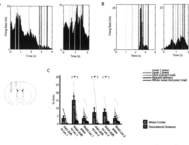
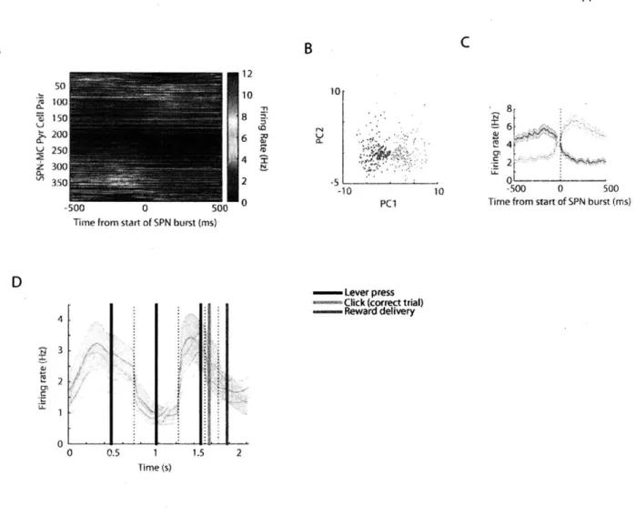
Motor cortex, but not dorsolateral striatum task-related neuronal activity can be accounted for by individual motor actions
The question of how neuronal representations are transformed with each successive node in the cortico-basal ganglia-thalamic circuit is a fundamental one for understanding the mechanisms of function of these circuits. In this study, I recorded from two such nodes, a forelimb area of the motor cortex (MC) and its target striatal projection zone in the dorsolateral striatum (DLS). For each recorded single unit, I assessed the pattern of firing rate modulation in the correct sequence and any incorrect sequences the rat performed within the given training session. Specifically, I addressed whether the neuron responded during lever 1 or lever 2 presses, or during transitions from one of the levers to the other lever, and whether the response to those events was similar whether they occurred at any point during the rewarded sequence or during other sequences of presses (See methods). If the unit did fire similarly in relation to a given lever press regardless of
when it occurred within the rewarded sequence and unrewarded sequences then I could argue that the spiking response of the unit could be accounted for by the occurrence of the given motor action alone. Many units in motor cortex did fit this criterion - one such example motor related unit is shown in Fig. 2A. However, a majority of the units in dorsolateral striatum, did not fulfill this criterion: they were more strongly modulated during the same motor events during some sequences than others. Two examples of such striatal neurons are shown in Fig. 2B and C. I assessed the relative distribution for such simple motor units in motor cortex and DLS by identifying sessions in which I had simultaneously recorded at least 10 putative motor cortex pyramidal neurons and 10 DLS spiny projection neurons (SPNs). I found that in across all the motor timepoints I analyzed (the start of the shift from lever 1 to lever 2, the end of the shift from lever 2 to lever 1, the completion of the resulting lever 1 press, and corresponding events for the transition from lever 1 to lever 2) motor cortex neurons were significantly more likely to fit the simple motor unit criterion than were the DLS units (p < 0.001, Mann-Whitney test) (Fig.
1D). This suggests different levels of task representations in motor cortex and striatum where
motor cortex neurons are more likely to directly represent movement parameters, whereas DLS neurons, although in the sensorimotor region of striatum, are more likely to modulate their responses based on the context of the behavior.
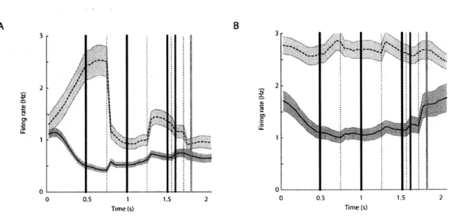
Dorsolateral striatum SPN firing reflects the boundaries of the learned action sequence across a variety of different learned lever press sequences
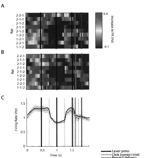
Given the minority of simple motor neurons in the DLS, I considered alternative accounts for the task-related firing rate modulations in DLS. One observation that became clear after examination of a large number of single unit response profiles was that there were neurons that responded in a similar manner across rats each of whom had learned a different lever press sequence. Most apparent were neurons that were highly and phasically activated around the time of the first lever press in the learned sequence, around the time of the last lever press in the learned sequence, or both. These neurons were present in rats which learned different sequences of lever presses (Fig. 3A).
r I ~ 0 1 2 Time (s) 35 B I cc C
J
(1 Time (s) 51 0 1 2 s 3 Time (s) 30 [*- F -1 25 20 0 Lever I press Lever 2 pressClick (correct trial) Reward delivery
White noise (incorrect trial)
Motor Cortex Dorsolateral Striatum
Figure 2. Motor-type neurons are prevalent in forelimb motor cortex but not dorsolateral striatum.
(A) An example of a motor type neuron recorded from motor cortex. Peri-event histograms for each of the events in the trial were pasted together with window sizes according to the median time between each successive event. Event markers are indicated with colored lines and the pasted window edges are indicated with dashed lines. This unit fired during lever 1 presses both in the correct sequence and during repetitive lever 1 pressing.
(B) An example of a non-motor type neuron in dorsolateral striatum. This neuron responded selectively during the initiation of the correct sequence, but not during the same lever press during repeated presses of the lever.
(C) The proportions of motor-type neurons in n = 14 sessions with 10 or more neurons recorded simultaneously in both motor cortex and DLS. Error bars indicate SEM. Asterisks indicate significant difference between motor cortex and DLS cell proportions. A cc C C 4
06
),' t I Time (s) 2A
3 35
c30 1220
0 1 2 3 4 5 0 1 2 3 4 5 0 1 2 3 4 5 6 7 8
Time (s) Time (s) Time (S)
C Correct
B
0 1 2 3 4 0 1 2
Time s) T im (s)
Time (s) Tirme (s)
Incorrect non-repeat Incorrect repeat press1 Incorrect repeat press 2
> -1. 2 J. 5~ 00 0 D 1.)-I I-. (tiC CC 0 C C ~t~ N. I C _____ ____- ___________ 1 _____ _____
LI
__ ~C 05 1 1% 0 C E I 4 /j. uF_{4.
_
5
3m (s)tre s 0 0.5 1 1.5 Time (s) lime "s) Lever 1 press Lever 2 press Click (correct trial) Reward delivery White noise (incorrect trial) - Lever press (either I or 2)Figure 3.
e
2 1 0 0.5 r- ** -Ir-**-7S
.5 2 C 0 5Figure 3. DLS SPN population spiking is concentrated around the initiation of the learned lever press sequence but not during incorrect lever press sequences, regardless of the lever press sequence learned.
(A) Examples of beginning-type (top tow) and end-type (bottow row) neurons in rats who learned different lever press sequences (1-1-2 first column, 1-2-2 second column, 2-1-2 third column).
(B) Top row, an example of a beginning-type neuron which fired during the first lever 1 press in the correct sequence, but not during the middle lever 1 press. This neuron did not respond during incorrect trials with repetitive lever 1 pressing. Bottom row, an example of an end-type neurons with fired during the last lever 2 press in the correct sequence but not during the middle lever 2 press during the correct sequence. This neuron responded weakly during repetitive lever 2 pressing.
(C) Average peri-event DLS SPN spiking during correct (first column), non-repeat incorrect (second column), lever 1 repeat (third column), and lever 2 repeat (fourth column) in 9 implanted rats who learned different lever press sequences indicated on the left.
(D) Average peri-event DLS SPN spiking across all rats during correct sequence performance (first column, n = 2501 putative SPNs), and non-repeat incorrect trials (second column, same n = 2501 putative SPNs), incorrect lever 1 repeat press trials (third column, n = 1143 SPNs), and incorrect lever 2 repeat press trials (fourth column, n = 1338 SPNs).
(E) Sub-groups of beginning-type (blue) and end-type (red) responsive SPNs in the same trial types. On right, the proportion of total beginning and end-type neuron sub-types and their overlap.
All error bars indicate SEM
Commonly these beginning or end neurons which fired in relation to the first or last lever press in the learned sequence, did not fire when the same lever was pressed in the middle of the trial. Similarly, when the same lever was pressed repetitively in an incorrect trial, these neurons were often not modulated or weakly modulated (Fig. 3B).
As a result of these strongly modulated beginning and end neurons the population firing pattern across nine rats, each of whom learned a single sequence, was concentrated around the time of the first lever press and around the time of the last lever press (Fig. 3C, first column). The population of all the recorded DLS SPNs increased firing rates from a baseline inter-trial interval rate of 0.4Hz to 1.3-1.4Hz during the first and last presses in the learned lever press sequence while the average firing rate during the middle lever press was 0.8Hz (p < 0.01 for first v. second
press, and second v. third press, Mann-Whitney tests) (Fig. 3C, first column). Although these rats learned different lever press sequences and developed individualized movement patterns in order to correctly complete the rewarded sequence, the population of SPNs accentuated the task-boundaries of the learned sequence across animals.
This task-boundary activity developed quickly, as soon as there were a minimum of 30 correct trials within training sessions and continued throughout training (Fig. Si), similarly to the early development of the beginning and end pattern found in the T-maze task (Barnes et al., 2005, Thorn et al., 2010). In addition to findings that this task-boundary activity occurred in rats each of whom learned different stereotyped movement sequences and in the T-maze task which required locomotion behavior distinct from the movements used during the lever press task; I found that overall speed of the animal was unlikely to account for the task-boundary activation due to the decrease of speed that occurred prior to each time the animal pressed a lever (Fig.
S2).
The timing of the peak population spiking varied across rats. This variability could have been produced by a multitude of factors, including the subsets of sampled neurons as the timing of the bursts of firing varied across single units within rats. How the learned sequence was encoded or the perceptual start point of the chunked action sequence could also vary across rats and be reflected in the variability in the spiking. However, in each of the rats, the DLS neuronal population was more active around the time of the first and last lever presses than around the time of the middle lever press, regardless of whether the middle lever press was a lever 1 or lever 2 press, or whether it matched the identity of the first or last lever in the sequence.
By contrast, these same units had a remarkably different pattern of firing when the animals were performing incorrect sequences within the same recording session. The beginning and end activity was absent across nine rats in cases when they were performing an alternative non-repeat sequence (Fig. 3C and D, second column) and in cases when they were non-repeatedly pressing lever 1 or lever 2 (Fig. 3C and D, third and fourth column). When the rats were pressing the levers in a sequence that was different from the rewarded lever press sequence, the population firing