doi: 10.1093/intimm/dxh098 Advance Access publication May 17, 2004
A chimeric T cell receptor with
super-signaling properties
Troels R. Petersen, Sven GuÈlland, Estelle Bettelli
1, Vijay Kuchroo
1, Ed Palmer
2and B. Thomas BaÈckstroÈm
Malaghan Institute of Medical Research, Wellington, New Zealand
1Center for Neurologic Diseases, Brigham and Women's Hospital, Harvard Medical School, Boston, MA, USA 2Laboratory of Transplantation Immunology and Nephrology, Department of Research, University Hospital
Basel, CH-4031 Basel, Switzerland
Keywords: antigen signaling, structure±function, T cell receptor Abstract
A key question yet to be resolved concerns the structure and function relationship of the TCR complex. How does antigen recognition by the TCR-ab chains result in the activation of distinct signal transduction pathways by the CD3-gde/z complex? To investigate which part of the TCR-b chain is involved in TCR signaling, we exchanged different domains of the constant regions of the TCR-b chain with the corresponding TCR-g chain domains. We show here that hybridoma cells expressing a chimeric TCR-b chain (bIII) containing intracellular and transmembrane TCR-g amino acids, together with a wild-type TCR-a (awt) chain, were 10 times more sensitive to antigenic stimulation compared to cells expressing TCR-awt/bwt chains. This super-signaling phenotype of the bIII chain was observed in two different TCRs. One speci®c for an alloantigen (I-Abm12) and
one for an autoantigen (I-Ab/MOG
35±55). We found that this chimeric awt/bIII TCR had normal
association with CD3-gde and z chains. To investigate the effect of the chimeric bIII chain in transgenic T cells, we made MOG35±55-speci®c TCR transgenic mice expressing either the awt/bwt
or chimeric awt/bIII TCR. Similar to what was observed in hybridoma cells, transgenic awt/bIII T cells showed a super-signaling phenotype upon antigenic stimulation. Further studies may help us understand the effect of increased TCR signaling on autoimmunity and may lead to the
identi®cation of signaling molecules that can be targeted to stop the progression of autoimmune disorders such as multiple sclerosis.
Introduction
Although the structure and function relationship of the T cell receptor (TCR) has been investigated for more than two decades, it remains unknown which of the extracellular, transmembrane or intracellular domains of the constant region are responsible for interactions with the different components of the TCR complex. More importantly, it is also not known which of these domains are responsible for TCR-induced proliferation, apoptosis and cytokine production.
T cells express a TCR consisting of clonotypic ab- or gd-heterodimers associated with the CD3-gde and z chain homodimers. The ab and gd TCRs show sequence homology but may still have different structural properties. For example, a CD3-e epitope detected with the mAb WT31 in the ab TCR complex is masked in T cells expressing the gd TCR (1,2). In addition, unlike the ab TCR, the gd TCR is predominantly expressed without CD3-d (3), and CD3-d de®cient mice lack
ab- but not gd-expressing T cells (4). These examples indicate that the gd TCR can be expressed at the cell surface in the absence of the CD3-d chain, and that the structural require-ment for cell surface expression may differ between ab- and gd-expressing T-cell subsets.
While the variable region of the TCR-ab and -gd chains is responsible for antigen recognition, intracellular immuno-receptor tyrosine-based activation motifs (ITAMs) in the CD3-gde and z chains are essential for TCR signaling. However, it is still not certain which domains in the constant regions of the TCR-ab chains are involved in signal trans-mission, or how the TCR links antigen recognition to ITAM phosphorylation and downstream signaling. Interestingly, structural differences between ab and gd T cells impact on receptor signaling and ab/gd lineage determination (5). Some evidence suggests that gd T cells may induce different
Correspondence to: B. T. BaÈckstroÈm; Email: [email protected]
downstream signaling events to ab T cells. For example, gd-expressing T cells do not depend on CD4 or CD8 for activation, and have different requirements for PLC-g1 and protein tyrosine kinase activation of the guanine nucleotide-exchange factor Vav (6±9).
To better understand the signaling capabilities of the TCR, it is important to study domains in the TCR-ab that are not directly involved in antigen recognition, but may in¯uence the association with the CD3-gde and z chains. To do this, we took advantage of the similarities and differences between the ab and gd TCR constant regions by making chimeric a and b chains containing different lengths of TCR-d and g chain amino acids, respectively. By studying some of these chimeric TCR chains, we have previously identi®ed a motif in the connecting peptide domain of the TCR-a chain (aCPM), and shown that it is involved in antigen-signaling and association with the CD3-d chain (10,11). Interestingly, disruption of aCPM affects posi-tive but not negaposi-tive selection of thymocytes (10,11), indicat-ing that the different domains of the TCR carry out specialized functions. Furthermore, TCR-b connecting peptide domain is also involved in signaling, since hybridoma cells possessing mutations within this domain are unresponsive to superantigen stimulation. In spite of the pronounced signaling defect found, the interactions of the chimeric TCR-b chain with the CD3-gde and z chains are preserved (11,12).
The T- and B-cell receptors contain a conserved antigen receptor transmembrane (CART) motif, suggesting an import-ant role for this domain in import-antigen receptor assembly and function (13). To study the role of the TCR-b chain transmem-brane domain on TCR structure and function, we utilized a previously generated chimeric TCR-b chain (bIII) which contains TCR-g amino acids downstream of the conserved transmembrane Y145 residue (11). We demonstrate that T
hybridoma and transgenic T cells expressing the chimeric TCR-bIII chain show super-signaling properties upon stimula-tion through the TCR with antigen, but not upon CD3 cross-linking. As the association with the CD3-gde and z chains is intact, the super-signaling TCR-bIII complex appears to have normal structure but altered function.
Methods Cell lines
All cell lines were grown in IMDM supplemented with 5% FCS, 2 mM glutamine, 100 U/ml penicillin G, 100 mg/ml streptomycin sulphate and 5 3 10±5M 2-mercaptoethanol (all from Gibco
BRL, Auckland, New Zealand), referred to as complete medium unless otherwise stated. The human B cell line LG-2 (14) expressing MHC class II DR1 molecule was utilized to present the superantigen staphylococcal enterotoxin B (SEB) to T hybridoma cells. The 58a±b±and 58hCD4a±b±have been
previously described (11). The 58a±b± and 58hCD4 are T
hybridoma cell lines which do not express TCR-ab, and in addition 58hCD4 has been transfected with the human CD4 protein. The Phoenix Eco packaging cell line (a gift from Garry Nolan, Stanford University School of Medicine, CA) was cultured in complete medium containing 10% FCS. The indicator cell line HT-2 (15) was grown in complete medium with the addition of 20 U/ml IL-2.
Generation of DNA constructs and myelin oligodendrocyte glycoprotein (MOG) peptide 35±55-speci®c transgenic mice The Va2.1 and Vb8.1 TCR cDNAs were isolated from the T-cell hybridoma 3BBM74 as previously described (11). They confer reactivity to the I-Abm12alloantigen and the SEB superantigen.
Cloning of the MOG35±55-speci®c T-cell receptor 2D2 (Va3.2
and Vb11) has previously been described (16). For construc-tion of wild-type and chimeric TCR tg mice, the VJ segments of the 2D2 a and VDJ of the b chain were ampli®ed by PCR using 2D2 cDNA as template and the following primers: Va-5¢: GATCGAATTCGTCGACATGCTCCTGGCGCTCC, Va-3¢: GT-TCTGGGTTCTGGATGTTGGGCTTGATAGATAACTTG, Vb-5¢: GATCGAATTCGTCGACATGGCCCCCAGGCTCCTT, Vb-3¢: GTCACATTTCTCAGATCCTCTACAACTGTGAGTCTGG. The constant a and b chains were ampli®ed by PCR from LXSN-Va2.1, LXSP-Vb8.1 and LXSP-Vb8.1-bIII (11) as templates for the constant a, constant b and constant bIII (chimeric b/g) chains respectively. The following primers were used: Ca-5¢: CAAGTTATCTATCAAGCCCAACATCCAGAACCCAGAAC, Cb-5¢: CCAGACTCACAGTTGTAGAGGATCTGAGAAATGT-GAC, LXSN/P-3¢: GGGCGGGACTATGGTT. The variable and constant chain PCR products were sewn together in a PCR using either the Va or Vb upstream primer with LXSN/P-3¢ and the resulting PCR products were TA cloned in pCR2.1 (Invitrogen, Carlsbad, CA) in accordance with the manufac-turer's recommendations. The cloned PCR products were sequenced, excised from the cloning vector with SalI/BamHI, and cloned into the SalI/BamHI sites of the pHSE3¢ vector (17). For construction of transgenic mice, the DNA fragment coding for the TCR chains were excised from pHSE3¢ with XhoI, gel puri®ed, and microinjected into C57BL/6J oocytes as previ-ously described (16).
Retroviral infection of 58hCD4 cells
The generation of virus-containing supernatants by transfec-tion of Phoenix cells and retroviral infectransfec-tion of hybridoma cells was done as previously described (11). Brie¯y, 2.5 3 105
Phoenix cells/well were seeded in 6-well plates and trans-fected with 2 mg of retroviral vector the day after using 6 ml of Fugene6 (Roche Diagnostics, Auckland, NZ) according to the manufacturer's recommendations. The supernatant, 2 ml, containing the retroviral particles was supplemented with 10 mg/ml of polybrene (Sigma, Global Science, Auckland, New Zealand) and added to 2.5 3 105 hybridoma cells. For
selection of positive cells, LXSP-infected cells were added 3 mg/ml of Puromycin (Sigma), and LXSN infected cells 1 mg/ml G418 (Merck, Palmerston North, New Zealand). The selective drug was added to the cell culture 24 h after infection. Bulk-infected cells were sorted by ¯ow cytometry for equal expression of the TCRs and human CD4 chains and then used in indicated experiments.
Electroporation of 58a±b±T hybridoma cells
For expression of 2D2 TCR and murine CD4 in 58a±b± T
hybridoma cells, pcDNA3/neo (for G418 selection) and pcDNA/L3T4 (murine CD4) were linearized with PvuI and ScaI respectively, while pHSE3¢/2D2awt, pHSE3¢/2D2bwt and pHSE3¢/2D2bIII were linearized with XhoI. The linearized DNA was precipitated and dissolved in PBS. For electroporation,
5 3 10658a±b±T hybridoma cells were electroporated in 200 ml
PBS containing 2 mg pcDNA3/neo, 4 mg pcDNA3/L3T4, 10 mg pHSE3¢/2D2awt and 10 mg of either pHSE3¢/2D2bWT or pHSE3¢/2D2bIII at 960 mF and 0.25 kV using a Bio-Rad Gene Pulser (Bio-Rad, San Diego, CA). Surviving cells were cultured for 24 h in complete medium after which they were seeded at 5 3 103 cells/well in 96-well plates in complete medium
containing 1 mg/ml of G418. Surviving clones were screened for Va3.2, Vb11 and mCD4 expression using ¯ow cytometry. One 2D2 wt and one 2D2 bIII expressing clone was found to respond to MOG35±55 and therefore selected for this study.
These clones were stained with anti CD4/PE and anti TCR-b/ FITC and sorted for equal expression of TCR and mCD4, using a FACSVantage Flow Cytometry System (Becton Dickinson). Antibodies, peptides and superantigen
The MOG35±55 peptide was synthesized by Mimotopes
(Clayton, Australia) and was more than 90% pure, con®rmed by HPLC. The peptide sequence for mouse MOG35±55 is
MEVGWYRSPFSRVVHLYRNGK. SEB was purchased from Toxin Technology, Inc. (Sarasota, FL). The antibodies B20.1 (anti Va2.1), RR3-16 (anti Va3.2), H28±710 (anti TCR-a constant region), F23.1 (anti Vb8), RR3-15 (anti Vb11), H57± 597 (anti TCR-b constant region), 145±2c11 (anti CD3-e), H129.19 (anti mouse CD4), RPA-T4 (anti human CD4), or H146±968A (anti z) were used. All mAbs were either pur-chased from BD PharMingen (San Diego, CA) or puri®ed from culture supernatants. Goat anti-rabbit-HRP (Southern Biotechnology Associates, AL) and goat anti-Armenian hamster-HRP (Jackson ImmunoResearch Laboratories, Inc.) were used as secondary antibodies for their respective primary antibodies.
Hybridoma stimulation assay
For hybridoma stimulation assays, 5 3 104/well hybridoma
cells were seeded in 96-well plates. Hybridoma cells were stimulation with indicated concentration of the MOG35±55
peptide and 5 3 105/well irradiated (2000 Rad) C57BL/6J
splenocytes. For stimulation with SEB, 2 3 104/well LG-2 cells
and SEB at indicated concentrations were added. The cultures were incubated for 24 h, after which culture super-natants were harvested. To quantify the concentration of IL-2 in the culture supernatants, 5 3 103HT-2 cells in 100 ml were
cultured with 100 ml culture supernatant for 24 h and 0.5 mCi/ml of [3H]thymidine added for the ®nal 6 h of incubation. The
amount of IL-2 was expressed as the HT-2 stimulation index (SI) (proliferation of HT-2 cells with culture supernatant from stimulated cells/proliferation of HT-2 cells with culture super-natant from unstimulated cells). Alternatively, recombinant IL-2 was added as a standard to calculate total units of IL-2. T-cell stimulation and proliferation assay
Splenocytes, 4 3 106cells/well in 24-well plates, from
wild-type and bIII 2D2 mice were stimulated with 10 mg/ml MOG35±55, and IL-2 (50 U/ml) added on day 2, 5 and 8. On
day 9, cultures were washed twice and 3 3 104T cells/96-well
mixed with 5 3 105irradiated splenocytes and antigen. The
T-cell cultures were incubated for 72 h and 0.5 mCi/ml of [3H]thymidine added for the ®nal 9 h of incubation.
Results
Exchanging transmembrane and intracellular domains of TCR-bfor TCR-ggenerates a super-signaling TCR
To identify domains in the constant region of the TCR-b chain important for TCR signal transduction, we have previously made a number of chimeric TCR-b chains by replacing domains of the b-chain constant region with homologous domains of the TCR-g chain. We have previously found that T cells expressing chimeric TCR-b chains containing TCR-g-derived amino acids in the TCR-b connecting peptide domain are defective in response to superantigen. Point mutations revealed that a single amino acid residue, Q136, located within
the connecting peptide domain of the TCR-b chain controls the ability of the ab TCR to transmit a full signal (12). In addition, we have shown that the short intracellular domain of the TCR-a chain, but not the b-chain, is important for TCR downregulation but not antigen signaling (18). In this paper, we study the chimeric TCR-b/g chain, bIII, containing TCR-b-derived sequences up to and including the conserved transmembrane tyrosine at position 145, followed by the TCR-g sequence encoding the remaining transmembrane and intracellular domains (Fig. 1) (11). The chimeric bIII chain was ®rst introduced in the 3BBM74 TCR, which confers reactivity to the I-Abm12 alloantigen and SEB (19). Cells were sorted to
achieve comparable TCR expression level of hybridoma cells possessing wild-type or chimeric bIII chains. Sorted wild-type and chimeric bIII chain expressing cells stained with TCR-a, TCR-b or CD3-e speci®c mAbs showed similar levels of cell surface expression (data not shown). Upon stimulation with SEB, T-cell hybridoma cells expressing TCR-awt with chimeric TCR-bIII responded to almost 10-fold lower concentrations of SEB compared to hybridoma cells expressing TCR-awt/bwt (Fig. 2A). Interestingly, upon cross-linking the TCR with plate-bound anti-CD3 mAb, cells expressing the bwt or chimeric bIII chains responded similarly (Fig. 2B). Thus, hybridoma cells with the chimeric TCR-bIII chain are hyper-responsive to antigenic but not cross-linking signals, indicating that these stimuli activate distinct signaling pathways.
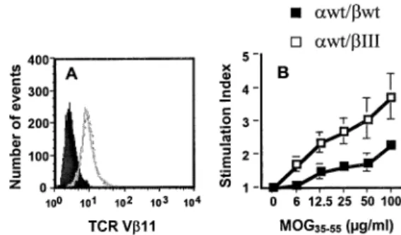
To further investigate whether this chimeric TCR also shows super-signaling properties to an autoantigen, we introduced the chimeric bIII chain into the TCR-b chain of the 2D2 TCR (Va3.2/Vb11), which speci®cally recognizes the CNS-derived MOG35±55 peptide (16). We then electroporated 58a±b±
T hybridoma cells with CD4, 2D2 awt/bwt or 2D2 awt/bIII TCRs and selected for drug-resistant clones. We sorted the hybridoma cells for equal numbers of surface-expressed 2D2 awt/bwt or 2D2 awt/bIII TCR and CD4 (Fig. 3A, and data not shown). When these cells were tested for responsiveness to MOG35±55, hybridoma cells expressing the chimeric bIII TCR
responded better to antigenic challenge, and thus showed a super-signaling phenotype compared to hybridoma cells expressing 2D2 awt/bwt TCR (Fig. 3B).
Similar structure ofawt/bwt andawt/bIII TCRs
We have previously shown that the awt/bIII TCR associates normally with the CD3-gde and z chains at the cell surface, using biotinylation and immunoprecipitation techniques (11). Furthermore, we immunoprecipitated the 2D2 awt/bwt or awt/ bIII TCR with an antibody speci®c for TCR-b and subsequently
probed western blots with antibodies speci®c for the TCR-a, CD3-g, -d, -e, or z chains and found no differences in the assembly of TCR-ab chains with the TCR complex (data not shown).
Super-signaling MOG35±55-speci®c TCR transgenic T cells
In order to evaluate the super-signaling chimeric bIII TCR in a more physiological system, we generated TCR transgenic mice expressing either the bwt or chimeric bIII chain together with the wild-type 2D2 Va3.2 chain. We stimulated spleno-cytes from 2D2 TCR transgenic mice with MOG35±55and then
expanded the T cells with IL-2 for 9 days. Naive splenocytes from both 2D2 awt/bwt and awt/bIII mice, and cells cultured for 9 days with IL-2, expressed similar levels of the Va3.2, Vb11, CD3e and CD4 molecules at the cell surface (Fig. 4A, and data not shown). These short-term T-cell lines were then tested for proliferative responses to antigen and superantigen stimulation. We found that the T cells expressing the awt/bIII TCR proliferated better in response to MOG35±55(Fig. 4B) and
SEB (Fig. 4C), compared to T cells carrying the awt/bwt TCR. These results indicate that the chimeric bIII chain renders T cells hyper-responsive to antigenic signals delivered through the TCR-ab chains, and that the phenotype is not due to increased TCR expression.
Discussion
The fact that antigen stimulation through the same TCR-ab chains can be translated into a number of different effector functions makes the TCR complex a fascinating cell surface receptor to study. To determine whether the different domains
Fig. 3. Response of awt/bwt or awt/bIII chimeric MOG35±55-speci®c TCR to MOG35±55. The 58a±b± hybridoma cell line was electroporated with mouse CD4, 2D2-awt/bwt or awt/bIII TCRs. Antigen-speci®c clones, one of each, were selected and sorted by ¯ow cytometry. TCR expression levels of 2D2 awt/bwt (thick line) or awt/bIII (broken line) cells, or no antibody (solid area) were determined using an antibody speci®c for TCR-Vb11 (A). Clones were stimulated with irradiated splenocytes and MOG35±55. Supernatants from quadruplicate cultures of each antigen concentration were assayed for IL-2 concentration using the HT-2 bioassay (B). The y-axis represents the stimulation index of HT-2 cells (CPM with antigen/CPM with no antigen). One representative experiment out of three is shown.
Fig. 4. Proliferative response of 2D2 transgenic T cells to MOG35±55 and SEB. Splenocytes from 2D2 awt/bwt or awt/bIII TCR transgenic mice were stimulated with MOG35±55 and expanded with IL-2 for 9 days. TCR-Vb11 expression of 2D2 awt/bwt (thick line) or awt/bIII (broken line) cells, or no antibody (solid area) after 9 days is displayed (A). Similar results were found using anti-TCR-Va3.2 or CD3-e mAbs (data not shown). Proliferation of 2D2 awt/bwt or awt/ bIII T cells in response to MOG35±55 (B) and SEB (C) is depicted. One representative experiment out of three is shown.
Fig. 1. Amino acid sequence of TCR-b, TCR-g or chimeric TCR-b/g (bIII) chains. The C-terminal sequences of the TCR-b, TCR-bIII and TCR-g chains are shown using the single-letter amino acid code. Only the sequence of the connecting peptide, transmembrane, and intracellular domains are shown. The box indicates the sequence derived from TCR-g. For more information see (11).
Fig. 2. Response of awt/bwt or awt/bIII I-Abm12-speci®c TCR to SEB
and anti CD3. The 58hCD4 hybridoma cell line was infected with I-Abm12-speci®c awt/bwt or awt/bIII TCRs, sorted for similar TCR and
CD4 expression, and then stimulated either with LG-2 cells and SEB (A), or plate-bound anti CD3e mAb (B). Supernatants from quadruplicate cultures of each antigen concentration were assayed for IL-2 concentration using the HT-2 bioassay. One representative experiment out of four is shown.
of the TCR chain constant regions may be involved in different effector functions, we have replaced part of the extracellular connecting peptide, transmembrane and intracellular domains of the TCR-ab with the corresponding domains of the TCR-gd chains. In this way, we have previously found that the connecting peptide domains of the TCR-a and -b chains control antigen responsiveness (11,12).
Here, we further analyze the role of the chimeric TCR-bIII chain, which contains TCR-g amino acids downstream of the conserved transmembrane Y145 residue. Introducing the
chimeric bIII TCR together with the wild-type TCR-a chain renders hybridoma cells and transgenic T cells ~10-fold more sensitive to antigen stimulation (MOG35±55and SEB), but not to
stimulation by anti-CD3 cross-linking. We have previously shown that the awt/bIII TCR associates with the CD3-gde and z chains at the cell surface, using biotinylation and immuno-precipitation techniques [(11) and data not shown]. Thus, the super-signaling phenotype of the chimeric bIII TCR is not due to a change in the assembly of the TCR with CD3-gde or z chains or higher surface expression of the chimeric TCR (Figs 3A and 4A). The phenotype of the chimeric bIII chain is most likely mediated by changes in the transmembrane domain of the TCR-b chain, since we have previously dem-onstrated that the short intracellular domain of the TCR-b chain is dispensable for TCR signaling (18).
The transmembrane domain of the TCR-b chain has previ-ously been implicated to be involved in TCR signaling. By introducing point mutations, it has been revealed that TCR-b transmembrane residues contribute towards both T-cell acti-vation and apoptosis (20,21). Mutating one or both of the two conserved tyrosines (Y145and/or Y155) to phenylalanine in the
transmembrane region of the b chain causes a marked reduction in antigen responsiveness in mouse hybridoma cells. Likewise, mutating the conserved transmembrane C-terminal tyrosine (Y155) to leucine in Jurkat cells affects the
association of the z chain with the TCR complex. Interestingly, these Jurkat cells show normal IL-2 secretion, but reduced apoptosis and FasL upregulation in response to anti-CD3 stimulation (22±24). However, the super-signaling phenotype of the chimeric TCR-bIII might be due to a different mechan-ism, since both the TCR-b and TCR-g chains possess the transmembrane Y145 and Y155 residues (11). In addition, a
chimeric TCR-b chain, bII, containing TCR-g-derived amino acids from the conserved transmembrane residue K151and
downstream paired with the wild-type TCR-a chain does not show any super-signaling properties [(11) and data not shown]. The only difference between these two chimeric TCR-b chains is between positions 146±150 in the transmem-brane domain, where bII encodes the TCR-b-derived amino acids EILLG, and bIII the TCR-g derived amino acids LLLLL. Thus, the super-signaling phenotype is most likely attributed to the replacement of the conserved E146and G150amino acids
in the TCR-b chain to L146and L150.
Transmembrane domains often possess an a-helix structure with every three/four amino acids ending up in the same vertical plane (13). The spacing between E146and G150is four
amino acids and they are therefore likely to face the same side. BaÈckstroÈm et al. (11) have previously reported that, whereas single point mutations within the aCPM do not affect TCR signaling, substituting three amino acid residues within
the same motif renders the resulting TCR non-functional. This indicates that small changes are tolerated without affecting the function of the TCR. It is possible that the E146 and G150
residues of the TCR-b chain are part of an important TCR-b chain transmembrane motif and interact directly with the CD3/ z complex and that the interface is made up by a number of additional amino acids, all of which contribute to the inter-action. Substituting E146 and G150 with the corresponding
TCR-g leucine residues in the bIII chimera could therefore be structurally tolerated, but with altered TCR signaling. On the other hand, the corresponding TCR-g leucine residues, in conjunction with residues found further downstream, might be part of an important motif in the gd TCR complex which makes the bIII chain interact more ef®ciently in comparison to bII (or the wild-type TCR-b chain) and thus renders this TCR hyper-active. Another possibility is that the TCR-g chain leucine residues are less ef®cient, in comparison to the wild-type TCR-b chain, in interacting with a transmembrane-resident molecule involved in TCR-induced inhibition, and therefore cells expressing the bIII TCR show a ®ctional hyper-proliferative response.
In summary, the bIII chain generates a TCR with an apparent super-signaling phenotype which is not limited to one TCR, since both alloantigen (I-Abm12) and autoantigen-speci®c
(MOG35±55) TCRs expressing this chimeric bIII chain are
hyper-responsive upon antigen stimulation (Figs 2 and 3). We are currently in the process of investigating how the chimeric bIII chain affects downstream signaling pathways. In addition, the super-signaling MOG35±55-speci®c C57BL/6J TCR
trans-genic mice may represent a novel model for studying how changes in TCR signaling affect the development of an autoimmune disorder, using the experimental autoimmune encephalomyelitis model for human multiple sclerosis. Acknowledgements
We thank colleagues at the Malaghan Institute of Medical Research for critical reading of the manuscript. We are grateful to Robert McGill and Joanna Roberts for the sorting of hybridoma cells. This work was supported by the Marsden Fund administered by the Royal Society of New Zealand and New Zealand Lottery Grants Board. B.T.B. is the recipient of the Wellcome Trust Senior Research Fellowship in Medical Science, New Zealand.
Abbreviations
CART conserved antigen receptor transmembrane CPM connecting peptide motif
ITAM immunoreceptor tyrosine-based activation motif MOG myelin oligodendrocyte glycoprotein
SEB Staphylococcal enterotoxin B TCR T-cell receptor
References
1 van de Griend, R. J., Borst, J., Tax, W. J. and Bolhuis, R. L. 1988. Functional reactivity of WT31 monoclonal antibody with T cell receptor-gamma expressing CD3+4±8± T cells. J. Immunol.
140:1107.
2 Salmeron, A., Sanchez-Madrid, F., Ursa, M. A., Fresno, M. and Alarcon, B. 1991. A conformational epitope expressed upon association of CD3-epsilon with either CD3-delta or CD3-gamma is the main target for recognition by anti-CD3 monoclonal antibodies. J. Immunol. 147:3047.
3 Hayes, S. M. and Love, P. E. 2002. Distinct structure and signaling potential of the gamma delta TCR complex. Immunity 16:827. 4 Dave, V. P., Cao, Z., Browne, C., Alarcon, B., Fernandez-Miguel,
G., Lafaille, J., de la Hera, A., Tonegawa, S. and Kappes, D. J. 1997. CD3 delta de®ciency arrests development of the alpha beta but not the gamma delta T cell lineage. EMBO J. 16:1360. 5 Hayes, S. M., Shores, E. W. and Love, P. E. 2003. An architectural
perspective on signaling by the pre-, alphabeta and gammadelta T cell receptors. Immunol. Rev. 191:28.
6 Swat, W., Xavier, R., Mizoguchi, A., Mizoguchi, E., Fredericks, J., Fujikawa, K., Bhan, A. K. and Alt, F. W. 2003. Essential role for Vav1 in activation, but not development, of gammadelta T cells. Int. Immunol. 15:215.
7 Kadlecek, T. A., van Oers, N. S., Lefrancois, L., Olson, S., Finlay, D., Chu, D. H., Connolly, K., Killeen, N. and Weiss, A. 1998. Differential requirements for ZAP-70 in TCR signaling and T cell development. J. Immunol. 161:4688.
8 Hayday, A. C. 2000. gd cells: a right time and a right place for a conserved third way of protection. Annu. Rev. Immunol. 18:975. 9 Aguado, E., Richelme, S., Nunez-Cruz, S., Miazek, A., Mura, A.
M., Richelme, M., Guo, X. J., Sainty, D., He, H. T., Malissen, B. and Malissen, M. 2002. Induction of T helper type 2 immunity by a point mutation in the LAT adaptor. Science 296:2036.
10 BaÈckstroÈm, B. T., Muller, U., Hausmann, B. and Palmer, E. 1998. Positive selection through a motif in the alphabeta T cell receptor. Science 281:835.
11 BaÈckstroÈm, B. T., Milia, E., Peter, A., Jaureguiberry, B., Baldari, C. T. and Palmer, E. 1996. A motif within the T cell receptor alpha chain constant region connecting peptide domain controls antigen responsiveness. Immunity 5:437.
12 BaÈckstroÈm, B. T., Hausmann, B. T. and Palmer, E. 1997. Signaling ef®ciency of the T cell receptor controlled by a single amino acid in the beta chain constant region. J. Exp. Med. 186:1933. 13 Campbell, K. S., Backstrom, B. T., Tiefenthaler, G. and Palmer, E.
1994. CART: a conserved antigen receptor transmembrane motif. Semin. Immunol. 6:393.
14 Accolla, R. S. 1984. Analysis of the structural heterogeneity and polymorphism of human Ia antigens. Four distinct subsets of molecules are coexpressed in the Ia pool of both DR1,1 homozygous and DR3,W6 heterozygous B cell lines. J. Exp. Med. 159:378.
15 Watson, J. 1979. Continuous proliferation of murine antigen-speci®c helper T lymphocytes in culture. J. Exp. Med. 150:1510. 16 Bettelli, E., Pagany, M., Weiner, H. L., Linington, C., Sobel, R. A. and Kuchroo, V. K. 2003. Myelin oligodendrocyte glycoprotein-speci®c T cell receptor transgenic mice develop spontaneous autoimmune optic neuritis. J. Exp. Med. 197:1073.
17 Pircher, H., Mak, T. W., Lang, R., Ballhausen, W., Ruedi, E., Hengartner, H., Zinkernagel, R. M. and Burki, K. 1989. T cell tolerance to Mlsa encoded antigens in T cell receptor V beta 8.1 chain transgenic mice. EMBO J. 8:719.
18 BaÈckstroÈm, B. T., Rubin, B., Peter, A., Tiefenthaler, G. and Palmer, E. 1997. T cell receptor alpha-chain tail is required for protein kinase C-mediated down-regulation, but not for signaling. Eur. J. Immunol. 27:1433.
19 DiGiusto, D. L. and Palmer, E. 1994. An analysis of sequence variation in the beta chain framework and complementarity determining regions of an allo-reactive T cell receptor. Mol. Immunol. 31:693.
20 Fuller-Espie, S., Hoffman Towler, P., Wiest, D. L., Tietjen, I. and Spain, L. M. 1998. Transmembrane polar residues of TCR beta chain are required for signal transduction. Int. Immunol. 10:923. 21 Kunjibettu, S., Fuller-Espie, S., Carey, G. B. and Spain, L. M. 2001.
Conserved transmembrane tyrosine residues of the TCR beta chain are required for TCR expression and function in primary T cells and hybridomas. Int. Immunol. 13:211.
22 Rodriguez-Tarduchy, G., Sahuquillo, A. G., Alarcon, B. and Bragado, R. 1996. Apoptosis but not other activation events is inhibited by a mutation in the transmembrane domain of T cell receptor beta that impairs CD3zeta association. J. Biol. Chem. 271:30417.
23 Sahuquillo, A. G., Roumier, A., Teixeiro, E., Bragado, R. and Alarcon, B. 1998. T cell receptor (TCR) engagement in apoptosis-defective, but interleukin 2 (IL-2)-producing, T cells results in impaired ZAP70/CD3-zeta association. J. Exp. Med. 187:1179. 24 Teixeiro, E., Fuentes, P., Galocha, B., Alarcon, B. and Bragado, R.
2002. T cell receptor-mediated signal transduction controlled by the beta chain transmembrane domain: apoptosis-de®cient cells display unbalanced mitogen-activated protein kinases activities upon T cell receptor engagement. J. Biol. Chem. 277:3993.