Université de Montréal
The role of ThPOK and T cell receptor
signaling in CD4
+versus CD8
+T-cell lineage fate
par
Nabil Zeidan
Département de Microbiologie, Infectiologie et Immunologie
Faculté de Médecine
Thèse présentée à la faculté des études supérieures
en vue de l’obtention du grade de Philosophiae Doctor (Ph.D.)
en Microbiologie et Immunologie
Septembre 2019
Résumé
Les lymphocytes T sont au cœur du système immunitaire adaptatif et leur dérégulation est à la base de pathologies. Les cellules T se développent dans le thymus et passent par de nombreuses étapes de maturations identifiables par l'expression des corécepteurs CD4+/CD8+ à
la surface des cellules. À leur sortie du thymus, les cellules T sont divisées en deux sous-types principaux: les cellules T auxiliaires CD4+ spécifique aux antigènes présentés sur complexe
majeur d'histocompatibilité (CMH) de classe II et les cellules T cytotoxiques CD8+
reconnaissant un antigène présenté sur un CMH-I. Toutes les cellules T proviennent d’un précurseur commun. Leur différenciation en cellule T CD4+ et T CD8+ est influencée par
l'intensité et la durée de la signalisation du récepteur des cellules T (RCT) et des cytokines. Cette signalisation résulte en l’expression des facteurs de transcription ThPOK pour la différenciation de cellule T CD4+ et Runx3 pour les cellules T CD8+. Il a été démontré que ThPOK est à la fois
nécessaire et suffisant pour le développement des lymphocytes T CD4+, puisque le gain et la
perte de la fonction de ThPOK favorise le développement de cellules lymphocytes T CD4+ et
CD8+, respectivement. Ma thèse vise à approfondir notre compréhension du choix de la lignée
CD4+/CD8+ en explorant les mécanismes moléculaires de la voix de signalisation de ThPOK et
du RCT.
Dans cette étude, nous avons étudié l'impact d'un gain-de-fonction de ThPOK sur la différenciation des thymocytes, en utilisant trois lignées transgéniques exprimant des niveaux variables de ThPOK. Une analyse approfondie de ces transgènes chez des souris dont le RCT est restreint soit au CMH de classe I ou de classe II, a démontré que, comparés aux thymocytes restreints au CMH-II, les thymocytes restreints au CMH-I requéraient des niveaux plus importants de ThPOK pour se différencier en CD4+. L’introduction d’un transgène exprimant
un niveau moins élevé de ThPOK comparé aux deux autres transgènes, mais un niveau plus élevé de ThPOK par rapport au niveau endogène dans les cellules CD4+ WT, n'induit qu'une
réorientation partielle des cellules T CD8+ en CD4+, ce qui a mené à la génération, à la fois de
lymphocytes T CD4+, DN (doubles négatifs) et CD8+ matures. L'analyse génotypique, plus
précisément celle des cellules DN chez les souris porteuses du transgène ThPOK et dont le RCT est restreint au CMH-I, a révélé que l’inhibition des gènes spécifiques à la lignée CD8+
nécessitait des niveaux d'expression différents de ThPOK comparés à ceux requis pour l’induction des gènes spécifiques à la lignée CD4+. En effet, cette étude nous a permis de
démontrer que l’intensité du signal dérivé du RCT ainsi que sa spécificité pour un CMH donné jouent un rôle essentiel dans le choix de différentiation CD4+/CD8+ induit par ThPOK. Ainsi,
la réorientation CD8+/CD4+ chez les souris exprimant le transgène ThPOK-H est
significativement augmentée par l'amplification de l’intensité du signal dérivé du RCT dans les cellules spécifiques aux CMH-I. De plus, la fréquence des cellules CD4+ était plus élevée
lorsqu’une quantité identique de ThPOK était exprimée dans des lymphocytes T spécifiques au CMH-II, suggérant qu’il existe un aspect qualitatif quant à la régulation de la différenciation des lymphocytes T CD4+ par la signalisation induite par le RCT.
Nous avons également tenté d’étudier la voie de différenciation CD4+ en l’absence de
ThPOK, à la suite de la perturbation physiologique de la voie de signalisation induite par le RCT, par rapport à la perte de fonction de ThPOK. Bien que nous ayons observé une réorientation des thymocytes spécifiques au CMH-II vers la lignée CD8+, aussi bien à la suite
d'une délétion de Thpok, qu’à la perturbation de la signalisation RCT les deux modes de redirections semblent toutefois être différents. En effet, notre investigation a démontré qu’en l’absence de ThPOK, la signalisation induite par le RCT dans les cellules restreintes au CHM-II induit l’activation de certains gènes, suggérant ainsi leur implication dans la voie de différenciation CD4+. Ces résultats suggèrent également que la contribution de la signalisation
du RTC dans la différenciation des thymocytes restreints au CMH-II ne se limitait pas à l'induction de ThPOK. Étonnamment, seul un effet synergique limité a été observé sur la différenciation des thymocytes restreints au CMH-I, lorsque Gata3, un autre facteur de transcription également induit dans les thymocytes restreints au CMH-II, et ThPOK étaient surexprimés en même temps dans ces cellules, suggérant peu de chevauchement fonctionnel entre ces deux facteurs de transcription. L’ensemble de ces résultats indique que ThPOK et la signalisation induite par le RCT fonctionnent en synergie durant le développement des lymphocytes T CD4+.
Mots-clés : ThPOK, lymphocyte T, RCT, choix de la lignée CD4+/CD8+, CMH, thymus,
Abstract
T lymphocytes are at the core of the adaptive immune system and their dysfunction is associated with several disorders and pathologies, which are at times fatal. The two main types of T-cells in mice and man are: the major histocompatibility complex (MHC) class-II-restricted CD4+ helper T-cells, and the MHC-I-restricted CD8+ cytotoxic T-cells. Developmental stages
of the two types of T-cells occurs in the thymus in multiple sequential maturation stages that are identified by cell-surface CD4+/CD8+ co-receptor expression. Differentiation of the two types
of T-cells in the thymus from a common precursor is influenced by the intensity and duration of signals derived from the T-cell receptor (TCR) and cytokines secreted by the thymic stromal cells. These signals lead to the activation of ThPOK or Runx/CBF transcription factors, which control the transcriptional network regulating CD4+ and CD8+ lineage fate, respectively. Studies
have demonstrated that ThPOK is both necessary and sufficient for CD4+ T-cell development
as gain- and loss-of-ThPOK function redirects positively selected MHC-I- and MHC-II-restricted thymocytes into CD4+ and CD8+ T-cell lineage fate, respectively. However, the role
of TCR signaling and the extent to which ThPOK expression influences CD4+ lineage choice
remains to be investigated. My thesis aims to elucidate the fundamental basis the CD4+/CD8+
lineage choice by exploring the molecular mechanism of action of ThPOK and TCR signaling in CD4+ lineage fate of MHC-I- and MHC-II-specific thymocytes.
In this study, we have characterized gain-of-function of ThPOK in three independent transgenic mouse lines expressing varying amounts of ThPOK. Extensive analysis of the three ThPOK transgenic lines expressing MHC-I- and MHC-II-specific monoclonal TCR indicated that MHC-I-restricted, compared to MHC-II-restricted, thymocytes required significantly more ThPOK for efficient differentiation into the CD4+ lineage. Interestingly, the founder line with
the lowest transgene expression, despite expressing significantly higher amounts of ThPOK compared to the endogenous levels in WT CD4+ T cells, induced a partial CD8+ to CD4+
redirection of MHC-I-restricted cells, leading to the generation of mature CD4+, DN and CD8+
T-cells in the same mouse. Lineage specific gene expression analysis, specifically in DN mature T cells from ThPOK transgenic mice expressing MHC-I-specific TCR, showed that, compared to induction of helper program, suppression of cytotoxic program required lower amount of ThPOK. Further investigation showed that TCR signal strength and MHC specificity of
developing thymocytes played a critical role in determining ThPOK-induced CD4+ lineage fate.
While increase in TCR signal strength augmented the efficiency of ThPOK-induced CD4+
lineage choice of MHC-I-restricted thymocytes in part via endogenous ThPOK induction, it appeared to have ThPOK independent function as well as judged by significantly different CD4+
T-cell frequencies in OTI mice expressing the same amount of ThPOK but transduced quantitatively different TCR signal. Importantly, the efficiency of CD4+ lineage choice of
MHC-I-specific thymocytes with augmented TCR signal strength was still significantly lower compared to the efficiency of CD4+ lineage choice of MHC-II-restricted thymocytes expressing
only the transgene-encoded ThPOK suggesting a qualitative role for TCR signaling as well in CD4+ lineage choice.
We then evaluated CD4+ lineage fate decision in the absence of ThPOK induction in
physiologically relevant alteration in TCR signaling versus loss of ThPOK function. While we observed CD4+ to CD8+ lineage redirection of MHC-II-specific thymocytes due to
Thpok-deficiency as well as lack of ThPOK induction due to disruption of TCR signaling, the two modes of lineage redirection appeared to be due to different mechanisms. Our investigation demonstrates that TCR signaling in MHC-II-restricted thymocytes induces the expression of select genes in loss-of-function of ThPOK model suggesting potential role for these genes in establishing the CD4+ helper program. These results also suggest that the contribution of
MHC-II-specific TCR signaling in driving CD4+ lineage choice is not limited to Thpok induction.
Interestingly, only a limited synergistic effect was observed when both Gata3, which is also induced in MHC-II-signaled thymocytes, and ThPOK were overexpressed in MHC-I-restricted thymocytes suggesting a limited functional overlap between the two transcription factors. Collectively, these data indicate that ThPOK and TCR signaling work synergistically to promote the development of CD4+ T-cells with some ThPOK independent function for TCR signaling.
Keywords: ThPOK, TCR signaling, CD4+/CD8+ lineage choice, MHC, thymus, T
Table of contents
Table of Contents
Résumé ... 1 Abstract ... 3 Table of contents ... 5 List of figures ... 10 List of abbreviations ... 12 Acknowledgments... 18 Chapter 1: Introduction ... 19 1.1 Hematopoiesis ... 20 1.1.1 General Overview ... 20 1.1.2 T-cell progenitors ... 22 1.2 T-cell development ... 221.2.1 Early T-cell development ... 24
1.2.1.1 Characteristics of the early T-cell progenitors (ETPs) ... 24
1.2.1.2 Formation and maintenance of the T-cell identity ... 24
1.2.1.3 Transcriptional control of early T-cell development ... 25
1.2.1.3.1 Phase 1 ... 27
1.2.1.3.2 Phase 2 ... 28
1.2.1.3.3 Phase 3 ... 29
1.2.2 Positive/negative thymic selection ... 30
1.2.2.1 Transcription factors regulating positive selection ... 31
1.2.2.1.1 Bcl11b ... 31
1.2.2.1.2 Tox ... 31
1.2.2.1.3 Gata3 ... 31
1.2.2.1.4 Notch signaling ... 32
1.2.2.1.5 Nuclear factor of activated T-cells (NFAT)... 32
1.2.2.2 Transcription factors regulating negative selection ... 32
1.2.2.2.1 Nur77 ... 32
1.2.2.3 Transcription factors regulating positive and negative selection ... 33
1.2.2.3.1 ID3, IRF1 and NF-κB ... 33
1.2.2.3.2 HDAC7 ... 33
1.2.3 CD4+/CD8+ Lineage fate of positively selected thymocytes ... 34
1.2.3.1 Stochastic model of CD4+/CD8+ lineage choice... 35
1.2.3.2 Instructive CD4+/CD8+ lineage choice models ... 36
1.2.3.2.1 Strength-of-signal ... 36
1.2.3.2.2 Duration-of-signal ... 37
1.2.3.3 Kinetic signaling model ... 39
1.2.3.3.1 Cytokine signaling ... 40
1.2.3.3.2 Regulation of Cd4 and Cd8 expression ... 41
1.2.3.3.2.1 Cd4 gene regulation ... 42
1.2.3.3.2.2 Cd8 gene regulation ... 42
1.2.3.3.3 Co-receptor reversal ... 44
1.2.3.3.4 TCR signaling ... 45
1.2.3.4 The network of transcription factors in CD4+/CD8+ lineage fate decision... 45
1.2.3.4.1 Gata3 ... 45
1.2.3.4.2 Tox ... 46
1.2.3.4.3 Bcl11b ... 46
1.2.3.4.4 Runx proteins ... 47
1.2.3.4.5 MAZR ... 47
1.2.4 ThPOK, the master regulator of CD4+/CD8+ lineage choice... 49
1.2.4.1 Study of the helper deficient (HD) mutation and the discovery of ThPOK ... 50
1.2.4.2 General Structure of BTB-POZ domain of ThPOK ... 52
1.2.4.1.1 BTB domain ... 52
1.2.4.1.2 Zinc finger domain ... 53
1.2.4.3 Transcriptional regulation of ThPOK induction in positively selected thymocytes ... 54
1.2.4.4 Upstream regulatory pathway involved in regulating ThPOK expression ... 57
1.2.5 Structural biology of the TCR complex ... 61
1.2.5.1 The αβTCR-CD3 complex ... 61
1.2.5.2 Lck and regulation of TCR signaling ... 63
1.2.5.3 TCR signaling pathway... 65
1.2.5.4 TCR signaling threshold in immature and mature T-cells ... 66
1.2.5.5 MHC class-I- vs MHC class-II-specific TCR signaling ... 67
1.3 Rationale ... 70
1.4 Hypothesis and aims ... 71
Chapter 2: Critical role for TCR signal strength and MHC specificity in ThPOK-induced CD4+ helper lineage choice (Manuscript #1) ... 73
2.1 Résumé ... 74
2.1 Abstract ... 76
2.2 Introduction ... 77
2.3 Materials and methods ... 80
2.3.1 Mice ... 80
2.3.2 Flow cytometry ... 81
2.3.3 Quantitative RT-PCR (QPCR) ... 82
2.3.4 Functional assays ... 83
2.3.5 Luciferase reporter Assay ... 84
2.4 Results ... 85
2.4.1 Characterization of ThPOK transgenic mice ... 85
2.4.2 Impact of ThPOK dose on the CD4+ lineage choice of MHC-I-signaled thymocytes ... 86
2.4.3 Functionality of mature T-cell subsets in OTI+ThPOK-H+ mice ... 90
2.4.4 Role of endogenous ThPOK in the CD8+ to CD4+ lineage redirection in OTI+ ThPOK-H+ mice ... 91
2.4.5 Evaluating role of ThPOK-H in the CD4+ lineage choice of MHC-II-specific thymocytes ... 92
2.4.6 Impact of augmented TCR signal strength on the ThPOK-induced CD4+ lineage choice of MHC-I-signaled thymocytes ... 93
2.4.7 Evaluating contribution of transgenic and endogenous ThPOK in the CD8+ to CD4+
lineage redirection in the presence of augmented TCR signaling ... 95
2.5 Discussion ... 100
2.6 Figures and figure legends ... 105
Chapter 3: Sustained TCR signaling protects the helper fate and suppresses the cytotoxic developmental program independently of ThPOK (manuscript #2) ... 131
3.1 Résumé ... 133
3.2 Abstract ... 134
3.3 Introduction ... 135
3.4 Materials and methods ... 136
3.4.1 Mice ... 136
3.4.2 Flow cytometry ... 137
3.4.3 Quantitative RT-PCR (Q-PCR) ... 137
3.4.4 Functional assays ... 138
3.4.5 Retroviral transduction of peripheral murine T cells ... 138
3.4.6 Statistical analyses ... 139
3.5 Results ... 139
3.5.1 Disruption of TCR signaling affects lineage fate ... 139
3.5.2 MHC-II redirected CD8+ T-cells following disruption in TCR-signals can be rescued by transgenic ThPOK expression ... 140
3.5.3 MHC-II-specific redirected CD8+ T-cells in ThPOK knock out mice show some functional and transcriptional differences compared to genuine MHC-I-specific CD8+ T-cells ... 141
3.5.4 Sustained TCR signaling activates helper program and supresses cytotoxic program independently of ThPOK ... 142
3.5.5 Impact of constitutive Gata3 expression on the ThPOK-induced lineage redirection ... 143
3.6 Discussion ... 145
3.7 Figures and figure legends ... 148
4.1 Role of TCR signaling in ThPOK-mediated CD4+ lineage commitment (manuscript#1)
... 163
4.1.1 The dose dependent effect of ThPOK ... 163
4.1.2 Role of TCR specificity in ThPOK-mediated CD4+ lineage redirection ... 170
4.2 TCR-activated downstream pathways in CD4+ T-cell development (manuscript #2) . 173 4.2.1 CD4+/CD8+ lineage fate is susceptible to changes in selecting ligand density ... 173
4.2.2 Role of TCR signaling on the integrity of the helper phenotype ... 174
Chapter 5: Conclusions ... 177
Chapter 6: Future Directions ... 180
References ... i
List of figures
Figure 1. Schematic representation of hematopoietic lineage differentiation and
specification ... 21
Figure 2. The three different phases of early T-cell development and role of cytokines and transcription factors ... 28
Figure 3. Previous models of CD4/CD8 lineage fate ... 39
Figure 4. The kinetic signaling model... 41
Figure 5. Cd4 and Cd8 gene structure and regulation ... 44
Figure 6. Nuclear proteins and environmental factors that regulate CD4/CD8 lineage choice ... 49
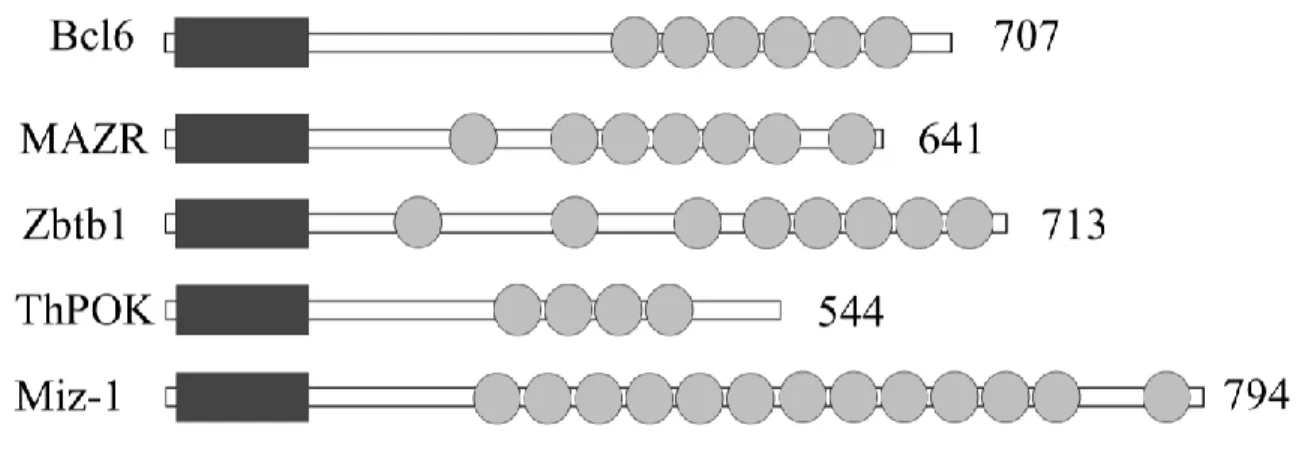
Figure 7. Protein structure of BTB-ZF transcription factors ... 53
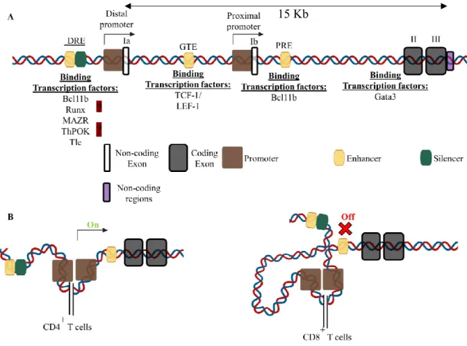
Figure 8. Mouse Thpok gene structure and regulating transcription factors ... 56
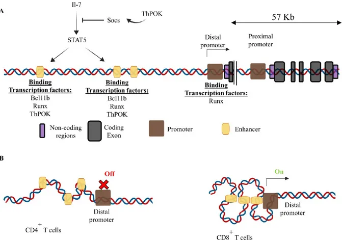
Figure 9. Mouse Runx3 gene structure... 60
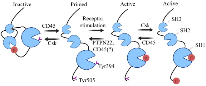
Figure 10. Regulation of Lck function ... 64
Figure 11. Overview of the most important TCR signaling pathways ... 69
Figure 12. ThPOK dose impacts the CD8+ to CD4+ lineage redirection ... 107
Figure 13. ThPOK-induces partial CD8+ to CD4+ lineage redirection in MHC-II-/- mice ... 109
Figure 14. ThPOK modulates lineage specific gene expression in T-cell subsets ... 111
Figure 15. Insignificant contribution of endogenous ThPOK in the CD8+ to CD4+ lineage redirection ... 113
Figure 16. ThPOK-H completely rescues CD4+ development in OTII+Thpok-/- mice. .... ... 115
Figure 17. Augmenting TCR signal strength enhances the CD8+ to CD4+ lineage redirection ... 117
Figure 18. Evaluating contribution of endogenous and transgenic ThPOK in CD4+ lineage choice in OTI+dLGF+ThPOK-H+ mice ... 119
Figure 19. Comparison of CD4+ mature T-cell frequency and TCR signal strength in
ThPOK-Figure 20. Characterization of ThPOK transgenic mice ... 124 Figure 21. Mature DN T-cells in OTI+ThPOK-H+ mice are not innate like T-cells and
transdifferentiate mostly from CD4+ thymocytes ... 126
Figure 22. Impact of ThPOK-H on lineage choice in P14 mice, Nur77 reporter and function of mature T-cells ... 128 Figure 23. The impact of individual ThPOK transgene on CD4+ development in
ThPOK-/- mice, the impact of augmented TCR signal strength on the CD8+ to CD4+ lineage
redirection in OTI+ThPOK-163+ and CD4+ lineage gene expression analysis ... 130
Figure 24. Disruption of MHC-II TCR signaling during lineage commitment impairs CD4+ lineage commitment ... 150
Figure 25. Lineage redirection in MHC-II impaired TCR signaling can be rescued by constitutive expression of transgenic ThPOK ... 152 Figure 26. ThPOK deficiency does not affect class-II TCR signaling in redirected T-cells ... 154
Figure 27. Strong TCR signaling can induce helper function and repress cytotoxic program independently of ThPOK ... 156 Figure 28. Constitutive Gata3 differentially affects the frequency and number of redirected T-cells in OTI+ThPOK-H+ mice ... 158
Figure 29. Enforced GATA3 differentially affects the frequency and number of redirected T-cells in OTI+ThPOK-H+ mice ... 160
Figure 30. ThPOK expression profile in DP thymocytes determines lineage fate .... 166 Figure 31. Dose-dependent gain of function of ThPOK in periphery ... 169 Figure 32. Representative model for the role of TCR specificity in ThPOK- mediated lineage redirection. ... 172
List of abbreviations
γc: Common γ chain A: α-helix
Abs: Antibodies
ADAP: adhesion and degranulation promoting adaptor protein Ade: Adenine
AGM: Aorta-gonad-mesonephros AP-1: Activator protein-1
APC: Antigen presenting cell Arg: Arginine
ATM: Ataxia telangiectasia mutated B: β-strand
BAC: Bacterial artificial chromosome
BACH: Broad-complex, tramtrack and bric-à-brac domain and cap'n'collar homolog BAF: Barrier-to-autointegration factor
Bcl: B cell chronic lymphoma Bcl-XL: B cell lymphoma-2-like 1 BCoR: B cell lymphoma 6 corepressor bHLH: Basic helix-loop-helix
BM: Bone marrow
BTB: Broad-complex, tramtrack and bric-à-brac BTB-ZF: BTB/ Pox virus zinc finger
C terminus: Carboxyl terminus
C/EBPα: Cytosine cytosine adenine adenine thymine (CCAAT)-enhancer-binding proteins α C2H2: Cysteine 2 histidine 2
CARMA1: CARD-containing MAGUK protein 1 Cbf: Core-binding factor
Cbfb: Core-binding factor subunit β CD: Cluster of differentiation
CDC42: Cell division control protein 42 homologue cDNA: Complementary deoxyribonucleic acid CDR1-3: Complementary determining regions (1-3) CCR7: Chemokine cysteine-cysteine motif receptor 7 CCR9: Chemokine cysteine-cysteine motif receptor 9
ChIA-PET: Chromatininteraction analysis by paired-end tag sequencing
ChIP: Chromatin immunoprecipitation CLP: Common lymphoid progenitor CMP: Common myeloid progenitor
CRAC: calcium release-activated calcium channel CSK: C-Terminal sarcoma (Src) Kinase
CTCF: Cytosine cytosine cytosine thymine cytosine (CCCTC)-binding factor cTEC: Cortical thymic epithelial cells
CXCL12: Cytosine-x-cytosine motif chemokine ligand 12 CXCR4: C-X-C motif chemokine receptor type 4
DAG: Diacylglycerol
DHS: Deaxyribonuclease I hypersensitive sites DLL4: Delta-like ligand 4
DC: Dendritic cell
DN: CD4-CD8- double negative
DNA: Deoxyribonucleic acid
dNTP: Deoxynucleoside triphosphate DP: CD4+CD8+ double positive
DRE: Distal regulatory element DUSP: Dual Specificity Phosphatase
E2A: E-box E12/E47-α; also known as Transcription factor 3 (Tcf3) E2F1: E2 transcription factor 1
E8I-V: Enhancer elements (I – V)
ELP: Early lymphoid progenitors ER: Endoplasmic reticulum
ERK: Extracellular signal regulated kinase ETP: Early T-lineage progenitors
Ets: E-twenty-six
FACS: Fluorescence-activated cell sorting FBS: Fetal bovine serum
Flt3: Fms-like tyrosine kinase 3 Flt3L: Flt3 ligand
FOXA: Forkhead box protein A
GADS: Growth factor receptor-bound protein 2-related adaptor downstream of Shc Gata: [Adenine thymine] guanine adenine thymine adenine [adenine guanine]
([AT]GATA[AG]) binding protein
GFI1: Growth factor independent 1 transcriptional repressor GFP: Green fluorescent protein
GLUT1: Glucose transporter 1 Gly: Glycine
GTE: General T enhancer
Grb2: Growth factor receptor-bound protein 2 Gua: Guanine
Gzmb: Granzyme b HD: Helper deficient
HDAC: Histone deacetylase
Hes1: Hes Family bHLH Transcription Factor 1 HMG box: High mobility group box
HSC: Hematopoietic stem cells IEL: Intraepithelial lymphocytes IKZF3: IKAROS Family Zinc Finger 3 ID3: Inhibitor of DNA Binding 3 Ifn: Interferon
Ig: Immunoglobulin IL: Interleukin
ILC: Innate lymphocytes InsP3: Inositol trisphosphate IRF: Interferon regulatory factor
ITAM: Immunoreceptor tyrosine-based activation motif ITK: IL-2-inducible T-cell kinase
JNK: Jun N-terminal kinase Kb: Kilo base
KitL: Kit Ligand
KLF4: Krüppel like factor 4 Lin: Lineage marker
Lck: lymphocyte-specific protein tyrosine kinase LSK: Lin−Sca-1+c-Kit+
MALT1: Mucosa-associated lymphoid tissue lymphoma translocation protein 1
MAZR: Myelocytomatosis viral oncogene-associated zinc finger protein related factor MAP: Mitogen-activated protein
MEKK: MAP/ERK kinase kinase MFI: Mean fluorescence intensity
MHC: Major histocompatibility complex miR: MicroRNA
MPP: Multipotent precursors N terminus: Amino terminus
NCoR1: Nuclear receptor corepressor 1 NF-κB: Nuclear factor κ B
NFAT: Nuclear factor of activated Thymus-derived cell (T-cell) NK: Natural killer
Nor-1: Neuron-derived orphan receptor 1
NR4A1: nuclear receptor subfamily 4 group A member 1 P-selectin: Platelet-selectin
p19Arf: Alternate reading frame protein 19 PBS: Phosphate-buffered saline
PKCθ: Protein kinase Cθ
PLCγ1: Phosphoinositide-specific phospholipase C PLZF: Promyelocytic leukaemia zinc finger
PI3K: Phosphatidylinositol 3-kinase
pMHC: Self-peptide/major histocompatibility complex POZ: Pox virus zinc finger domain
PSGL1: P-selectin glycoprotein ligand 1 pTα: Pre-T cell receptor α (TCRα) chain
PtdIns(4,5)P2: Phosphatidylinositol 4,5-bisphosphate
PTPN22: Protein Tyrosine Phosphatase Non-Receptor Type 22
PTP-PEST: protein tyrosine phosphatase - rich in proline, glutamic acid, serine, and
threonine
PRE: Proximal enhancer element Prf: Perforin
PU.1: Purine-rich box1
QPCR: Quantitative polymerase chain reaction R: Arginine
Rac1: Ras-related C3 botulinum toxin substrate 1 RAG: Recombination activating gene
RASGRP1: Ras guanyl-releasing protein 1 Rb: Retinoblastoma
RFI: Relative fluorescence intensity rmIL: Recombinant murine IL
RORγt: Retinoic acid-related orphan receptor γ t
RT-PCR: Reverse transcription polymerase chain reaction Runx: Runt-related transcription factor
Sca-1: Stem cell antigen
SCN4B: Sodium voltage-gated channel β subunit 4 SH: Src homology
Socs: Suppressor of cytokine signaling Sos: Son of sevenless
Sos1: Son of sevenless homologue 1 SOX2: Sex determining region Y-box 2 SP: Single positive
SMRT: Nuclear receptor co-repressor 2 SWI/SNF: SWItch/Sucrose non-fermentable
STAT: Signal transducer and activator of transcription T-cell: Thymus-derived cell
TCF: T-cell factor TF: Transcription factor Tg: Transgenic
THEMIS: Thymocyte-expressed molecule involved in selection ThPOK: T helper Inducing POZ-krüppel like factor
Th: CD4+ T helper cell
TM: Transmembrane
TNFα: Tumor necrosis factor α
TOX: Thymocyte selection-associated high mobility group box factor Treg: Regulatory T lymphocyte
Tyr: Tyrosine
V(D)J: Variable, diversity, joining recombination Vα: Variable α-chain
VGSC: Voltage-gated sodium channels Vs: Versus
WT: Wild-type
Zap70: ζ chain of T-cell receptor associated protein kinase 70 ZBTB: Zinc finger and BTB
Zc: Zinc ZF: Zinc finger
Acknowledgments
I express my most sincere thanks to my Ph.D. advisors, Dr. Vibhuti Dave and Dr. Denis Claude Roy for their guidance and support during my Ph.D. study. I thank them for the opportunity I was given to contribute to the scientific success of the laboratory and be part of the story I am so proud of. I feel grateful to have had two Ph.D. advisors that have helped me combine fundamental and translational science. The scientific education and skills that I have gained under their guidance will undoubtedly have a significant positive impact on my scientific career and my contribution in the field of immunology.
I would like to thank my colleagues: Jean-Philippe Bastien, Ines Adassi, Céline Leboeuf, Hassan Damen, Jessica Trottier, Christian Tebid, Marie-Paule Lachambre and, Anabelle Minguy, for their support, and advice for the better part of the past five years. I would also like to thank Dr. Heather Melichar for her mentorship and strong mental support, Martine Dupuis for her cordial and knowledgeable assistance with my FACS analysis and sorting, Marie-Philippe Boucher and Karel Prud’Homme for their generous help with animal care.
I would like to express my gratitude to the University of Montréal’s graduate program in Immunology and Microbiology and the member of my thesis committee for agreeing to review the thesis, including Dr. Jacques Thibodeau, Dr. Subburaj Ilangumaran, Dr. John Stagg and Dr. Heather Melichar. Additionally, I would like to thank Line Beauregard and Youance Lamercie for their help navigating all the forms, deadlines, and coursework that accompanies a graduate student.
Huge thank to present and past members of our lab, as well as my colleagues of the research centre for the time spent together.
Lastly, I would like to thank my better half, my wife, for standing there by my side, my parents and my friends for believing in me.
1.1 Hematopoiesis
1.1.1 General Overview
Differentiation of progenitor stem cells into specialized cells endowed with unique biological functions is one of the most fundamental and critical biological processes for normal development of multicellular organisms including man. Amongst the various types of cells formed, blood lineage cells are essential for healthy living as they form the innate and adaptive arms of our immune system and play vital role in warding off myriads of pathogens that we encounter in our lifetime. Constant replenishment of cells of blood lineage is achieved by a process called hematopoiesis. In mammals, hematopoiesis is a structured process in which the hematopoietic stem cell (HSC) sits at the top of the hierarchy that self-renews and gives rise to all types of mature blood cells [1, 2].
The most reliable procedure that is routinely employed for identifying HSCs is to conduct an in vivo assay to evaluate the multi-lineage differentiation and self-renewal potential of these cells in primary and secondary irradiated hosts [3]. Despite considerable progress made in the purification and molecular characterisation of HSCs in recent years, no singular gene/molecular signature specific to HSCs has been identified so far. This is not surprising considering the extensive heterogeneity that exists within the HSC population.
HSC, which are of mesodermal origin, develop in localised niches that change over the course of a lifespan. As HSC progress through series of well-defined differentiation and proliferation stages, they gradually lose their self-renewal and multi-lineage potential [4, 5].
Figure 1. Schematic representation of hematopoietic lineage differentiation and specification
LSKFLT3−, are self-renewable, multipotent HSC that reside in the bone marrow. CLP, which
develop from LSK with the help of IKAROS, can give rise to all lymphoid subsets. Important cytokines for the development and lineage specification of CLP progenitors are depicted in green brackets, while black brackets represent transcription factors/cell intrinsic signaling molecules that regulate the differentiation of the various subsets. CLP, common lymphoid progenitor; CMP, common myeloid progenitor; ETP, early thymic progenitor; HSC, hematopoietic stem cell; IL, interleukin; LSK, Lin−Sca1+c-Kit+; LSKFLT3−, Lin−Sca1+
1.1.2 T-cell progenitors
The multi-lineage potential of HSCs, first described by James Till and Ernest McCulloch, proceeds in a hierarchical fashion through a stepwise loss of lineage potential [6-10]. The hematopoietic progenitors are defined as lineage markers negative (Lin-, which refer
to specific lineage defining cell surface antigens, such as TER119, Mac1, Gr1, B220, CD3, CD4, CD8) and expressing stem cell antigen (Sca-1) and stem cell growth factor called c-Kit (CD117 antigen). Thus, cells with lin-sca1+c-Kithi (LSK) phenotype are the least differentiated
hematopoietic progenitor cells in adult BM [11, 12]. LSK can be further differentiated based on surface expression of fms-related tyrosine kinase 3 (FLT3): LSKFLT3- are multipotent cells
with self-renewing potential, whereas LSKFLT3+ are multipotent progenitors (MPP) that lack
self-renewing potential. [5, 13]. MPPs can differentiate into common myeloid progenitor (CMP) that produce cells of myeloid and erythroid lineages (monocytes, erythrocytes, thrombocytes, granulocytes) or common lymphoid progenitor (CLP) that gives rise to lymphoid cells (T and B cells and NK cells) [14-17] (Fig1).
Myeloid and lymphoid progenitors branch out early during hematopoiesis. CLPs arise from MPPs along a pathway that requires transcription factor IKAROS and FLT3 ligand [18, 19]. Surprisingly deletion of IKAROS, did not affect T-cell development, suggesting that CLP may not be the only progenitors with T lineage potential [18, 20]. Actually, a fraction of MPP have been shown to express IL-7Rα and the lymphoid specific recombination-activating gene 1 (Rag1), both needed for T-cell development [21-26].
Although mechanisms governing thymic migration of progenitors is not fully elucidated, it requires expression of different receptors such as glycoprotein ligand 1 (PSGL1), CD44, CCR7 and CCR9 and integrins such as platelet-selectin (P-selectin). [27, 28].
1.2 T-cell development
The thymus is a specialized primary lymphoid organ that provides a microenvironment suitable for the differentiation of T-cells from progenitor cells. Although the identity of the earliest thymus seeding progenitors is not clear, interactions of developing thymocytes with thymic epithelial cells drive the multistep differentiation process that result in gradual loss of
non-T cell lineage capacity of the progenitors and eventually establish the T-cell identity. This process begins during the early stages of T-cell development commitment of and persist through out many subsequent cycles [29, 30]. αβT-cells, which form the vast majority mature T-cell pool in man and mice, can be identified by the presence of a T-cell receptor (TCR) αβ heterodimer on their cell surface that can recognize foreign or self-protein-derived peptides presented by the major histocompatibility complex (MHC) class-I or -II. TCRα and TCRβ-chain encoded by Tcra and Tcrb genes, respectively, consists of several variable, joining and diversity (for TCRβ-chain) segments that rearrange somatically and join with constant region to produce diverse TCR repertoire [31]. As thymocytes lack self-renewing potential, continued thymopoiesis relies on the constant seeding of hematopoietic progenitor cells present in the BM.
Early thymocyte development is independent of antigen receptor engagement, whereas the specificity of the TCR for self-peptide/MHC (pMHC) ligand is a determining factor in the outcome of several steps during the later stages of T-cell development. While a vast majority of thymocytes fail to interact with pMHC on thymic epithelial cells and die by neglect, a fraction of them undergo thymic selection. Depending on the strength of the TCR with pMHC interactions, developing thymocytes survive and further differentiate in a process termed positive selection or eliminated by apoptosis in a process termed negative selection. Those thymocytes with strong affinity TCR for pMHC typically undergo negative selection although some differentiate into regulatory T-cells, while those expressing TCR with weak affinity for pMHC are positively selected and differentiate into the CD4+CD8low (CD4+8lo) intermediate
thymocytes before maturing into either CD4 helper or CD8 cytotoxic single positive T-cells [32]. This thymic selection checkpoint helps ensure that only thymocytes with “useful” TCR and devoid of potentially auto-reactive T-cells enter the circulation. The vast majority of MHC-I-restricted thymocytes differentiate into CD8SP cytotoxic T-cells, whereas MHC-II-restricted thymocytes typically become CD4 SP helper T-cells. Below I describe various stages of thymocyte development and critical players regulating the process of generation of mature T-cells that surveillance our body for infection or transformed T-cells.
1.2.1 Early T-cell development
1.2.1.1 Characteristics of the early T-cell progenitors (ETPs)
The early T-cell progenitors (ETPs; LinloCD25-Kithi) constitute approximately 1:10,000
cells in the thymus and are capable of more than 10,000-fold expansion [18, 33]. Different subsets of ETPs have been identified based on CD24, C-C chemokine receptor 9 (CCR) and FLT3 expression, with CCR9+FLT3+CD24- ETP being the more efficient precursors [34-37].
They constitute ~10% of the ETP pool and have ten-fold greater expansion potential than FTL3lo
ETPs [35, 38]. Following expansion and differentiation, ETP downregulate CCR9 and FLT3, and lose B-cell-lineage potential [39]. Surprisingly, cell-surface expression of CD4 can be detected on more than half of ETP that enter the thymus [40-43]. This suggests that CD4 expression is either rapidly downregulated following seeding the thymus or that a different developmental pathway is adopted by CD4+ ETP.
1.2.1.2 Formation and maintenance of the T-cell identity
Thymus seeding progenitors lose their ability to adopt alternate lineage fate and initiate the T-cell differentiation program through a gradual process that is regulated by the thymic environment. The CD4-CD8- (double negative-DN) thymocytes can be subdivided into four
distinct sequential subsets based on the differential surface expression of CD25, CD44 and CD117 (DN1 to DN4). The most immature DN1 cells (CD117hiCD44hiCD25−CD24−/lo) consists
of a heterogeneous population, which includes the ETP, and amounts to 0.01% of the total pool of thymocytes [18, 36]. Cells at this stage reside at the corticomedullary junction where they spend around 10 days and undergo massive proliferation [44]. DN1 to DN2 (CD117hiCD44hiCD25hiCD24+) differentiation is triggered by stimulatory signals from cortical
thymic epithelial cells (cTECs) and fibroblasts in the subcapsular cortex region [45, 46]. The DN2 stage is characterized by the induction of lymphoid lineage specific genes, like Rag1/2 (coding for Rag1/2) and T lineage specific tyrosine kinase Lck (coding for Lck), and the loss of potential to differentiate into dendritic cells (DC) [47]. As the cells progress from DN2 to DN3, the recombinase enzymes Rag1/2 mediate random recombination of V(D)J elements at the TCRβ locus, and thereby contribute to diverse TCR repertoire. Productively rearranged TCRβ
expression and oligomerization triggers cell autonomous signal transduction with critical role for Lck in this process [48-52]. DN3 (CD117loCD44loCD25+CD24+) thymocytes encounter the
first important checkpoint during T-cell development, which ensures the functionality of the rearranged TCRβ chain [47, 53, 54]. This developmental checkpoint, referred to as β-selection, results in downregulation of Rag1/2 and is influenced by at least two additional signals: C-X-C motif chemokine receptor type 4 (CXCR4) and Notch signaling [55-58]. Notch suppresses the activity of the E proteins factors (E-box E12/E47-α [E2A] and HEB, encoded by T-cell factor 3 (Tcf3) and Tcf12, respectively), which impair thymocyte proliferation by regulating cell survival and metabolism. CXCR4, on the other hand, regulates β-selection and proliferation by associating with the preTCR and influencing localization of developing thymocytes in thymic sub-compartments [55-59]. DN4 thymocytes (CD24+CD25−CD44−CD117−) migrate away from
the subcapsular region deeper into the cortex of thymus. At the DN4 stage, thymocytes experience a proliferation burst followed by the re-expression of Rag genes to initiate recombination at the Tcra locus [44].
The thymus is a separate and specialized organ with a unique microenvironment that largely supports T-cell development. Signals from the thymus environment trigger multiple rounds of proliferation of progenitors, while simultaneously initiating the T-cell specification program [45, 60-66]. Early events of T-cell development are marked by the consecutive and stepwise loss of potential for alternative cell lineage fates and concomitant acquisition of T-cell identity. The ubiquitous presentation of Notch ligands, mainly delta-like ligand 4 (DLL4), is a key feature that characterizes the thymic environment and that drives all direct and indirect events critical for establishing the T-cell identity [64, 66, 67].
1.2.1.3 Transcriptional control of early T-cell development
Transcriptional regulation of hematopoiesis serves as a model system for studying developmental biology. Understanding the basis of immune cell development from multi-lineage progenitors by transcriptional regulators has shed significant light on the molecular mechanisms governing developmental biology [61, 68]. T-cell development, from early progenitor settling events in the thymus to CD4+/CD8+ lineage commitment, occurs in a
T-cell development, like many other developmental processes, needs to be tightly regulated by a network of transcription factors to avoid detrimental physiological consequences. The transcriptional regulation of temporal and spatial gene expression helps ensure that the right gene is expressed at the right time and in right amount. It is important to note that transcription factors recruited to cis-regulatory motifs in eukaryotes can control promoter activity over large genomic distances. Chromatin looping is a common mechanism for long-range regulation of gene expression that in some cases is mediated by the CCCTC-binding transcription factor (CTCF). By binding to multiple sequences throughout the genome, homodimerized CTCF can regulate long-range DNA looping and gene expression [69].
The access of the transcription factors to their target sites is regulated by the acetylation and methylation status of histones (at specific lysine residues). Thus, the activating or repressing multi-step process initiated by a transcription factor, requires chromatin structure remodeling in advance, which mostly implicates nucleosome unpacking mediated by the chromatin remodeling complex SWItch/Sucrose non-fermentable (SWI/SNF) and by epigenetic modifications [70]. Recent studies have found that several transcription factors, referred to as “pioneer factors”, can open up chromatin without the help of chromatin remodeling or histone modifying complexes. Some of these pioneer factors include the forkhead box protein A (FOXA) factors, purine-rich box1 (PU.1), [AT]GATA[AG] (Gata) binding factors, krüppel like factor 4 (KLF4) and sex determining region Y-box 2 (SOX2) [71, 72]. These pioneer factors influence gene expression in two ways: 1) by regulating chromatin accessibility and 2) by regulating the actual transcription of a given gene [71, 72]. Although it is speculated that pioneer factors are recruited to DNA sites that are marked with epigenetic features such as H3K4me1, H3K4me2 and H3K9me3, their mechanism of actions remains poorly elucidated [71].
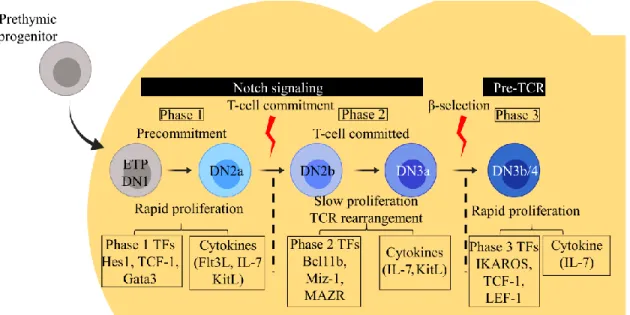
Based on the expression of different surface molecules and transcription factors, early T-cell development can be divided into three phases (Fig. 2). While the first two, phase 1 and phase 2 depend on signals from the Notch receptor, the phase 3 differentiation requires pre-TCR signaling. While phase 1 is characterized by the expansion of multipotent progenitors, and in phase 2 differentiation to the T-cell lineage occurs by establishing the cells’ ability to respond to the pre-TCR signaling in the differentiation of DN3 to DN4 during the phase 3. The three
1.2.1.3.1 Phase 1
In the first phase, Notch signaling triggers the differentiation of IL-7Rαlo ETP
progenitors into IL-7Rαhi DN2a cells.
Notch signaling (Notch1-4) is an evolutionarily conserved signaling pathway, with Notch1 being the functionally predominant receptor in cell development [73-76]. While T-cell differentiation in Notch1-deficient ETP is impaired at the DN1 stage, experimental studies showed that Notch signaling is also needed for DN2 to DN3 transition and in the β-selection [75-77]. Notch1-signaling is needed to antagonize the development of ETP into alternative non-T-lineages and to induce transcription factors such as Hes1 (codes for Hes1), B cell chronic
lymphoma 11b (Bcl11b codes for Bcl11b), Gata3 (codes for Gata3) and Tcf7 (codes for Tcf1)
that regulate the T-cell identity [78-80]. Notch1-mediated inhibitory effect on non-T-cell lineage development of ETP occurs in a stepwise fashion. First, Notch signaling shuts off B-cell development by inducing the expression of Gata3 in ETP [81]. Following this, the potential of DN2a to develop into DC, macrophages and innate lymphocytes (ILC), is antagonized by sustained Notch signaling via Hes1 expression so that DN2b thymocytes are T-committed [82-90].
TCF-1-induced Gata3 plays a critical role in the establishment of the T-cell-regulatory network of transcription factors [91-95]. Gata3’s role is not restricted to the early steps of T-cell development, but functions throughout T-cell development and mature T-cell function in the periphery [95-99]. However, in ETP, Gata3 supports T-cell specification mainly by blocking B-cell lineage potential in ETP thymocytes and inducing the expression Bcl11b [81, 85, 95, 99-102]. Later during T-cell development, Gata3 is needed for activating the TCRβ locus and for the production of CD4+ SP T-cells [96]. Interestingly, Gata3 has a very limited range of
dose-response, in DN cells at least. While increased levels of Gata3 is tolerated in periphery in CD4+
Th2 cells, overexpression of Gata3 in ETPs is just as toxic to cells as Gata3 deficiency [85]. Several posttranslational modifications have been found to regulate Gata3 function [103-106]. IL-7 drives the proliferation of DN2a thymocytes and their differentiation into DN2b during phase 1 to phase 2 transition [22, 23, 107-111]. IL-7 regulates T-cell proliferation at several stages of development and function by activating both phosphoinositide 3-kinase (PI3K) and
signal transducer and activator of transcription 5 (STAT5) signaling pathways [22, 23, 107-111].
Figure 2. The three different phases of early T-cell development and role of cytokines and transcription factors
Notch signaling activates the T-cell differentiation program in T-cell progenitors. The early events of T-cell development consist of the sequential progression of early thymic progenitors (ETP or DN1) through the consecutive DN2a, DN2b, DN3a, DN3b/4 stages before they develop into double positive (DP) thymocytes. Based on Notch or pre-TCR signaling, early T-cell development can be divided into phases with distinct cytokine signaling and transcription factors (TF) that regulate proliferation rate and T-lineage commitment program. DN, Double negative; IL, interleukin; TCR, T-cell receptor. Figure adapted from Hosokawa and Rothenberg 2018 [79].
1.2.1.3.2 Phase 2
The transition from phase 1 to phase 2 is marked by a dynamic shift in the expression of several family of transcription factor [101]. The activation of the zinc-finger transcriptional repressor Bcl11b is one of the crucial transcriptional changes that marks the phase 2 [112-114]. By downregulating Kit expression and repressing all phase 1 specific transcription factors, Bcl11b prevents deviation to alternative cell lineage fates and dedifferentiation of developing thymocytes [112-114]. Bcl11b-mediated exclusion of myeloid/NK lineage fate and activation of Notch-signaling propels the DN2b cells into the T-committed DN3a stage in which T-cell restricted genes like Rag1, Rag2, Ptcra, and Cd3e are expressed by a process that is largely
mediated by Notch and E proteins (E2A and HEB, encoded by Tcf3 and Tcf12, respectively) [115-119]. Alongside their role in the expression of T-cell specific genes, E proteins also regulate Rag-mediated recombination of the Tcrb locus by inducing the suppressor of cytokine signaling (Socs) genes, which uncouple growth factor receptors, such as IL-7R, from their signaling pathway and induce cell-cycle arrest needed for Rag-mediated recombination at the
Tcrb locus [117, 120].
Myelocytomatosis viral oncogene-associated zinc finger protein related factor (MAZR) is a transcription factor that regulates gene expression in a context-dependent manner [121-125]. MAZR plays an important role in silencing Cd8 gene expression by recruiting the nuclear coreceptor nuclear receptor corepressor 1 (NCoR1), during the DN3a to double positive (DP) transition [124]. MAZR was first described as a corepressor that functioned by recruiting the B-cell and T-B-cell regulatory factor, broad-complex, tramtrack and bric-à-brac domain and cap'n'collar homolog (Bach2) [121, 126-128]. Moreover, MAZR deficient mice are smaller in size and show increased risk of developing Bcl6-dependent lymphomas [129].
1.2.1.3.3 Phase 3
During the third phase, Notch-signaling, is quickly turned off by the newly formed pre-TCR signaling complex in an Ikaros-dependent manner [57, 130-132]. Thymocytes that have undergone a productive β-selection, experience a massive expansion and upregulate IKAROS
family zinc finger 3 (Ikzf3) and retinoic acid-related orphan receptor γ t (Rorγt), needed for the
development of DP thymocytes [79, 133-135].
Repressive epigenetic marks accumulate at the promoters of Notch target genes and other phase 1-related regulatory loci as DN4 cells proliferate/differentiate into DP thymocytes [101, 136]. At the same time, multiple DP thymocyte-specific genes are epigenetically activated, making the T-cell differentiation process irreversible [101, 136, 137]. The newly formed regulatory network of transcription factors that includes mainly, TCF-1, HEB/E2A and RORγt supports survival of DP cells by inducing the anti-apoptotic molecule B cell lymphoma-2-like 1 (BCL-XL) [138-141].
1.2.2 Positive/negative thymic selection
Immature DP cells make up around 90% of developing thymocytes and can be separated into several stages. After a successful β-selection checkpoint, thymocytes differentiate into the highly proliferative pre-TCR+ DP blasts. This is followed by a more quiescent phase during
which DP thymocytes contract in size and downregulate pre-TCR complex [142]. Rearrangement at the Tcra locus is initiated at the small DP pre-TCR- stage before a fraction of
cells audition for thymic selection. As mentioned earlier, the strength of the TCR interaction with pMHC complex will determine the fate of the developing thymocytes[142, 143][142, 143][142, 143][142, 143]. While strong interactions result in negative selection, positive selection is promoted by weak interactions. In some cases, strong affinity and/or avidity promotes the development of TReg. Ordinarily, deviation into the TReg cell lineage occurs when
the strength of the interaction is not strong enough to cause negative selection [144]. When the TCR on developing thymocytes fails to engage in a productive interaction with the pMHC on the stromal cells, cells undergo a type of programmed cell death, called death by neglect.
Recently, genome wide analyses have uncovered a dynamic gene expression pattern that is unique to each DP subsets (pre-TCR+, pre-TCR-, αβTCR+) [143]. The mapping of the
transcriptional landscape of the DP subpopulations revealed, among other things, that cells destined for positive selection versus apoptotic deletion display unique gene signatures. Equally important, results showed a large-scale transcriptional shutdown of several genes that accompanies the differentiation of the proliferative pre-TCR+ DP blasts to the resting pre-TCR
-DP cells of smaller size. Then, the relatively transcriptionally quiescent stage is followed by another major transcriptional modification that is initiated by the TCR-mediated positive selection signaling. Some of these modifications include the reactivation of several important signaling pathways, such as the canonical TCR pathway, the metabolic pathway and distinct positive and negative selection-related genes, like Id2 and Id3 [145-147]. The transition of the small resting DP thymocytes into the transcriptionally dynamic CD69+ DP thymocytes is
associated with increased glycolytic and oxidative phosphorylation activity [143].
Signaling downstream of the newly formed TCR complex leads to the regulation of several factors involved in positive and negative selection. Some of these factors are involved
(IRF1, encoded by Irf1), and nuclear factor κ-B (NF-κB), while others, described below, function specifically in one or the other selection process.
While this section covers the details of the complex regulatory network of transcription factors regulated by TCR-mediated signaling at the DP stage, the structural components of the TCR complex and the signaling pathways downstream of the TCR signaling will be discussed later.
1.2.2.1 Transcription factors regulating positive selection
1.2.2.1.1 Bcl11b
At the DP stage, Bcl11b expression has also been shown to control positive selection efficiency [148]. Bcl11b-deficient mice displayed defective proximal TCR signaling events, leading to dysregulated expression of genes involved in positive selection as well as CD4+/CD8+
lineage commitment [148]. Interestingly, introduction of the antiapoptotic Bcl2 transgene, but not a TCR transgene, rescued the phenotype in Bcl11b-deficient mice [148].
1.2.2.1.2 Tox
Thymocyte selection-associated high mobility group box protein (TOX, encoded by
Tox) is induced following TCR-stimulation. Its deficiency severely impairs positive selection of
developing thymocytes with a predominant impact on the development of MHC-II-restricted CD4+ thymocytes [149-151].
1.2.2.1.3 Gata3
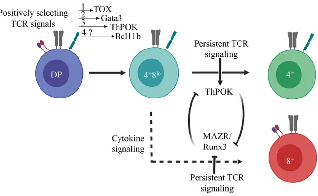
Although Gata-3 deficiency does not impair positive selection (as identified by CD69 upregulation on DP thymocytes), it is upregulated by the TCR-mediated positive selection signals and required for survival of selected MHC-II-restricted thymocyte. Gata-3 is induced in positively selected thymocytes immediately after TOX expression and increases gradually as cells undergo positive selection before peaking at the CD4+8lo stage, which is when CD4+/CD8+
lineage choice occurs [152]. Subsequently, while Gata-3 expression is maintained in MHC-II restricted CD4+ T-cells, it is downregulated in MHC-I-signaled CD8 committed thymocytes
1.2.2.1.4 Notch signaling
The role of Notch-signaling in positive selection remains controversial [154-156]. A study showing an inhibitory effect of Notch on TCR signaling during thymic selection was later challenged by a study indicating that Notch-signaling potentiated TCR signaling by regulating late (or delayed) responding genes during positive selection [157, 158]. It was later elucidated by high-throughput screening that Notch- and positive-selection-induced genes (which interestingly could not be activated by in vitro TCR activation) overlapped significantly [158].
1.2.2.1.5 Nuclear factor of activated T-cells (NFAT)
Similar to Notch signaling, the role of nuclear factor of activated T-cells (NFAT) in positive selection has been controversial. While three of the four members of the NFAT family of transcription factors are expressed by the immune cells (NFATc1, NFATc2, and NFATc3), only NFATc3 was shown to be potentially involved in positive selection [159]. Surprisingly, the detrimental effect of Nfatc3-deficiency on thymic selection was not aggravated by the additional loss-of-function of Nfatc2. As for NFATc1, experimental results so far are inconclusive [159-162]. Nevertheless, NFAT reporter mice are commonly used to monitor TCR signaling [163].
1.2.2.2 Transcription factors regulating negative selection
DP and SP expressing a TCR with high affinity for self-peptide/MHC complexes are eliminated by negative selection to ensure that self-reactive T-cells are prevented from entering circulation [164, 165]. Important regulators of negative selection include transcription factors such as Nur77 (encoded by nuclear receptor subfamily 4 group A member 1 [Nr4a1]) and Bim (encoded by Bcl2l11).
1.2.2.2.1 Nur77
The transcription factor Nur77 belongs to the steroid nuclear hormone receptors superfamily of transcription factors that includes two other members, Nurr1 (encoded by Nr4a2) and neuron-derived orphan receptor 1 (Nor-1 – encoded by Nr4a3) [166, 167]. Nur77 orphan receptor is a dynamic transcription factor that is induced in response to TCR stimulation and
dominant negative version of Nur77, or its downregulation, correlated with reduced pro-apoptotic activity [169, 170]. The Nur77 transcription factor has been shown to mediate apoptosis by two main mechanisms: 1) by transcriptionally regulating its downstream gene
Ndg1, which codes for a protein that can trigger apoptosis through caspase-8, and 2) by a
transcriptional independent mechanism involving depolarisation of the mitochondria, through the transformation of Bcl2 into a toxic protein [166, 171-177]. While overexpression of Nur77 results in overt apoptosis of DP thymocytes, Nur77-deficient mice show no perturbation of clonal deletion, probably due to redundancy with Nurr1 or Nor-1 [170, 178].
1.2.2.2.3 Bim
The proapoptotic factor Bim, which functions by inhibiting Bcl2, induces apoptosis by regulating mitochondrial permeability [179, 180]. Surprisingly, loss-of-function of Bim, does not result in autoimmune diseases, suggesting that other redundant mechanisms maintain peripheral tolerance [179-181].
1.2.2.3 Transcription factors regulating positive and negative selection
1.2.2.3.1 ID3, IRF1 and NF-κB
ID3 is a Helix-Loop-Helix inhibitor protein that generally regulates E protein function by antagonising the DNA binding potential of E2A (encoded by TCF3) and HEB (encoded by
TCF12) [182]. Although more rigorous research is needed, expression profile and genetic
manipulations show that, unlike ID3, IRF1 and NF-κB control mainly positive and negative selection of MHC-I-restricted CD8+ cytotoxic T-cells [183-187].
1.2.2.3.2 HDAC7
A high-throughput screen identified histone deacetylase 7 (HDAC7) as a potential regulator of thymic selection. The introduction of various dominant negative mutant forms of HDAC7 not only impaired thymic selection, but failed to rescue thymic selection in Hdac7-deficient mice as well [188].
The induction of the regulatory nuclear factors discussed above form a part of a broad transcriptional modifications induced by TCR-mediated positive selection signals. Thorough
investigations are needed to better elucidate the complete transcriptional landscape governing positive and negative selection.
Following positive selection, MHC-I- and MHC-II-restricted thymocytes terminate Cd8 transcription to become CD4+8lo intermediates, and differentiate into CD4+ and CD8+ single
positive thymocytes by a process that is influenced by co-receptor, TCR and cytokine signaling [189]. Surprisingly, the differentiation of TCR-signaled thymocytes into CD4+ helper or CD8+
cytotoxic lineage is accompanied by differences in the expression of only a few lineage specific genes. These genes include Runx3 and Eomes for CD8+ cytotoxic lineage fate, and
zinc-finger-and-broad-complex, tramtrack and bric-à-brac-domain containing 7 (Zbtb7b also called
“Thpok” here, encoding ThPOK protein) and Gata3 for CD4+ helper lineage [143].
1.2.3 CD4
+/CD8
+Lineage fate of positively selected thymocytes
Following positive selection, developing thymocytes undergo a crucial lineage fate decision to differentiate into either CD8+ cytotoxic or CD4+ helper T-cells. DP thymocytes are
unique among the developing T-cell subsets in that they express both CD4 and CD8 co-receptors and are unresponsive, due to high expression of SOCS proteins, to the pro-survival cytokine IL-7 [189-191]. The intracellular domains of both co-receptors are bound by the lymphocyte specific tyrosine kinase Lck. Engagement of TCR/co-receptor with MHC brings the co-receptor associated Lck in close proximity to the TCR complex leading to a cascade of phosphorylation events [192]. The extracellular domains of CD4 and CD8 co-receptors, on the other hand, bind to the MHC-II and MHC-I molecules, respectively [193, 194]. TCR specificity for MHC then determines thymocytes developmental fate; thymocytes expressing MHC-I- and MHC-II-specific TCR invariably differentiate into CD8+ and CD4+ SP mature T-cells, respectively. The
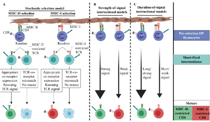
development of DP thymocytes into either of the two lineages is a classical example of bipotential lineage-fate development of precursor cells. The last two decades have witnessed a significant progress in our understanding of the cellular and molecular mechanisms underlying the CD4+/CD8+ lineage choice, leading to development of various models as described below.
It was previously thought that uncommitted positively selected DP thymocytes downregulated one or the other of the co-receptors to ultimately give rise to either CD4+ or
the same TCR-signals in DP thymocytes regulated, simultaneously, positive selection and lineage commitment, resulting in the irreversible termination of either one of the Cd4 or Cd8 co-receptor genes [189]. Opinions, however, were divided over whether co-receptor termination was “stochastic” or ‘instructive’ (Fig. 3). The kinetic signaling model, on the other hand, is a non-classical model and is currently the most widely accepted model of CD4+/CD8+ lineage
choice. It proposes that positively selected DP thymocytes develop into CD4+8lo uncommitted
intermediates before differentiating into either MHC-II-restricted CD4+ or MHC-I-restricted
CD8+ SP mature thymocytes (Fig. 4) [195].
1.2.3.1 Stochastic model of CD4+/CD8+ lineage choice
The stochastic model of lineage choice proposes that TCR induces random termination of one of the co-receptors during positive selection, which generates a pool of SP thymocytes with matching and mismatching TCRs and co-receptors. Only selected thymocytes with matching co-receptors and TCRs, capable of transmitting a productive TCR signal, would proceed to differentiate into mature T-cells. Thymocytes with mismatching co-receptors and TCRs, which in theory should be observed in 50% of selected cells, are destined to die by apoptosis (Fig. 3). Actually, the presence of a significant number of MHC class-II-specific CD4+CD8+ (DP) mature T cells in CD4 co-receptor transgenic mice supported this model
[196-199]. This prompted the authors to argue that forced CD4 expression rescued MHC class-II-restricted SP thymocytes that had incorrectly terminated Cd4 co-receptor expression and, hence, had died by apoptosis. However, the number of DP T-cells in periphery was fewer than 15%, much lower than the 50% frequency predicted by the stochastic model [196, 198, 200]. Studies of thymic selection using TCR transgenic mice have helped shed light on this matter. Positive selection efficiency in several TCR transgenic mice can reach up to 90%, which would not be feasible if co-receptor termination was a random event [201]. Another observation arguing against the stochastic lineage commitment model is the fact that long-lived and functionally mature TCR/co-receptor mismatched T-cells can be generated even in normal mouse [202, 203]. So, why does forced CD4 expression lead to the development of DP mature T-cells? It is possible that early transgenic co-receptor expression interfered in thymic selection leading to generation of mismatched thymocytes [196, 197, 204]. When investigators addressed this issue by placing a CD4 co-receptor transgene under the control of the CD8 enhancer I (E8I), which is
active only in positively selected CD8+-committed T-cells, mature T-cells expressing both the
co-receptors were not produced contradicting the original study [205]. Taken together, these experimental observations have challenged the core principles of the stochastic model and demonstrate that lineage fate is neither error-prone nor stochastic [204, 206, 207].
1.2.3.2 Instructive CD4+/CD8+ lineage choice models
An alternate model explaining CD4+/CD8+ lineage choice proposes that TCR specificity
for pMHC instructs positively selected thymocytes to develop into CD4+ or CD8+ lineage. Thus,
MHC-II- and MHC-I-specific thymocytes almost always develop into CD4+ helper and CD8+
cytotoxic T-cells, respectively. Subsequent studies led to refinement of the instructive models as described below.
1.2.3.2.1 Strength-of-signal
The strength-of-signal model proposes that in the positively selected DP thymocytes a strong and weak TCR-signal terminates Cd8 and Cd4 transcription, respectively. As tyrosine kinase Lck binds the cytosolic tail of CD4 with more affinity than CD8 co-receptor [192, 208], MHC-II-restricted thymocytes would be predicted to receive quantitatively stronger signal compared to MHC-I-restricted thymocytes. Thus, according to this model, the relative strength of the TCR signaling instructs co-receptor transcription termination [201]. Redirection of MHC-I-specific thymocytes to the CD4+ lineage upon introduction of a transgenic form of CD8α
co-receptor engineered to express the cytosolic domain of CD4 (CD8.4) provided the first evidence that TCR-signal strength influenced the lineage fate of selected thymocytes [209]. A similar pattern was observed when components of the TCR complex, such as Lck, ζ-chain-associated protein kinase 70 (Zap70), C-terminal SRC kinase (CSK) and extracellular signal–regulated kinase (ERK), were manipulated to affect strength-of-signal in developing thymocytes in mice [210-218]. Basically, when a component downstream of TCR signaling was modulated to augment TCR-signal strength more CD4+ T-cells were generated, while modifications leading