Non-typhoidal Salmonella blood stream
infection in Kuwait: Clinical and
microbiological characteristics
M. John AlbertID1*, Dieter Bulach2, Wadha Alfouzan1,3, Hidemasa Izumiya4, Glen Carter2,
Khaled Alobaid5, Fatemah Alatar6, Abdul Rashid Sheikh5, Laurent Poirel7
1 Department of Microbiology, Faculty of Medicine, Kuwait University, Jabriya, Kuwait, 2 Microbiological
Diagnostic Unit, Public Health Laboratory, Peter Doherty Institute for Infection and Immunity, The University of Melbourne, Victoria, Australia, 3 Microbiology Unit, Department of Laboratories, Al Farwaniya Hospital, Sabah Al-Nasser, Kuwait, 4 National Institute of Infectious Diseases, Tokyo, Japan, 5 Department of Microbiology, Al Amiri Hospital, Sharq, Kuwait, 6 Microbiology Unit, Muabarak Al Kabeer Hospital, Jabriya, Kuwait, and 7 Department of Medicine, University of Fribourg, Switzerland
Abstract
Non-typhoidal Salmonella (NTS) bacteremia is a significant cause of morbidity and mortality worldwide. It is considered to be an emerging and neglected tropical disease in Africa. We studied this in two tertiary hospitals–Al Farwaniya and Al Amiri–in Kuwait, a subtropical country, from April 2013-May 2016. NTS bacteremia was present in 30 of 53,860 (0.75%) and 31 of 290,36 (1.33%) blood cultures in the two hospitals respectively. In Al Farwaniya hospital, one-third of the patients were from some tropical developing countries of Asia. About 66% of all patients (40/61) had diarrhea, and of these, 65% had the corresponding blood serovar isolated from stool culture. A few patients had Salmonella cultured from urine. Patients were either young or old. Most of the patients had co-morbidities affecting the immune system. Two patients each died in both hospitals. The number of different serovars cultured in each hospital was 13, and most infections were due to S. Enteritidis (all sequence type [ST]) 11) and S. Typhimurium (all ST19) except in a subgroup of expatriate patients from tropical developing countries in Al Farwaniya hospital. About a quarter of the isolates were multidrug-resistant. Most patients were treated with a cephalosporin with or without other antibiotics. S. Enteritidis and S. Typhimurium isolates were typed by pulsed field-gel electrophoresis (PFGE) and a selected number of isolates were whole-genome sequenced. Up to four different clades were present by PFGE in either species. Whole-genome
sequenced isolates showed antibiotic-resistance genes that showed phenotypic correlation, and in some cases, phenotypes showed absence of specific genes. Whole-genome
sequenced isolates showed presence of genes that contributed to blood-stream infection. Phylogeny by core genome analysis showed a close relationship with S. Typhimurium and
S. Enteritidis from other parts of the world. The uniqueness of our study included the finding
of a low prevalence of infection, mortality and multidrug-resistance, a relatively high preva-lence of gastrointestinal infection in patients, and the characterization of selected isolates of
S. Typhimurium and S. Enteritidis serovars by whole-genome sequencing that shed light on
phylogeny, virulence and resistance. Similarities with studies from developing countries
a1111111111 a1111111111 a1111111111 a1111111111 a1111111111 OPEN ACCESS
Citation: Albert MJ, Bulach D, Alfouzan W, Izumiya
H, Carter G, Alobaid K, et al. (2019) Non-typhoidal Salmonella blood stream infection in Kuwait: Clinical and microbiological characteristics. PLoS Negl Trop Dis 13(4): e0007293.https://doi.org/ 10.1371/journal.pntd.0007293
Editor: Ruifu Yang, Beijing Institute of Microbiology
and Epidemiology, CHINA
Received: January 16, 2019 Accepted: March 12, 2019 Published: April 15, 2019
Copyright:© 2019 Albert et al. This is an open access article distributed under the terms of the Creative Commons Attribution License, which permits unrestricted use, distribution, and reproduction in any medium, provided the original author and source are credited.
Data Availability Statement: All relevant data are
contained within the manuscript and/or supporting information files.
Funding: The authors received no specific funding
for this work
Competing interests: No authors have competing
especially Africa included infection in patients with co-morbidities affecting the immune sys-tem, predominance of S. Typhimurium and S. Enteritidis serovars and presence of drug-resistance in isolates.
Author summary
Salmonella organisms are classified into typhoidal Salmonella (causing enteric fever) and
non-typhoidalSalmonella (NTS) (causing infections other than enteric fever). Apart from
causing other infections, NTS causes blood-stream infection (bacteremia and septicemia). NTS blood stream infection (NTS-BI) is considered to be an emerging and neglected trop-ical disease in Africa. It causes a very high morbidity and mortality in Africa. The individ-uals affected in Africa are children, malnourished people, patients with malaria or HIV etc. These conditions affect the immune system and make them vulnerable to infection with NTS. In these patients, diarrheal disease due to NTS is rare. The majority of infec-tions are due to two types of NTS: Typhimurium and Enteritidis. There is a very high prevalence of multidrug-resistance in NTS making the infection difficult to treat. NTS-BI is also present in other parts of the world including developed countries albeit at a lower prevalence. Kuwait is a high-income, subtropical country in transition (from a developing to developed country), located in the Middle East. We studied NTS-BI in Al Farwaniya and Al Amiri hospitals in Kuwait during April 2013 to May 2016. Out of nearly 30,000 to more than 50,000 blood cultures done in these hospitals, NTS was present in 0. 75 to 1.33% of blood cultures, representing a very small proportion of blood cultures, unlike in Africa. This showed that 31 patients in Al Farwaniya hospital and 30 patients in Al Amari hospital had NTS-BI. Most of these patients had underlying illnesses such as diabetes, lung infection, cancer etc. that affect the immune system, as in Africa. Many patients also had diarrheal disease caused by the same NTS that caused blood stream infection, unlike in Africa. Only two patients in each hospital died, a low mortality, unlike in Africa. The majority of the isolates belonged to Typhimurium and Enteritidis as in Africa. Even though resistance to drugs was a problem, about quarter of the isolates only were multi-drug-resistant, a lower prevalence compared to in Africa. In Kuwait, we performed a detailed genetic study of a selected number of Typhimurium and Enteritidis isolates by a modern technique called whole genome sequencing. This revealed genetic determinants encoding drug-resistance and virulence causing blood-stream infection. This type of study was not performed in African isolates. Thus, our study revealed similarities and dif-ferences with studies of NTS-BI in Africa.
Introduction
Salmonella enterica subspecies enterica serovar Typhi and Salmonella Paratyphi A, B and C
cause enteric fever, a systemic febrile illness that occurs only in humans. There are more than 2500 serovars of non-typhoidal Salmonella (NTS). NTS infects a variety of hosts and are fre-quently zoonotic in origin [1]. It mainly causes self-limiting gastroenteritis in humans. NTS has also been recognized as a major cause of extra-intestinal invasive bacterial infection in young children and immunocompromised patients worldwide [2,3]. It is a cause of severe bac-teremia [1]. It has been estimated that globally about 3.4 million cases of bacbac-teremia due to NTS occur every year [4]. The estimated worldwide mortality from NTS infection is 155,000
per year [1]. Invasive NTS disease is considered to be an emerging and neglected tropical dis-ease in Africa [1].
Kuwait is a high-income subtropical country situated in the Middle East. It is neither a developing nor a developed country, but is a country in transition. There are no systemic stud-ies of bacteremia due to NTS in Kuwait even though NTS is a major cause of diarrhea in Kuwait [5,6]. Antimicrobial resistance is also a problem among diarrheal stool isolates of NTS in Kuwait [7]. Also, there are no data on serovars of NTS causing infection in Kuwait. There-fore, the primary objective of the study was to assess the case-fraction of NTS bacteremia among bacteremia cases in two tertiary hospitals in Kuwait. The secondary objective was to determine the serovars of NTS isolates and their antibiotic susceptibilities. A study was con-ducted in two tertiary hospitals in Kuwait. We determined the serovars of NTS isolates and their antibiotic susceptibilities. Moreover, all isolates ofS. Enteritidis and S. Typhimurium
ser-ovars were typed by pulsed-field gel electrophoresis (PFGE) to study their relatedness. In addi-tion, selected isolates were subjected to whole genome sequencing for further characterization. We also compared our results with those from Africa. The results are presented in this communication.
Materials and methods
Patients. The only criterion for inclusion in the study was the isolation of a NTS from blood
culture irrespective of any accompanying conditions and age. The indication for doing a blood culture was a suspicion of sepsis. Isolates were collected from Al Farwaniya hospital (which has pediatric and adult wards) from July 2013 until May 2016. The period of collection of iso-lates from Al Amiri hospital (which has neonatal, pediatric and adult wards) was from April 2013 until December 2015. The distance between the two hospitals is 26 km (16.2 miles). Al Farwaniya hospital is a 900-bed tertiary hospital which serves a population of 1.2 million peo-ple. Even though it serves both Kuwaitis and non-Kuwaitis, about 80% of the population served are non-Kuwaitis. Al Amiri hospital is a 418-bed tertiary hospital and it serves a popula-tion of 50,000 people. Although the hospital caters to both Kuwaitis and non-Kuwaitis, most of the people treated are Kuwaitis. All patients including diabetic patients and cancer patients have access to these hospitals, although for cancer treatment itself, there is a separate hospital. The patients included in our study were admitted in these hospitals, because of gastrointestinal symptoms and/or fever. Even though there are general guidelines for antimicrobial therapy, the antimicrobial therapy administered to patients in both hospitals was left to the discretion of attending physician. Antibiotics were given based on severity of infection, comorbidities and immune status of the patients.
Since typhoid is not endemic in Kuwait, typhoid vaccine is not offered to its residents. However, the vaccine is offered to residents travelling to high-risk areas.
Blood culture. Blood culture was done using BD BACTEC system (Becton Dickinson, MD,
USA). From adults, 20 ml blood was drawn and 10 ml was inoculated into an aerobic bottle and 10 ml into an anaerobic bottle. From children, 1–5 ml of blood was drawn and inoculated into the aerobic bottle only. After inoculation of the bottle in the wards, they were immediately transported at ambient temperature to the hospital microbiology laboratory. Aerobic bottle signalling growth was subcultured on sheep blood agar, MacConkey agar and chocolate agar. The first two plates were incubated aerobically and the last plate in 5% CO2 atmosphere at 37˚C for 24 h. Anaerobic bottle signaling growth was subcultured on brucella agar plate and the plate was incubated anaerobically at 37˚C for 48 h. Identification ofSalmonella and the
ini-tial antibiotic susceptibility were done from lactose non-fermenting colonies on MacConkey agar by Vitek 2 system (BioMerieux, Marcy l’Etoile, France). For grouping ofSalmonella, the
colony was inoculated into triple sugar iron agar and if the reaction was suggestive of Salmo-nella, the growth was used in a slide agglutination test against Salmonella polyvalent and
group-specific antisera (Denka Seiken, Tokyo, Japan). As soon asSalmonella was detected in
blood culture, empirical antimicrobial therapy for the patient was initiated. Further identifica-tion ofSalmonella and antibiotic susceptibility testing were done as described below.
Stool was cultured forSalmonella from all Salmonella blood culture-positive patients.
When warranted, urine culture was done. Stool was inoculated onto MacConkey agar and Sal-monella-Shigella agar (SSA) and enriched in selenite F broth. Selenite F broth was subcultured
onto SSA after 20 h of incubation. When urinary tract infection was suspected, urine was cul-tured on CLED (cysteine, lysine, electrolyte-deficient) agar and blood agar. All plates were incubated for 20–24 h at 37˚C. SuspectedSalmonella colonies were subjected to identification
as above. Subcultures of Salmonella isolates on MacConkey agar plates from the hospital microbiology laboratories were sent to the reference laboratory at the Faculty of Medicine, Kuwait University, where further tests, listed below, were arranged or carried out.
Typing ofSalmonella isolates. Internal fragments of seven housekeeping genes (thrA, purE,
sucA, hisD, aroC, hemD, dnaN) were amplified by PCR [8]. Sequences of the genes were trimmed to the required lengths (399–501 bps). Sequence types were assigned in accordance with theSalmonella enterica database,http://mlst.ucc.ie/mlst/dbs/Senterica. From sequence types, serovars were assigned according to the scheme of Achtman et al [9]. Isolates belonging to
S. Tyhimurium and S. Enteritidis serovars were further confirmed by specific PCR assays [10].
Antibiogram. Antibiotic susceptibility test was carried out by E test (Biomerieux) and
interpreted by the criteria laid down by Clinical and Laboratory Standards Institute (CLSI) [11]. The breakpoints (μg/ml) (intermediate resistance, resistance) for various antibiotics were as follows: ciprofloxacin (0.12 to 0.5, �1), chloramphenicol (16, �32), trimethoprim/sulfa-methoxazole (not applicable, �4), gentamicin (8, �16), tetracycline (8, �16), ampicillin (16, �32), ceftazidime (8, �16), cefotaxime (2, �4), ceftriaxone (2, �4), imipenem (2, �4), mero-penem (2, �4) and piperacillin/tazobactam (32 to 64, �128). Resistant and intermediate-resis-tant minimum inhibitory concentrations were categorized as resisintermediate-resis-tant in accordance with clinical practice.
When there was a necessity of testing resistance to other antibiotics (streptomycin, neomy-cin, carbenicillin, trimethoprim and sulfonamide), this was done by Kirby-Bauer disc diffusion test [12].
Results were interpreted according to the criteria of CLSI [13].Escherichia coli ATCC
25922 strain was used as a quality control strain.
Extended spectrum
β-lactamase (ESBL) production
E test. Isolates suspected of producing ESBL were tested for clavulanic-inhibitable ESBL
pro-duction with E test ESBL strips–ESBL CT/CTL 16/1, ESBL TZ/TZL 32/4, ESBL PM/PML- as per manufacturer’s instructions (BioMerieux).
Vitek 2 test. The Vitek 2 ESBL test (bioMe´rieux) is based on the simultaneous assessment
of the antibacterial activity of cefepime, cefotaxime and ceftazidime, measured either alone or in the presence of clavulanate. This test relies on card wells containing 1.0 mg/L of cefepime, or 0.5 mg/L of cefotaxime or ceftazidime, either alone or associated with 10 or 4 mg/L of clavu-lanate, respectively. After inoculation, cards were introduced into the Vitek 2 machine, and for each antibiotic tested, turbidity was measured at regular intervals. The proportional reduction of growth in wells containing a cephalosporin combined with clavulanate was then compared with that achieved by the cephalosporin alone and was interpreted as ESBL- positive or–nega-tive through a computerized expert system (Advanced Expert System) [14].
Detection of genes encoding ESBL. PCR assays were performed to detect genes encoding
blaCTX-M,blaTEMandblaSHV[7] andblaPSE-1[15].
AmpC disk test and modified three dimensional test (M3DT) for detection of AmpC β-latcamases. This test was performed as described by Coskun et al [16]. Enhanced growth of bacteria around blank disks and AmpC disk or intersected growth in the zone of inhibition was considered positive.
Pulsed-field gel electrophoresis (PFGE). PFGE was performed according to the CDC
pro-tocol (http://www.cdc.gov/pulsenet/propro-tocols.htm) usingXbaI restriction enzyme (Roche,
Mannheim, Germany) and a CHEF-DR III PFGE system (Bio-Rad, Munich, Germany). Sal-monella Braenderup strain H9812(ATCC BAA 664) was used as a reference strain. The bands
were visualized under UV light with Gel DOC(Bio-Rad) and analyzed with Bionumerics soft-ware (Applied Maths NV, Belgium). Pairwise similarities between patterns were calculated by DICE’s similarity coefficient. Clustering was based on unweighted pair-wise group method with averages (UPGMA) setting tolerance and optimization each at 1.5%.
Whole genome sequencing (WGS). Based on differences in banding patterns,S. Enteritidis
andS. Typhimurium isolates were selected for whole genome sequencing. Sequencing libraries
were prepared using the Nextera XT DNA sample preparation kit (Illumina, San Diego, CA, USA) and the sequence read data were produced on the Illumina NextSeq instrument (paired end, 150 base reads). Read data were submitted to the sequence read archive under project number PRJNA363099 (between 70x and 140x read depth coverage for each isolate). De novo assembly of the read data from each isolate was performed using MegaHit [17]. The resulting draft genome sequences were used to derive MLST (MLST:https://github.com/tseemann/mlst PubMLST:https://pubmlst.org/).
Abricate (https://github.com/tseemann/abricate) was used to detect virulence genes ([VFDB]: [18]). Antibiotic resistance gene profile was determined using Abricate and the Resfinder database [19].
Data management and analysis. Data on NTS bacteremia were obtained from laboratory
records. Patient charts were traced, and relevant information was extracted. Results of bacterial analysis including serovars, antibiogram and WGS data were linked to patient data and tabu-lated in the reference laboratory. This dataset was used for analysis as appropriate.
Statistics. The difference in the prevalence of resistance to antibiotics in the two hospitals
was calculated by Chi square test. A P value of �0.05 was considered significant.
Ethical approval. Bacterial isolates studied were a part of the routine collection at the
Enteric Microbiology Reference Laboratory, Kuwait University, for further studies and archiv-ing. It was not possible to get informed consent of patients as the clinical data were retrospec-tively collected. No additional specimens were collected for this study and patient identity was kept anonymous. Therefore, a waiver for informed consent, and approval for the study were granted by the Ethics Committee of Ministry of Health, State of Kuwait (permit number 898/ 2018).
Results
NTS from blood culture. The isolation of NTS from blood cultures of both hospitals is shown
inFig 1. In Al Farwaniya hospital, 53,860 blood cultures were done and 3981 were positive for microorganisms. Of the positive cultures, 30 were positive for a non-typhoidalSalmonella
ser-ovar (0.75%). There were 13 differentSalmonella serovars, but 50% of them belonged to S.
Enteritidis (all sequence type [ST]11) andS. Typhimurium (all ST19). In Al Amiri hospital, 29,
036 blood cultures were done and 2,331 were positive for microorganisms. Of the positive cul-tures, 31 were positive for a non-typhoidal serovar (1.33%). There were 13 different serovars of
Salmonella infecting the patients, but 19 isolates (61.3%) belonged to S. Enteritidis (all ST11)
andS. Typhimurium (all ST19).
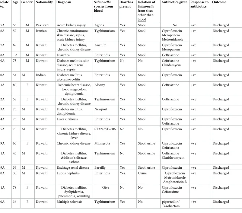
Patient characteristics,Salmonella serovars isolated, response to therapy and patient
out-come in Al Farwaniya and Al Amiri hospitals are presented inTable 1. Isolate numbers with suffix F are from Al Farwaniya hospital and those with suffix A are from Al Amiri hospital. Patients were both Kuwaitis and expatriates of many nationalities in both hospitals. Median age for all 61 patients was 58 y and 62.3% of patients were >50 y old. Eight patients (13.1%) were children <5 y old. Approximately 67% of patients had chronic diseases such as diabetes mellitus, cancer, blood disorder, kidney disease or lung disease. Forty patients (65.6%) had diarrhea and 26 of them (65%) had aSalmonella (identical to the corresponding blood isolate)
cultured from the stool. Four of these 26 patients had also the correspondingSalmonella
sero-var cultured from the urine. One patient who had diarrhea did not haveSalmonella cultured
from the stool, but urine culture was positive for the corresponding blood isolate. None of the non-diarrheal patients hadSalmonella cultured from the stool. Most patients responded to
antibiotic therapy and were discharged. Many classes of antibiotics were used, but many received a cephalosporin. In addition, six patients (22A, 36A, 51A, 59A, 60A, 70A) received steroids. Only four patients died.
Antibiogram. The prevalence of resistance to all antibiotics was similar in both hospitals
(P>0.05 for all comparisons) (S1A & S1B Table). Therefore, resistance for isolates from both hospitals was combined and is presented inTable 2. The prevalence of resistance to ampicillin, ciprofloxacin and tetracycline ranged between 36.1 to 50.8%. The prevalence of resistance to other antibiotics was either negligible or absent.
Fig 1. Non-typhoidalSalmonella isolation from blood cultures of patients in Al Farwaniya and Al Amiri hospitals. A
fraction (0.75–1.33%) belonged to non-typhoidalSalmonella (NTS). https://doi.org/10.1371/journal.pntd.0007293.g001
Table 1. Details of patients from Al Farwaniya hospital and Al Amiri hospital from whomSalmonella species were isolated from blood cultures.
Isolate No.
Age Gender Nationality Diagnosis Salmonella
species from blood Diarrhea present Isolation of Salmonella from sites other than blood
Antibiotics given Response to antibiotics
Outcome
1F 41 M Indian Abdominal pain Livingston No No Cefotaxime,
Metronidazole +ve Discharged 2F 81 M Kuwaiti Cerebrovascular accident Unspeciated type 2641
No No Ceftriaxone Uncertain Died
3F 38 F Kuwaiti Diarrhea Enteritidis Yes No Cefotaxime +ve Discharged
5F 58 M Indian Abdominal pain, fever,
vomiting
Infantis No No Piperacillin/
tazobactam
+ve Discharged 7F 2.5 M Kuwaiti Intestinal obstruction,
diarrhea
Enteritidis Yes No Ceftriaxone Uncertain Transferred to
surgical unit
9F 5 M Saudi Abdominal pain, sickle
cell anemia
Typhimurium No No Cefotaxime +ve Discharged
10F 27 M Indian Pancytopenia Enteritidis No No Ceftriaxone Uncertain Transferred to
Infect Dis Hospital for? HIV infection 11F <1 F Egyptian Fever, bronchiolitis,
diarrhea
Typhimurium Yes No Cefotaxime,
gentamicin
+ve Discharged
12 F 72 M Kuwaiti Diabetes mellitus,
ischemic heart disease, diarrhea, vomiting,
fever
Enteritidis Yes No Piperacillin/
tazobactam, metronidazole
+ve Discharged
13F 30 M Filipino Fever, diarrhea Enteritidis Yes No Cefotaxime,
metronidazole
+ve Discharged
14F 45 M Egyptian Diabetes mellitus,
chronic renal failure, fever
Livingston No No Cefotaxime +ve Discharged
15F 65 M Pakistani Diabetes mellitus, fever, jaundice
Livingston No No Cefotaxime,
clarithromycin
+ve Discharged
16F 2 F Kuwaiti Diarrhea Minnesota Yes No None, oral
rehydration only
+ve Discharged
18F 56 M Bangladeshi Diabetes, eczema, diarrhea
Kentucky Yes No Ciprofloxacin +ve Discharged
19F 54 F Iraqi Diabetes, diarrhea Typhimurium Yes No Ciprofloxacin,
metronidazole
+ve Discharged
46F 73 M Kuwaiti Diabetes mellitus,
ischemic heart disease, fever, vomiting,
diarrhea
Typhimurium Yes No Meropenem +ve Discharged
47F 76 M Kuwaiti Diabetes mellitus, colon cancer, acute kidney injury, loss of appetite
Bareilly Yes No Ceftriaxone +ve Discharged
48F 47 F Kuwaiti Sickle cell anemia, fever Enteritidis No No Ciprofloxacin +ve Discharged
52F 52 M Egyptian Colon cancer, weight loss
Typhimurium No No Ciprofloxacin,
amoxicillin/ clavulanic acid
+ve Discharged
53F 70 F Jordanian Ischemic heart disease, chronic obstructive pulmonary disease,
sinusitis
Enteritidis No No Ceftriaxone Uncertain Transferred to
neurosurgery for suspected brain
abscess
55F 24 F Kuwaiti Fever, vomiting,
diarrhea
Braenderup Yes Stool None, oral
rehydration only
+ve Discharged (Continued )
Table 1. (Continued) Isolate
No.
Age Gender Nationality Diagnosis Salmonella
species from blood Diarrhea present Isolation of Salmonella from sites other than blood
Antibiotics given Response to antibiotics
Outcome
57F 47 F Kuwaiti Thalassemia, sickle cell anemia, upper respiratory infection, abdominal pain, chills
Poona No No Ceftriaxone, azithromycin +ve Discharged 58F 46 M Indian Fever, thrombocytopenia, diarrhea
Telaviv Yes Stool Piperacillin/
tazobactam, metronidazole
Uncertain Discharged against recommendation
62F 65 M Kuwaiti Acute kidney injury,
dehydration
Typhimurium No No Meropenem,
ceftriaxone
+ve Discharged
64F 1 M Kuwaiti Diarrhea, fever Enteritidis Yes Stool Cefotaxime +ve Discharged
66F 59 M
Non-Kuwaiti�
Diabetes mellitus, end-stage renal disease,
sepsis Poona No No No information No information No information 68F 1 M Non-Kuwaiti�
Diarrhea Anatum Yes Stool None, oral
rehydration only
+ve Discharged
69F 66 M Pakistani Diabetes mellitus,
cellulitis of thigh, diarrhea
Minnesota Yes Stool Piperacillin/
tazobactam, cefotaxime, clindamycin
+ve Discharged
71F 59 M Indian Hyponatremia,
ruptured aortic valve
Enteritidis No No Cefotaxime -ve Died
74F 1 M Kuwaiti Diarrhea Uganda Yes No None, oral
rehydration only
+ve Discharged 21A 79 F Kuwaiti Chronic kidney disease,
liver chirrosis
Typhimurium Yes No Ciprofloxacin -ve Died
22A 75 F Kuwaiti Diabetes mellitus,
thrombocytopenia, Darriers disease
Enteritidis No No Ceftriaxone +ve Discharged
23A 62 M Syrian Diabetes mellitus,
ischemic heart disease
Enteritidis Yes Stool, urine Ceftriaxone Metronidazole
+ve Discharged
24A 54 M Kuwaiti Diabetes mellitus,
Ormond’s disease, periaertitis
Typhimurium Yes No Ciprofloxacin
Ceftriaxone
+ve Discharged
25A 63 M Kuwaiti Acquired hemophilia ST516 Yes Stool Ceftriaxone +ve Discharged
26A 70 F Kuwaiti Diabetes mellitus Enteritidis Yes Stool Ciprofloxacin +ve Discharged
27A 81 M Kuwaiti Diabetes mellitus,
chronic kidney disease, liver cirrhosis
Enteritidis Yes Stool Ceftriaxone,
Meropenem
+ve Discharged
28A 85 M Kuwaiti Chronic kidney disease, ischemic heart disease,
dyslipidemia
ST27 Yes Stool Ceftriaxone +ve Discharged
29A 72 M Kuwaiti Diabetes mellitus Typhimurium Yes Stool Ceftriaxone +ve Discharged
30A 32 F Kuwaiti Ankylosing spondylitis, psoriasis
Enteritidis No No Ceftriaxone
Clindamycin
+ve Discharged
31A 83 M Kuwaiti Diarrhea New type Yes No Ceftriaxone +ve Discharged
32A 33 F Egyptian Sickle cell anemia,
hepatitis C virus infection
Enteritidis Yes Stool No Uncertain Died within hours
33A 69 M Kuwaiti Diabetes mellitus Enteritidis Yes Stool Ciprofloxacin +ve Discharged
34A 65 F Kuwaiti Chronic kidney disease Typhimurium No No Ceftriaxone +ve Discharged
The resistance phenotypes in both hospitals are shown inTable 3. A total of 52 of 61 isolates (85. 2%) were resistant to one or more antibiotics tested. Of the resistant isolates, 30(60%) were eitherS. Typhimurium or S. Enteritidis. Among the 16 multi-resistant (resistant to three
or more classes of antibiotics) isolates, 8 (50%) were eitherS. Typhimurium or S. Enteritidis.
Fourteen isolates (23%) were resistant to a cephalosporin.
ESBL production. One isolate (69F) from Al Farwaniya hospital was resistant to all three
cephalosporins tested. Two isolates (45A and 49A) from Al Amiri hospital were resistant to all three cephalosporins. All three isolates were negative for clavulanic acid-inhibitable ESBL Table 1. (Continued)
Isolate No.
Age Gender Nationality Diagnosis Salmonella
species from blood Diarrhea present Isolation of Salmonella from sites other than blood
Antibiotics given Response to antibiotics
Outcome
35A 53 M Pakistani Acute kidney injury Agona Yes Stool No +ve Discharged
36A 52 M Iranian Chronic autoimmune
skin disease, sepsis, acute kidney injury
Typhimuriam Yes Stool Ciprofloxacin
Meropenem Metronidazole
+ve Discharged
37A 69 M Kuwaiti Diabetes mellitus,
chronic kidney disease
Anatum Yes Stool Ciprofloxacin
Meropenem
+ve Discharged
38A 2 M Kuwaiti Diarrhea Enteritidis Yes Stool Ceftriaxone +ve Discharged
39A 73 M Kuwaiti Diabetes mellitus, skin disease, acute renal injury, sepsis
Typhimurium No No Ceftriaxone
Clindamycin
+ve Discharged
40A 54 M Indian Diabetes mellitus,
ulcerative colitis
Enteritidis Yes Stool Ciprofloxacin +ve Discharged
41A 80 F Kuwaiti Ischemic heart disease, toxic megacolon,
dyslipidemia
Albany Yes Stool Ceftriaxone +ve Discharged
42A 58 F Kuwaiti Diabetes mellitus,
chronic kidney disease
Typhimurium Yes Stool Ceftriaxone +ve Discharged
43A 73 M Kuwaiti Diabetes mellitus,
dyslipidemia
Newport Yes Stool Ciprofloxacin +ve Discharged
44A 75 M Kuwaiti Liver cirrhosis Enteritidis Yes Stool Ciprofloxacin
Ceftriaxone
+ve Discharged
45A 70 M Kuwaiti Diabetes mellitus,
chronic kidney disease, fever
ST324/ST2606 No No Ciprofloxacin +ve Discharged
49A 60 F Kuwaiti Chronic kidney disease Minnesota Yes Stool, urine Ciprofloxacin Ceftriaxone
+ve Discharged
51A 45 M Kuwaiti Diabetes mellitus,
Addison’s disease, asthma
Typhimurium No Stool, urine Ceftriaxone Clarithromycin
+ve Discharged
59A 36 M Kuwaiti Endstage renal disease Bareilly Yes Stool, urine Ciprofloxacin +ve Discharged
60A 30 M Kuwaiti Lupus nephritis Enteritidis Yes Urine Ciprofloxacin
Metronidazole Amphotericin B
+ve Discharged
61A 78 F Kuwaiti Diabetes mellitus,
dyslipidemia, pneumonia, vomiting
Give No No Ciprofloxacin
Cefotaxime
+ve Discharged
70A 36 F Kuwaiti Multiple sclerosis Typhimurium Yes No piperacillin/
Tazobactam
+ve Discharged
�Non-Kuwaiti, but nationality uncertain
production by E test and for specific genes encoding ESBL, but were positive for ESBL produc-tion by both Vitek 2 test and E test. Two isolates (69F and 49A) were positive for AmpC test.
PFGE. The dendrogram ofS. Enteritidis isolates from both Al Amiri and Al Farwaniya
hos-pitals is shown inFig 2. There were three clusters–cluster 1 comprised isolates 12F, 53F, and Table 2. Antibiotic susceptibility of non-typhoidalSalmonella isolated from blood cultures of 61 patients admitted in Al Farwaniya and Al Amiri hospitals,
Kuwait.
Antibiotic Range (μg/ml) MIC50 MIC90 No. (%) resistant
Ampicillin (AMP) 0.125 - >256 2 >256 22 (36.1) Ceftazidime (CAZ) 0.094–32 0.38 2 5 (8.2) Cefotaxime (CTX) 0.094–128 0.125 0.5 6 (9.8) Ceftriaxone (CRO) 0.032–24 0.094 1.5 5 (8.2) Imipenem (IPM) 0.032–1.5 0.19 0.5 0 (0.0) Meropenem(MEM) 0.004–0.38 0.032 0.125 0 (0.0) Piperacillin/Tazobactam (TZP) 1.0–8 2 4 0 (0.0) Tetracycline (TET) 1.5–256 8 256 31 (50.8) Gentamicin (GM) 0.38–48 0.5 0.75 2 (3.3) Trimethoprim—Sulfamethoxazole (SXT) 0.047–32 0.25 32 9 (14.8) Chloramphenicol (CHL) 0.032–256 6 16 7 (11.5) Ciprofloxacin (CIP) 0.016–1.5 0.047 0.5 24 (39.3) https://doi.org/10.1371/journal.pntd.0007293.t002
Table 3. Resistance phenotypes of non-typhoidalSalmonella isolates.
Resistance phenotype Number of isolates
Cip 1 Amp 2 Tet 16 Cip, Tet 7 Amp, Tet 3 Tet, Chl 2 Tet, Cro 1 Tet, Ctx 1 Cip, Sxt, Tet 2 Cip, Tet, Ctx 1 Cip, Chl, Tet 1 Amp, Caz, Ctx 2 Amp, Sxt, Tet 2
Amp, Tet, Caz 1
Amp, Ctx, Cro 1
Cip, Amp, Tet 1
Cip, Amp, Chl, Tet 1
Chl, Tet, Caz, Ctx, Cro 1
Cip, Amp, Chl, Sxt, Gent, Tet 1
Cip, Amp, Chl, Sxt, Tet, Caz 1
Cip, Chl, Tet, Caz, Ctx, Cro 1
Cip, Amp, Chl, Tet, Caz, Ctx, Cro 2
Cip, Amp, Chl, Sxt, Gent, Tet, Ctx, Cro 1
Cip: Ciprofloxacin, Amp: Ampicillin, Chl:Chloramphenicol, Cro: Ceftriaxone, Ctx: Cefotaxime Sxt = Trimethoprim-sulphamethoxazole, Gen = Gentamicin, Tet = Tetracycline, Caz = Ceftazidime https://doi.org/10.1371/journal.pntd.0007293.t003
13F; cluster 2 comprised isolates 64F, 10F, 72F, 71F, 22A, 44A, 7F, 23A, 33A, 27A, 30A, 40A, and 38A; and cluster 3 comprised isolates 32A, 3F and 26A. Isolates 48F and 60A were outliers.
The dendrogram ofS. Typhimurium isolates from both Al Amiri and Al Farwaniya
hospi-tals is shown inFig 3. There were three clusters–cluster 1 comprised of isolates 20F and 19F;
cluster 2 comprised isolate 36A; cluster 3 comprised isolates 51A, 11F, 54F, 52F, 62F, and 46F; cluster 4 comprised isolates 34A, 21A, 70A, 39A, 42A and 29A. Isolate 36A was an outlier.
WGS. FourS. Enteritidis isolates from Al Amiri hospital (23A, 32A, 38A, 60A) and two S.
Enteritidis isolates from Al Farwaniya hospital (12F, 48F) were sequenced. FiveS.
Typhimur-ium isolates from Al Amiri hospital (21A, 51A, 34A, 29A, 36A) and fourS. Typhimurium
iso-lates from Al Farwaniya hospital (46F, 52F, 11F, 19F) were sequenced. These isoiso-lates belonged to different clusters by PFGE.
The phylogenetic relationship among the sixS. Enteritidis isolates based on core genome
sequence is shown inFig 4. The genome ofS. Enteritidis strain P125109 was used as the
refer-ence genome sequrefer-ence. More than 97% of the referrefer-ence genome was present in the genomes of the six clinicalS. Enteritidis isolates. The core genome contained 1999 sites that varied in
the six clinical isolates. The pairwise distance was greatest (1670 SNPs [single nucleotide poly-morphisms]) between isolates 60A and12F. There were three clusters formed by 12F, 23A & 38A; 32A; and 48F & 60A.
Fig 2. PFGE dendrogram showing the relationship amongS. Enteritidis. The similarities between isolates were evaluated using Dice
coefficient and UPGMA clustering method. https://doi.org/10.1371/journal.pntd.0007293.g002
The phylogenetic relationship based on the core genomes of the six clinicalS. Enteritidis
isolates in relation to core genomes of 59S. Enteritidis strains from different parts of the world
for which there are closed genome sequences, is shown inS1 Fig. The greatest pairwise
dis-tance of 1833 SNPs was found between isolate 48F and OLF-SE9-10012, a clam isolate of 2010 from Canada. There were a total of seven clusters and Kuwaiti isolates exhibited three clusters: 12F, 23A & 38A; 32A; and 48F & 60A. The details of genomes of 60S. Enteritidis strains which
were used for comparative analysis are given inS2 Table.
The phylogenetic relationship of the nineS. Typhimurium isolates based on their core
genome sequence is shown inFig 5. The genome ofS. Typhimurium strain LT2 was used as
the reference genome. More than 95% of the genome of the reference strain was contained in the genomes of the nine clinicalS. Typhimurium isolates. The core genome contained 1979
sites that varied among the isolates. The greatest pairwise distance of 1017 SNPs was found between isolates 36A and 34A. Kuwaiti isolates formed five clusters: 11F, 51A &52F; 46F; 34A & 19F; 21A; 29A & 36A.
The phylogenetic relationship of the nineS. Typhimurium isolates based on their core
genomes in relation to 21S. Typhimurium strains from different parts of the world whose
closed genome sequences are known, is shown inS2 Fig. The greatest pairwise distance of
1333 SNPs was found between isolates 22792 (a cormorant isolate of 2008 from Canada) and RM10961 (an isolate from an agricultural produce in the USA, whose isolation details are not known). There were a total of 10 clusters and Kuwait isolates formed four clusters: 19F; 34A; Fig 3. PFGE dendrogram showing the relationship amongS. Typhimurium. The similarities between isolates were evaluated using
Dice coefficient and UPGMA clustering method. https://doi.org/10.1371/journal.pntd.0007293.g003
21A, 29A & 36A; 46F, 11F, 51A & 52F. The details of genomes of 21S. Typhimurium strains
which were used for comparative analysis are shown inS2 Table.
Virulence genes. The catalog of virulence genes identified in the whole genome-sequenced
S. Enteritidis and S. Typhimurium is shown inS3 Table. The organisms possessed genes encoding a variety of type III secretion system proteins that manipulate host cell transduction pathways and cellular processes to pathogen’s advantage. Other genes included were those for chemotaxis and different fimbriae; anti- inflammatory effector genes for enhancing coloniza-tion; genes for resistance to antimicrobial peptides, mouse cecal colonization and prolonged shedding, cellular invasion, enteritis, fluid secretion etc. Of note are:ste gene (for spread and
survival in host tissue),sse gene (for proliferation inside the macrophage and systemic
infec-tion in mice),sodC1 gene (for resistance to phagocytosis), rck gene (for serum resistance) and spv genes (for proliferation inside the macrophages and late apoptosis and spread of infection).
Antibiotic resistance genes. Antimicrobial resistance genes were found in some of the
whole genome- sequencedS. Typhimurium and S. Enteritidis isolates. These are shown in
Table 4. Based on the presence of an antimicrobial resistance gene, resistance to the correspond-ing antibiotic was found in the isolate as indicated in the footnote to the Table. Even thoughS.
Typhimurium isolate 34 A possessedblaCARB-2gene, it was susceptible to imipenem and mero-penem.S. Typhimurium isolate 34A resistant to ampicillin, chloramphenicol and tetracycline
had corresponding resistance genes. The same isolate was ciprofloxacin- intermediate resistant Fig 4. An unrooted tree showing the inferred relationship between the six S. Enteritidis clinical isolates. TheS. Enteritidis strain P125109 was used as the reference genome sequence for read mapping. More than 97% of the reference genome was included in the core genome derived for this set of isolates. The core genome contained 1,999 sites that varied in one or more of the clinical isolates. The tree was inferred using Fast Tree and the greatest pairwise distance between isolates is 1670 SNPs, e.g. between isolates 60A and 12F.
and had a substitution of asparagine (N) for aspartic acid (D) at position 87 in thegyrA gene.
Multi-resistantS. Typhimurium isolate 51A was ciprofloxacin-intermediate resistant and had a
substitution of tyrosine (Y) for serine (S) at position 83 in thegyrA gene [20].S. Enteritidis
iso-lates, 23A and 38A carried the ESBL gene,blaTEM-1Band were resistant to ampicillin [21].
Ceph-alosporin-intermediate-resistant isolates were not among the isolates that were subjected to whole genome sequencing.
Discussion
A small fraction (0.75–1.33%) of blood culture-positive isolates only accounted for NTS bac-teremia in Kuwait. The prevalence of NTS bacbac-teremia as a proportion of community-acquired bacteremia varies widely according to geographic areas. It was 8% in Southern Africa, 25% in Central Africa, 27% in East Africa, and 18% in Western Africa [22]. In a multicenter study cov-ering Indonesia, Thailand and Vietnam, the prevalence of NTS in individuals positive for any blood-stream bacterial infection was 27.5% among children, and 11.7% among adults [23]. In a study in Bangladesh, the prevalence of NTS in blood culture was 0.16% [24]. A Malaysian study found a prevalence rate of 16.2% with most of the cases occurring in children below 1 y Fig 5. An unrooted tree showing the inferred relationship between the nineS. Typhimurium clinical isolates. The S.
Typhimurium strain LT2 was used as the reference genome sequence for read mapping. More than 95% of the reference genome was included in the core genome derived for this set of isolates. The core genome contained 1,979 sites that varied in one or more of the clinical isolates. The tree was inferred using Fast Tree and the greatest pairwise distance between isolates is 1,017 SNPs between isolates 36A and 34A.
of age [25]. The risk factors contributing to NTS bacteremia are extremes of age, immunosup-pressive therapy, and underlying comorbidities such as diabetes mellitus, cancer, cardiovascu-lar diseases etc. that affect the immune system [26]. In Africa, the risk factors for children are: sickle cell disease and malnutrition, and the risk factors not associated with age are malaria, anemia and human immunodeficiency virus infection [22,26–32]. In Kuwait too, the affected patients were either old people or children. The patients also suffered from comorbidities such as diabetes mellitus, cancer, blood disorders, lung diseases and kidney diseases. In our case series, 4 out of 61 patients died with a case- fatality rate (CFR) of 6.6%. This low CFR in our study may be attributed to prompt and appropriate therapy of our patients including better management of underlying diseases. CFR was 20.6% in Sub-Saharan Africa [22]; 25% in Ban-gladesh [24]; 33% in Israel [33]; and 8.7% in Taiwan [34]. A higher CFR of 40.5% was seen among NTS bacteremic patients with malignancy compared to 17.7% among NTS bacteremic patients without malignancy in Taiwan [35]. In Al Farwaniya hospital, 16 patients (53.3%) had diarrhea, and of these, 5 patients (31.3%) had the same serovar ofSalmonella as the one in
blood culture isolated from the stool. There was an interval of 1–2 days between blood culture and stool culture. If blood culture of a patient was positive for a NTS, the patient was empiri-cally treated with antibiotics during this interval. This antibiotic treatment would have affected the recovery ofSalmonella in the stool culture of the patient. Approximately 5% of individuals
with gastrointestinal illness caused by NTS will develop bacteremia [36]. However, in primary NTS bacteremia, most of the patients do not develop diarrhea and NTS is also not cultured from stool [37]. This suggested that a higher proportion of Kuwaiti patients with NTS bacter-emia had gastrointestinal illness with the isolation of corresponding blood isolates from stools. Many different NTS serovars caused blood stream infection in Kuwaiti patients. However, 50– 60% of the infections were due toS. Enteritidis and S. Typhimurium species in the two
hospi-tals. Nonetheless, in Al Farwaniya hospital, most of the expatriate patients from South Asia and Southeast Asia had infection with neither of these serovars.
Table 4. Antibiotic resistance genes in sequencedS. Enteritidis and S. Typhimurium isolates.
Genea S. Enteritidis Isolates S. Typhimurium Isolates GenBank Resistance
12F 23A 32A 38A 48F 60A 11F 19F 21A 29A 34A 36A 46F 51A 52F
aac(6')-Iaa_1 100b 100 100 100 100 100 100 100 100 100 100 100 100 100 100 NC_003197 Aminoglycoside resistance
aph(3'')-Ib_5 -c - - - 100 - - - 98.1 100 100 AF321551 Aminoglycoside resistance
aph(3')-Ia_1 - - - 100 - - - V00359 Aminoglycoside resistance
aph(6)-Id_1 - - - 100 - - - 100 100 100 M28829 Aminoglycoside resistance
blaCARB-2_1d - - - - - - - - - - 100 - - - - M69058 Beta-lactam resistance
blaTEM-1B_1e - 100 - 100 - - - - 100 - - - JF910132 Beta-lactam resistance
dfrA5_1 - - - 100 - - - X12868 Trimethoprim resistance
floR_2 - - - 100 - - - - AF118107 Phenicol resistance
sul2_2 - - - 100 - - - 100 100 100 AY034138 Sulphonamide resistance
tet(A)_6 - - - 97.8 - - - 100 97.8 97.8 AF534183 Tetracycline resistance
tet(G)_2 - - - 100 - - - - AF133140 Tetracycline resistance
gyrA mutation - - - D87N - - S83Y - X78977 Fluoroquinolone resistance
aGene symbol as per ResFinder database bPercent gene coverage
cNegative for presence dAlternate name: PSE-1,
blaP1b
eAlternate name: Rbla TEM-1
Resistance to trimethoprim, sulphonamide and carbenicillin was tested by disk diffusion method. Resistance to other antibiotics was tested by E test https://doi.org/10.1371/journal.pntd.0007293.t004
Although many serovars of NTS can cause blood stream infection,S. Enteritidis and S.
Typhimurium serovars are the predominant serovars causing blood stream infection in many parts of the world [22,29,38–40]. NTS infection is usually zoonotic in origin contracted by contact with animals or consumption of contaminated water or food of animal origin [41]. However, there is also evidence of person- to- person transmission [42,43]. In the largely urban environment, coupled with the cultural context of Kuwait where pet animals such as dogs are a religious taboo, contact with animals is less likely, and the most likely routes are consumption of contaminated food and person- to- person contact.
Patients were treated with many antibiotic classes, but most were treated with a third-gen-eration cephalosporin–ceftriaxone, cefotaxime or ceftazidime. Most patients responded to these antibiotics. The antibiotic response concurs with the susceptibility data (Tables2and3). ESBL production has been reported in NTS worldwide including in Kuwait [7,44,45]. In the current study, three out of 13 cephalosporin- resistant isolates showed ESBL production. In two isolates, ESBL production may be mediated by AmpC. All the 13 resistant isolates were negative for specific genes. Resistance may also be due to other mechanisms such as an altered porin with decreased entry of the antibiotic into bacterial cell, with the bacteria showing inter-mediate susceptibility [46,47]. A combination of tests needs to be done for the detection of ESBL production [48]. Isolate 34A possessed the carbapenemase resistant gene,blaCARB-2_1, yet, it was susceptible to carbapenem. This concurs with a previous report [15]. Most of the isolates were resistant to one or more antibiotics, and 26.2% were multidrug-resistant. Half of the multidrug-resistant isolates belonged toS. Typhimurium or S. Enteritidis. Drug resistance
is a problem among NTS isolates in many parts of the world [39,27,28,49,50,51,52]. Studies in African countries showed a higher prevalence of multidrug-resistant bacteria (40 to 100%) [28,49–52] than in our study.
We typed all NTS isolates by PFGE and some selected isolates by WGS. There were discrep-ancies between the two typing methods. This is not unexpected as approaches to typing are dif-ferent in the two methods. Nevertheless, studies have shown that WGS is more discriminatory than traditional typing methods including PFGE [53,54]. Moreover, WGS gives a near thor-ough sequence information of all genes present in the bacteria. In our series, the MLST ofS.
Typhimurium was ST19 and that ofS. Enteritidis was ST11.
In Sub- Saharan Africa, the predominant ST ofS. Typhimurium was ST313 [55,56]. ST 19 was the predominant type found in both Europe and North America [57]. ST11 and ST19 were the predominant types causing gastroenteritis in Qatar, another Middle Eastern country [58]. ST19 was the predominant type in Iran being isolated from blood, urine and stool speci-mens [59]. It was also the predominant ST found in China [60].S. Enteritidis ST11 causes
diar-rhea and blood stream infection world-wide. It caused blood stream infections from Mozambique [61], Kenya [55], and Vietnam [62].
Sequencing of our NTS isolates showed that they carried a full complement of virulence genes. Of note are the presence of genes that contribute to systemic spread and survival in the blood stream—ste gene for spread and survival in the host tissue through multiplication in a
membrane-bound compartment, SCV [63],sse gene for survival and replication inside the
macrophages via a type III secretory system[64],sodC1 gene for protection of bacteria against
superoxide generated within phagocytes [65],rck gene for serum resistance [66], andspv genes
for virulence of NTS to cause extra-intestinal disease by cytotoxicity and apoptosis of macro-phages [67].
Thus, our data showed that unlike in sub-Saharan Africa and some parts of Asia, there was only a low-case fraction of NTS isolated from blood cultures done at these two hospitals in Kuwait. From a public health point of view, these patients need to be protected from contract-ing NTS infection by provision of thoroughly cooked foods of animal origin, and a high
standard of personal hygiene of caregivers. A significant proportion of our patients had gastro-intestinal illness, and mortality was negligible. Similarities with other studies included the fol-lowing: the patients affected were young or old; most patients had immunocompromising co-morbidities; an array of serovars ofSalmonella caused blood stream infection, but most of the
isolates wereS. Typhimurium and S. Enteritidis; and drug resistance was a problem in the
iso-lates, but most infections were treated with a third-generation cephalosporin with or without other antibiotics. Unlike other studies, we performed phylogenetic analysis of allS.
Typhimur-ium orS. Enteritidis isolates by PFGE and some selected PFGE typed isolates by WGS. These
two typing methods showed that the isolates showed closely related clusters. Phylogeny by core genome analysis showed a close relationship withS. Typhimurium and S. Enteritidis
from other parts of the world. WGS showed thatS. Typhimurium and S. Enteritidis had a
complement of virulence genes mediating extra-intestinal infection and antibiotic resistance genes mostly corresponding to the resistance phenotypes.
Our study has several limitations since it is a hospital-based study and cases were diagnosed by blood culture. For hospitalization, there is a selection bias towards more severe cases and associated conditions. Hospitalized cases are not truly representative of community cases where a spectrum of severity of cases can occur, and because of this, community population at risk cannot be properly defined. Therefore, our findings cannot be extrapolated to a general population. Hospital records are not primarily designed for research purposes because of incomplete and unstandardized information and diagnostic variability between hospitals. Ret-rospective studies may have inferior level of evidence compared with pRet-rospective studies, and may be subject to confounding variables that may be present, but not measured. Moreover, temporal relationships are difficult to assess in retrospective studies. The concentration of NTS in blood is about 1 cfu per ml [68]. Therefore, conventional blood culture and even polymerase chain reaction (PCR) method may not be sensitive enough to detect all true positive cases [69]. This is prompting investigators to design more sensitive methods [70].
Supporting information
S1 Fig. An unrooted tree showing the inferred relationship between the sixS. Enteritidis
clinical isolates and 59S. Enteritidis strains for which there is closed genome sequence. (TIF)
S2 Fig. An unrooted tree showing the inferred relationship between the nineS.
Typhimur-ium clinical isolates and 21S. Typhimurium strains for which there is closed genome
sequence.
(TIF)
S1 Table. A. Prevalence of antimicrobial resistance in non-typhoidalSalmonella from
blood cultures of 30 patients in Al Farwaniya hospital. B. Prevalence of antimicrobial resis-tance in non-typhoidalSalmonella from blood cultures of 31 patients in Al Amiri hospital. (DOCX)
S2 Table.S. enterica strains with complete genome sequences used for core genome
com-parisons.
(XLSX)
S3 Table.S. enterica virulence genes detected in the genome sequences of the S. Enteritidis
andS. Typhimurium isolates. (XLSX)
Author Contributions
Conceptualization: M. John Albert.
Data curation: Wadha Alfouzan, Khaled Alobaid, Abdul Rashid Sheikh. Formal analysis: M. John Albert, Dieter Bulach, Hidemasa Izumiya. Investigation: M. John Albert, Wadha Alfouzan, Khaled Alobaid.
Methodology: Hidemasa Izumiya, Glen Carter, Khaled Alobaid, Fatemah Alatar, Laurent
Poirel.
Project administration: M. John Albert. Resources: M. John Albert, Wadha Alfouzan. Supervision: M. John Albert.
Validation: Wadha Alfouzan, Khaled Alobaid, Abdul Rashid Sheikh, Laurent Poirel. Writing – original draft: M. John Albert.
Writing – review & editing: M. John Albert, Wadha Alfouzan, Hidemasa Izumiya, Glen
Carter, Khaled Alobaid, Fatemah Alatar, Abdul Rashid Sheikh, Laurent Poirel.
References
1. Feasey NA, Dougan G, Kingsley RA, Heyderman RS, Gordon MA. Invasive non-typhoidal salmonella disease: an emerging and neglected tropical disease in Africa. Lancet. 2012; 379: 2489–2499.https:// doi.org/10.1016/S0140-6736(11)61752-2PMID:22587967
2. Majowicz SE, Musto J, Scallan E, Angulo FJ, Kirk M, O’Brien SJ, et al. The global burden of nontyphoi-dal salmonella gastroenteritis. Clin Infect Dis. 2010; 50: 882–889.https://doi.org/10.1086/650733
PMID:20158401
3. Reddy EA, Shaw AV, Crump JA. Community-acquired bloodstream infections in Africa: a systematic review and meta-analysis. Lancet Infect Dis. 2010; 10: 417–432.https://doi.org/10.1016/S1473-3099 (10)70072-4PMID:20510282
4. Ao TT, Feasey NA, Gordon MA, Keddy KH, Angulo FJ, Crump JA. Global burden of invasive nontyphoi-dal salmonella disease, 2010. Emerg Infect Dis. 2015; 21: 941–949.
5. Sethi SK, Khuffash FA, Al-Nakib W. Microbial etiology of acute gastroenteritis in hospitalized children in Kuwait. Pediatr Infect Dis. 1989; 8: 593–597.
6. Albert MJ, Rotimi VO, Iqbal J, Chehadeh W. Evaluation of the xTAG gastrointestinal pathogen panel assay for the detection of enteric pathogens in Kuwait. Med Princ Pract. 2016; 25: 472–476.https://doi. org/10.1159/000447698PMID:27322647
7. Rotimi VO, Jamal W, Pal T, Sovenned A, Albert MJ. Emergence of CTX-M-15 type extended-spectrum b-lactamase-producing Salmonella spp. in Kuwait and the United Arab Emirates. J Med Microbiol. 2008; 57: 881–886.https://doi.org/10.1099/jmm.0.47509-0PMID:18566147
8. Kidgell C, Reichard U, Wain J, Linz B, Torydahl M, Dougan G, et al. Salmonella Typhi, the causative agent of typhoid fever, is approximately 50,000 years old. Infect Genet Evol. 2002; 2: 39–45. PMID:
12797999
9. Achtman M, Wain J, Weill F-X, Nair S, Zhou Z, Sangal V, et al. Multilocus sequence typing as a replace-ment for serotyping in Salmonella enterica. PLoS Pathog. 2012; 8: e1002776.https://doi.org/10.1371/ journal.ppat.1002776PMID:22737074
10. Soumet C, Ermel G, Rose V, Rose N, Drouin P, Salvat G, et al. Identification by a multiplex PCR-based assay of Salmonella Typhimurium and Salmonella Enteritidis strains from environmental swabs of poul-try houses. Lett Appl Microbiol. 1999; 29: 1–6. PMID:10432625
11. Clinical and Laboratory Standards Institute (CLSI): M100-S26. Performance standards for antimicrobial susceptibility testing; 26th informational supplement. Wayne, PA: CLSI, 2016.
12. Reller LB, Weinstein M, Jorgensen JH, Ferraro MJ. Antimicrobial susceptibility testing: a review of gen-eral principles and contemporary practices. Clin infect Dis. 2009; 49: 1749–1755.
13. Clinical and Laboratory Standards Institute. Performance standards for antimicrobial susceptibility test-ing; 25th informal supplement, Jan 2015, M100-S25, 35, no. 3, Wayne, PA, USA.
14. Drieux L, Brossier F, Sougakoff W, Jarlier V. Phenotypic detection of extended-spectrum beta- lacta-mase production in Enterobacteriaceae: review and bench guide. Clin Microbiol Infect. 2008; 14(Suppl 1): 90–103.
15. Poirel L, Guibert M, Bellais S, Naas T, Nordmann P. Integron- and carbenicillinase-mediated reduced susceptibility to amoxicillin-clavulanic acid in isolates of multidrug-resistant Salmonella enterica sero-type Typhimurium DT104 from French patients. Antimicrob Agents Chemother. 1999; 43: 1098–1104. PMID:10223920
16. Coskun S, Altanlar N, Keyvan E. Detection of plasmid mediated AmpC beta-lactamases in clinical iso-lates of Escherichia coli and Klebsiella pneumoniae. Sch J App Med Sci. 2016; 4: 444–450.
17. Li D, Liu CM, Luo R, Sadakane K, Lam TW. An ultrafast single node solution for large and complex metagenomics assembly via succinct de Bruijn graph. Bioinformatics. 2015; 31: 1674–1676.https:// doi.org/10.1093/bioinformatics/btv033PMID:25609793
18. Chen L, Zheng D, Liu B, Yang J, Jin Q. Hierarchical and refined dataset for big data analysis-10 years on. Nucleic Acids Res. 2016; 44: D694–D697.https://doi.org/10.1093/nar/gkv1239PMID:26578559 19. Zankari E, Hasman H, Cosento S, Vestergaard M, Rasmussen S, Lund O, et al. Identification of
acquired antimicrobial resistance genes. J Antimicrob Chemother. 2012; 67: 2640–2644.https://doi. org/10.1093/jac/dks261PMID:22782487
20. Karunakaran R, Tay ST, Rahim FF, Lim BB, Puthucheary SD. Molecular analysis of ciprofloxacin resis-tance among nontyphoidal Salmonella with reduced susceptibility to ciprofloxacin isolated from patients at a tertiary care hospital in Kuala Lumpur, Malaysia. Jpn J Infect Dis. 2014; 67: 157–162. PMID:
24858603
21. Delmani F-A, Jaran AS, Al Tarazi Y, Masaadeh H, Zaki O. Characterization of ampicillin resistant gene (blaTEM-1) isolated from Escherichia coli in northern Jordan. Asian J Biomed Pharmaceut Sci. 2017; 7: 11–15.
22. Uche IV, MacLennan CA, Saul A. A systematic review of the incidence, risk factors and case fatality rates of invasive nontyphoidal Salmonella (iNTS) disease in Africa (1966 to 2014). PLoS Negl Trop Dis. 2017; 11: e0005118.https://doi.org/10.1371/journal.pntd.0005118PMID:28056035
23. Southeast Asia Infectious Disease Clinical Research Network (SAIDCRN). Causes and outcomes of sepsis in southeast Asia: A multinational multicenter cross-sectional study. Lancet Glob Health. 2017; 5: e157–e167.https://doi.org/10.1016/S2214-109X(17)30007-4PMID:28104185
24. Shahunja KM, Leung DT, Ahmed T, Bardhan PK, Ahmed D, Qadri F, et al. Factors associated with non typhoidal Salmonella bacteremia versus typhoidal Salmonella bacteremia in patients presenting for care in an urban diarrheal disease hospital in Bangladesh. PLoS Negl Trop Dis. 2015; 9: e0004066.
https://doi.org/10.1371/journal.pntd.0004066PMID:26361076
25. Nor Azizah A, Fadzilah MN, Mariam M, Anis Siham ZA, Ariza A, Noor Shafina MN, et al. Community acquired bacteremia in paediatrics: epidemiology, aetiology and patterns of antimicrobial resistance in a tertiary care centre, Malaysia. Med J Malaysia. 2016; 71:117–121. PMID:27495884
26. Turgeon P, Murray R, Nesbitt A. Hospitalizations associated with salmonellosis among seniors in Can-ada, 2000–2010. Epidemiol Infect. 2017; 145:1527–1534.https://doi.org/10.1017/
S0950268817000292PMID:28228183
27. Mahon BE, Fields PI. Invasive infections with nontyphoidal Salmonella in Sub-Saharan Africa. Microbiol Spectr. 2016; 4:0015–2016.
28. Crump JA, Heyderman RS. A perspective on invasive Salmonella disease in Africa. Clin Infect Dis. 2015; 1: S235–S240.
29. Crump JA, Sjolund-Karlsson M, Gordon MA, Parry CM. Epidemiology, clinical presentation, laboratory diagnosis, antimicrobial resistance, and antimicrobial management of invasive Salmonella infections. Clin Microbiol Rev. 2015; 28:901–937.https://doi.org/10.1128/CMR.00002-15PMID:26180063 30. Park SE, Pak GD, Aaby P, Aaby P, Adu-Sarkodie Y, Ali M, et al. Incidence of invasive salmonella
dis-ease in sub-Saharan Africa: a multicentre population-based surveillance study. Lancet Global Health.2017; 5(3): e310–e323.https://doi.org/10.1016/S2214-109X(17)30022-0PMID:28193398 31. Kariuki S, Onsare RS. Epidemiology and genomics of invasive nontyphoidal Salmonella infections in
Kenya. Clin Infect Dis. 2015; 1: S317–S324.
32. Mastroeni P, Rossi O. Immunology, epidemiology and mathematical modelling towards a better under-standing of invasive nontyphoidal Salmonella disease and rational vaccination approaches. Expert Rev Vaccines. 2016; 15:1545–1555.https://doi.org/10.1080/14760584.2016.1189330PMID:27171941 33. Shimoni Z, Pitlik S, Leibovici L, Samra Z, Konigsberger H, Drucker M, et al. Nontyphoid Salmonella
bac-teremia: Age-related differences in clinical presentation, bacteriology, and outcome. Clin Infect Dis. 1999; 28:822–7.https://doi.org/10.1086/515186PMID:10825045
34. Huang C-F, Chen P-L, Liu M-F, Lee C-C, Lee N-Y, Chang C-M, et al. Nontyphoidal Salmonella bacter-emia in patients with connective tissue diseases. J Microbiol Immunol Infect. 2012; 45: 350–355.
https://doi.org/10.1016/j.jmii.2011.12.013PMID:22571997
35. Li C-W, Chen P-L, Lee N-Y, Lee H-C, Chang C-M, Lee C-C, et al. Non-typhoidal Salmonella bacteremia among adults: An adverse prognosis in patients with malignancy. J Microbiol Immunol Infect. 2012; 45:343–349. 41.https://doi.org/10.1016/j.jmii.2011.12.015PMID:22209686
36. Hohmann L. Nontyphoidal salmonellosis. Clin Infect Dis. 2001; 32: 263–269.https://doi.org/10.1086/ 318457PMID:11170916
37. Tennant SM, MacLennan CA, Levine MM. Invasive nontyphoidal Salmonella disease in Africa: current status. Expert Rev in Anti Infect Ther. 2013; 11: 443–446.
38. Eibach D, Al-Emran HM, Dekker DM, Krumkamp R, Adu-Sarkodie Y, Espinoza MC, et al. The emer-gence of reduced ciprofloxacin susceptibility in Salmonella enterica causing bloodstream infections in rural Ghana. Clin Infect Dis. 2016; 62 (S1): S32–S36.
39. Angelo KM, Reynolds J, Karp BE, Hoekstra RM, Scheel CM, Friedman C. Antimicrobial resistance among nontyphoidal Salmonella isolated from blood in the United States, 2003–2013. J Infect Dis. 2016; 214: 1565–1570.https://doi.org/10.1093/infdis/jiw415PMID:27609807
40. Dhanoa A, Fatt QK. Non-typhoidal Salmonella bacteraemia: Epidemiology, clinical characteristics and its association with severe immunosuppression. Ann Clin Microbiol Antimicrob. 2009; 8: 15.https://doi. org/10.1186/1476-0711-8-15PMID:19445730
41. Painter JA, Hoekstra RM, Ayers T, Tauxe RV, Braden CR, Angulo FJ, et al. Attribution of foodborne ill-nesses, hospitalizations, and deaths to food commodities by using outbreak data, United States, 1998 2008. Emerg Infect Dis. 2013; 19:407–415.https://doi.org/10.3201/eid1903.111866PMID:23622497 42. MacLennan CA, Simon R, Martin LB, Khan MI. Nontyphoidal salmonella disease: Current status of
vac-cine research and development. Vacvac-cine. 2016; 34: 2907–2910.https://doi.org/10.1016/j.vaccine. 2016.03.072PMID:27032517
43. Wiksvo ME, Kambhampati A, Shioda K, Walsh KA, Bowen A. Outbreaks of acute gastroenteritis trans-mitted by person to person contact, environmental contamination, and unknown modes of Transmis-sion-United States. MMWR. 2015; 64: 1–16.
44. Yates C, Amyes S. Extended-spectrumβ-lactamases in non-typhoidal Salmonella spp. isolated in the UK are now a reality: why the late arrival? J Antimicrob Chemother. 2005; 56: 262–264.https://doi.org/ 10.1093/jac/dki237PMID:16000403
45. Kim S, Hu J, Gautom R, Kim J, Lee B, Boyle DS. CTX-M extended-spectrum b-lactamases, Washing-ton State. Emerg Infect Dis. 2007; 13: 513–514.https://doi.org/10.3201/eid1303.060479PMID:
17552121
46. Thenmozhi S, Moorthy K, Sureshkumar BT, Suresh M. Antibiotic resistance mechanism of ESBL pro-ducing Enterobacteriaceae in clinical field. Int J Pure App Biosci. 2014; 2: 207–226.
47. Munita JM, Arias CA. Mechanisms of antibiotic resistance. Microbiol Spectr. 2016; 4(2): VMBF-0016-2015.
48. Robin F, Delmas J, Schweitzer C, Bonnet R. Evaluation of Vitek-2 extended-spectrum beta-lactamase test against non-duplicate strains of Enterobacteriaceae producing a broad diversity of well-character-ised beta-lactamases. Clin Microbiol Infect. 2008; 14: 148–154.https://doi.org/10.1111/j.1469-0691. 2007.01893.xPMID:18076663
49. Gordon MA, Graham SM, Walsh AL, Wilson L, Phiri A, Molyneux, et al. Epidemics of invasive
Salmo-nella enterica serovar enteritidis and S. enterica serovar typhimurium infection associated with
multi-drug resistance among adults and children in Malawi. Clin Infect Dis. 2008; 46: 963–969.https://doi. org/10.1086/529146PMID:18444810
50. Kariuki S, Revathi G, Kariuki N, Kiiru J, Mwituria J, Hart CA. Characterisation of community acquired nontyphoidal Salmonella from bacteremia and diarrhoeal infections in children admitted to hospital in Nairobi, Kenya. BMC Microbiol. 2006; 6: 101.https://doi.org/10.1186/1471-2180-6-101PMID:
17173674
51. Lunguya O, Lejon V, Phoba M-F, Bertrand S, Vanhoof R, Glupczynski Y, et al. Antimicrobial resistance in invasive nontyphoid Salmonella from the Democratic Republic of the Congo: emergence of
decreased fluoroquinolone susceptibility and extended-spectrum beta lactamases. PLos Negl Trop Dis. 2013; 7: 14.
52. Oneko M, Kariuki S, Muturi-Kioi V, Oteino K, Oteino VO, Williamson JM, et al. Emergence of commu-nity- acquired, multidrug-resistant invasive nontyphoidal Salmonella disease in rural Western Kenya, 2009–2013. Clin Infect Dis. 2015; 1: S310–S316.
53. Leekitcharoenphon P, Nielsen EM, Kaas RS, Lund O, Aarestrup FM. Evaluation of whole genome sequencing for outbreak detection of Salmonella enterica. PLOS ONE. 9(2): e87991.https://doi.org/10. 1371/journal.pone.0087991PMID:24505344
54. Deng X, Shariat N, Driebe EM, Roe CC, Tolar B, Trees E, et al. Comparative analysis of subtyping methods against a whole-genome-sequencing standard for Salmonella enterica serotype Enteritidis. J Clin Microbiol. 2015; 53: 212–218.https://doi.org/10.1128/JCM.02332-14PMID:25378576
55. Akullian A, Montgomery JM, John-Stewart G, Miller SI, Hayden HS, Radey MC, et al. Multi-drug resis-tant non-typhoidal Salmonella associated with invasive disease in western Kenya. PLOS Negl Trop Dis. 2018; 12(1): e0006156.https://doi.org/10.1371/journal.pntd.0006156PMID:29329299
56. Feasey NA, Cain AK, Msefula CL, Pickard D, Alaerts M, Aslett M, et al. Drug Resistance in Salmonella enterica ser. Typhimurium bloodstream infection, Malawi. Emerg Infect Dis. 2014; 20: 1957–1959.
https://doi.org/10.3201/eid2011.141175PMID:25340988
57. Haselbeck AH, Panzner U, Im J, Baker S, Myer CG, Marks F. Current perspectives on invasive nonty-phoidal Salmonella disease. Curr Opin Infect Dis. 2017; 30: 498–503.https://doi.org/10.1097/QCO. 0000000000000398PMID:28731899
58. Chang YC, Scaria J, Ibraham M, Doiphode S, Chang Y-F, Sultan A, et al. Distribution and factors asso-ciated with Salmonella enterica genotypes in a diverse population of humans and animals in Qatar using multi locus sequence typing (MLST). J Infect Pub Health. 2016; 9, 315–323.
59. Ranjbar R, Elhagi P, Shokoohizadeh L. Multilocus sequence typing of the clinical isolates of Salmonella enterica serovar Typhimurium in Tehran Hospitals. Iran J Med Sci. 2017; 42: 443–448. PMID:
29234176
60. Sun J, Ke B, Huang Y, He D, Li X, Liang Z, et al. The molecular epidemiological characteristics and genetic diversity of Salmonella Typhimurium in Guangdong, China, 2007–2011. PLOS ONE. 9(11): e113145.https://doi.org/10.1371/journal.pone.0113145PMID:25380053
61. Garcı´a V, Mandomando I, Ruiz J, Herrera-Leo´n S, Alonso PL, Rodicio MR. Salmonella enterica sero-vars Typhimurium and Enteritidis causing mixed infections in febrile children in Mozambique. Infect Drug Resist.2018; 11: 195–204.
62. Lan NPH, Phuong TLT, Huu HN, Le Thuy L, Mather AE, Park SE, et al. Invasive non- typhoidal
Salmo-nella infections in Asia: Clinical observations, disease outcome and dominant serovars from an
infec-tious disease hospital in Vietnam. PLOS Negl Trop Dis. 2016; 10(8): e0004857.https://doi.org/10. 1371/journal.pntd.0004857PMID:27513951
63. Domingues L, Holden DW, Mota LJ. The Salmonella effector SteA contributes to the control of mem-brane dynamics of Salmonella-containing vacuoles. Infect Immun. 2014; 82: 2923–2934.https://doi. org/10.1128/IAI.01385-13PMID:24778114
64. Nikolaus T, Deiwick J, Rappl C, Freeman JA, Schro¨der W, Miller SI, et al. SseBCD proteins are secreted by the Type III secretion system of Salmonella pathogenicity island 2 and function as a translo-con. J Bacteriol. 2001; 183: 6036–6045.https://doi.org/10.1128/JB.183.20.6036-6045.2001PMID:
11567004
65. Ammendola S, Ajello M, Pasquali P, Kroll JS, Langford PR, Rotilio G, et al. Differential contribution of
sodC1 and sodC2 to intracellular survival and pathogenicity of Salmonella enterica serovar
Cholerae-suis. Microbes Infect. 2005; 7: 698–707.https://doi.org/10.1016/j.micinf.2005.01.005PMID:15823516 66. Heffernan EJ, Harwood J, Fierer J, Guiney D. The Salmonella typhimurium virulence plasmid
comple-ment resistance gene rck is homologous to a family of virulence-related outer membrane protein genes, including pagC and ail. J Bacteriol. 1992; 174: 84–91. PMID:1729227
67. Guiney DG, Fierer J. The role of the spv genes in Salmonella pathogenesis. Front Microbiol. 2011; 2:129.https://doi.org/10.3389/fmicb.2011.00129PMID:21716657
68. Gordon MA, Kaukwatira AMK, Mwafulirwa G, Walsh AL, Hopkins MJ, Parry CM, et al. Invasive non-typhoid salmonellae establish systemic intracellular infection in HIV-infected adults: an emerging dis-ease pathogenesis. Cin Infect Dis. 2010; 50: 953–962.
69. Gordon MA. Invasive nontyphoidal Salmonella disease: epidemiology, pathogenesis and diagnosis. Curr Opin Infect Dis. 2011; 24: 484–489.https://doi.org/10.1097/QCO.0b013e32834a9980PMID:
21844803
70. Tennant SM, Zhang Y, Galen JE, Geddes CD, Levine MM. Ultra-fast and sensitive detection of non-typhoidal Salmonella using microwave-accelerated metal-enhanced fluorescence. PLOS ONE. 2011; 6: e18700.https://doi.org/10.1371/journal.pone.0018700PMID:21494634