LYMPHOID NEOPLASIA
Activating mutations in genes related to TCR signaling in
angioimmunoblastic and other follicular helper
T-cell
–derived lymphomas
David Vallois,1,* Maria Pamela D. Dobay,2,* Ryan D. Morin,3,4Franc¸ois Lemonnier,5Edoardo Missiaglia,1M ´elanie Juilland,6
Justyna Iwaszkiewicz,2Virginie Fataccioli,5Bettina Bisig,1Annalisa Roberti,1Jasleen Grewal,3Julie Bruneau,7
Bettina Fabiani,8Antoine Martin,9Christophe Bonnet,10Olivier Michielin,2,11Jean-Philippe Jais,12Martin Figeac,13
Olivier A. Bernard,14Mauro Delorenzi,2,11,15Corinne Haioun,16Olivier Tournilhac,17Margot Thome,6Randy D. Gascoyne,18
Philippe Gaulard,5,†and Laurence de Leval1,†
1Institute of Pathology, Centre Hospitalier Universitaire Vaudois, Lausanne, Switzerland;2SIB Swiss Institute of Bioinformatics, Lausanne, Switzerland; 3Department of Molecular Biology and Biochemistry, Simon Fraser University, Burnaby, BC, Canada;4Genome Sciences Centre, BC Cancer Agency, Vancouver, BC, Canada;5INSERM U955, Universit ´e Paris-Est, D ´epartement de Pathologie, H ˆopital Henri-Mondor, Cr ´eteil, France;6Department of Biochemistry, University of Lausanne, Lausanne, Switzerland;7Service d’Anatomie et cytologie pathologiques, H ˆopital Necker, Paris, France;8Service d’Anatomie et cytologie pathologiques, H ˆopital Saint-Antoine, Paris, France;9Service d’Anatomie pathologique, H ˆopital Avicenne, Bobigny, France; 10H ´ematologie clinique, CHU Li `ege, Li `ege, Belgium;11Department of Oncology, Centre Hospitalier Universitaire Vaudois, Lausanne, Switzerland;12Service de Biostatistiques, GH Necker-Enfants Malades, Paris, France;13Plate-forme de G ´enomique Fonctionnelle et Structurale, Institut Pour la Recherche sur le Cancer de Lille, Lille, France;14INSERM U1170, Institut Gustave Roussy, Villejuif, France;15Ludwig Center for Cancer Research, University of Lausanne, Epalinges, Switzerland;16H ´emopathies lymphoides, CHU Henri Mondor, Creteil, France;17H ´ematologie Clinique, CHU Estaing, Clermont-Ferrand, France; and18Centre for Lymphoid Cancer, BC Cancer Agency, Vancouver, BC, Canada
Key Points
• A high frequency of diverse activating mutations in costimulatory/TCR-related signaling genes occurs in AITL and other TFH-derived PTCL.
• Deregulated TCR activation may play a role in the pathogenesis of TFH-derived PTCL, paving the way for developing novel targeted therapies.
Angioimmunoblastic T-cell lymphoma (AITL) and other lymphomas derived from fol-licular T-helper cells (TFH) represent a large proportion of peripheral T-cell lymphomas (PTCLs) with poorly understood pathogenesis and unfavorable treatment results. We investigated a series of 85 patients with AITL (n5 72) or other TFH-derived PTCL (n 5 13) by targeted deep sequencing of a gene panel enriched in T-cell receptor (TCR) signaling elements. RHOA mutations were identified in 51 of 85 cases (60%) consisting of the highly recurrent dominant negative G17V variant in most cases and a novel K18N in 3 cases, the latter showing activating properties in in vitro assays. Moreover, half of the patients carried virtually mutually exclusive mutations in other TCR-related genes, most frequently in PLCG1 (14.1%), CD28 (9.4%, exclusively in AITL), PI3K elements (7%), CTNNB1 (6%), and GTF2I (6%). Using in vitro assays in transfected cells, we demonstrated that 9 of 10 PLCG1 and 3 of 3 CARD11 variants induced MALT1 protease activity and increased transcription from NFAT or NF-kB response element reporters, respectively. Collectively, the vast majority of variants in TCR-related genes could be classified as gain-of-function. Accordingly, the samples with mutations in TCR-related genes other than RHOA had transcriptomic profiles enriched in signatures reflecting higher T-cell activation. Although no correlation with presenting clinical features nor significant impact on survival was observed, the presence of TCR-related mutations correlated with early disease progression. Thus, targeting of TCR-related events may hold promise for the treatment of TFH-derived lymphomas. (Blood. 2016;128(11):1490-1502)
Introduction
Angioimmunoblastic T-cell lymphoma (AITL), one of the most
common peripheral T-cell lymphomas (PTCLs),1,2comprises CD41
neoplastic T cells with a T-follicular helper (TFH) immunophenotype,
and an important reactive microenvironment.3In addition, a subset of
PTCL not otherwise specified (PTCL-NOS) exhibiting a TFH-like
immunophenotype and overlapping characteristics with AITL are
believed to be part of the same disease spectrum.4,5The prognosis of
AITL remains poor, with a 5-year overall survival of ;30%.1,6
Although autologous stem cell transplantation may improve the
response to conventionalfirst-line therapy, early disease progression
Submitted February 18, 2016; accepted June 22, 2016. Prepublished online as Blood First Edition paper, July 1, 2016; DOI 10.1182/blood-2016-02-698977.
*D.V. and M.P.D.D. are considered co-first authors. †P.G. and L.d.L. are considered co-last authors.
The online version of this article contains a data supplement.
There is an Inside Blood Commentary on this article in this issue.
The publication costs of this article were defrayed in part by page charge payment. Therefore, and solely to indicate this fact, this article is hereby marked “advertisement” in accordance with 18 USC section 1734.
© 2016 by The American Society of Hematology
makes it frequently impossible.7Thus, pinpointing new druggable targets for developing specific therapies in TFH-derived lymphomas remains critical.
Recurrent mutations in the epigenetic regulators TET2, IDH2, and
DNMT3A have been detected in AITL and TFH-like PTCLs8,9but
are not likely sufficient to drive lymphomagenesis.10,11A highly
recurrent dominant-negative G17V mutation in the RHOA GTPase was recently discovered in as much as 70% of AITL and TFH-like
PTCLs.12-14 RHOA is activated downstream of T-cell receptor
(TCR) engagement in mature T cells and is linked to cytoskeleton
reorganization after T-cell activation.12,13Although their exact role
in human T-cell transformation remains uncertain, RHOA muta-tions tend to co-occur with TET2 mutamuta-tions, suggesting that they
represent a second event in multistep AITL lymphomagenesis.15
Finally, AITL also bears a variety of other less recurrent genetic
alterations.12,13,16
In B-cell lymphomas, constitutive B-cell receptor (BCR) signaling via somatic mutation-induced or antigen-mediated activation of key
BCR components leads to neoplastic B-cell survival and expansion.17
Several lines of evidence suggest that TCR activation might be similarly relevant to PTCL pathogenesis. For example, NPM1-ALK chimeric proteins induce an intracellular cascade of events substituting
to TCR signaling in anaplastic large cell lymphoma.18The ITK-SYK
fusion present in rare PTCLs likewise drives lymphomagenesis by triggering antigen-independent activation of TCR signaling pathways
in mice.19Mutations or gene fusions in costimulatory/TCR signaling
genes as CD28, FYN, PLCG1, or CARD11 have also been reported
in several PTCLs.12,20-25However, alterations in the TCR signaling
pathway have not yet been extensively studied in TFH-related lymphomas. Here, we focused on investigating mutations in the costimulatory/TCR signaling cascade in a series of AITL and TFH-like PTCLs using a targeted deep-sequencing approach. Besides frequent RHOA mutations, we demonstrated recurrent activating and virtually mutually exclusive mutations in costimulatory/TCR pathway components in 49% of the cases. Notably, we found several mutations in PLCG1 and CARD11 that we characterized as function-ally activating in vitro. Integrated analysis with gene expression profiles indicated that TCR-mutated samples were enriched for molecu-lar signatures, reflecting higher T-cell activation and proliferation. Clinically, TCR-mutated patients receiving anthracyclin-based chemotherapy showed an increased early progression risk compared with patients without such mutations. These results indicate the potential of using drugs that reduce TCR signaling for treating these lymphomas.
Methods
Patients and tumor samples
Discovery cohort. Eleven paired tumoral and normal samples from 10 clinically annotated AITL patients were subject to whole-genome sequencing (WGS, n58) or whole-exome sequencing (WES, n53)(supplementalTable1,availableonthe Blood Web site).
Extended cohort. Targeted deep sequencing (TDS) was performed on DNA extracted from frozen tissue biopsies of an extended cohort of 85 previously untreated patients (72 AITL, 13 TFH-like PTCL-NOS). In 3 patients, paired samples were analyzed. These cases and associated clinical annotations (supplemental Table 2) were collected in the framework of the Tenomic LYSA consortium. Diagnoses were confirmed by expert hematopathologists. Criteria used to define TFH-like PTCL-NOS were previously reported.8A subset of these patients was analyzed for TET2, IDH2, and DNMT3A mutations in previous studies.8,9The study was approved by the local ethics committee (CPP Ile de France IX 08-009).
WGS, WES, and TDS
Libraries were constructed for WGS following a standard protocol.26 100-bp paired-end reads were generated using an Illumina HiSequation 2000 Sequencer (version 3 chemistry). Reads were mapped to the NCBI Build 36.1 reference genome using the Burrows-Wheeler Aligner alignment tool version 0.5.7, algorithm aln. Polymerase chain reaction duplicates were eliminated using Picard version 1.38. Indels and single-nucleotide variants (SNVs) were called using Strelka version 1.0.14, removing false-positive calls internally. Variants were independently identified for each tumor-germline pair, using the germline as reference. GRCh37 coordinates of thefiltered indels and SNVs were obtained using LiftOverVCF. Variants were annotated with VEP, and their read counts were obtained using in-house scripts. Copy number variations (CNVs) were identified using HMMcopy. Commonly deleted or amplified regions from WGS were visually identified using the Integrative Genome Viewer Software (IGV, https://www.broadinstitute.org/igv/). Deleted loci and genes having SNVs and/or CNVs in at least 2 patients are listed in supplemental Tables 3 and 4.
TDS of 69 genes (supplemental Table 5) was performed on the extended cohort. Systematic sequencing errors were removed by adding 2 randomly selected blood samples from 4 healthy volunteers to each run. Libraries were prepared using the True-Seq Custom Amplicon kit (Illumina) and sequenced on a MiSeq machine (Illumina, 10003 mean depth, mean coverage 94%). Variants were called using MiSeq reporter using default settings and were visually inspected on IGV. All synonymous variants and known SNPs with an allelic frequency.0.1% in public databases (dbSNP131, 1000 Genomes Project, 5000 Exomes project) werefiltered out. We excluded variants found in normal samples, in ambiguously mapped regions from captured pseudogenes, and in regions of low complexity. Candidate variants were independently validated in a second round of TDS: we selectively amplified the amplicons covering the positions of candidate mutations (IonAmpliseq kit, Life Technol-ogies) and sequenced them on an Ion PGM System (Life Technologies; 13053 mean depth, mean coverage 91.64%). Variants were called using default settings of the IonReporter software and reviewed with IGV. Thefinal variants list (supplemental Table 6) was generated applying the followingfiltering criteria: (a) variants called by both platforms; (b) variants with minor allele frequency (MAF),0.4; and (c) variants already described as somatic in the COSMIC database. Filter (b) was introduced to take into account the low tumor content and the unavailability of matched normal samples. The threshold was defined basedon the MAF range of well-characterized AITL mutations, such as in RHOA and CD28.12-14,25Variants were annotated using GeneTalk (http://www.gene-talk.de/). Cloning and functional validation
A detailed description is provided in the supplemental Material. Briefly, we performed site-directed mutagenesis of RHOA, PLCG1, and CARD11 mutations on cloned cDNAs into pcMV6-MycFlag (RHOA, PLCG1) (Origene, C-terminal tag) and pcDNA3-HA (CARD11) (N-terminal tag)27vectors. RHOA variants were functionally assessed in vitro with rhotekin pull-down assays12-14and SRE (Serum Responsive Element) (Promega, Madison, WI) luciferase reporter assay using HEK293T cells.
PLCG1 and CARD11 variants were functionally tested byfluorescence resonance energy transfer (FRET)-based determination of their ability to induce MALT1 protease activity in HEK293T cells28and in luciferase-based assays using a NFAT reporter in HEK293T cells (forPLCG1) or a NF-kB reporter in Jurkat cells deficient in CARD1129(kindly provided by Dr. Xin Lin) (for CARD11) .
Western blot analysis
The following primary antibodies were used in western blotting: anti-HA (Covalab Biotechnology), anti-Myc (clone 4A6, Millipore), anti-CARD11 (clone 1D12, Cell Signaling), and anti-MALT128and anti-Actin (ab3280, Abcam). Secondary antibodies were purchased from Jackson Laboratories. Gene expression and enrichment analysis
Gene expression profiles generated by hybridization on Affymetrix U133 Plus 2.0 were available for the extended cohort.3Rotation testing using mean ranks
(ROMER, R package limma) was used to determine the gene set signature enrichments in TCR_Mut compared with TCR_WT patients. A total of 304 signatures, including 23 of 50 hallmark signatures and 9 signatures from the curated and immunogenic signature collection of the Molecular Signatures Database (MSigDB, http://software.broadinstitute.org/gsea/msigdb), all signatures of interest in lymphoid biology (Staudt, lymphochip.nih.gov/ signaturedb/), and 2 manually annotated sequences linked to TCR signaling, were tested.
Clinical correlations and survival analysis
Frequency differences of categorical variables were analyzed using the Fisher exact test, whereas differences in continuous variables were tested with the Wilcoxon rank-sum test. Overall survival (OS) was defined as time from diagnosis to death or last follow-up, whereas progression-free survival (PFS) was defined as time from treatment initiation to death or progres-sion or last follow-up. Analyses of OS and PFS were performed using the 0 0 0 0 0 0 0 0 0 0 0 0 0 0 0 0 0 0 0 0 0 0 0 0 0 0 0 0 0 0 0 0 0 0 0 0 0 0 0 0 0 0 0 0 0 0 0 0 0 RHOA 10 20 30 cumulative frequencies (%) 40 50 60 TET2 IDH2 DNMT3a PLCG1 CD28 CTNNB1 GTF2I Missense AITL TFH-like PTCL Mutations Functional Groups PIK3R1 PDPK1 VAV1 FYN CARD11 KRAS NLRP4 STAT3 MDN1 LCK TRAF6 AKT1 PIK3R5 VAV2 MAPK3 WDFY3 CDK11B CEP170 LRP6 JAK2 JAK3 PIK3CA JAK1 MYD88 Frameshift Indel Inframe Indel Nonsense Splicing Site TCR signaling JAK/STAT and TLR Epigenetic Other
Figure 1. Mutational landscape of nodal TFH-derived lymphomas. The results of targeted deep sequencing of 69 genes in 72 AITL (light gray) and 13 TFH-like PTCL (dark gray) are presented. Ten cases (8 AITL and 2 TFH-like PTCL) with no mutations detected are not represented. TET2, DNMT3A, and IDH2 mutations available for a subset of the cases reported in previous studies8,9are also shown. Case-mutation pairs for which data are not available are indicated by a 0. Mutated
genes (rows) are arranged by decreasing order of mutation frequency. Patients (columns) are arranged from left to right based on their mutational status following gene ranking. ICOS CD28 P P P P P P P P P CELL MEMBRANE cytoskeleton remodelling NUCLEUS inhibition activation Drug Targetable Mutated >5% Mutated <5% WT Not tested P P P P P PI3K PDK1 AKT1 GSK3ββ CTNNB1 RELB NF-kB1 IRF4 RLTPR FYN PLCG1 PKCθ CARD11 BCL10 MALT1 TRAF6 A20 IKKs
NFAT1 c-JUN STAT3 c-FOS
Calcineurin TCR CD3 CD4 ZAP70 ITK LAT SOS GRB2 GADS SLP76 VAV1 VAV2 KRAS CDC42 BRAF GTF2I JNK1 JNK2 MEK1 MEK2 ERK2 ERK1 GTPases pathway
PI3K pathway NF-kB/NFATpathway AP-1/MAPK pathway
RAC1 ROCK RHOA NLCK
Figure 2. Mutations of TCR signaling–related genes in nodal lymphomas of TFH origin. The intracellular pathways after TCR ligation and costimulatory activation were reconstructed using the Ingenuity pathway analysis (IPA) tools, the KEGG database, and other re-ferences. Four main pathways are individualized, from left to right: (1) PI3K pathway after CD28/TCR-dependent FYN phosphorylation and ultimately resulting in CTNNB1 translocation into the nucleus; (2) NF-kB/NFAT pathway proximally initiated by ITK-dependent PLCG1 activation and resulting in NFAT1, NF-kB, and IRF4 activation; (3) AP-1/MAPK pathway that comprises ITK-dependent GTF2I activation, MALT1-induced JNKs activation, and PLCG1-GRB2/SOS–induced MAPK components ac-tivation; and (4) GTPase-dependent pathway, including RHOA, responsible for cytoskeleton remodeling upon costimulatory/TCR activation. The main positive inter-actions are indicated by solid green arrows, whereas inhibitory effects are indicated in red. The TCR signaling elements are depicted in yellow or red if the coding genes were mutated in,5% or $5% cases, respectively. The most frequently mutated genes (PLCG1, CD28, PI3K components, CTNNB1, and GTF2I) were part of cos-timulatory, NF-kB/NFAT, PI3K, and AP-1/MAPK intracel-lular signaling pathways. Proteins corresponding to WT genes are indicated in blue, and genes that were not sequenced are in gray. ERK1, ERK2, JNK1, JNK2, and PDK1 are protein names for MAPK1, MAPK3, MAPK8, MAPK9, and PDPK1 genes, respectively.
Kaplan-Meier method, and hazard ratio differences were computed with the log-rank test.
Results
Whole-exome/genome sequencing reveals alterations in T-cell signaling genes in AITL
Eleven tumor samples from 10 AITL patients were subject to WGS or WES and compared with germline DNA (supplemental Figure 1;
supplemental Table 1). On average, 91% of the genome had.103
coverage, with 68% having.303. A mean of 27 SNVs per patient
(range 11-50) was observed in coding regions, except in a sample from a relapsing patient with a chromothripsis pattern of structural variation (supplemental Figure 2) and 225 SNVs. In one patient refractory to first-line therapy, WGS performed on tumor biopsies at diagnosis and after 5 cycles of chemotherapy revealed a similar profile of alterations, with identical TET2 and ANKRD62 SNVs in both samples. CNV analysis revealed multiple loci deleted in a number of patients, with 8 minimal common regions (supplemental Table 3), mostly in 3 patients. A total of 39 genes had SNVs and/or CNVs in at least 2 patients (supplemental Table 4). Apart from RHOA G17V and TET2 mutations, 9
of 39 (23%) altered genes were related to TCR (n5 7), Toll-like receptor
(n5 1), or Wnt (n 5 1) signaling. Other mutations were found in genes
involved in the cell cycle (n5 5), autophagy (n 5 2), or other pathways
(n5 6). Six additional genes relevant to TCR or JAK/STAT signaling
(CTNNB1, VAV1, MTOR, CREBBP, PIK3C2G, JAK1) were altered in one patient each. Collectively, T-cell signaling alterations were found in 7 of 10 patients.
Frequent mutations of RHOA and other TCR-related genes are confirmed in an extended cohort of TFH-derived lymphomas
We designed a panel of 69 genes for TDS, partly based onfindings in
the discovery cohort (supplemental Table 5; supplemental Figure 1) for application on our extended cohort (supplemental Table 2). After
orthogonal cross-validation and conservativefiltering (supplemental
Table 6; supplemental Figure 3), we validated 70 variants in 29 genes (Figure 1), including missenses (61), frameshifts (5), nonframeshift deletions (2), and stop-gain mutations (2). Mutations were detected in
70 of 85 samples (82%, mean mutations per sample5 1.8). RHOA and
other TCR signaling–related genes accounted for 19 of 29 (65.5%)
mutated genes (Figures 1 and 2).
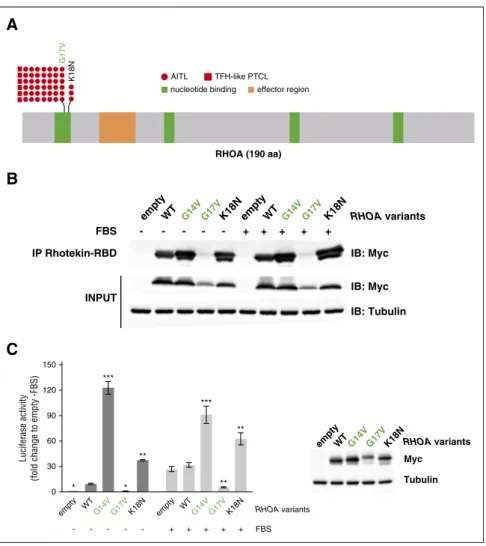
RHOA mutations include a novel activating K18N variant RHOA was the most frequently mutated gene among those analyzed (51/85; 60% of patients), with a similar prevalence in AITL and
TFH-like PTCL. Consistent with previous reports,12,13 the G17V
variant was found in most of the cases. We also identified a novel K18N mutation (variant frequency [VF] [5.5%; 7.4%; 10%]) in 3
AITL patients (Figure 3A; Table 1).30-44Both mutations occur in the
highly conserved GTP binding site of RHOA. We tested Myc-tagged 150
Luciferase activity
(fold change to empty -FBS)
120 90 60 30 0 empty WTG14V G17VK18N empty WTG14V G17VK18N * *** ** *** ** ** * - - - + + + + + FBS RHOA variants
C
B
AITLnucleotide binding effector region TFH-like PTCL K18N G17V RHOA (190 aa) RHOA variants RHOA variants IB: Myc Myc Tubulin - - - - - + + + + + IB: Myc IB: Tubulin IP Rhotekin-RBD FBS empty empty empty K18N K18N K18N WT WT WT G14V G14V G14V G17V G17V G17V INPUT
A
Figure 3. RHOA mutations in AITL and TFH-like PTCL. (A) Overview of the RHOA protein structure, showing G17V (42 AITL, 6 TFH-like PTCL) and the novel K18N (3 AITL) variants that target the highly conserved GTP/GDP binding site of RHOA. (B) Protein blot analysis of GTP-bound RHOA-Myc in rhotekin pull-down assay from HEK293T cells expressing indicated RHOA con-structs. IP, immunoprecipitation. Representative of 6 independent experiments. (C) SRE (serum-responsive element) luciferase reporter assay monitoring the activity of RHOA K18N mutant, compared with WT, G14V, or G17V mutants, previously characterized as activating and dominant-negative, respectively. Cells were stimulated (light gray) or not (dark gray) with fetal bovine serum for 6 hours. Data are represented as mean6 standard error of the mean (SEM) from 4 independent experiments. Significant differences in activation activity were deter-mined using 2-way analysis of variance (ANOVA) with repeated measurement (**P # .01; ***P # .001 compared with WT). Representative western blot from a luciferase assay experiment. Ectopic myc-tagged RHOA expression is revealed by anti-Myc. Anti-Actin blotting serves as loading control.
Table 1 . Characteristics of the 70 variants identified by targeted deep sequencing of TCR, JAK/STAT, and TLR-related genes in 8 5 TFH-derived PTCL sam ples Functional groups Gene Amino acid change Mutation type Domain New/repo rted Effect References Costimulatory and pro ximal TCR sign aling CD28 D124V Missense Extracellular AITL, ATLL Gain of function (F) 20,25 CD28 D124E Missense Extracellular AITL, ATLL Gain of function (F) 20,25 CD28 T195P Missense Cytoplasmic AITL, ATLL Gain of function (F) 20,25,30 LCK N446K* Missense Kinase New Probably gain o f functio n (P) LCK P447R* Missense Kinase New Probably gain o f functio n (P) FYN Q527X Stop gain ATLL Probably gain o f functio n (P) 20 FYN 52 5_525del† Frameshift deletion New Probably gain o f functio n (P) FYN S186L† Missense SH2 New Probably gain o f functio n (P) FYN K108fs* Frameshift deletion New Probably loss of functio n (P) FYN E107S* Missense SH3 New Probably loss of functio n (P) NK-kB/NF -AT path way PLCG1 E47K Missense PH 1 ATLL Gain of function (F) 20 PLCG1 R48W Missense PH 1 ATLL, SS No effect (F) 20,24 PLCG1 D342G Missense PI-PLC X-box ATLL, MF Gain of function (F) 20 PLCG1 S345F† Missense PI-PLC X-box ATL, MF, SS, ATLL Gain of function (F) 20-22,24 PLCG1 S520F† Missense PH 2; first part MF, SS, ATLL Gain of function (F) 20,21,24 PLCG1 E730K Missense SH2 2 New Gain of function (F) PLCG1 G869E Missense Near SH3 New Gain of function (F) PLCG1 E1163K Missense C2 ATLL, SS Gain of function (F) 20,24 PLCG1 D1165H Missense C2 ATLL, SS Gain of function (F) 20,24 PLCG1 D1165G Missense C2 ATLL Gain of function (F) 20 CARD11 F902C Missense ATLL Gain of function (F) 20 CARD11 S547T Missense New Gain of function (F) CARD11 F176C Missense C oiled coil New Gain of function (F) TRAF6 Q347X Stop gain Coiled coi l /MATH New NA PI3K pathway PIK3R1 K141R Missense RHO-GAP New Probably gain o f functio n (P) PIK3R1 Q475P Missense iSH2 New Probably gain o f functio n (P) PIK3R1 T576A Missense iSH2 New Probably gain o f functio n (P) PIK3R1 G680S Missense SH2 2 New Probably gain o f functio n (P) PIK3R1 V704M Missense SH2 2 New Probably gain o f functio n (P) PIK3R5 A259V Missense Carc inoma (liver) Probably gain o f functio n (P) 31 PIK3CA L1001P Missense PI3K/PI4K New Probably gain o f functio n (P) PDPK1 19_20del Inframe deletion New NA PDPK1 151_152de l Frameshift deletion Protein kinase New Probably loss of functio n (P) PDPK1 R324Q Missense Protein kinase New Probably gain o f functio n (P) PDPK1 P340Q Missense Protein kinase New Probably gain o f functio n (P) AKT1 G294R Missense Protein kinase New Probably gain o f functio n (P) CTNNB1 T41A† Missense Phospho by GSK3b Hepatocellul ar carcinoma, acute lymphoblastic leu kemia, breast cancer, Wilms’ tumor Gain of function (F) 32 CTNNB1 H36P Missense Hepatocellul ar carcinoma, acute lymphoblastic leu kemia, breast cancer, Wilms’ tumor Gain of function (F) 32 CTNNB1 S45F† Missense Hepatocellul ar carcinoma, acute lymphoblastic leu kemia, breast cancer, Wilms’ tumor Gain of function (F) 32 CTNNB1 K335T Missense Hepatoce llular carcinoma Gain of function (F) 33 Genes are organized by function al groups. AITL, angioimmunoblas tic T-cell lymp homa; ATLL, adult T-cell lymphoma/leuk emi a; D LBCL, diffuse large B-cell lymphoma; F, functional va lidation ; MF, mycosis fungoides; P, literature-and model s-based; SS, S ´ezary s yndrome. *Gene variants found in the same patie nt a nd in the same allele. †Gene variants found in the same patie nt b ut in different alleles.
Table 1 . (continued) Functional groups Gene Amino acid change Mutation type Domain New/repo rted Effect References AP-1/MAP K path way KRAS I36M Missense Hematop oeitic neoplasms Gain of function (F) 34 KRAS A18D Missense GTP binding Lymphoma Gain of function (F) 34 KRAS G13D Missense GTP binding Solid tumors Gain of function (F) 34 MAPK3 R278Q Missense Protein kinase New Probably gain o f functio n (P) STAT3 E616G Missense SH2 Lymphoma Gain of function (F) 26,35 STAT3 E616K Missense SH2 Lymphoma Gain of function (F) 26,35 GTF2I D317E Missense New Probably gain o f functio n (P) GTF2I N340S Missense New Probably gain o f functio n (P) GTF2I R523S Missense GTF2I-like New Probably gain o f functio n (P) GTF2I L607F Missense GTF2I-like New Probably gain o f functio n (P) GTF2I R702Q Missense New Probably gain o f functio n (P) GTPases path way RHOA K18N Missense GTP binding New Gain of function (F) RHOA G17V Missense GTP binding AITL, ATLL Dominant neg ative (F) 12-14,20,36 VAV1 151_158de l Frameshift deletion New Probably loss of functio n (P) VAV1 778_783de l Frameshift deletion New Probably gain o f functio n (P) VAV1 D797G Missense SH3 2 New Probably gain o f functio n (P) VAV1 Y826S Missense SH3 2 New Probably gain o f functio n (P) VAV2 Y214C Missense DH Gastri c ca rcinoma Proba bly dominant negative (P) JAK/STA T an d TLR pa thways JAK1 D831E Missense Kinase New Probably gain o f functio n (P) JAK2 V617F Missense Myelopro liferative disorders Gain of function (F) 37-43 JAK3 A699V Missense Kinase Lymphoma, carcinoma Gain of function (F) 37-43 MYD88 S219C Missense TIR DLBCL, lymphocytic leukemia Probably gain o f functio n (P) 44 Genes are organized by function al groups. AITL, angioimmunoblas tic T-cell lymp homa; ATLL, adult T-cell lymphoma/leuk emi a; D LBCL, diffuse large B-cell lymphoma; F, functional va lidation ; MF, mycosis fungoides; P, literature-and model s-based; SS, S ´ezary s yndrome. *Gene variants found in the same patie nt a nd in the same allele. †Gene variants found in the same patie nt b ut in different alleles.
G14V (constitutively active45), G17V (dominant-negative), K18N, and wild-type (WT) RHOA in a pull-down assay using Rhotekin-RBD (r binding domain) beads (Figure 3B). Although RHOA G17V
already characterized as dominant-negative12-14did not bind RBD
beads, even in the presence of fetal bovine serum, compared with WT, RHOA K18N showed a marked increase of RBD beads binding similarly to RHOA G14V (Figure 3B). Moreover, unlike RHOA G17V that did not activate and even repressed transcription from the SRE under serum activation, RHOA K18N markedly enhanced its transcription (Figure 3C).
Mutations in other TCR signaling–related genes are variably recurrent and diverse
Apart from RHOA, mutations in genes involved in TCR costimulation or signaling were detected in 42 of 85 patients (49%) (Figures 1 and 2; Table 1).
CD28 and TCR-proximal signaling genes. Three different
CD28 mutations—T195P, D124V, and D124E—all described
previously13,25,30were identified in 4, 2, and 2 AITL patients, respectively
(8/85, 9.4%) (Figure 4A). All TFH-like PTCL were CD28-WT. T195P and D124V mutations are known to enhance TCR/CD28-induced NF-kB
activity in vitro.25,30Three patients (2 AITL, 1 TFH-like PTCL; 3.5%)
harbored mutations in FYN, a TCR- and CD28-proximal kinase component (Figure 4B). Mutations in the SH2 domain (S186L) and the absence of phosphorylated tyrosine 531 (T524fs or Q527X mutations) probably confer enhanced kinase activity by disrupting their inhibitory
interaction.12The 2 amino acid substitutions in the tyrosine kinase domain
of LCK observed in one AITL patient is expected to enhance its kinase
activity.46We did notfind mutations in ICOS, ZAP70, LAT, or ITK.
NF-kB/NFAT pathway. Ten different missense mutations
span-ning the coding region of PLCG1 (VF [2.1%; 38%]) were identified in 12 of 85 patients (8 AITL, 4 TFH-like PTCL; 14.1%) (Figure 5A). Apart from previously reported variants in adult T-cell leukemia/lymphoma
(ATLL) or in other PTCL,20-24we identified 2 novel variants (E730K and
G869E). We generated all mutant constructs and tested their activity against WT PLCG1 in a FRET-based reporter assay of MALT1
protease activity27,28(Figure 5B) and in a NFAT luciferase reporter assay
(Figure 5C). Both experiments confirmed that the S345F and S520F
variants were activating.21All 6 variants in the PI-PLC, SH2, SH3, and C2
domains were also activating, increasing the FRET signal by 1.7- to
threefold and the luciferase expression by four- tofivefold. Of the 2 PH1
domain variants, R48W had no effect in both assays, whereas E47K increased NFAT reporter activity by 2.1-fold and FRET signal by 1.5-fold. Point mutations in CARD11, which encodes a scaffolding protein downstream of PLCG1 required for CD28/TCR-induced NF-kB
D
A259V
PIK3R5 (880 aa) heterodimerization binding to Ggamma-beta
G
H36P T41A(a) S45F(a) K335T
CTNNB1 (781 aa) GSK3β inhibitory region armadillo repeats
H
I36M
A18D
G13D
KRAS (189 aa) nucleotide binding
I
D317E N340S R523S L607F R702Q
GTF2I (997 aa) GTF2I-like
J
Y826S A777fs I149fs D797G VAV1 (845 aa) CH DH PH SH3 SH2 SH3F
T402M P340Q R324Q T148fs Δ C20 PDK1 (556 aa) kinase PHB
E107S(b) K108fs (b) S186L(a) T524fs (a) Q527X
FYN (537 aa) SH3 SH2 kinase D124V D124E T195P CD28 (220 aa)
A
extra-cellular TM cytoplasmicE
L1001P PIK3CA (1068 aa)ABD RBD C2 helical kinase
AITL TFH-like PTCL missense inframe indel frameshift indel stop gain
C
K141R Q475P T576A G680S V704M
PIK3R1 (724 aa)
SH3 Rho-GAP SH2 1 iSH2 SH2 2
Figure 4. Mapping of variants in TCR signaling genes mutated in at least 3 patients. (A) CD28: the D124V/E variants involve the extracellular part of the receptor, whereas T195P lies in the intracellular, C-terminal, domain between the 2 domains, allowing interaction with PIK3R1 (YVKM sequence) or GRB2/VAV (PRRP sequence) proteins. (B) FYN: the five mutations indicated occurred in three patients; 1 patient harbored 2 mutations (a); 2 mutations in adjacent positions in the SH3 domain (b) were observed on the same allele in another patient. (C-E) PI3K subunits: when appropriate, cellular stimuli are present, the nSH2 and cSH2 domains of PIK3R1 bind phosphorylated tyrosines (YXXM motif) in activated receptors (CD28) and adapter proteins, thereby activating the PIK3CA (p110a) catalytic subunit without releasing the PIK3R1 (p85a) interaction with p110a through their iSH2 and ABD domains, respectively. The K141R missense affects the r-GAP domain, whereas iSH2 and the second SH2 domains of PIK3R1 bore 2 missenses each (Q475P, T576A and G680S, V704M, respectively). The A259V mutation, described as somatic in COSMIC, affects a linker region of PIK3R5. Finally, the L1001P point mutation affects the PIK3CA kinase domain. (F) PDK1: one inframe INDEL, one frameshift INDEL, and 3 missense affect PDK1 protein. (G) CTNNB1: 3 previously described activating missenses affect the GSK3b inhibitory domain (exon 3), whereas the K335T activating mutation affects the armadillo repeats region. (H) KRAS: 3 missense mutations alter 2 N-terminal nucleotide binding regions. (I) GTF2I: 5 missense mutations affect the GTF2I transcription factor, among which two are found in different GTF2I-like domains. (J) VAV1: 2 frameshift deletions and 2 missenses affect VAV1, three being localized in the C-terminal SH3 domain of the protein. In all figure panels, previously described activating mutations are in green boldface, and mutations previously described but not functionally tested are underlined. PDK1 is the protein name of PDPK1 gene.
activation,28were found in 2 AITL and 1 TFH-like PTCL (3.5%). The F176C variant affects the coiled-coil domain of the protein, commonly
mutated in diffuse large B-cell lymphoma (DLBCL).27The S547T
and F902C variants map to linker regions with undefined structure
(Figure 5D). In a FRET-based assay,28F176C and S547T enhanced
MALT1 proteolytic activity by two- and fourfold, respectively,
whereas the F902C mutant, previously reported in ATLL patients,20
had no detectable effect (Figure 5E). When transfected into Jurkat cells
deficient for CARD11,29
we found that, compared with WT CARD11, all 3 variants induced enhanced NF-kB reporter activity in response to phorbol myristate acetate/ionomycin stimulation (Figure 5F).
PI3K pathway. Six patients (3 AITL, 3 TFH-like PTCL; 7%)
showed mutations in PI3K genes encoding the regulatory subunits
PIK3R1 (n5 5), PIK3R5 (n 5 1), or the catalytic subunit PIK3CA
(n 5 1) (Figure 4C-E). These mutations likely enhance the
catalytic subunit activity or increase PIK3R1 binding to CD28.31
Five AITL patients (5.9%) had mutations in PDPK1 (PDK1), a master serine/threonine kinase with multiple targets including AKT. Three missense mutations were found on or near its kinase
domain, suggesting an activating effect47 (Figure 4F). Four
different CTNNB1 mutations, known to occur in a variety of
carcinomas and Wilms’ tumors, were identified in 5 patients
(4 AITL, 1 TFH-like PTCL; 5.9%) (Figure 4G). All have been previously characterized as activating or stabilizing variants that induce persistent signaling and increased proliferation, and have been linked to both poor treatment response and frequent tumor
relapse in various tumors.32,33
AP-1/MAPK pathway. Eleven patients (8 AITL, 3 TFH-like
PTCL; 13%) had mutually exclusive activating mutations in the MAPK pathway, which regulate the activity of the AP-1 transcription factor
family. Activating KRAS34and STAT326,35mutations were detected in
3 and 2 patients, respectively (Figure 4H). We also found 5 missense
mutations in GTF2I (TFII-I)48(Figure 4I), which are likely activating as
those reported in thymic epithelial tumors.49
GTPases pathway. Apart from highly recurrent RHOA
muta-tions, 4 patients (3 AITL, 1 TFH-like PTCL; 4.7%) harbored previously undescribed frameshift deletions or missense mutations in VAV1, a guanosine exchange factor for RAC1, CDC42, and RHOA (Figure 4J). Two missense mutations were on the second SH3 domain, where a D797 residue mutation was previously shown to activate VAV1-mediated transformation via cell-cell
contact deregulation.50
Analysis of sequential biopsies. Analysis of paired samples in
4 refractory or relapsed patients (supplemental Table 8) showed that the
mutations identified at diagnosis were also present in the second biopsy,
with overall very similar VF. Within the limit of the panel of genes examined, 2 patients presented one additional mutation at relapse (CTNNB1 and VAV1), at somewhat lower VF than those present at diagnosis. Although this analysis is limited to a few pairs of samples, it indicates that the mutational heterogeneity that characterizes many AITL samples appears to be preserved and is essentially not modified by the treatments administered.
Activating mutations in TCR signaling–related genes correlate with molecular signatures reflecting higher T-cell activation and with response to therapy
Sixty-six of 85 patients harbored mutations in TCR signaling genes. To assess the biological and clinical impact of the tumor mutational status, we focused on patients with mutations in TCR signaling–related genes other than RHOA, designated hereafter as “TCR_Mut” (42/85 cases,
49%), and compared them with those with only RHOA or no detectable
mutation, designated“TCR_WT” (43/85 cases, 51%) (Figure 6).
Mutations in TCR-related genes other than RHOA were virtually nonoverlapping, and the vast majority (47/56 distinct variants, 84%)
were either functionally validated (n5 24) or predicted (n 5 23) to be
biologically gain-of-function mutants (Figure 6). Consequently, nearly all TCR_Mut patients (39/42 patients, 93%) harbored at least 1 activating mutation.
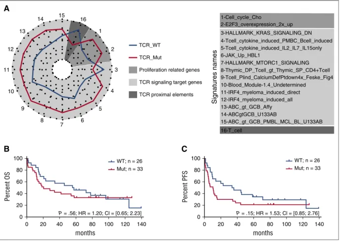
There were no significantly differentially expressed genes between TCR_Mut and TCR_WT samples (supplemental Figure 4). However,
the molecular signatures of TCR_Mut were significantly enriched in 15
gene sets compared with TCR_WT by enrichment analysis (Figure 7A;
supplemental Table 7; supplemental Figure 5), reflecting the activation
of signaling pathways like PI3K, NF-kB,17IRF4, and JAK-STAT, as
well as the upregulation of calcium-signaling target genes. This points toward TCR signaling activation in TCR_Mut PTCLs. TCR_Mut
samples also showed enrichment in cell-cycle signatures reflecting
increased proliferative activity. Finally, there was a trend for TCR_Mut samples to have downregulated proximal TCR signaling genes (signature “T_cell”) such as TCR (TCRa, TCRb; CD3d, CD3g), signaling molecules (LAT, TRIM, SAP, FYB, ZAP70), and cell surface markers
(adjusted P 5 .10), a feature consistent with sustained T-cell activation.51
Then, when comparing the clinical features and outcome of
TCR_Mut and TCR_WT patients, no significant differences in
main clinical characteristics at presentation (supplemental Table 9) or in OS (18 vs 40 months median OS; 5-year OS 20% vs 29%; P 5 .37) were found (supplemental Figure 6). Strikingly, however, among the 59 patients (49 AITL and 10 TFH-like PTCLs) who received anthracyclin-based induction chemotherapy, 11 of 33 (33%) TCR_Mut patients vs 2 of 26 (8%) TCR_WT (P 5 .02, supplemental Table 9) showed early
progression, relapse, or no response to treatment within thefirst 6
months after diagnosis. This, however, did not translate into a
statistically significant difference in PFS at 5 years (21% vs 24% for
TCR_Mut vs TCR_WT patients; P 5 .15) (Figure 7B-C).
Discussion
In this study, we confirmed the high prevalence of RHOA mutations in
TFH-derived PTCL, with most samples bearing the dominant-negative
G17V variant. Interestingly, we identified a new K18N variant in 3% of
the patients that presented activating features in in vitro assays. This
intriguingfinding of distinct RHOA mutations, with apparently opposite
functional properties in the same disease, was also recently documented
in ATLL.36,52Further studies are warranted to understand how these
variants may contribute to the pathogenesis of PTCLs.
Apart from RHOA alterations, we identified activating and virtually mutually exclusive mutations in diverse TCR signaling genes in half of TFH-derived lymphomas. Individually, the frequencies of these mutations were variable, with the 5 most mutated genes (PLCG1, CD28, PIK3 elements, GTF2I, CTNNB1) being altered in 14% to 5% of the patients. CD28 and FYN mutations in AITL were previously
described in a small number of cases.12-14Our study significantly
expands these previous results and strongly supports a role of activated TCR signaling in the pathogenesis of TFH-derived PTCLs, drawing strong parallels with the role of BCR signaling in B-cell lymphomas. Mutations in genes related to TCR costimulation and signaling were recently reported in other PTCL entities, like cutaneous T-cell
lympho-mas, PTCL-NOS, and ATLL.20-24,53Nonetheless, mutations in specific
genes are variably recurrent in distinct entites,24,25and for a given gene,
G869E E730K S520F S345F E1163K D1165G D1165H D342G R48W E47K
PH1 EF PI-PLCX-box PI-PLCY-box
PLCG1 (1301 aa) AILT TFH-like PTCL AILT TFH-like PTCL
SH2 SH2 SH3 C2
nPH2 cPH2
A
F176C S547T F902C
CARD11 (1154 aa)
CARD Coiled-coil PDZ SH3 GUK
D
0 PLCG1-myc HA-CARD11 Strep-MALT1 Malt1 HA Myc PLCG1-myc Actin Myc empty WT E47K R48W D342G S345F WT L244P F176CS547T F902Cempty WT E47KR48W D342GS345F S520F empty WTE730KG869E E1163KD1165GD1165H S520F E730K G869E E1163K D1165GD1165H
Tubulin MALT1 PLCG1 variants *** *** *** *** *** *** *** *** ** + + + + + + + + + + + + MALT1 empty WT 1 2 3 4
E47KR48WS342GS345FS520FE730KG869EE1163KD1165GD1165H
E47K D342G E1163K D1165H MAL T1 cleavage activity (fold change to PLCG1 WT)
B
CARD11 variants ** ** ** + + + + + + + + + + + 0 MALT1 2 4 6 8 emptyWTL244PF176CS547TF902C MAL T1 cleavage activity(fold change to CARD11 WT)
E
0 PLCG1 variants 1 2 3 4 5 6 7 8 9 ** ** ** ** ** ** * ** * WT S345FS520FE730KG869E D1165G R48W Luciferase activity(fold change to empty)
C
0 20 40 60 80 100 120 CARD11 variants ** ** *** * * + -- + + + + + PMA/IONO emptyWTL244PF176CS547TF902Cempty WT L244PF176CS547TF902C Luciferase activity(fold change to empty DMSO)
F
Figure 5. Mapping and functional analysis of PLCG1 variants and CARD11 variants. (A) Schematic representation of PLCG1 protein with mapping of the 10 missense mutations identified in AITL (circles) or TFH-like PTCL (squares) cases. Previously described activating mutations are in green boldface and mutations previously described but not functionally tested are underlined. (B) Monitoring of PLCG1-mediated MALT1 activation via a FRET-based reporter assay. Data are represented as mean6 SEM from 3 independent experiments. Significant differences in activation activity were determined using 1-way ANOVA (**P # .01; ***P # .001). Representative western blot from a MALT1 activation experiment. PLCG1 expression is revealed by anti-Myc tag blotting, whereas MALT1 expression is shown by anti-MALT1 antibody. (C) NFAT luciferase reporter assay monitoring activity of PLCG1 mutants, compared with previously reported activating mutants (green). Data are represented as mean6 SEM from 7 independent experiments. Significant differences in activation activity were determined using 1-way ANOVA (*P # .05; **P # .01). Representative western blot from a luciferase assay experiment. Ectopic myc-tagged PLCG1 expression is revealed by anti-Myc. Anti-Actin blotting serves as loading control. (D) Schematic representation of CARD11 protein with mapping of the 3-point mutations found in 2 AITL (circles) and 1 TFH-like PTCL-NOS (square) patients. A previously described mutation is underlined. (E) Monitoring of CARD11-mediated MALT1 activation via a FRET-based reporter assay. Data are
heterogeneous. For instance, PLCG1 and CARD11 mutations are highly prevalent (up to 18% and 15% of the cases, respectively) in cutaneous T-cell lymphoma, in which conversely CD28 mutations
are not found.20CARD11 mutations are more frequent in ATLL
(24%) and S´ezary syndrome (10.9%) than in this series of TFH-derived lymphomas. Whether the observed mutations are sufficient to induce constitutive activation of the respective pathways, and how their effect may be dependent or influenced by antigen-driven or other stimulatory signals remains unknown. Interestingly, some of the TCR-related genes may be altered through point mutations or small indels as well as by major structural rearrangements,
amplifications, and deletions.20,23,24 Collectively, these findings
strongly suggest that genetic alterations affecting TCR signaling operate as a common pathogenic mechanism in several PTCL entities.
Our analysis also explored the JAK/STAT and TLR pathways. Mutation-induced activation of the JAK/STAT pathway, highly prevalent in myeloid neoplasms, was recently identified as a major oncogenic mechanism in several T-cell leukemias and lymphomas
derived from innate immune cells.20,37-43,53 Our results suggest,
however, that JAK/STAT mutations do not have the same importance in the pathogenesis of TFH-derived lymphomas, because only 4 of 85 patients bore a single activating mutation each in JAK1, JAK2, JAK3, or
MYD88 (Table 1).44 Except for JAK2 V617F, the 3 others were
observed in PLCG1-mutated cases.
Our data further support that AITL and TFH-like PTCL are
closely related at the molecular level5and share common oncogenic
mechanisms. We and others have reported the occurrence of TET2, DNMT3A, and RHOA mutations in a large proportion of
both AITL and TFH-like PTCL patients.5,8,9,12-14 This study
extends the molecular overlap across TFH-derived neoplasms to genetic alterations in various TCR-signaling. Interestingly, CD28 mutations were exclusively identified in AITL patients, consistent
with recent reports.25,30 Similarly, IDH2 mutations were rarely
detected in PTCL cases,9,54suggesting that the distribution of some
genetic features might be relevant in distinguishing AITL from other TFH-derived nodal lymphomas, which otherwise share many
common features.5
In the proposed multistep model of pathogenesis for TFH-derived PTCL, mutation-induced epigenetic deregulation, possibly arising in early hematopoietic precursors, may promote the emergence of premalignant cells, which requires additional genetic events to acquire a
definitively malignant phenotype.13,55It has been thus suggested that
RHOA alterations occur as a second event in TET2- and/or
DNMT3A-mutated cells.13,55,56The prevalence of mutations in TET2, DNMT3A, and
IDH2 analyzed in a subset of our cases was 52% (34/65 cases), 29% (17/
56 cases), and 30% (21/71 cases), respectively.8,9These mutations were
not mutually exclusive, and in fact tended to co-occur with each other, as
previously reported.12,13We also found a tendency for RHOA mutations
to associate with epigenetic alterations, because RHOA mutations were detected in 31 of 41 (76%) cases mutated in TET2, DNMT3A, and/or IDH2, but in only 8 of 23 (35%) cases without epigenetic mutations (P 5 .003). Because the proportion of neoplastic cells in AITL is typically
low and variable from case to case,3the comparison of VF of different
TCR mut, RHOA mut or wt
(n = 42/85)
TCR_Mut (49%) TCR_WT (51%)
RHOA
AITL TFH-like PTCL
Missense Frameshift Indel Inframe Indel Nonsense Activating mutations
PIK3CA MAPK3 VAV2 PIK3R5 AKT1 TRAF6 LCK STAT3 KRAS CARD11 FYN VAV1 PDPK1 PIK3R1 GTF2I CTNNB1 CD28 PLCG1 TCR wt, RHOA mut (n = 24/85) TCR wt, RHOA wt (n = 19/85)
Figure 6. Mutual exclusivity of TCR signaling variants. The mutational status of TCR-related genes is represented for the 85 patients of the extended cohort. Genes other than RHOA are ranked by decreasing mutation frequency and show an essentially mutually exclusive mutation pattern. In total, 49% of cases were mutated in 1 or several TCR-related gene(s) other than RHOA (hereafter considered as TCR_Mut), 28.5% were mutated in RHOA only, and 22.5% harbored no mutation in any of the genes tested (collectively considered as TCR_WT).
Figure 5 (continued) represented as mean6 SEM from 6 independent experiments. Significant differences in activation activity were determined using 1-way ANOVA (*P # .05; **P # .01). The known activating L244P variant (green) was used as a positive control for the experiment. Representative western blot from a MALT1 activation experiment. CARD11 expression is revealed by anti-HA tag blotting, whereas MALT1 expression is shown by anti-MALT1 antibody. (F) NF-kB luciferase reporter assay in Jurkat cells deficient for CARD11 monitoring activity of CARD11 mutants, compared with previously reported activating mutants (green). Data are represented as mean6 SEM from 4 independent experiments. Significant differences in activation activity were determined using 2-way ANOVA (*P # .05; **P # .01; ***P # .001 compared with WT PMA/IONO).
mutated genes was performed for each individual sample. In 13 cases harboring mutations in both TET2 and/or DNMT3, and RHOA or IDH2 with available VF, the TET2/DNMT3A VF was significantly higher than for RHOA or IDH2 (supplemental Figure 7A). There was no obvious association of RHOA mutations with other TCR-related genes, because 27 of 51 (53%) RHOA-mutated and 15 of 34 (44%) RHOA-WT cases were mutated in other TCR-related genes (P 5 .51). In 27 cases mutated in both RHOA and one or several genes of the TCR, JAK/STAT, or TLR pathways, no significant difference in VF means was observed between RHOA and other genes (supplemental Figure 7B).
Although our results warrant confirmation in cohorts of patients treated on prospective clinical trials, they suggest that the muta-tional status of TCR-related genes may have important clinical implications, predicting early treatment failure with anthracyclin-based chemotherapy in TFH-related PTCL patients. Importantly, several activating mutations found in PI3K or NF-kB pathways could be targeted by idelalisib or the proteasome inhibitor bortezomib, respectively. It is of interest to determine their efficacy in TCR_Mut
patients,12,21possibly in combination with demethylating agents.57
Thus, similar to the importance of targeting BCR signaling in B-cell
lymphomas with the BTK inhibitor ibrutinib,17characterization of
the TCR mutational status might open new avenues to design specific and more effective therapies. In clinical practice, given the high number of genes involved and the diversity of mutations found, targeted deep sequencing with high depth/coverage appears to be the method of choice for selecting patients.
Acknowledgments
The authors acknowledge Catherine Chapuis (Pathology, Lausanne) and Caroline Communaux from the LYSA-Pathology for their technical assistance; K. Harshman and the Lausanne Genomic Technology Facility for their technical support; and C´eline Villenet and Sabine Quief from the plate-forme de g´enomique fonctionnelle et structurale, Lille University, for the WES experiments.
This study was supported by grants received from the Plan Cancer (Belgium), the Ligue du Cancer (Switzerland), the Institut National du Cancer (INCa AAP PLBIO13-085, INCa-DGOS 2010-085, and INCa-Plan Cancer 2013), and the MEDIC foundation.
Authorship
Contribution: D.V., R.D.M., M.T., R.D.G., L.d.L., and P.G. conceived and designed the study; D.V. and M.P.D.D. developed the methodologies; D.V., M.P.D.D., J.B., B.F., A.M., C.B., F.L., M.J., M.T., J.I., A.R., M.F., and B.B., acquired the data; D.V., M.P.D.D., E.M., F.L., M.J., O.M., O.T., C.H., M.D., O.A.B., J.-P.J., and J.G. analyzed and interpreted the data; D.V., M.P.D.D., F.L., E.M., L.d.L.,
1-Cell_cycle_Cho 2-E2F3_overexpression_2x_up 3-HALLMARK_KRAS_SIGNALING_DN 4-Tcell_cytokine_induced_PMBC_Bcell_induced 5-Tcell_cytokine_induced_IL2_IL7_IL15only 6-JAK_Up_HBL1 7-HALLMARK_MTORC1_SIGNALING 8-Thymic_DP_Tcell_gt_Thymic_SP_CD4+Tcell 9-Tcell_Plind_CalciumDefPtdown4x_Feske_Fig4 10-Blood_Module-1.4_Undetermined 11-IRF4_myeloma_induced_direct 12-IRF4_myeloma_induced_all 13-ABC_gt_GCB_Affy 14-ABCgtGCB_U133AB 15-ABC_gt_GCB_PMBL_MCL_BL_U133AB 16-T_cell Signatures names 0 0 20 40 60 80 Per cent OS 100
B
20 40 60 months P = .56; HR = 1.20; Cl = [0.65; 2.23] 80 100 WT; n = 26 Mut; n = 33 WT; n = 26 Mut; n = 33 120 140 0 0 20 40 60 80 Per cent PFS 100C
20 40 60 months P = .15; HR = 1.53; Cl = [0.85; 2.76] 80 100 120 140 1 2 3 4 5 6 7 8 9 10 11 12 13 14 15 16 TCR_WT TCR_MutProliferation related genes TCR signaling target genes TCR proximal elements
A
Figure 7. Biological significance and clinical relevance of TCR signaling–related mutations. (A) Spider plot representation of gene sets differentially enriched in patients with or without mutations in genes related to TCR signaling (TCR_Mut vs TCR_WT). Genes tested in the enrichment analysis were selected from signatures relevant in T- and B-cell differentiation and activation. Statistical significance of the enrichment was reached for gene sets 1 to 15 (P, .05); for gene set 16, marginal significance was observed (P5 .1). (B-C) Overall survival (B) and progression-free survival (C) of patients with (red) or without (blue) mutations in TCR signaling–related genes. Analyses are restricted to the 59 patients treated with anthracyclin-based chemotherapy. Mutated patients show a trend toward a shorter PFS (11 vs 36 months) than WT patients (P5 .15).
and P.G. wrote the manuscript; V.F. was the administrative, technical, and material support; and L.d.L. and P.G. supervised the study.
Conflict-of-interest disclosure: The authors declare no competing financial interests.
ORCID profiles: D.V., 0000-0003-0703-6762.
Correspondence: Laurence de Leval, CHUV Institut de Patho-logie, Rue du Bugnon 25, 1011 Lausanne, Switzerland; e-mail: laurence.deleval@chuv.ch.
References
1. Federico M, Rudiger T, Bellei M, et al. Clinico-pathologic characteristics of angioimmunoblastic T-cell lymphoma: analysis of the international peripheral T-cell lymphoma project. J Clin Oncol. 2013;31(2):240-246.
2. de Leval L, Parrens M, Le Bras F, et al. Angioimmunoblastic T-cell lymphoma is the most common T-cell lymphoma in two distinct French information data sets. Haematologica. 2015;100(9): e361-e364.
3. de Leval L, Rickman DS, Thielen C, et al. The gene expression profile of nodal peripheral T-cell lymphoma demonstrates a molecular link between angioimmunoblastic T-cell lymphoma (AITL) and follicular helper T (TFH) cells. Blood. 2007;109(11):4952-4963.
4. Gaulard P, de Leval L. Pathology of peripheral T-cell lymphomas: where do we stand? Semin Hematol. 2014;51(1):5-16.
5. Dobay MP, Lemonnier F, Missiaglia E, et al. A PTCL, NOS subset with molecular and clinical features similar to AITL. Hematol Oncol. 2015; 33(S1):100-180.
6. Mourad N, Mounier N, Bri `ere J, et al; Groupe d’Etude des Lymphomes de l’Adulte. Clinical, biologic, and pathologic features in 157 patients with angioimmunoblastic T-cell lymphoma treated within the Groupe d’Etude des Lymphomes de l’Adulte (GELA) trials. Blood. 2008;111(9): 4463-4470.
7. Corradini P, Vitolo U, Rambaldi A, et al. Intensified chemo-immunotherapy with or without stem cell transplantation in newly diagnosed patients with peripheral T-cell lymphoma. Leukemia. 2014; 28(9):1885-1891.
8. Lemonnier F, Couronn ´e L, Parrens M, et al. Recurrent TET2 mutations in peripheral T-cell lymphomas correlate with TFH-like features and adverse clinical parameters. Blood. 2012;120(7): 1466-1469.
9. Cairns RA, Iqbal J, Lemonnier F, et al. IDH2 mutations are frequent in angioimmunoblastic T-cell lymphoma. Blood. 2012;119(8):1901-1903. 10. Jaiswal S, Fontanillas P, Flannick J, et al.
Age-related clonal hematopoiesis associated with adverse outcomes. N Engl J Med. 2014;371(26): 2488-2498.
11. Muto H, Sakata-Yanagimoto M, Nagae G, et al. Reduced TET2 function leads to T-cell lymphoma with follicular helper T-cell-like features in mice. Blood Cancer J. 2014;4(12): e264.
12. Palomero T, Couronn ´e L, Khiabanian H, et al. Recurrent mutations in epigenetic regulators, RHOA and FYN kinase in peripheral T cell lymphomas. Nat Genet. 2014;46(2): 166-170.
13. Sakata-Yanagimoto M, Enami T, Yoshida K, et al. Somatic RHOA mutation in
angioimmunoblastic T cell lymphoma. Nat Genet. 2014;46(2):171-175.
14. Yoo HY, Sung MK, Lee SH, et al. A recurrent inactivating mutation in RHOA GTPase in angioimmunoblastic T cell lymphoma. Nat Genet. 2014;46(4):371-375.
15. Cleverley SC, Costello PS, Henning SW, Cantrell DA. Loss of Rho function in the thymus is accompanied by the development of thymic lymphoma. Oncogene. 2000;19(1):13-20.
16. Odejide O, Weigert O, Lane AA, et al. A targeted mutational landscape of angioimmunoblastic T-cell lymphoma. Blood. 2014;123(9): 1293-1296.
17. Wilson WH, Young RM, Schmitz R, et al. Targeting B cell receptor signaling with ibrutinib in diffuse large B cell lymphoma. Nat Med. 2015; 21(8):922-926.
18. Inghirami G, Chan WC, Pileri S; AIRC 5xMille consortium ‘Genetics-driven targeted management of lymphoid malignancies’. Peripheral T-cell and NK cell lymphoproliferative disorders: cell of origin, clinical and
pathological implications. Immunol Rev. 2015; 263(1):124-159.
19. Pechloff K, Holch J, Ferch U, et al. The fusion kinase ITK-SYK mimics a T cell receptor signal and drives oncogenesis in conditional mouse models of peripheral T cell lymphoma. J Exp Med. 2010;207(5):1031-1044. 20. Kataoka K, Nagata Y, Kitanaka A, et al.
Integrated molecular analysis of adult T cell leukemia/lymphoma. Nat Genet. 2015;47(11): 1304-1315.
21. Vaqu ´e JP, G ´omez-L ´opez G, Mons ´alvez V, et al. PLCG1 mutations in cutaneous T-cell lymphomas. Blood. 2014;123(13):2034-2043. 22. Manso R, Rodr´ıguez-Pinilla SM, Gonz´alez-Rinc ´on
J, et al. Recurrent presence of the PLCG1 S345F mutation in nodal peripheral T-cell lymphomas. Haematologica. 2015;100(1):e25-e27. 23. da Silva Almeida AC, Abate F, Khiabanian H,
et al. The mutational landscape of cutaneous T cell lymphoma and S ´ezary syndrome. Nat Genet. 2015;47(12):1465-1470.
24. Wang L, Ni X, Covington KR, et al. Genomic profiling of S ´ezary syndrome identifies alterations of key T cell signaling and differentiation genes. Nat Genet. 2015;47(12):1426-1434.
25. Rohr J, Guo S, Huo J, et al. Recurrent activating mutations of CD28 in peripheral T-cell lymphomas. Leukemia. 2016;30(5):1062-1070. 26. Morin RD, Mendez-Lago M, Mungall AJ, et al.
Frequent mutation of histone-modifying genes in non-Hodgkin lymphoma. Nature. 2011;476(7360): 298-303.
27. Lenz G, Davis RE, Ngo VN, et al. Oncogenic CARD11 mutations in human diffuse large B cell lymphoma. Science. 2008;319(5870): 1676-1679.
28. Pelzer C, Cabalzar K, Wolf A, Gonzalez M, Lenz G, Thome M. The protease activity of the paracaspase MALT1 is controlled by monoubiquitination. Nat Immunol. 2013;14(4):337-345.
29. Wang D, You Y, Case SM, et al. A requirement for CARMA1 in TCR-induced NF-kappa B activation. Nat Immunol. 2002;3(9):830-835.
30. Lee SH, Kim JS, Kim J, et al. A highly recurrent novel missense mutation in CD28 among angioimmunoblastic T-cell lymphoma patients. Haematologica. 2015;100(12):e505-e507. 31. Huang C-H, Mandelker D, Schmidt-Kittler O,
et al. The structure of a human p110a/p85a complex elucidates the effects of oncogenic PI3Kalpha mutations. Science. 2007;318(5857): 1744-1748.
32. Austinat M, Dunsch R, Wittekind C, Tannapfel A, Gebhardt R, Gaunitz F. Correlation between b-catenin mutations and expression of
Wnt-signaling target genes in hepatocellular carcinoma. Mol Cancer. 2008;7(1):21. 33. Pilati C, Letouz ´e E, Nault J-C, et al. Genomic
profiling of hepatocellular adenomas reveals recurrent FRK-activating mutations and the mechanisms of malignant transformation. Cancer Cell. 2014;25(4):428-441.
34. Stolze B, Reinhart S, Bulllinger L, Fr¨ohling S, Scholl C. Comparative analysis of KRAS codon 12, 13, 18, 61, and 117 mutations using human MCF10A isogenic cell lines. Sci Rep. 2015;5:8535. 35. Ohgami RS, Ma L, Monabati A, Zehnder JL, Arber DA. STAT3 mutations are present in aggressive B-cell lymphomas including a subset of diffuse large B-cell lymphomas with CD30 expression. Haematologica. 2014;99(7):e105-e107. 36. Nagata Y, Kontani K, Enami T, et al. Variegated
RHOA mutations in adult T-cell leukemia/ lymphoma. Blood. 2016;127(5):596-604. 37. Koskela HL, Eldfors S, Ellonen P, et al. Somatic
STAT3 mutations in large granular lymphocytic leukemia. N Engl J Med. 2012;366(20): 1905-1913.
38. Jerez A, Clemente MJ, Makishima H, et al. STAT3 mutations unify the pathogenesis of chronic lymphoproliferative disorders of NK cells and T-cell large granular lymphocyte leukemia.
Blood. 2012;120(15):3048-3057.
39. Koo GC, Tan SY, Tang T, et al. Janus kinase 3-activating mutations identified in natural killer/ T-cell lymphoma. Cancer Discov. 2012;2(7): 591-597.
40. Kiel MJ, Velusamy T, Rolland D, et al. Integrated genomic sequencing reveals mutational landscape of T-cell prolymphocytic leukemia. Blood. 2014;124(9):1460-1472.
41. Bellanger D, Jacquemin V, Chopin M, et al. Recurrent JAK1 and JAK3 somatic mutations in T-cell prolymphocytic leukemia. Leukemia. 2014; 28(2):417-419.
42. Nicolae A, Xi L, Pittaluga S, et al. Frequent STAT5B mutations in gd hepatosplenic T-cell lymphomas. Leukemia. 2014;28(11):2244-2248. 43. K ¨uc¸ ¨uk C, Jiang B, Hu X, et al. Activating
mutations of STAT5B and STAT3 in lymphomas derived from gd-T or NK cells. Nat Commun. 2015;6:6025.
44. Avbelj M, Wolz O-O, Fekonja O, et al. Activation of lymphoma-associated MyD88 mutations via allostery-induced TIR-domain oligomerization. Blood. 2014;124(26):3896-3904.
45. Ihara K, Muraguchi S, Kato M, et al. Crystal structure of human RhoA in a dominantly active form complexed with a GTP analogue. J Biol Chem. 1998;273(16):9656-9666.
46. Laham LE, Mukhopadhyay N, Roberts TM. The activation loop in Lck regulates oncogenic potential by inhibiting basal kinase activity and restricting substrate specificity. Oncogene. 2000; 19(35):3961-3970.
47. Chinen Y, Kuroda J, Shimura Y, et al. Phosphoinositide protein kinase PDPK1 is a crucial cell signaling mediator in multiple myeloma. Cancer Res. 2014;74(24): 7418-7429.
48. Sacrist ´an C, Schattgen SA, Berg LJ, Bunnell SC, Roy AL, Rosenstein Y. Characterization of a novel interaction between transcription factor TFII-I and the inducible tyrosine kinase
in T cells. Eur J Immunol. 2009;39(9): 2584-2595.
49. Petrini I, Meltzer PS, Kim I-K, et al. A specific missense mutation in GTF2I occurs at high frequency in thymic epithelial tumors. Nat Genet. 2014;46(8):844-849.
50. Razanadrakoto L, Cormier F, Laurient ´e V, et al. Mutation of Vav1 adaptor region reveals a new oncogenic activation. Oncotarget. 2015;6(4): 2524-2537.
51. Singh NJ, Schwartz RH. The strength of persistent antigenic stimulation modulates
adaptive tolerance in peripheral CD41 T cells. J Exp Med. 2003;198(7):1107-1117. 52. Ishikawa S. Opposite RHOA functions
within the ATLL category. Blood. 2016;127(5):524-525. 53. Kiel MJ, Sahasrabuddhe AA, Rolland DCM, et al. Genomic analyses reveal recurrent mutations in epigenetic modifiers and the JAK-STAT pathway in S ´ezary syndrome. Nat Commun. 2015;6:8470. 54. Wang C, McKeithan TW, Gong Q, et al.
IDH2R172 mutations define a unique subgroup of patients with angioimmunoblastic T-cell lymphoma. Blood. 2015;126(15): 1741-1752.
55. Sakata-Yanagimoto M. Multistep tumorigenesis in peripheral T cell lymphoma. Int J Hematol. 2015; 102(5):523-527.
56. Quivoron C, Couronn ´e L, Della Valle V, et al. TET2 inactivation results in pleiotropic hematopoietic abnormalities in mouse and is a recurrent event during human lymphomagenesis. Cancer Cell. 2011;20(1):25-38.
57. Cheminant M, Bruneau J, Kosmider O, et al. Efficacy of 5-azacytidine in a TET2 mutated angioimmunoblastic T cell lymphoma. Br J Haematol. 2015;168(6):913-916.