1ID 1994; 170 (November) Concise Communications 1319
ment conducive enough for us to run 24-h shifts; the mothers and their infants; the entire staff of the Medical Research Coun-cil Laboratories, The Gambia; Melissa E. Hickman and Marcia A. Rench (Infectious Disease Section, Department of Pediatrics, Baylor College of Medicine) for their contributions; and Richard R. Facklam (CDC) for providing 13-lysin-producing
Staphylococcus aureusS5-697.
References
I. Baker CJ. Edwards MS. Group B streptococcal infections. In: Reming-ton JL. Klein JO. eds. Infectious diseases of the fetus and newborn infant. 4th ed. Philadelphia: WB Saunders. 1994 (in press). 2. anile BA. Group B streptococcal carriage in Nigeria. Trans R Soc Trop
Med Hyg 1980;74:367-70.
3. Faye-Kette AH. Dosso M, Kacou A, et al. Genital carriage of Strepto-coccusgroup B in the pregnant women in Abidjan (Ivory Coast). Bull Soc Pathol Exot 1991; 84:532-9.
4. Baker CJ, Barret FF. Transmission of group B streptococci among par-turient women and their neonates. J Pediatr 1973; 83:919-25.
5. Sunna E, el-Daher N, Bustami K. Na'wasT.A study of group B strepto-coccal carrier state during late pregnancy. Trop Geogr Med
1991;43: 161-4.
6. Ferrieri P, Cleary PP. Seeds AE. Epidemiology of group B streptococcal carriage in pregnant women and newborn infants. J Med Microbiol
1977; 10:103-14.
7. Badri MS, Zaweneh S. Cruz AC, et at. Rectal colonization with group B Streptococcus:relation to vaginal colonization in pregnant women. J Infect Dis 1977; 135:308-12.
8. Hogkamp-Koistanje JAA, GeraldsLJ, Cals BP. Maternal carriage and neonatal acquisition of group B streptococci. J Infect Dis 1982; 145:800-3.
9. Franciosi RA, Knostman JD, Zimmerman RA. Group B streptococcal neonatal and infant infections. J Pediatr 1973; 82:707-18. 10. Aber RC, Allen N, Howell JT, Wilkinson HW, Facklam RR.
Nosoco-mial transmission of group B streptococci. Pediatrics 1976; 58: 346-53.
II. Anthony BF, Okada DM. Hobel CJ. Epidemiology of group B Strepto-coccus: longitudinal observations during pregnancy. J Infect Dis
1978; 137:524-30.
12. Mani V. Jadhar M, Siradasan K, Thang-velu CP, Rachel M, Prasha J. Maternal and neonatal colonization with group BStreptococcusand neonatal outcome. Indian Pediatr 1984; 21 :357-63.
Lipopolysaccharide (LPS)-Binding Protein in Human Serum Determines the
Tumor Necrosis Factor Response of Monocytes to LPS
Philippe Gallay,
*
Catherine Barras, Peter S. Tobias, Thierry Calandra, Michel Pierre Glauser,and Didier Heumann
Department of Internal Medicine. Division of Infectious Diseases. Lausanne. Switzerland: Department ofImmunology, Research Institute
ofScripps Clinic. La Jolla. California; Picower Institute. New York
Lipopolysaccharide (LPS)-binding protein (LBP) and CD14 represent key elements in mono-cyte activation by LPS. The mean concentration of LBP was 18.1 Ilg/mL in normal serum and 40-60 J,Lg/mL in serum of patients with septic shock, independent of the fact that patients had gram-negative or other infections. Ten percent normal serum presented large concentrations of LPS (in the microgram range) to monocytes. Only when diluted 1:100 was LBP in plasma a limiting factor for monocyte activation, as measured by tumor necrosis factor (TNF) release. When LBP was depleted from serum with anti-LBP antibodies, the resulting serum did not support TNF release of monocytes upon LPS challenge. In conclusion, monocyte activation resulting in TNF secretion was related to LBP, which is abundantly present in normal serum, and elevated two to three times in patients with septic shock.
Evidence from in vitro studies implicates serum lipopoly-saccharide (LPS)-binding protein (LBP) and monocyte CD 14 as major factors contributing to the LPS-induced
acti-Received 7 March 1994; revised 3 June 1994.
Financial support: Fonds National Suisse de la Recherche Scientifique (32-30265.90 and 3200-039763.93).
Informed consent was obtained from patients or their relatives for blood collection.
Reprints or correspondence: Dr. Didier Heumann. Division ofInfectious Diseases, BH-19 CHUV-l 0 11 Lausanne, Switzerland.
*Current affiliation: Salk Institute, La Jolla. California.
The Journal of Infectious Diseases 1994;170:1319-22
©1994byThe University of Chicago. All rights reserved. 0022-1899/94/7005-0044$01.00
vation of monocytes [1-4]. However, it is not clear whether the interaction ofLPS with LBP and CD 14 is the major mech-anism in triggering monocytes or whether other mechmech-anisms ofLPS recognition involving other monocyte receptors [5] or other serum mediators [6] could also be operational. At low concentrations of LPS, tumor necrosis factor (TNF) release by human monocytes appeared to be mostly dependent upon the presence ofLBP or septin and CD 14 [1-3,6]. How-ever, the presence of CD 14 and LBP as a prerequisite for LPS-induced monocyte activation has been questioned. In-deed, secretion of TNF and interleukin-I (IL-l) has been observed in the absence of LBP and in CD 14-deficient pa-tients [4], implicating the participation of receptors other than CD 14. Using sera from normal donors and patients
1320 Concise Communications JID 1994; I 70 (November)
with septic shock, we investigated the process of monocyte activation leading to TNF release by human monocytes with respect to CD 14 and LBP, examining the relationships be-tween the concentration of serum and the presence of CD 14.
Materials
andMethods
Blood. Serum was collected in endotoxin-free tu bes at study entry and at day 10 from 77 patients with septic shock [7]. In addition, fresh and outdated samples from 60 normal blood do-nors were used. Serum was stored at -70°(' and underwent up to two freeze-thaw cycles before being assayed.
Materials. 0 I I I LPS, unlabeled or labeled with fluorescein isothiocyanate (FITC) was from Sigma (St. Louis), and anti-CD 14 monoclonal antibody (MAb) MY 4 was from Coulter Im-munology (Luton, UK). Human recombinant LBP (rh-LBP) was a gift ofR. J. Ulevitch (Scripps Clinic).
Blood from normal donors was collected in endotoxin-free heparin to prepare peripheral blood mononuclear cells (PBMC) by centrifugation over Ficoll-Paque (Pharrnacia, Uppsala, Swe-den).
LBP depletion. LBP was depleted from serum using IgG from a goat immunized with rh-LBP [8]. Serum (I mL) was treated overnight with 10 mg of protein-G goat IgG or, as a control, normal goat immune IgG. Immune complexes were eliminated by centrifugation at 13,000 g for 10 min at 4°C.
Binding assay ofFITC-LPS tomonocvtes. PBM(' (106 ) were
incubated with IJlg/mL FITC-LPS in I mL of medium enriched with human serum (1.0% or 0.1 %) or with 4% albumin. Binding of FITC-LPS to monocytes was assessed by flow cytometry [3].
RIA and ELISA for measuring plasma levels ofLBP. LBP was measured by a sandwich RIA using monophosphoryl lipid A (Ribi Chemicals, Hamilton, MT) as the solid phase and 12sl_la_ beled IgG directed against rh-LBP to detect captured LBP[9].
LBP was also measured by ELISA made up of two MAbs to LBP [10].
TNF assays. PBMC (0.5 X 10⁶
) in 200 JlL of medium enriched with 1.0% and O. I%serum were stimulated with unla-beled LPS at a concentration of I ng/mL. Supernatants were collected for TNF measurements after 4 h of culture at 37°C using a bioassay set up with WEHI cells [11].
Statistical analysis. Measurements ofLBP were presented as mean± SO. Differences between groups were estimated using a two-tailed Student's t test. Regression analysis was estimated according to the transformation of Fisher [12].
Results
At entry, the mean concentration of LBP was 58.1
±
13.7 Jig/mL in patients who survived gram-negative shock and 52.0 ± 7.6 Jig/mL in patients who died. In patients with septic shock from causes other than gram-negative bacteria, the concentrations ofLBP at entry were 52.0± 3.9 Jig/mL in patients who survived and 41.2 ± 12.1 Jig/mL in patients who died. These values were statistically similar among groups. Furthermore, these LBP concentrations were in the same range for samples obtained 10 days after the onset of shock: 54.3 ± 14.6 Jig/mL in patients with gram-negative shock; 46.8 ± 12.7 Jig/mL in patients with septic shock from other causes. By statistical analysis, no differences were ob-served when LBP values were correlated with outcome or with a specific type of infection. In addition, neither the ini-tial level nor the level 10 days after the onset of the disease had a predictive value for survival in individual patients (data not shown).Correlation ofLBP levels measured by RIA and byflowcyto-metric assay. We previously reported that LBP could be measured in plasma by using its ability to present FITC-LPS to CD 14 on monocytes [3, 9]. Serum-mediated binding of FITC-LPS to monocytes was found to be correlated with LBP concentrations measured by RIA for both normal do-nors(r= .87,P< 10-3
) and for patients with septic shock at
study entry(r = .91, P< 10-3
) .Repeated freeze-thaw cycles of the samples or use of fresh versus outdated plasma did not modify LBP measurements (data not shown).
We pooled normal donor sera (3-6 serum samples/pool) with high (pool A) and low (pool B) LBP activity. By RIA and ELISA, respectively, we measured 39
±
3 and 41±
16 Jig/mL LBP in pool A and 10.5± 2.1 and 11± 5 Jig/mL LBP in pool B.Effect of LBP depletion on theflowcytometric assay. We confirmed that the flow cytometric assay actually measured LBP by depletion experiments (table 1). Treatment of serum with anti-LBP but not with control IgG suppressed the serum-mediated binding of FITC-LPS to monocytes to a level similar to that observed in monocytes preincubated
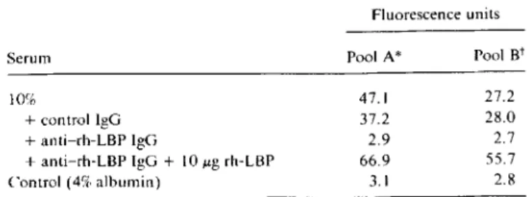
Table1. Effect of LBP depletion and supplementation on serum-mediated binding of fluorescein isothiocyanate-Iabeled LPS to monocytes as assessed by flow cytometry.
Fluorescence units
NOTE: rh = recombinant human.
*High LBP.
tLow LBP.
Determination of LBP concentrations. Patients in septic shock had higher LBP concentrations in serum than did nor-mal volunteers, as measured by RIA. The mean concentra-tion of LBP was 18. I ± 4.0 Jig/mL in normal sera, 54.1 ±
10.2 Jig/mL in sera collected at entry from patients with gram-negative shock, and 45.4 ± 8.7 Jlg/mL in sera of pa-tien ts with shock caused by other organisms or with no micro-biologic documentation. LBP levels in these 2 groups of pa-tients were higher than levels in normal donors(P < 10-s).
Serum 10%
+control IgG +anti-rh-LBP IgG
+anti-rh-LBP IgG+ 10foLgrh-LBP Control (4% albumin) Pool A* Pool Bt 47.1 27.2 37.2 28.0 2.9 2.7 66.9 55.7 3.1 2.8
lID 1994; 170(November) Concise Communications 1321
with anti-CD 14 MAb. The specificity of the immunodeple-tion experiments was assessed by adding back rh-LBP, which restored FITC-LPS binding to monocytes.
Effects of variable LBP concentrations on the binding 10
monocvtes. We previously showed that the binding of FITC-LPS to monocytes can be inhibited by preincubation of serum with nonfluorescent LPS [3]; therefore, we preincu-bated 10% serum with increasing concentrations ofnonfluo-rescent LPS for 30 min before adding PBMC with FITC-LPS for an additional 30 min and assessing binding ofFITC-LPS to monocytes by flow cytometry. When 50-75 ~g/mLLPS was first added to 10% serum, there was still partial binding of the 1-~g/mLfluorescent probe to monocytes, suggesting an absence of total saturation of LBP in the preincubation period. Only when using I00 ~g/mL unlabeled LPS was FITC-LPS binding suppressed.
Using this protocol, the ICso (i.e., the concentration of unlabeled LPS that reduced by 50% the maximal binding of FITC-LPS) was related to the concentration of LBP. The ICsowas threefold higher in pool A (12.3± 1.5~g/mL)than in pool B (4.3 ± 0.8 ~g/mL; mean±
so
of 3 experiments). These experiments showed that FITC-LPS binding was re-lated to the concentration ofLBP. In other experiments (not shown), the limiting factor for LPS binding was actually LBP, not CD 14. Indeed, monocytes first incubated with 100~g/mL LPS did not bind FITC-LPS. However, the same monocytes were able to bind FITC-LPS if fresh LBP was subsequently added, but this binding was suppressed by CD 14 blockade.
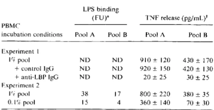
Effects of LBP on TNF release by tnonocytes. Since bind-ing experiments showed that the amount of LPS bound to monocytes was dependent upon the amount of LBP present in serum, we analyzed the involvement ofLBP in the
indue-Table 2. Effect of serum dilution and LBP blockade on binding of fluorescein isothiocyanate-Iabeled LPS to monocytes and on tu-mor necrosis factor (TNF) release by peripheral blood mononu-clear cells upon LPS challenge.
LPS binding
(FU)* TNF release (pg/mL)t PBMC
incubation conditions Pool A Pool B Pool A Pool B
Experiment I If+ pool ND ND 910± 120 430 ± 170 +control IgG NO ND 920± 150 420± 130 +anti-LBP IgG ND ND 20±25 30±25 Experiment2 I(tpool 38 17 800±220 380±35 O.lfX pool 15 4 360± 140 70±30
NOTE. Pool A= high LBP; Pool B= low LBP; ND= not done. * Binding was2.5fluorescence units (FU) on cells incubated with control albumin. Assessed by flow cytometry.
t Stimulus was I ng/mL LPS. No TNF was detected by bioassay when cells were incubated in medium alone.
tion of TNF release by monocytes upon LPS challenge. As shown in table 2. the presence of LBP was a prerequisite for TNF secretion, since treatment of serum with anti-LBP IgG (but not control IgG) suppressed TNF release. The concen-tration ofLBP also determined the response when serum was highly diluted: 0.1 % pool B was less efficient than 0.1 % pool A in its ability to present FITC-LPS to monocytes and to sustain a TNF response.
Discussion
The data support the view that LBP is a key serum compo-nent in the process of monocyte activation by LPS. The de-pletion of LBP from sera suppressed both the serum-me-diated LPS binding and the TNF production of monocytes stimulated with LPS. In the activation experiments. we used LPS concentrations that are usually found in septic shock patients (nanogram per liter range) [13]. The role played by septin, which could bind LPS and which is inhibited by CD14 blockade [6], appeared not to be directly linked to TNF production in our experiments, since the mere deple-tion of LBP from serum efficiently suppressed TNF produc-tion. Furthermore, addition of exogenous rh-LBP in the serum depleted from LBP restored LPS binding to mono-cytes. Thus. activation of monocytes by low LPS concentra-tions through pathways not related to CD 14 and LBP ap-peared to be of minor importance.
The data showed that very high quantities of LPS can be presented to monocytes through LBP. In 10% serum, prein-cubation with 4-12~g/mLLPS was required to diminish by half the maximal subsequent LBP-mediated binding of LPS to monocytes. This demonstrated that LBP in normal plasma was able to enhance LPS binding to monocytes in concentra-tions far above those found in patients presenting with shock. Furthermore, the factor that limited the binding of LPS to monocytes was the presence of LBP not CD 14. In experiments in which monocytes were preincubated with very large concentrations of LPS, followed by washing and incubation with fresh LBP. the LBP could restore LPS bind-ing to monocytes.
The mean concentration ofLBP was 18.1~g/mLin serum of normal donors. This concentration was similar to that pre-viously measured in plasma (17.8~g/mL)[9]. Both ELISA and RIA gave similar values of LBP in serum. Our data also showed that the ability of LBP to present LPS-LBP com-plexes to monocytes could be estimated by flow cytometry [9). LBP levels were higher in blood of septic patients, and the response was not specifically related to gram-negative infection. LBP levels did not have a predictive value for sur-vival in individual patients. and LBP levels are likely to repre-sent a measure of the acute-phase response in these patients. Normal serum contains 10 to 100 times more LBP than the level needed for monocyte activation. During physio-logic conditions and even more during the acute-phase
reac-1322 Concise Communications JID 1994; I 70 (November)
tion, LBP is not a limiting factor for LPS-induced monocyte activation. Our data showed that LBP was the only factor present in serum responsible for TNF release of monocytes upon LPS stimulation.
On the other hand, serum contains many factors that can interfere with the presentation ofLPS to monocytes, includ-ing LPS antibodies and lipoproteins [14, 15], and with mono-cyte activation, such as antiinflammatory cytokines, includ-ing IL-l 0, IL-13 and IL-4. Different levels of these factors could partially explain the variability of the response of se-creted TNF, depending on the donors. However, the present data suggest that when LBP was limiting in highly diluted serum, the serum pool with low LBP concentration was less active than the pool with high LBP concentration in its abil-ity both to present LPS and to sustain TNF release. Thus, this observation and the fact that LBP blockade suppressed TNF release suggest that the level ofLBP was a major factor, although the involvement of other factors could not be ex-cluded. Measurements of LBP in biologic and tissue fluids will help determine if LBP is present in sufficient concentra-tion to activate tissue monocytes in the presence of LPS or gram-negative bacteria.
References
I. Schumann RR, Leong SR. Flaggs GW. et al. Structure and function of lipopolysaccharide binding protein. Science 1990;249: 1429-31. 2. Wright SO, Ramos RA, Tobias PS, Ulevitch RJ, Mathisonrc.CDI4, a
receptor for complexes of lipopolysaccharide (LPS) and LPS binding protein. Science 1990;249: 1431-3.
3. Heumann 0,Gallay P, BarrasC.et al. Control of LPS binding and LPS-induced TNF secretion in human peripheral blood monocytes. J Immunol 1992; 148:3505-12.
4. CouturierC.Jahns G, Kazatchkine MD, Haeffner-Cavaillon N. Mem-brane molecules which trigger the production of interleukin-I and
tumor necrosis factor-a by lipopolysaccharide-stimulated human monocytes. Eur J Immunol 1992;22: 1461-6.
5. Lei MG, Morrison DC. Lipopolysaccharide/lipid A receptors on lym-phocytes and macrophages. Int Rev Immunol 1990;6:223-35. 6. Wright SO, Ramos RA, Patel M. Miller OS. Septin: a factor in plasma
that opsonizes lipopolysaccharide-bearing particles for recognition by CDI4 on phagocytes. J Exp Med 1992;176:719-27.
7. Calandra T, Gerain J, Heumann 0, Baumgartner JD, Glauser MP, Swiss-Dutch J5 Immunoglobulin Study Group. High circulating lev-els of interleukin-6 in patients with septic shock: evolution during sepsis, prognostic value and interplay with other cytokines. Am J Med 1991;91:23-9.
8. Pugin J, Schurer-Maly Cc. Leturcq 0, Moriarty A, Ulevitch RJ, Tobias PS. Lipopolysaccharide activation of human endothelial and epithe-lial cells is mediated by lipopolysaccharide-binding protein and solu-ble CDI4. Proc Nat! Acad Sci USA 1993;90:2744-8.
9. Heumann 0, Gallay P, Le Roy 0, Glauser MP. Radioimmunoassay versus flow cytometric assay to quantify LPS-binding protein (LBP) concentrations in human plasma. J Immunol Methods 1993;
171:169-76.
10. Leturcq0, VanHook P, Smith R, Tobias P, Ulevitch R, MoriartyA.
Generation of monoclonal antibodies to human LBP and their use in the detection of LBP protein in serum. J Cell Biochem 1992; 16C (suppl): 161.
II. Baumgartner JD, Heumann 0, Gerain J, Weinbreck P, Grau GE, Glauser MP. Association between protective efficacy ofanti-li po poly-saccharide (LPS) antibodies and suppression of LPS-induced tumor necrosis factoraand interleukin 6. Comparison of0 side chain-spe-cific antibodies with core LPS antibodies. J Exp Med 1990; 171 :889-96.
12. Fisher RA. On the probable error of a coefficient of correlation deduced from a small sample. Metron 1921;I:3-31.
13. van Deventer SJH, Buller HR, Ten Cate 1M, Stark A, Pauw W. Endo-toxemia: an early predictor of septicemia in febrile patients. Lancet 1988; I:605-8.
14. Flegel WA, Wolpl A, Manuel ON, Northoff H. Inhibition of endo-toxin-induced activation of human monocytes by human lipopro-teins. Infect Immun 1989;57:2237-45.
15. Warren HS, Novitsky TJ, Ketchum PA, Roslansky PF, Kania S, Siber GR. Neutralization of bacterial lipopolysaccharide by human plasma. J Clin Microbiol 1985;22:590-5.