ARTICLE
A NEW RHAMPHORHYNCHID PTEROSAUR FROM THE UPPER JURASSIC OF XINJIANG,
CHINA, AND THE PHYLOGENETIC RELATIONSHIPS OF BASAL PTEROSAURS
BRIAN ANDRES,*,1JAMES M. CLARK,2and XU XING3
1Department of Geology and Geophysics, Yale University, New Haven, Connecticut 06520, U.S.A., [email protected]; 2Department of Biological Sciences, George Washington University, Washington, D.C. 10024, U.S.A., [email protected];
3Institute of Vertebrate Paleontology & Paleoanthropology, Chinese Academy of Sciences, Beijing 100044, China, [email protected]
ABSTRACT—A new rhamphorhynchid pterosaur species, Sericipterus wucaiwanensis, gen. et sp. nov., is described from the Upper Jurassic part of the Shishugou Formation in the Xinjiang Autonomous Region of northwest China. Pterosaurs from this unit are the earliest and only records of pterosaurs in the Jurassic of northwest China. The individual specimen is one of the largest known among ‘rhamphorhynchoids,’ or non-pterodactyloid pterosaurs. The holotype comprises an as-sociated skeleton of mostly disarticulated, largely three-dimensional material. Although partly crushed, the preservation in this specimen reveals morphology rarely seen in non-pterodactyloid pterosaurs. This includes a distinct cervical intervertebral articulation morphology that is proposed to be widespread among the non-pterodactyloids. The skull of this new specimen is most similar to that of other rhamphorhynchids, Angustinaripterus longicephalus and Harpactognathus gentryii, found in terrestrial deposits. A phylogenetic analysis of 18 non-pterodactyloid pterosaurs and the Pterodactyloidea places Sericipterus wucaiwanensis with these species within the Rhamphorhynchinae and a monophyletic Rhamphorhynchidae. Unlike previous phylogenetic analyses, the Dimorphodontidae is paraphyletic, the Campylognathoididae is polyphyletic, and the Anurognathi-dae is the sister group of the Pterodactyloidea. Sericipterus wucaiwanensis, Angustinaripterus longicephalus, Harpactognathus gentryii represent a clade of large pterosaurs that likely lived in the terrestrial settings in which they preserved.
INTRODUCTION
From 2001 to 2006, joint paleontological expeditions from the Institute of Vertebrate Paleontology and Paleoanthropology, Beijing, and The George Washington University, Washington, D.C., surveyed the Junggar Basin of the Xinjiang Autonomous Region, People’s Republic of China. The most productive fos-sil vertebrate locality found during these annual expeditions has been the Wucaiwan area in the eastern part of the Junggar Basin (Fig. 1). Here, the remains of the basal tyrannosauroid Guanlong
wucaii (Xu et al., 2006b), ceratosaurs (Xu et al., 2009), a giant
theropod (Xu and Clark, 2008), the basal ceratopsian Yinlong
downsi (Xu et al., 2006a), the stegosaur Jiangjunosaurus jung-garensis (Jia et al., 2007), tritylodonts, and multiple
crocodylo-morphs such as the ‘sphenosuchian’ Junggarsuchus sloani (Clark et al., 2004a) were excavated from the Shishugou Formation. Three pterosaur specimens have also been reported from the same area and formation (Andres and Clark, 2005), the most complete of which is described here.
Pterosaurs have already been reported from Xinjiang but, pre-viously, were confined to the Lower Cretaceous Tugulu Group (Young, 1964, 1973; Buffetaut, 1996; Maisch et al., 2004). These include the species Dsungaripterus weii Young, 1964, Noripterus
complicidens Young, 1973, and Lonchognathosaurus acutirostris
Maisch et al., 2004, which is likely an individual of Dsungaripterus
weii (B. Andres, pers. observ.). The pterosaur discoveries from
the Wucaiwan locality are the earliest and only record of sic pterosaurs from Xinjiang and one of only a handful of Juras-sic pterosaur localities from China. The new specimens include a ‘rhamphorhynchoid,’ or non-pterodactyloid pterosaur, that is de-scribed here, an isolated wing phalanx from the upper part of the
*Corresponding author.
Shishugou Formation, and a fragmentary pterodactyloid from the lower part of the same formation.
Traditional classifications (e.g., Wellnhofer, 1978) divided pterosaurs into two suborders: the primitive long-tailed “Rham-phorhynchoidea,”and the derived short-tailed Pterodactyloidea. The “Rhamphorhynchoidea” has been shown to be paraphyletic with respect to the Pterodactyloidea (e.g., Howse, 1986; Kell-ner, 2003; Unwin, 2003a). Therefore, in phylogenetic literature it has been informally termed non-pterodactyloid pterosaurs (e.g., Jensen and Padian, 1989; Andres and Ji, 2006).
The Wucaiwan non-pterodactyloid pterosaur is described here, named Sericipterus wucaiwanensis, gen. et sp. nov., compared to the rhamphorhynchids, and its phylogenetic relationships to the other basal pterosaurs are delineated. The new specimen presents sufficient morphology to be described, diagnosed as a new species, and used to help determine the phylogenetic relationships of the non-pterodactyloid pterosaurs. It is com-pared to the other taxa identified as rhamphorhynchids in the phylogenetic analysis: Angustinaripterus longicephalus He et al., 1983, from the Middle Jurassic of central China,
Harpactog-nathus gentryii Carpenter et al., 2003, from the Upper Jurassic
of Wyoming, Rhamphorhynchus muensteri (sensu Bennett, 1996) and Scaphognathus crassirostris Wagner, 1861, from the Upper Jurassic of Germany, Cacibupteryx caribensis Gasparini et al., 2004, from the Upper Jurassic of Cuba, and Dorygnathus
ban-thensis Theodori, 1830, from the Lower Jurassic of Germany.
MATERIALS AND METHODS
The skeleton is described in anatomical position of the wings outstretched laterally as they would be in flight. Therefore, what might be termed the medial and lateral aspects of the more proximal wing bones are instead referred to as the ventral and dorsal aspects, respectively. The anatomical directions of mesial 163
FIGURE 1. Locality of Sericipterus wucai-wanensis holotype (arrow), upper part of Shishugou Formation in the Wucaiwan area. Viewed from the southeast.
and distal are used for orientating along the jaw margins as opposed to anterior and posterior, respectively (e.g., Unwin, 2003a). Muscle scars in the appendicular skeleton are identified with respect to the attachments recognized by Bennett (2003) in
Campylognathoides liasicus.
The procedure of calculating wingspans is taken from Bennett (2001b), which consists of summing of the lengths of the forelimb bones excluding the carpus and then multiplying by a factor of two. The omission of the width across the pectoral girdle and the carpus is intended to offset the flexures along the wing in the total wingspan. This is a repeatable and conservative method to calculate wingspans, which have tended to be reported as larger in the literature.
The wings of Sericipterus are missing the wing metacarpals as well as having incomplete radii/ulnae and third wing phalanges. A minimum wingspan estimate of 1.73 m was calculated by summing the lengths of the complete wing bones, the estimated minimum length of the third wing phalanx, and the estimated length of the missing radius/ulna and wing metacarpal. The left and right third wing phalanges are missing their proximal and distal ends, respectively, so that it is not possible to ascertain the total length of these elements. The more complete of these two phalanges, the left phalanx, is missing its proximal end, which through comparison with the proximal expansion of right phalanx would add a minimum of 30 mm to its length. The length of the radius/ulna and the wing metacarpal were calculated from the av-erage ratio of these bones to the humerus in Rhamphorhynchus
muensteri, the species most closely related to Sericipterus with a
completely preserved wing skeleton in the analysis. All measure-ments were made using a pair of Mitutoyo calipers, accurate to 0.02 mm.
The results of 20 phylogenetic analyses of pterosaur intrarela-tionships have been published at the time of acceptance, 15 with published data matrices (Howse, 1986; Bennett, 1989, 1994, 2007; Unwin, 1992, 1995, 2002, 2003a, 2003b; Unwin and L ¨u, 1997; Viscardi et al., 1999; Kellner, 2003, 2004; Maisch et al., 2004; Wang et al., 2005, 2008; L ¨u and Ji, 2006; Martill and Naish, 2006; Andres and Ji, 2008; L ¨u et al., 2008). Only eight of these analyses have addressed the relationships of the basal pterosaurs. Char-acters, codings, and terminal taxa from this previous work were
integrated into this analysis and were recoded as little as possible to provide a consensus of previous work. Inapplicable character states were reductively coded. In other words, characters depen-dent on the presence of a particular state in another character were coded as missing data for taxa in which the particular state is absent (i.e., a character complex). Inapplicable states are marked by a dash (–), which phylogenetic analysis programs treat the same as missing data (Strong and Lipscomb, 2000). Parsimony uninformative characters were omitted resulting in a list of 75 characters (Appendix 1). Eighteen non-pterodactyloid species including Sericipterus wucaiwanensis, the three outgroups used by Wang et al. (2005), and a supraspecific taxon representing the Pterodactyloidea were used as terminal taxa. The character states for the Pterodactyloidea were obtained by optimizing the characters of this analysis to the base of the Pterodactyloidea on the topology recovered by Andres and Ji (2008). Characters with ambiguous optimizations were coded as polymorphic for this taxon.
The character matrix (Appendix 2) was analyzed using PAUP∗4.0 b10 (Swofford, 2003) both with and without am-biguous branch support (amb and amb− parsimony options, respectively). Tree searches included a Branch-and-Bound search and 10,000 random addition-sequence Tree-Bisection-Reconnection heuristic searches. All characters were unordered and equally weighted (Fitch optimality criterion). Bootstrap and Bremer support values were generated using the same settings as the heuristic parsimony analysis. Tree lengths and tree scores were calculated in PAUP∗, and the index file for calculating Bremer support values in PAUP∗ was generated in MacClade 4.07 (Maddison and Maddison, 2005).
SYSTEMATIC PALEONTOLOGY PTEROSAURIA Owen, 1842 RHAMPHORHYNCHIDAE Seeley, 1870 RHAMPHORHYNCHINAE SENSU Unwin, 2003a
SERICIPTERUS, gen. nov.
Type Species—Sericipterus wucaiwanensis, sp. nov. Diagnosis—As for type and only species.
Etymology—The generic name is based on the Latin word
ser-icum (L.), meaning silk in reference to the ancient Silk Road that
passed through what is now the Xinjiang Autonomous Region, and pteros (Gr.), meaning wing, a traditional ending for pterosaur names.
SERICIPTERUS WUCAIWANENSIS, sp. nov.
(Figs. 2–6)
Holotype—IVPP V14725 (Institute of Vertebrate
Paleon-tology and Paleoanthropology, Beijing, People’s Republic of China): an incomplete skeleton including a disarticulated skull; partial mandible; at least 12 isolated teeth; partial vertebral col-umn (six cervicals, nine dorsals, two sacrals); the right scapula and coracoid; both humeri; the ends of the right ulnae and radii; the distal end of the left ulna; a proximal manual phalanx; the left first, right second, both third, and both fourth wing phalanges; an ischiopubic plate; two metapodial elements, and a probable pedal phalanx fragment.
Etymology—The specific name is derived from the Wucaiwan
area in which this pterosaur was found. In Chinese it means ‘five-color bay’ and refers to the striking variegated ‘five-colors of the rocks in the area (Fig. 1).
Distribution—Alluvial facies of the Upper Shishugou
Forma-tion, between tuffs dated at 161.2± 0.2 Ma and 158.7 ± 0.3 Ma (Clark et al., 2006), equivalent to the Oxfordian, Upper Jurassic; Wucaiwan locality, eastern Junggar Basin, Xinjiang Autonomous Region, People’s Republic of China.
Diagnosis—Largest rhamphorhynchid with a wingspan of at
least 1.73 m. Apomorphies in comparison with other non-pterodactyloid pterosaurs: terminal rostral expansion includes only two pairs of teeth; nasal process of the maxilla with T-shaped cross-section; large, U-shaped quadratojugal has broad contact with the ventral margin of the skull; large lateral processes of the parietals abut the postorbital processes of frontals; low parietal crest extending most of length of parietal; transverse crest at fron-toparietal contact; scapula length subequal to coracoid; wing pha-langes with oval cross-section twice as wide as deep; expanded ends of first and second wing phalanges wider than twice their mid-width; fourth wing phalanx slightly longer than second wing phalanx; enlarged metatarsals with subterminal distal condyles.
DESCRIPTION
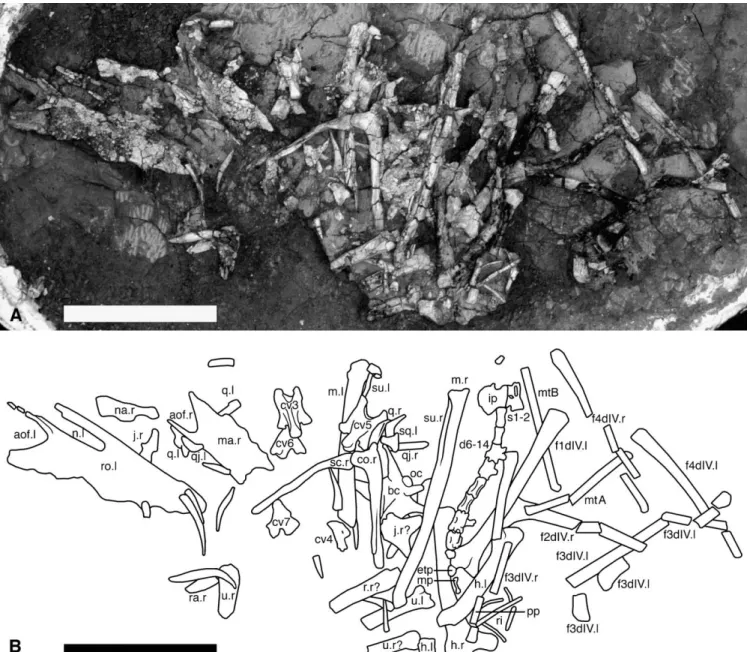
IVPP V14725 is a partial skeleton preserved over a one-half by one-quarter-meter area and collected in a single field jacket (Fig. 2). The specimen is disarticulated save for bones of the braincase and the dorsal and sacral vertebral series. The speci-men was removed from the surrounding matrix with the excep-tion of the bones of the braincase and temporal region of the skull for which full preparation would have undermined phys-ical integrity of these elements. The quality of preservation of the individual bones varies greatly over the specimen. It was found on a deflated surface so that the skeletal elements are all in some manner crushed, broken, or fractured; however, the tex-ture of the bone is generally well preserved and most elements are three-dimensional. This type of preservation is more typical of pterosaur specimens recovered from terrestrial sediments, in-stead of the thinly bedded lagerst ¨atten that preserve the majority of pterosaur specimens.
Ontogeny
This individual is considered to be an osteological sub-adult be-cause most but not all of the elements known to fuse during on-togeny of pterosaurs remain unfused in this specimen. Sutures are not visible between some elements of skull whereas others have become disarticulated. In the postcranium, the scapula and
cora-TABLE 1. Measurements of the skull elements of IVPP V14725 (in mm).
Element Left Right
Preserved rostrum length >146.8 Preserved braincase length >54.0 Skull length anterior to external
naris ∼63.2
Skull height at anterior margin of external naris
36.0
Skull maximum width ∼58.5
External nares length > 45.9 > 50.6
External nares height — 9.1
Antorbital fenestra length > 28.6 > 34.1 Antorbital fenestra height > 15.3 > 29.9
Tooth row length > 84.1
Mandible length > 113.2 > 146.4 —= missing element; > = preserved length; ∼ = approximate.
coid, sacral ribs, and the extensor tendon process of the first wing phalanx remain unfused. Some neural arches of dorsal vertebrae are separated from their lateral lamina, suggesting incomplete fu-sion between centra and the neural arches. However, sutures are not visible on the more complete dorsal or any other vertebrae.
Skull
The skull of IVPP V14725 is preserved as two distinct accumu-lations and approximately 18 isolated tooth fragments that could represent a minimum of 12 distinct teeth (Figs. 2–4; Table 1). The rostrum is detached from the posterior region of the skull and split into left and right halves just distal to the tip of the rostrum on the left side (Fig. 3). These have come to lie upon the right jugal and the left quadrate, respectively. The preserved posterior region of the skull includes the braincase, several of the tempo-ral bones, and the right quadrate (Fig. 4), as well as most of the right and the posterior half of the left mandible (Fig. 1). The skull likely broke into anterior and posterior regions, and subsequently the rostrum split into two halves with the jugal and quadrate com-ing to lie between them. The degree of disarticulation of the skull is unusual for pterosaurs and reveals aspects of the skull not nor-mally visible. The nasal process of the left maxilla is the poste-rior extent of the preserved rostrum, whereas the anteposte-rior extent of the preserved braincase is the orbit dorsal margin. These two extremities would flank the ascending process of the jugal. Sum-ming the length of the preserved rostrum, braincase, and width of the ascending process of the jugal provides a minimum skull length estimate of about 210 mm. The missing portions of the frontals, prefrontals, lacrimals, nasals, and maxillae would have to occupy only five mm for this skull to be larger than the previ-ously largest known non-pterodactyloid skull, present in
Dimor-phodon macronyx Buckland 1829 (Wellnhofer, 1978).
Rostrum—The two halves of the rostrum were found lying on
their lateral surfaces (Fig. 2). They are highly fractured, espe-cially at their posterior ends where they have been eroded. Col-lectively, they preserve the anterior ends of the external narial and antorbital fenestrae, jugal and nasal processes of the max-illae, maxillary process of the left jugal, and the premaxillary bar.
The left rostral fragment is more complete than the right and was found associated with part of the right jugal (Fig. 3AB). It includes the premaxillary bar, the anterior-most end of the right half of the rostrum, and the anterior end of the maxillary process of the left jugal. The left half of this fragment preserves the an-terior portions of the left external naris, antorbital fenestra, the jugal and nasal processes of the maxilla, and up to four alveoli. A small, attached fragment of the right rostrum preserves the first two right alveoli. The anterior end of the maxillary process of
FIGURE 2. Sericipterus wucaiwanensis, gen. et sp. nov. (IVPP V14725). A, photograph; B, line drawing illustrating the arrangement of skeletal elements as they were collected. Teeth are not labeled. Abbreviations: aof, antorbital fenestra; bc, braincase; co, coracoid; cv, cervical vertebra; d, dorsal vertebra; etp, extensor tendon process of the first wing phalanx; fXdY, phalanx X of digit Y; h, humerus; ip, ischiopubic plate; j, jugal; m, mandible; ma, maxilla; mp, manual phalanx; mt, metatarsal; n, external naris; na, nasal; oc, occipital condyle; pp, pedal phalanx; q, quadrate; qj, quadratojugal; ra, radius; ri, ribs; ro, rostrum; s, sacral vertebra; sc, scapula; sq, squamosal; su, surangular; u, ulna; X.l, left element; and X.r, right element. Teeth are not labeled. Scale equals 10 cm.
the left jugal was found articulated with the left maxilla but was removed in Figure 3. The right rostral fragment is less crushed and preserves presumably only the right maxilla (Fig. 3C). The ventral margin of the external naris, the anterior end of the antor-bital fenestra, and the nasal process of the maxilla that separates these two openings dominate the preserved morphology. A skull fragment located between the halves of the rostrum is identified as the right nasal (Fig. 2).
The preserved portions of the anterior skull outline an elon-gate rostrum. The premaxillae are fused along their preserved lengths. The anterior tip of the skull bears a laterally compressed rostral process. This process projects anteriorly from the midline of the skull and is missing its tip to reveal an elliptical
cross-section in anterior view. The preserved portion of the process is rather short, has a depth twice its width, and does not seem to taper. The process connects posteriorly with a low sagittal crest extending along the dorsal midline of the rostrum. The crest ex-tends for half a centimeter before it is broken off and continues posteriorly as a broken base. Posterior to the rostral process, the rostrum is not compressed and does not expand evenly towards the jaw articulations as in most pterosaurs. In dorsal and ventral views, the jaw margins can be seen to rapidly increase in width to the second alveolus forming an approximate 70◦angle with their anterior end (Fig. 3D). This is the extent of the preservation on the right side of the tip of the rostrum, but on the left side this ex-pansion is immediately followed by a concave lateral jaw margin
FIGURE 3. Photograph and drawing of the rostral region of Sericipterus wucaiwanensis, gen. et sp. nov. (IVPP V14725). A, left rostral fragment in lateral view; B, left rostral fragment in medial view; C, right maxillary fragment in medial view; D, tip of rostrum of left rostral fragment in ventral view. Abbreviations: aj, ascending process of the jugal; aof, antorbital fenestra; j, jugal; jm, jugal process of maxilla; l, left alveolus; L, left tooth; mj, maxillary process of the jugal; mm, medial process of the maxilla; n, external naris; nf, medial flange on nasal process of the maxilla; nm, nasal process of the maxilla; oj, lower orbital bar on the jugal; pb, premaxillary bar; r, right alveolus; rr, rugose ridge; R, right tooth; rc, rostral crest; X.l, left element;
X.r, right element. Scale equals 5 cm.
FIGURE 4. Photograph and line drawing of the temporal and occipital regions of Sericipterus wucaiwanensis, gen. et sp. nov. (IVPP V14725), in A, dorsal; B, ventral view; and photographs of the C, right; D, left quadrate in anterior view. Abbreviations: aj, ascending process of the jugal; f, frontal;
fo; foramen; j, jugal; lp, laterosphenoid and prootic region; oc, occipital condyle; op, opisthotic; pcr, parietal crest; pf, pneumatic foramen; pn, palatine; pr, fused parietals; qj, quadratojugal; qq, articular facet for quadratojugal on the quadrate; so, supraoccipital; sq, squamosal; su, sulcus; t, tooth; X.l,
left element; X.r, right element. Stippled regions represent matrix or matrix covered bone. Dashed lines represent the break between the left jugal and the ascending and maxilla processes of the jugal. Scale equals 5 cm.
FIGURE 5. Photographs of the cervical vertebrae in Sericipterus wucai-wanensis, gen. et sp. nov. (IVPP V14725). A, cervical 3; B, cervical 5; C, cervical 7; D, cervical 8; in i, anterior; ii, posterior; iii, right lateral; iv, ven-tral; v, left lateral; and vi, dorsal view. Abbreviations: ac, anterior cotyle;
di, diapophysis; ep, epipophysis; lt, lateral tubercle; ns, neural spine; pa,
parapophysis; pc, posterior condyle; pf, pneumatic foramen; pl; postlat-eral projection of centrum; vk, ventral keel of centrum. Scale equals 2 cm.
denoting a constriction in the skull posterior to an expanded tip of the rostrum.
It is difficult to discern the width of the skull posterior to the rostrum. The less crushed right maxilla is mediolaterally broad and has its nasal process dorsomedially oriented, indicating that the skull was rather broad at this region. All of the teeth pre-served in the specimen curve along an axis perpendicular to the long axis of their cross-sections with the exception of the first tooth-pair. The alveoli and in situ teeth in the jaw margins indicate that this long axis is the mesiodistal axis as in other pterosaurs. To accommodate the occlusion of this lingually curv-ing dentition, the jaw margins would have to incorporate some lateral orientation. Therefore, the teeth must have been directed laterally to some degree but the exact angle is not known. The alveoli project from the jaw margin, tracing a sinuous outline. The premaxillae and maxillae apparently do not contribute to the palate.
While being prepared, the premaxillary bar of IVPP V14725 was shifted slightly dorsally, giving the skull the higher posterior outline seen in Figure 3. The premaxillary bar is fragmented and may represent the disassociation of the left and right premaxillae from one another in this region. The anterior part of the external naris is preserved in both halves of the rostra. It is a remarkably elongate, anteriorly inclined, and narrow opening. Its dorsal and ventral margins are nearly straight and parallel to one another. The rounded anterior- and ventral-most margin of the naris lies well above the ventral margin of the antorbital fenestra. The an-torbital fenestra is a much deeper opening with a larger, more rounded anterior margin.
The contact between the premaxillae and maxillae is not visi-ble in medial or lateral view and they are likely fused. On both halves of the rostrum, the nasal process of the maxilla is nearly straight and inclined about 30◦from the horizontal. This process is T-shaped in cross-section due to a flange extending along the medial aspect of this process (Fig. 3). The contact surface on the left maxilla for the jugal is preserved, indicating these bones were not fused. At this contact, the maxillary process of the jugal is directed anterodorsally to extend over the jugal process of the maxilla (removed in Figure 3). The contact surface on the maxilla curves anteromedially to reach the anterior margin of the antor-bital fenestra on its medial aspect.
On the medial aspect of both maxillae, a flat shelf of bone ex-tends posteroventrally from the anterior end of the antorbital fenestra orthogonal to the surface of the maxilla proper. This can be seen best on the right maxilla (Fig. 3C), whereas on the left half it remains as a broken process near the base of the nasal process of the maxilla and has been shifted laterally into the antorbital fenestra. This shelf contacts the medial flange on the nasal process forming an internal fossa, presumably hous-ing a paranasal diverticulum (Witmer, 1997), at the anterior mar-gin of the antorbital fenestra. A rugose ridge extends anteriorly under the external naris from this point. This shelf tapers pos-terolaterally and somewhat resembles pterosaur palatines in this manner. However, this would imply both a more dorsal position and greater inclination for the palatines than is seen in other pterosaurs. In addition, these elements do not seem to be sep-arate bones from the maxillae. These shelves are termed the me-dial processes of the maxilla. They may be the original maxillary contributions to the palate but have subsequently been displaced dorsally due to the posterior expansion of the palatines, diagnos-tic for pterosaurs (Romer, 1956).
The midsection of the right jugal was found lying under the left rostral fragment and was left attached to its medial surface during preparation (Fig. 3A–B). The maxillary process, ascending pro-cess, and the lower bar of the orbit are present, but incomplete. These processes are perpendicular to one another. In addition, the jugal proper is missing a piece of its ventral margin at the junction of these processes and interrupting an otherwise straight ventral margin in this region. On the lower orbital bar of the jugal, a crescentic articular facet curves anteroventrally along the
FIGURE 6. Photographs of the appendicular elements of Sericipterus wucaiwanensis, gen. et sp. nov. (IVPP V14725). A, right scapulocoracoid in lateral view; B, proximal end of right radius in anterior view; C, proximal end of right ulna in anterior view; D, distal end of left ulna in anterior view; right humerus in E, ventral, F, dorsal, and G, proximal view; H, left first wing phalanx in dorsal view without extensor tendon process; I, right second wing phalanx in dorsal view; J, distal end of left third wing phalanx in dorsal view; K, proximal end of right third wing phalanx in dorsal view;
L, proximal end of left fourth wing phalanx in dorsal view; M, right fourth wing phalanx in dorsal view; and metatarsal A in N, lateral, O, dorsal P,
proximal, and Q, distal view. Abbreviations: ap, acrocoracoid process; co, coracoid; dc, dorsal condyle; dco, dorsal cotyle; dp, deltopectoral crest; ect, ectepicondyle; gf, glenoid fossa; gr, groove; hh, humeral head; ms, muscle scar; nf, nutrient foramen; pf, pneumatic foramen; ppr, posterior process; sa, sternal articulation; sc, scapula; tub, tubercle; uc, ulnar crest; vco, ventral cotyle. Scale equals 5 cm.
posterior aspect of a short dorsal process and onto the medial aspect of the jugal. The lateral margin of this short dorsal pro-cess is not confluent with the lateral margin of the jugal proper, which is damaged and likely constituted part of the posterior ex-pansion of the jugal. If so, the short dorsal process would be part of the anterior margin of the posterior expansion. The medial facet likely is the contact for the pterygoid or ectopterygoid and would indicate that the maxilla did not extend this far posteri-orly along the medial side of the jugal. A similar condition has been reported in Pteranodon where a small dorsal process lies just anterior to the ectopterygoid contact with the jugal (Bennett, 2001a).
An isolated bone lying between the two halves of the rostra is identified as the right nasal (Fig. 2). It is an elongate, wedge-shaped bone with long and short edges separated by the margin of an oval fenestra on one end. These edges have a slight sinusoidal outline. If this identification is valid, then the long edge would correspond to the contact between the left and right nasals, and the free margin would correspond to the posterior margin of the right external naris. The overall shape of this bone also resem-bles the palatine of pterosaurs. However, the palatine contacts a fenestrae, the internal naris, posteriorly and not on one of the convergent edges as in this bone.
Among non-pterodactyloid pterosaurs, a midline process at the anterior tip of the rostrum can only be seen as present in Rhamphorhynchus, Angustinaripterus, and Harpactognathus. These processes are elliptical in cross-section in Sericipterus
and Angustinaripterus, but triangular in Rhamphorhynchus and Harpactognathus. In Sericipterus, Angustinaripterus, and
Harpactognathus, this rostral process is laterally compressed and
connects posteriorly to a low premaxillary sagittal crest, collec-tively labeled a rostral crest in Figure 3. The posterior extent of the sagittal crest is not preserved in any of these pterosaurs. Premaxillary sagittal crests are widespread among pterodacty-loids. They are also reported in the non-pterodactyloids
Austri-adactylus cristatus (Dalla Vecchia et al., 2002) and Raeticodacty-lus filisurensis (Stecher, 2008), but these crests are much higher
and have a straight anterior margin in these species. Rostral ex-pansions are reported here in Sericipterus and the other non-pterodactyloids, Angustinaripterus and Harpactognathus. Rostral expansions are present in some ctenochasmatid (Unwin, 2002) and in the anhanguerid pterodactyloids (Campos and Kellner, 1985; Bakhurina and Unwin, 1995; Unwin, 2003a). The rostral expansions in the non-pterodactyloids differ in their relative size;
Sericipterus incorporates two pairs of teeth, Angustinaripterus has
three pairs, and Harpactognathus has four pairs of teeth in its expansion. These three pterosaurs also share a sinuous den-tal margin, formerly listed as a diagnostic character of
Harpactognathus by Carpenter et al. (2003). Sericipterus and the
other rhamphorhynchines (sensu Unwin, 2003a) have elongate, parallel-sided external nares. Other non-pterodactyloids have more triangular external nares that, even when elongate and an-teriorly inclined, maintain a flat base and a concave ventral mar-gin. An antorbital fenestra with a ventral margin situated below
the external nares is present in all rhamphorhynchids identified in the current phylogenetic analysis.
The condition of a T-shaped cross-section for the nasal process of the maxilla cannot be assessed in many pterosaurs. However, disarticulated maxillae in Campylognathoides, Dorygnathus, and
Dimorphodon; fortuitous breaks in Harpactognathus and An-gustinaripterus; and CT radiology of Rhamphorhynchus (CMNH
11434) indicate that it is absent in these taxa (B. Andres, pers. observ.) and therefore an apomorphic condition for Sericipterus.
Harpactognathus has a similar but distinct condition in that the
nasal process of its maxilla is dorsally thickened. A similar situ-ation occurs with assessing the condition of the contact between jugal and the maxilla. A jugal that spirals around the maxilla to reach the medial aspect of the jaw margin as in Sericipterus can be observed in some pterodactyloid species (e.g., Dsungaripterus
weii) (B. Andres, pers. observ.)). However, the distribution of
this feature among the non-pterodactyloids is not known because the three-dimensional preservation required to assess this mor-phology is rare among these taxa. Shelf-like palatal processes on the maxilla can be seen in Angustinaripterus, but further distribu-tion of this feature is not known.
Braincase and Temporal Region—The posterior region of the
skull of IVPP V 14725 was preserved lying on its dorsal sur-face under the mandibles and right coracoid (Fig. 2). The brain-case, frontals, fused parietals, portions of the left jugal, left squamosal, right quadrate, right quadratojugal, and possibly one of the palatines are present (Fig. 4). This region has been greatly fractured and crushed dorsoventrally so that identification of the other fragments and their margins is not possible, but these pre-sumably belong to the missing palatal and temporal bones. In general, the cranium fractured through the supraoccipital with the other bones of the occiput rotated forward and the ventral braincase being dorsally displaced into the portion of the cra-nial cavity formed by the frontals. This left a small dorsal portion of the supraoccipital and parietals extending further backwards than the rotated occiput, visible as a small, rectangular flange in Figure 4.
The identifiable elements of the chondrocranium are largely limited to the bones of the occiput. In ventral view, the brain-case narrows anteriorly from the occiput to where it is obscured by the frontals. The prootics and laterosphenoids presumably oc-cupy this region and would include some of the fragments in this area, but their margins are not discernable. The occipital condyle is semicircular in cross-section with a slight neck and is appar-ently comprised only of the basioccipital. A slight groove ex-tends along the otherwise flat dorsal flat margin of the condyle. A small piece of the right half of the condyle has become dis-placed slightly along a crack that extends through the region. Short lateral flanges attach to the sides of the condylar neck. Similar structures were identified as the exoccipitals in
Rham-phorhynchus by Wellnhofer (1975). The left example of these
two flanges has been detached and rotated, but no sutures are visible on either side to confirm that these are separate bones. The basioccipital is broken off ventrally at an apparent constric-tion in this bone leaving no trace of the basisphenoid. The ven-tral preserved end of the basioccipital is flanked laterally by two foramina, the ‘foramen ovale’ of Wellnhofer (1975) (Fig. 4B). However, these openings do not correspond to the fifth nerve opening between the prootics and laterosphenoids that is termed the foramen ovale in other taxa. They may be comparable to the subcondylar recess of theropods such as tyrannosaurids (Wit-mer, 1997). The opisthotics form large plate-like structures with round lateral margins and are missing their squamosal contacts. The left paroccipital process is more complete, extending out be-neath the posterolateral process of the parietal to the region of the squamosal contact. The opisthotics contact the parietals dor-sally and wrap around the ‘foramen ovale’ ventrally. The foramen magnum is relatively small and semicircular. It has a thickened
dorsal rim formed laterally by the opisthotics and dorsally by the supraoccipital. The supraoccipital is rather small and has a low midline ridge.
The preserved skull roof in this region includes the frontals and parietals (Fig. 4A). The frontals and parietals are unfused to each other and are oriented coplanar to one another. The large frontals dominate this region of the skull and are complete save for the missing tips of their anterior and postorbital processes. A midline dorsal notch in the anterior margin of the frontals is pre-sumably the contact for the premaxillary. The nasals do not ex-tend posteriorly as processes in pterosaurs. The frontals contact the parietals along a broad, posteriorly convex transverse crest. The frontals have a nearly flat dorsal surface that extends lat-erally behind the large orbits as wide postorbital processes. In ventral view, the cristae cranii are broad and have ventrolaterally facing surfaces.
The fused parietals are much smaller relative to the frontals but still meet along the midline of the skull to exclude the frontals from contacting the supraoccipital, unlike the reconstruction of
Rhamphorhynchus by Wellnhofer (1975). The parietals are
dor-sally arched above the braincase forming laterally sloping sides for the medial walls of the supratemporal fenestrae between the lateral and posterior processes of the parietal. A sizable distance between the posterolateral end of the parietal and the postor-bital process of the frontal indicates that the supratemporal fen-estra was large. A lateral process of the parietal abuts the postor-bital processes of the frontals posteriorly, but is shorter and ter-minates before it would contact the postorbitals. The fused pari-etals bear a low, narrow sagittal crest. However, this crest does not extend above the dorsal margin of the skull and is therefore not equivalent to the parietal crests present in some pterodacty-loid peterosaurs. This crest contacts the similar transverse crest that extends along the frontoparietal contact. The posterior pro-cesses of the parietals are tapered and oriented posterolaterally to where they would contact the squamosals.
A bell-shaped bone lying next to the left squamosal process of the parietal is identified as the left squamosal (Figure 4). Its overall form is of a broken process with two concave margins ter-minating in large, convex, laterally curving end. The only other bone in the skull with a similar shape would be the postorbital, which is more triradiate and has much narrower processes. If it is the squamosal, the broken process would correspond to the postorbital process, and the two concave margins as the ven-tral and dorsal margins of the infratemporal and supratempo-ral fenestrae, respectively. Because the more concave margin would correspond to the infratemporal fenestra and this bone curves slightly laterally, this element can be identified as the left squamosal.
An adjacent element is identified as the right quadratojugal. It is a rather large, U-shaped bone with tall anterior and poste-rior ascending processes. These processes would broadly contact the jugal and the quadrate. This bone traces the ventral margin of the infratemporal fenestra, which would therefore be rather large and have a round ventral margin. A raised edge traces the medial margin of the ventral and posterior margins of this bone. The anterior of the two ascending processes is straighter and about twice the length of the more posterior process. The lateral edge of this anterior process has been quite damaged. The more posterior process forms a sharp, hooked end that matches exactly a distinct anteriorly oriented facet on the lat-eral margin of the quadrate (Fig. 4C). The hooked process is all that remains of the left quadratojugal, which was found ly-ing on top of the left quadrate and underneath the right maxilla (Fig. 4D). On the medial surface of the anterior end of the ventral margin is a narrow groove that may have articulated with a slen-der process of the jugal. The quadratojugal would have a broad contact with the ventral margin of the skull and exclude the ju-gal from contacting the quadrate. The inclination of the posterior
hooked process indicates that the quadrate was relatively inclined with respect to the ventral margin of the rostrum.
Lying between the anterior and posterior processes of the right parietal is an elongate bone that is visible in dorsal and ventral views of the braincase. Though poorly preserved, its elongate, triangular outline and large size suggest that this is one of the palatines.
On the dorsal aspect of the skull is an incomplete bone with a greatly expanded end, a middle constriction, and a straight pro-cess that has been broken off at its base but connects at a right angle to the long axis of the rest of the bone. This bone is identi-fied as the left jugal. The expanded end corresponds to the poste-rior margin that would contact the anteposte-rior ascending process of the quadratojugal. A process would have extended dorsally from this posterior margin to contact the postorbital, but this has been broken off at its base. The middle constriction corresponds to the ventral-most margin of the orbit, and the elongate, broken pro-cess would be the ascending propro-cess of the jugal. The exact orien-tation of the ascending process cannot be determined. The bro-ken base of the maxillary process is evident just ventral to the as-cending process. The asas-cending process of the left jugal preserves part of its dorsal-most expansion and possibly includes portions of the lacrimal and/or prefrontal. This expansion would lie near the skull roof so that taking the entire height of the preserved jugal provides a minimum skull height estimate at the front of the orbit of 51.5 mm. The posterior end of the jugal is expanded ventrally. This ventral expansion is likely the posterior process of the jugal found in other pterosaurs but oriented vertically in this specimen. A large flat bone with two divergent processes lying on the ventral aspect of the frontals is likely the posterior end of the right jugal. This element is highly fractured and its identification is not certain.
The right quadrate was found in contact with the right quadra-tojugal, and the left quadrate was found underneath the right rostral fragment in contact with the posterior end of the left quadratojugal (Fig. 2, but removed in Fig. 3A–B). Both quadrates are missing their dorsal ends but are otherwise well preserved (Fig. 4C–D). The quadrates are anteroposteriorly compressed and would have had very little exposure on the lateral surface of the skull. This exposure is limited to the laterally offset lat-eral condyle of the jaw articulation and the thin flange extend-ing dorsally from its posterior margin. The flange forms an an-teriorly oriented vertical groove to receive the posterior margin of the quadratojugal. This facet is wider near its base, giving it an overall squat, subtriangular shape. The rest of the lateral sur-face is straight and recessed from what would be the lateral mar-gin of the skull. The quadrates have well-developed obliquely aligned, double condyles. The lateral condyle is well offset lat-erally to the extent that the margins of the condyles do not over-lap in anterior or posterior view. This is in contrast to the rela-tive elongate shape of the condyles, whose long axis is more than three times the length of the short axis. The condyles and the strong sulcus that divides them are oriented 45◦ anteromedially from the sagittal plane. A small, presumably pneumatic, foramen dives dorsally into the anterior aspect of the quadrate just lateral to the quadrate midline and level with the dorsal termination of the quadratojugal articulation. A sharp, anteromedially curving process extends from the medial side of the left quadrate’s ven-tral end. This process would presumably contact the pterygoid. The quadrates are laterally thicker in cross-section. On the right quadrate fragment, the lateral thickening shifts slightly medially near the dorsal end so that the lateral edge becomes a flange dor-sally. The medial margin is poorly preserved in both quadrates, but can be seen to expand medially in the right quadrate to where it would eventually contact the occiput.
The posterior aspect of the skull in non-pterodactyloids is ef-fectively described from a single specimen of Rhamphorhynchus (CMNH 11434, see Wellnhofer, 1975; http://www.digimorph.
org/specimens/Rhamphorhynchus muensteri/). The occipital re-gion of IVPP V 14725 largely agrees with Wellnhofer’s (1975) reconstruction of Rhamphorhynchus, with the exception of a much smaller supraoccipital and conversely larger parietals. The supraoccipital therefore does not contact the frontals as recon-structed by Wellnhofer (1975). The parietals have a significant contact with the postorbital processes of the frontal, but are still dwarfed by the frontals. Large, V-shaped quadratojugals are reported in Dorygnathus (Padian and Wild, 2008) and recon-structed in Parapsicephalus purdoni (Newton, 1888), but these are much shorter anteroposteriorly and do not have a broad contact with the ventral margin of the skull. Sericipterus, how-ever, has longer, U-shaped quadratojugals that are as long as they are tall, unlike the shorter quadratojugals in these other taxa. Inclined quadrates are present in all but the basal-most pterosaurs (Unwin, 2003b). Quadrates with oblique condyles sep-arated by a posterolaterally oriented groove for articulation with the mandible have been coded in some pterodactyloids such as
Quetz alcoatlus (Kellner and Langston, 1996) and termed helical
jaw joints (Eaton, 1910). They are also present in Sericipterus,
Dorygnathus, and Rhamphorhynchus. They cannot be observed
in Cacibupteryx and Angustinaripterus. An expanded posterior end with a ventral process on the jugal is also present in
Angusti-naripterus. The midline sagittal crest on the parietals has not been
reported in non-pterodactyloids and is most similar to the blunt sagittal crest coded in the anhanguerids (Kellner, 2003). A ridge at the contact between the frontals and the parietals has been reported in the pterodactyloid Gegepterus changi (Wang et al., 2007).
Mandible—The right and left mandibular rami are present
though severely cracked and distorted (Fig. 2). The right ramus is more complete but is twisted along its length and missing the anterior end. This ramus is relatively straight, dorsoventrally low, laterally compressed, and tapers slightly anteriorly. The remains of two crushed alveoli are visible anteriorly in the right ramus, but it is not known how many more teeth or how much more of the mandible is missing. The left ramus is very poorly pre-served and comprises only the posterior end of the ramus. The adductor fossa, the cranial articulation, and a detached surangu-lar are the only features that can be recognized on this element. The adductor fossae, best preserved on the right, are elongate, narrow, and elliptical in shape. The right fossa is about 53 mm long and reaches nearly the entire length of the surangular to terminate just anterior to the cranial articulation. The surangu-lar is an elongate, thin bone positioned along the dorsal edge of the posterior third of the preserved ramus. There is no dis-tinct coronoid process. The prearticular extends from the cra-nial articulation along the medial aspect of the ramus and ven-tral to the fossa. The contact between the articular and sple-nial is not discernable, but the splesple-nial can be seen to extend anteriorly from the fossa to terminate as a sharp wedge about halfway down the preserved length of the mandible. The mar-gins of the articular, angular, and posterior terminus of the den-tary are not visible. The cranial articulation, best preserved on the left, is gently concave dorsally and nearly square in outline. The anterior edge of the articulation surface comprises robust medial and lateral processes separated by a depression. The me-dial process is the larger of the two, nearly twice as broad as the lateral process, and about equal in breadth to the depression be-tween them. These processes form the anterior margins of the posterolaterally facing medial and lateral articular facets. A very slight posterior buttress is present but is far enough posterior that some anteroposterior movement of the mandible may have been possible. A short, blunt, and triangular retroarticular process is present.
Dentition—Eighteen tooth fragments that could at minimum
represent 12 teeth are preserved around the two skull regions. They have smooth, glossy enamel that is stained blue in some
fragments. Most of the preserved teeth are isolated and consist only of portions of the crowns. The right second and the left third teeth remain in their alveoli, but the left third tooth is broken off at its base. A tooth found lying on the right aspect of the left rostral fragment next to the right tooth is identified as the right third tooth based on its similar position, size, and cross-section as the left third tooth, which was found in the largest rostral alveo-lus (Fig. 3A–B). The largest isolated tooth in IVPP V 14725 is of similar size and was found next to the proximal right ulna and ra-dius (Fig. 2). The rostral alveoli increase in size to the third alve-olus and then decrease in size distally. A tooth closely associated with the tip of the rostrum (Fig. 2) is relatively less compressed and similar in cross-sectional size to the first alveoli pair and is most likely the left or right first tooth. The largest isolated tooth is likely the largest tooth from the mandible and from a similar position in the tooth row.
Alveolar spacing increases distally along the rostrum with the distance between successive teeth always exceeding the diame-ters in respective teeth. It is not known how far back in the skull the tooth row extended. The tooth rows begin about three mm behind the preserved tip of the rostral process where they lie very close to the midline of the skull. The left half of the rostrum preserves the mesial-most three alveoli, but the poorly preserved outline of a fourth alveolus is likely present in the sinuous outline of the jaw margin. An alveolus in a similar position on the right maxillary fragment and second alveolus distal to it would bring the tooth count along each side of the rostrum to at least five. If the tooth row terminated under the antorbital fenestra as in other Jurassic non-pterodactyloids, the tooth row could contain up to seven teeth bringing the rostral tooth count to anywhere between 10 and 14, and the total tooth count to between 20 and 28 teeth assuming a similar number of teeth present in the mandible. The rostral tooth row extends past the anterior margin of the external naris, or at least 40% of the minimum estimated skull length.
The rostral tooth row has raised borders on the mesial and dis-tal edges of each alveolus giving the jaw margin a sinuous outline. All alveoli are labiolingually compressed but are less compressed mesially. The putative first tooth is the least recurved tooth in the specimen, suggesting that the rostral teeth increase in curva-ture to the third tooth. All other teeth are curved. The anterior-most alveoli are inclined anteriorly about 30◦from the horizontal plane, the left second about 45◦, the left third about 75◦, and right fourth and fifth are subvertical.
The teeth are elongate, labiolingually compressed, and termi-nate in sharp tips. Tooth lengths range from 24 to 53 mm, of which half is erupted height. Short, isolated teeth are present so it is most probable that the teeth decreased in height as well in diam-eter distal to the third tooth. With the possible exception of the mesial-most teeth, the teeth curve strongly lingually over their entire erupted height at right angles to their mesiodistal long axis. The roots are essentially straight and end in a small, circular nu-trient foramen. The possible right third tooth has the largest cur-vature in IVPP V14725. Its 10.4-mm ventral displacement along its curvature indicates that the tip would have extended well ven-tral to its alveolus. The mesial rosven-tral teeth are partially later-ally oriented, but it is most probable that all of the dentition had some degree of lateral orientation to accommodate their curva-ture. The mesial and distal edges of the teeth have thin, sharp enamel keels extending along the length of the erupted tooth to the tip. The distal keel is distinctly sharper, forming a long cutting surface.
Compared to the 10–14 rostral teeth of Sericipterus,
Angusti-naripterus has 18 (He et al., 1983), Harpactognathus has at least
12 (Carpenter et al., 2003), Rhamphorhynchus has 17,
Doryg-nathus has 22, and ScaphogDoryg-nathus has 14 (Wellnhofer, 1978).
All non-pterodactyloids except for the anurognathids and
Sor-des pilosus have labiolingually compressed teeth. In Sericipterus,
all of the dentition is labiolingually compressed, whereas the
other rhamphorhynchids have teeth circular in cross-section at the mesial end of their dentitions. Slender teeth are present in all the rhamphorhynchids, which become more elongate in the rhamphorhynchines. Procumbent and strongly curved teeth are present in all rhamphorhynchines, but it is only in
Rham-phorhynchus that the entire dentition is procumbent. The mesial
and distal enamel keels with a sharper distal ridge, as in the teeth of Sericipterus, have been reported in Rhamphorhynchus (Wellnhofer, 1975) and seem to be to be present in
Angustinar-ipterus. In Sericipterus and Angustinaripterus, the teeth are
di-rected to some degree laterally and curve lingually, whereas the teeth of the other rhamphorhynchids are upright and recurve pos-teriorly. This lingual curvature of the teeth is perpendicular to the direction of curvature from the rest of the non-pterodactyloids. The cross-sectional long axes of the mesial-most teeth are not parallel to the sagittal plane in Sericipterus. The similar position of the mesial and distal enamel keels and mesiodistal long axis of the teeth confirm that this is a novel direction of curvature as opposed to the rotation of the teeth within their alveoli with re-spect to other pterosaurs. Harpactognathus has laterally directed alveoli, but because no teeth were found with the only known specimen, it is not known if it shares a lingually curving, keeled dentition.
Axial Skeleton
Remains of six cervical, nine dorsal, and at least two sacral ver-tebra are preserved in the axial skeleton in IVPP V14725 (Figs. 2 and 5, Table 2). The six cervical vertebrae are very similar in morphology and so represent cervicals 3 to 8, the longest series of similarly shaped vertebrae within the cervical series. Cervical 8 is obscured and not shown in Figure 2. The dorsal vertebrae can be recognized as the posterior end of the dorsal series be-cause of their contact with the pelvis. They are identified as dor-sals 6 through 14. If the first vertebra that bears a large rib that articulates with the sternum is identified as the first dorsal ver-tebra as suggested by Bennett (2007), then non-pterodactyloid pterosaurs typically have 13 to 15 dorsal vertebrae (modified from Romer, 1956). Specimens of Anurognathus ammoni have, however, been reported with as few as 11 or 12 dorsal vertebrae (Bennett, 2007). The successive closest relatives to Sericipterus,
Rhamphorhynchus and Dorygnathus, have 14 (modified from
Wellnhofer, 1975) and 13 (after Padian and Wild, 2008) reported dorsals, respectively. Because Rhamphorhynchus shares a closer relationship, its dorsal count was used to assign positional iden-tity, but it is possible that the original identities could be off by up to two positions. The two sacrals are identified as sacrals 1 and 2 based on the unfused cotyle of the first sacral vertebrae and the orientation of their sacral ribs. All vertebrae with recognizable intervertebral articulations are procoelous. No sutures are visible on any of the vertebrae. A number of elongate elements, aver-aging about 2 mm in diameter, preserved adjacent to the humeri, are likely parts of dorsal ribs based on their size, position, and shape.
Cervical Vertebrae—The cervical vertebrae were found
dis-articulated and lying on their dorsal surfaces. These dorsal sur-faces are very poorly preserved and so are not figured with the exception of cervical 8 (Fig. 5). The cervical vertebrae in this specimen have similar overall morphology but display variation along the series that, along with the relative size of the pre-served elements, were used to assign their positional identity. Pterosaurs are interpreted as having nine cervicals (sensu Ben-nett, 2007): an atlas-axis complex, five middle-series cervicals, and two posterior-series cervicals. The two post-cervicals resem-ble the vertebrae of the dorsal series to varying degrees across pterosaur phylogeny. IVPP V 14725 preserves the mid-cervical series and the anterior of the two post-cervicals so that only the atlas-axis complex and cervical 9 are missing. Cervicals 3, 5, 7, and
TABLE 2. Measurements of the identified elements of the vertebral column of IVPP V14725 (in mm).
Element Centrum length Mid-width
Cervical 3 23.3 15.1 Cervical 4 > 19.3 > 14.6 Cervical 5 24.5 18.7 Cervical 6 ? > 10.5 Cervical 7 > 17.1 ? Cervical 8 22.5 25.2 Dorsal 5 > 8.8 7.4 Dorsal 6 14.3 5.7 Dorsal 7 14.6 6.1 Dorsal 8 14.7 7.3 Dorsal 9 14.3 6.2 Dorsal 10 14.0 7.2 Dorsal 11 12.7 7.5 Dorsal 12 ∼ 12.6 ∼ 7.0 Dorsal 13 ∼ 12.6 ∼ 5.3 Sacral 1 8.5 16.9 Sacral 2 8.2 17.8
?= cannot be measured; > = preserved length; ∼ = approximate.
8 are the best preserved and are illustrated in Figure 5. Cervicals 4 and 7 each consist of only the posterior condyle and portions of the neural arch. No cervical ribs can be identified. However, distinct rib facets present on the cervical vertebrae indicate their presence in the living organism. The size of the rib facets is sug-gestive of elongate cervical ribs being present, but this cannot be confirmed.
Cervical 3 differs from the other mid-cervical vertebrae in be-ing relatively smaller and narrower (Fig. 5A). Though there is variation in length along the mid-cervical series, these vertebrae are subequal in length, ranging between 1.6 and 1.8 times the av-erage length of preserved dorsal vertebra. Cervical 3 shares with the other mid-cervicals a lateral, transverse crest extending from the prezygapophyses to the postzygapophyses. A ventral lip ex-tends along the lateral margin of the transverse crest at least an-terior to the rib articulations. Posan-terior to the rib articulations the transverse crest and postzygapophyses are too poorly pre-served to resolve their morphology. Anteriorly, the ventral lip of the transverse crest contacts the base of the prezygapophy-seal articulation to form a small, deep fovea. In other vertebrae, this pit may be filled with matrix and resemble a foramen, but they are all identified as foveae here. The prezygapophyses bear expanded, subcircular, nearly flat articular surfaces oriented an-teromedially at an angle about 45◦ from horizontal. The dorsal aspect of cervical 3 is poorly preserved but the anterior end of the neural spine is preserved. The neural spine is a thin crest that reaches the anterior margin of the neural arch, but its height and posterior extent cannot be seen. The anterior margin of the dor-sal laminae of the neural arch overhangs the anterior cotyle. The anterior margins of both the neural arch and centrum bound a triangular recess that houses a flat-bottomed neural canal. There are no traces of lateral or a medial pneumatic foramina flanking the neural canal. The anterior cotyle is a broad ellipse in ante-rior view, but in ventral view it is a deep, semicircular crescent. The anterolateral ends of the anterior cotyle form buttresses with the bases of the prezygapophyses and the lateral laminae of the neural arch. This is the position of the parapophysis that lies ven-tromedial to its sister diapophyses on the transverse crest at the base of the prezygapophysis. The left prezygapophysis is well pre-served; it forms a broad anteromedially facing surface with only a slight upward tilt as preserved. A narrow sulcus separates the rib articulations, but no more of their shape can be resolved on this vertebra. The rib articulations are at the widest point of the centrum from which the centrum rapidly decreases in width to a near constant width over its posterior half, giving the entire
cen-trum a T-shape in ventral view. The entire vertebra including the posterior condyle is straight. At its widest, the centrum laterally contacts the lateral neural arch, but posteriorly the lateral lami-nae of the neural arch are positioned dorsally. This posterior lat-eral surface is pierced by small, elliptical pneumatic foramina in the region just anterior to the base of the postzygapophysis, one on each side. These foramina lie in shallow lateral excavations of the neural arch. The right side of cervical 3 has been damaged in this region, making the foramina appear larger on this side. Just posterior to the anterior cotyle, a flat-bottomed ventral keel ex-tends posteriorly along the midline of the centrum, giving the cen-trum an inverted triangle cross-section in this region. Posteriorly, this ridge terminates at the intersection with a posteroventrally oriented concave surface. This concave surface angles up to the posterior condyle to form a posterodorsally oriented, ventrally curved lip at its contact with the condyle. This lip is mirrored by another ventrally curved lip at the dorsal margin of the condyle so that the entire articular surface is an inverted crescent (Fig. 5A). The entire posterior condyle resembles the saddle-shaped hete-rocoelous articulation of birds. It differs from the avian condition, however, in that the articular surface curves ventrally instead of expanding at its lateral margins, and lacks a lateral lip, merging instead with the lateral surface of the centrum.
Cervical 4 is preserved only as the posterior condyle of the cen-trum and the lateral lamina of the left neural arch (Fig. 2). It bears a distinct, elliptical lateral pneumatic foramen on the neural arch in a similar position to that seen in the other mid-cervical ver-tebrae. It is identified based on similarities in its morphology to cervical 3 and the slightly larger size of the posterior condyle.
Cervical 5 is broadly similar to the previous cervicals, but larger and better preserved (Fig. 5B). It is the longest vertebra preserved in the specimen. The anterior margin of the neural arch does not overhang the anterior cotyle and the thin neu-ral spine does not reach this anterior margin. Rib articulations are present and more visible than in cervical 3. The parapoph-ysis is an elongate, concave, and semicircular articular surface. The diapophysis is similar in shape except that it is laterally thin-ner and forms a ventral emargination where is intersects the lip of the lateral transverse crest. The ventral lip of the transverse crest extends the entire distance from the prezygapophysis to the postzygapophysis. No middle constriction is apparent in the lat-eral margins of the transverse crests, the mid-sections of which are straight. The zygapophyses project from these straight lat-eral margins, with the postzygapophyses projecting more later-ally than the prezygapophyses. The posterior condyle extends well posterior to the postzygapophyses as in the other cervi-cal vertebrae. In cervicervi-cal 5, the posterior condyle has been bro-ken at its based and shifted ventrally. Originally, the posterior condyle would have pointed directly posteriorly as in the other cervicals. The zygpapophyseal articulations are slightly laterally compressed and more oval than on cervical 3. They are ro-bust and inclined about 20◦ from vertical, the prezygapophyses facing slightly dorsally and the postzygapophyses facing slightly ventrally. Distinct epipophyses are developed as processes that overhang the dorsolateral margins of the postzygapophyseal ar-ticular surfaces. A shallow groove extends around the base of the epipophyses. The ventral keel is much thinner than in cervical 3. A posteroventral concave surface again separates the ventral keel from the posterior condyle, but this surface does not form a continuous ventral lip with the condyle. The dorsal lip is present, but all that is present of the ventral lip is the right posterolat-eral extension of the articular surface that connects with a ven-trolateral ridge extending along the lateral margin of the ventral concave surface. The left ventral part of the condyle is damaged, but these projections were presumably paired structures as in the succeeding vertebrae. The combined ridge and extension of the posterior condyle are termed here postlateral projections of the centrum. The lateral surfaces of the posterior condyle contact a
lateral sulcus extending anterior along the centrum from the pos-terior condyle. The outline of the pospos-terior condyle in ventral view is still convex.
Cervical 6 is identified on the basis of its relative size and is the most poorly preserved of the cervical vertebrae. It contains the eroded portions of the posterior condyle, right prezygapophysis and right postzygapophysis.
Cervical 7 is very poorly preserved with the exception of the posterior condyle (Fig. 5C). This cervical was found under cervi-cal 3 and presumably crushed by the overlying vertebra (Fig. 2). No measurements could be taken on this vertebra. Besides the condyle, the two prezygapophyses, and the left postzygapophysis bearing a sharp epipophysis, can be recognized. The articular sur-face of the slender left prezygapophysis sur-faces dorsomedially at an angle of about 45◦, unlike the more vertical zygapophyses ar-ticulations in the preceeding cervicals. The posterior condyle is quite distinct from that of the preceding vertebrae. There is a de-pression immediately anterior to the posteroventral surface. The postlateral projections are more robust, extending both ventrally and posteriorly so that they would have contacted the succeed-ing vertebra both ventrally and anteriorly. Though distinct, the margins of these projections are confluent with their surrounding surfaces so that no grooves, sulci, or any other linear structures denote these as separate processes in any cervical. These projec-tions give the condyle a biconvex shape in ventral and posterior view. Deep sulci extend along the lateral faces of the centrum from the condyle.
Cervical 8 is identified as a posterior-series cervical vertebra because it is wider than the centrum length and resembles a dor-sal vertebra more than any other cervical (Fig. 5D). Its identifi-cation as the eighth in the cervical series is based upon the ab-sence of small, closely situated prezygapophyses, the abab-sence of thin transverse processes, and the absence of an anteroposteri-orly short centrum, all of which are found in the typical non-pterodactyloid cervical 9 (B. Andres, pers. observ.). This verte-bra was crushed obliquely so that the main portions of the neural arch were shifted laterally to the right. The left prezygapophysis has had its base broken off and shifted onto the ventral aspect of the centrum. The postzygapophyses are missing, but if the iden-tification as cervical 8 is correct, they would extend directly pos-teriorly near the midline. A neural spine broken off at its base is visible on the dorsal surface of the vertebra. This spine becomes wider anteriorly. It is not known if this spine has a different shape from the other vertebrae. The anterior margin of the neural arch does not overhang the anterior cotyle. The large, widely spaced prezygapophyses dominate this vertebra as in the eighth cervi-cal of other pterosaurs. The transverse crests extend posteriorly from the prezygapophyses at least as far as the mid-section of the vertebra, but it cannot be determined whether they contact the postzygapophyses. Rib facets in the same shape and position as in cervical 5 can be seen on the ventral surface and are sepa-rated by a narrow sulcus. No ventral ridge can be seen, though the anterior half of the ventral aspect of the centrum has been crushed. A posteroventrally oriented, concave surface cannot be seen on the posterior end of the centrum. Stout postlateral pro-jections form the entire ventral lip of the posterior condyle giv-ing the posterior margin a rounded, biconvex appearance. This is mirrored by more widely spaced projections of the dorsal lip of the condyle. Distinct sulci extend along the lateral surfaces of posterior centrum, halfway along which are lateral tubercles on the ventral surface of the centrum that may be attachment sites for collateral ligaments.
Overall, the cervical vertebrae of Sericipterus resemble larger and more robust versions of the cervicals of other non-pterodactyloid pterosaurs. Basal pterosaurs are typified by the presence of similarly shaped and sized centra throughout the mid-cervical series, distinct rib articulations, a square outline to the neural arch in dorsal view, and a centrum that is wider
anteriorly. The rib articulations of Rhamphorhynchus are illus-trated as a pair of sharp tubercles (Bonde and Christiansen, 2003; Wellnhofer, 1975:fig. 4b). In Sericipterus, however, the rib artic-ulations are distinct, concave facets. It is possible that the sharp tubercles figured in Rhamphorhynchus are the broken off capit-ula and tuberccapit-ula of the cervical ribs. Hypapophyses have been reported on the mid-cervical vertebrae of a specimen of
Rham-phorhynchus (Geological Museum of Copenhagen 1891.738)
(Bonde and Christiansen, 2003) at the position of the anterior ter-mination of the ventral keel on the centra of Sericipterus. The hy-papophyses of Rhamphorhynchus, however, are quite short and do not extend posteriorly. Pneumatic foramina in the lateral lam-ina of the neural arch are present in Rhamphorhynchus,
Doryg-nathus, and Sordes (B. Andres, pers. observ.). This last pterosaur
has foramina piercing the arch near the base of the prezygapoph-ysis instead of near the base of the postzygapophprezygapoph-ysis, and so it is possibly an independent development of cervical pneumatic-ity. In their description of the pneumaticity present in MGUH 1891.738, Bonde and Christiansen (2003) reported two additional pneumatic foramina on the lateral surface of the mid-cervical centra, but this has not been confirmed in other specimens.
Dorsal Vertebrae—Portions of nine dorsal vertebrae are
pre-served in IVPP V14725. The total number of original dorsals is not known and their identity is assigned on the basis of the dor-sal vertebral count in Rhamphorhynchus. The preserved dordor-sal series is 107.7 mm long. Averaging the lengths of the preserved vertebrae and increasing to a total of 14 vertebrae would indi-cate an entire dorsal series approximately 168 mm long. Most of the dorsal vertebrae consist of preserved centra with varying amounts of their neural arches remaining. They are rather elon-gate and about twice as long as their widths or depths. Though subject to crushing and distortion, these vertebrae are subequal in length, varying less than two mm in length or width along the series.
The first vertebra in the series, identified as dorsal 6, consists of a posterior condyle with the neural arch missing, so that only the concave internal aspect of the neural canal is visible. The next dorsal is better preserved, though it has a large crack ex-tending through its posterior end. This second vertebra has its neural arch preserved, but the transverse processes are broken off at their bases. The neural spine is a long, low, and thin crest. The original spine was most likely higher and has been broken off. The pre- and postzygapophyses are distinct, slim processes. The prezygapophyses are more slender, elongate, curving, and widely spaced than the postzygapophyses. There is a lateral fossa between the base of the transverse process and centrum proper but it cannot be seen to communicate with an internal space. The succeeding vertebra, identified as dorsal 8, is the best preserved in the series. The neural spine reaches nearly the entire length of the neural canal. It has a slightly inclined appearance due to the convex and concave margins of the anterior and posterior mar-gins of the spine, respectively. Again, the spine appears broken off above its base. The lateral lamina of the arch is rather short extending about half the length of the canal. The left transverse process is present and is the best preserved in the series. The rib parapophysis lies slightly anterior and medial in the same hori-zontal plane as the diapophyses but is not immediately anterior to the diapophysis. This morphology also corresponds to dorsal 8 in
Rhamphorhynchus (Wellnhofer, 1975:fig. 6), assuming nine
cer-vical vertebrae are present instead of eight (sensu Bennett, 2007). The fossa on the lateral lamina of the neural arch is the most dis-tinct in the dorsal series and somewhat resembles a pneumatic foramen, but it does not communicate with an internal space. As can be seen in the following two vertebrae, which have had their neural arches sheared off at their lateral laminae, these laminae curve inward to create the lateral fossae as well as an hourglass shape for the neural canal in dorsal view. A break in the element identified as dorsal 10 appears to form a small hole in its right