Identification of FosA8, a Plasmid-Encoded Fosfomycin
Resistance Determinant from Escherichia coli, and Its Origin in
Leclercia adecarboxylata
Laurent Poirel,
a,b,cXavier Vuillemin,
aNicolas Kieffer,
aLinda Mueller,
bMarie-Christine Descombes,
dPatrice Nordmann
a,b,c,e aMedical and Molecular Microbiology Unit, Department of Medicine, Faculty of Science, University of Fribourg, Fribourg, SwitzerlandbINSERM European Unit (IAME, France), University of Fribourg, Fribourg, Switzerland
cSwiss National Reference Center for Emerging Antibiotic Resistance (NARA), University of Fribourg, Fribourg, Switzerland dLaboratoire Proxilis, Meyrin, Switzerland
eInstitute for Microbiology, University of Lausanne and University Hospital Centre, Lausanne, Switzerland
ABSTRACT
A plasmid-located fosfomycin resistance gene, fosA8, was identified
from a CTX-M-15-producing Escherichia coli isolate recovered from urine.
Identifica-tion of this gene was obtained by whole-genome sequencing. It encoded FosA8,
which shares 79% and 78% amino acid identity with the most closely related FosA2
and FosA1 enzymes, respectively. The fosA8 gene was located on a transferable 50-kb
plasmid of IncN type encoding high-level resistance to fosfomycin. In silico analysis
and cloning experiments identified fosA8 analogues (99% identity) in the genome of
Leclercia decarboxylata, which is an enterobacterial species with natural resistance to
fosfomycin. This finding adds L. decarboxylata to the list of enterobacterial species
that are a reservoir of fosA-like genes which have been captured from the
chromo-some of a progenitor and are then acquired by E. coli.
KEYWORDS
fosfomycin, FOS, Escherichia coli, fosA
F
osfomycin is a phosphonic acid-derived antibiotic with a broad-spectrum
bacteri-cidal activity used as a first-line oral agent for uncomplicated urinary tract infections
(1). It is receiving renewed interest worldwide as an antibiotic that contributes to the
sparing of carbapenems in the treatment of infections due to extended-spectrum
-lactamase (ESBL)-producing isolates (2, 3). These ESBL producers are increasingly
reported worldwide, particularly in Escherichia coli (4). Therefore, surveillance studies
aiming to evaluate the spread and to identify the nature of the fosfomycin resistance
determinants are becoming of growing importance for preserving the efficacy of that
antibiotic.
Chromosomally encoded fosfomycin resistance mechanisms in E. coli include
re-duced drug uptake due to mutations in the genes encoding GlpT and UhpT
transport-ers or mutations in the fosfomycin target which is the enzyme catalyzing the first step
in peptidoglycan biosynthesis, namely, MurA. Reduced drug uptake remains the most
frequent fosfomycin resistance mechanism (1, 5). Transferable and plasmid-encoded
resistance mechanisms in E. coli are the FosA metalloenzymes, responsible for
fosfo-mycin inactivation by catalyzing the conjugation of glutathione to fosfofosfo-mycin (1, 5).
FosA enzymes are Mn
2⫹and K
⫹-dependent glutathione S-transferases that have been
shown to inactivate fosfomycin in several Gram-negative bacterial species.
Among the seven known FosA variants, four (FosA3, FosA4, FosA5, and FosA6) have
been identified as acquired resistance determinants in E. coli isolates (6–15). The FosA3
variant is the most widespread fosfomycin resistance determinant in E. coli, reported in
human and animal isolates from Asia, Europe, and the United States (6, 7, 16–19). The
http://doc.rero.ch
Published in "Antimicrobial Agents and Chemotherapy 63(11): , 2019"
which should be cited to refer to this work.
natural reservoir of some plasmid-mediated fosA determinants has been identified. The
fosA3, fosA5, fosA
Tn2921, and fosA6 genes originate from Kluyvera georgiana, Klebsiella
pneumoniae, Enterobacter cloacae, and Klebsiella pneumoniae, respectively (9, 10).
Recently, by performing a screening of fosfomycin resistance determinants among
a collection of ESBL-producing E. coli isolates from Switzerland, we identified a single
isolate harboring a plasmid-mediated fosfomycin resistance determinant that did not
correspond to any known fosA gene (20). Therefore, the purpose of the study was to
characterize this fosfomycin resistant determinant and to identify its origin.
RESULTS
Identification of the
fosA8 gene. E. coli isolate 376 was resistant to broad-spectrum
cephalosporins, fluoroquinolones, kanamycin, tobramycin, and tetracycline (Table 1). It
was also resistant to fosfomycin, according to the result of the rapid fosfomycin NP test
(21), and had a fosfomycin MIC value of 1,024
g/ml. This MIC value was reduced by
16-fold when phosphonoformate (PPF) was added, suggesting the presence of a FosA
group glutathione-S-transferase activity (22). Using whole-cell DNA from E. coli 376 as
the template, PCR amplifications were negative for the known fosA genes (fosA1 to
fosA7 genes). Whole-genome sequencing (WGS) of E. coli 376 was then performed to
possibly identify the mechanism that is responsible for fosfomycin resistance. A gene
with a significant homology to fosA-like genes was identified. This novel fosA gene,
which was named fosA8, encoded the FosA8 enzyme, which was 141 amino acids long
and shared 66% to 79% amino acid identity with the FosA1 to FosA7 enzymes, the most
closely related being FosA2. A BLAST search revealed that the same fosA8 gene was
present in two E. coli strains isolated in the United States (GenBank accession no.
CP041527.1
and
CP019910.1
).
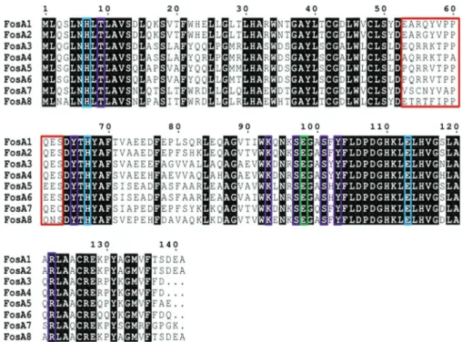
Phylogenetic analysis performed with FosA8 and the other FosA type enzymes
revealed that FosA8 was distantly related to the FosA enzymes, representing a distinct
cluster of FosA proteins (Fig. 1). FosA8 possessed conserved residues of FosA proteins
that are implicated in the dimer formation of the FosA proteins, of Mn
2⫹and K
⫹binding, and to fosfomycin binding (1) (Fig. 2).
Genetic context of the
fosA8 gene. Analysis of the WGS of E. coli 376 showed that
the fosA8 gene was located on a plasmid that also carried a kanamycin resistance
marker. Mating-out assays showed that this plasmid was self-conjugative, at a
fre-quency that was estimated to be ca. 10
⫺5. This plasmid did not carry the bla
CTX-M-15
ESBL gene that was identified in E. coli 376. PCR-based replicon typing and plasmid
analysis following extraction by the Kieser method (23) performed with isolate 376
confirmed the WGS data and identified an IncN natural plasmid 50 kb in size. The fosA8
gene was inserted into the sprT gene and was bracketed by two direct repeat
se-quences (Fig. 3). The sprT gene encoded a hypothetical protein of unknown function
(Fig. 3). The occurrence of identical direct repeats, TATTA, on each side of the DNA
fragment encompassing the fosA8 gene strongly suggested that the insertion of the
fosA8 gene onto its plasmid support resulted from a transposition event (Fig. 3).
TABLE 1 Strains and plasmids used in this study
Strain or plasmid Relevant genotype or phenotype Reference or source
Strains
E. coli
J53 Recipient strain for conjugation experiment; azide resistant 34
TOP10 Recipient strain for cloning experiments Invitrogen
376 Clinical isolate resistant to extended-spectrum cephalosporins,
fluoroquinolones, kanamycin, tobramycin, tetracycline, and fosfomycin
This study
L. adecarboxylata DSMZ5077 Natural fosfomycin-resistant enterobacterial species This study
Plasmids
pACYC184 Cloning plasmid; chloramphenicol and tetracycline resistant New England Biolabs p376 Natural ca. 50-kb and IncN plasmid of E. coli 376 This study
pFosA8 Recombinant plasmid pACYC184 with a 789-bp PCR fragment (fosA8) This study pFosALA Recombinant plasmid pACYC184 with a 789-bp PCR fragment (fosALA) This study
Origin of the
fosA8 gene. In silico analysis using the GenBank databases confirmed
that fosA-like genes are present in the chromosomes of several enterobacterial species,
such as those of Klebsiella pneumoniae, Enterobacter cloacae, Serratia marcescens, and
Kluyvera ascorbata (9). FosA8 showed the highest percentage of amino acid identity
(98%, differing in 3 amino acids) with a FosA protein, named FosA
LA, which is encoded
by a gene identified in the chromosome of the enterobacterial species Leclercia
adecarboxylata strain USDA-ARS-USMARC-60222 (GenBank accession no.
CP013990.1
).
Accordingly, the close sequences located upstream and downstream of the fosA8 gene
shared significant identity at the nucleotide level (99%) with those of L. adecarboxylata
USDA-ARS-USMARC-60222 (Fig. 3).
FIG 1 Phylogenetic tree obtained for the FosA proteins identified by distance method using the
neighbor-joining algorithm (SeaView version 4 software [33]). Branch lengths are drawn to scale and are proportional to the number of amino acid substitutions with 500 bootstrap replications. The distance along the vertical axis has no significance. The enterobacterial species known as the natural reservoirs of these FosA proteins are shown on the right.
FIG 2 Amino acid alignment of plasmid-mediated FosA proteins. Red, blue, green, and purple boxes
bracket amino acids of the dimer interface loop and those implicated in Mn2⫹coordination, in the K⫹ binding loop, and in fosfomycin binding, respectively (11).
Highly similar fosA
LA-like genes were identified in six chromosomes of Leclercia
species available in the genome databases, including two L. adecarboxylata isolates
(GenBank accession no.
CP036199.1
and
LR590464.1
) and four Leclercia species isolates
(GenBank accession no.
CP026387.1
,
CP026167.1
,
CP031104.1
and
CP031101.1
), sharing
from 85% to 97% nucleotide identity with fosA
LA.
The enterobacterial species L. adecarboxylata is rarely identified as a source of
human infections and is naturally resistant to fosfomycin (24, 25).
In order to characterize the fosfomycin resistance conferred by Fos
LA, the L.
adecar-boxylata strain DSMZ5077 was analyzed. It carried a fos
LAgene identical to that of L.
adecarboxylata strain USDA-ARS-USMARC-60222. The fosfomycin resistance phenotype
observed in L. adecarboxylata DSMZ5077 was antagonized by the addition of
phos-phonoformate (with the fosfomycin MIC value being decreased by 16-fold), indicating
the role of FosA
LAfor conferring fosfomycin resistance in that species. Both fosA8 and
fosA
LAgenes were cloned and expressed in E. coli TOP10. They conferred identical levels
of resistance to fosfomycin (fosfomycin MIC, 1,024 mg/liter).
DISCUSSION
We report here the identification of a novel plasmid-mediated and transferable
fosfomycin resistance determinant, FosA8, in E. coli. This enzyme conferred a high level
of resistance to fosfomycin. Its
⬍80% amino acid identity with known FosA proteins
indicates the diversity of fosA genes that may circulate, remaining undetected when
screening surveys based on molecular tools are performed. It is noteworthy that
analysis of GenBank databases showed that this gene is present in two E. coli isolates
recovered in the United States, indicating that it is already present on another
conti-nent.
The concomitant identification of the natural reservoir of the fosA8 gene, L.
decar-boxylata, adds to the growing list of enterobacterial species that naturally possess a
fosA-like gene and from which those genes are translocated onto plasmids in E. coli (9,
10). In contrast to other enterobacterial species that possess these fosA-like genes, such
as K. pneumoniae (9), fosA8-like genes are likely expressed at a high level in L.
decarboxylata, since this species is naturally resistant to fosfomycin (25) and the
fosfomycin resistance is inhibited by the addition of phosphonormate. The driving
forces at the origin of transfer and the spread of fosA genes from their natural reservoir
to a plasmid location in an enterobacterial species, E. coli, which does not possess a fosA
gene, remain to be investigated.
This novel FosA determinant is reported from an E. coli isolate belonging to
sequence type 40 (ST40) that is recognized as an emerging extraintestinal E. coli clone
(26). Of note, the fosA8 and bla
CTX-M-15genes were not associated on the same plasmid.
This is in contrast to the most commonly identified fosA gene, fosA3, which is always
identified on plasmids carrying either ESBL genes, AmpC-type
-lactamase genes, or
carbapenemase genes (16–19).
Nonetheless, identification of the FosA8 determinant from a CTX-M-producing
FIG 3 Genetic structures surrounding the fosALAgene on the chromosome of L. adecarboxylata (A) and
on the fosA8 gene on plasmid p376 of E. coli 376 (B). The grey-shaded sequences indicate sequence identity between plasmid p376 and L. adecarboxylata DSMZ5077. TATTA direct repeats bracketing fosA8 on plasmid p376 are also indicated.
isolate further highlights that ESBL producers are important reservoirs of transferable
fosfomycin resistance determinants. This result may be a source of concern, since
fosfomycin is becoming a first-line therapy for treating cystitis due to ESBL producers
and the prevalence of ESBL producers is still rising worldwide.
MATERIALS AND METHODS
Bacterial strains and plasmids. E. coli isolate 376 was isolated from urine in the context of screening
1,225 ESBL-producing E. coli isolates from a Swiss collection obtained from 2012 and 2013 (20). The list of strains and plasmids used in the study is shown in Table 1.
Fosfomycin resistance determination.Two techniques were used to assess fosfomycin suscepti-bility. First, the agar dilution method using cation-adjusted Mueller-Hinton agar (MHA-CA, reference 64884; Bio-Rad, Marnes-la-Coquette, France) supplemented with 25g/ml glucose-6-phosphate was used to determine fosfomycin MICs, as recommended by Clinical and Laboratory Standards Institute (CLSI) guidelines (27). The breakpoints of the European Committee on Antimicrobial Susceptibility Testing (EUCAST) were used as a reference (28). E. coli isolates with MICs of⬎32g/ml were categorized as resistant. Second, the rapid fosfomycin/E. coli NP test was performed as described by Nordmann et al. (21). The results of this test were read after 1 h 30 min of incubation at 35⫾ 2°C.
FosA-like role in fosfomycin resistance. The contribution of a potential FosA-like enzyme in
fosfomycin resistance was assessed first by using the disk diffusion method as described by Nakamura et al. (22). Briefly, the fosfomycin-resistant E. coli 376 and the L. decarboxylata DBZ isolate at an⬃0.5 McFarland standard were inoculated onto Mueller-Hinton agar plates supplemented with 25g/ml of glucose-6-phosphate. Two disks containing 200g of fosfomycin, with and without 0.5 mg of the FosA inhibitor sodium phosphonoformate (PPF) (Sigma-Aldrich), were added to the plates, which were incubated at 35⫾ 2°C overnight. Actually, PPF selectively inhibits the FosA-like proteins, which allows differentiation between plasmid-mediated FosA and other mechanisms of resistance to fosfomycin. An increase in the diameter of the growth inhibition zone byⱖ5 mm in the presence of PPF is interpreted as FosA-related resistance.
Molecular analyses. DNA was extracted from E. coli strain 376 with the QIAamp DNA minikit and the
QIAcube workstation (Qiagen, Courtaboeuf, France), according to the manufacturer’s instructions. PCR amplification (Microsynth, Balgach, Switzerland) was performed to detect any known plasmid-mediated
fosA genes (fosA1 to fosA7), as previously described (21). PCR conditions were 95°C for 5 min, followed
by 30 cycles of 95°C for 30 s, 52°C for 30 s and 72°C for 30 s, with a final extension of 72°C for 5 min. Sequences were analyzed with CloneManager Professional (Sci-Ed Software, Denver, CO, USA) (29). Whole-genome DNA of the fosA8-positive E. coli isolate and its transconjugant was extracted using a Sigma-Aldrich GenElute bacterial genomic DNA kit. Genomic libraries were assessed using a Nextera XT library preparation kit (Illumina Inc., San Diego, CA), and sequencing was performed using an Illumina MiniSeq system with 300-bp paired-end reads and a coverage of 50 times. The generated FastQ data were compiled and analyzed using the CLC Genomic Workbench (version 7.5.1; CLC Bio, Aarhus, Denmark). Reads were de novo assembled with automatic bubble and word size, and contigs with a minimum contig length of 800 nucleotides were generated using the mapping mode map reads back to contigs. The resulting contigs were uploaded into the Center for Genomic Epidemiology server (http:// www.genomicepidemiology.org/). The plasmid replicon type and multilocus sequence type according to the Warwick typing scheme were determined using the PlasmidFinder (version 1.3) and MLST (version 1.8) programs, respectively (26, 30, 31). Sequence alignments was performed with ESPript 3.0 software, and construction of phylogenetic trees was performed with the SeaView alignment tool (version 4; Prabi, La Doua, Lyon, France) (32).
The fosA8 gene from E. coli 376 and the fosLAgene from L. adecarboxylata were cloned into the
pACYC184 vector by using primers listed in Table 2 and expressed in E. coli TOP10.
Plasmid analysis and conjugation.Plasmid analysis was performed using the Kieser extraction method (23), followed by gel electrophoresis, in order to further confirm the size of the plasmid containing the fosA8 gene using E. coli strain 50192 harboring four plasmids of 154, 66, 48, and 7 kb as plasmid size markers (33). The determination of the plasmid incompatibility group was confirmed by PCR-based replicon typing (PBRT) (30). Conjugation experiments were performed using E. coli strain 376 and azide-resistant strain E. coli J53 as donor and recipient strains, respectively, as described previously (20). Both donor and recipient strains were cultured in exponential phase and then mixed on solid Luria Broth agar using filters at a 1:10 donor/recipient ratio. After 5 h of incubation, the filters were resuspended in 0.85% NaCl and the bacterial mixture was plated onto agar plates supplemented with fosfomycin (25g/ml) and sodium azide (100 g/ml). The susceptibility of all E. coli transconjugants to
TABLE 2 Oligonucleotide sequences designed for this studya
Primer Sequence
fosA8 BamHI forward 5=-GATGATGGATCCAGCGTGAAATGGAAGTGTGG-3=
fosA8 SalI forward 5=-GATGATGTCGACTAATACTGGTGATCCACGGC-3=
fosA8 intern forward 5=-AACCATCTGACCCTTGCTGT-3=
fosA8 intern reverse 5=-CGAGAAAATAGAACGACGCC-3=
aRestriction sites are underlined; intern, internal to the fosA gene.
fosfomycin was assessed by disk diffusion with azide- and fosfomycin-containing disks and by the PPF test (22).
Accession number(s). The sequence data corresponding to the fosA8 gene reported in this paper
was submitted to the GenBank nucleotide database under accession no.MN150127. In addition, the contigs corresponding to the whole genome of isolate 376 have been deposited as a BioProject under accession no.PRJNA558667.
ACKNOWLEDGMENTS
This work was funded by the University of Fribourg and by the Laboratoire Européen
Associé INSERM Emerging Antibiotic Resistance in Gram-Negative Bacteria, University
of Fribourg, Fribourg, Switzerland.
REFERENCES
1. Silver LL. 2017. Fosfomycin: mechanism and resistance. Cold Spring Harb Perspect Med 7:a025262.https://doi.org/10.1101/cshperspect.a025262. 2. Falagas ME, Vouloumanou EK, Samonis G, Vardakas KZ. 2016.
Fosfo-mycin. Clin Microbiol Rev 29:321–347.https://doi.org/10.1128/CMR .00068-15.
3. Gupta K, Hooton TM, Naber KG, Wullt B, Colgan R, Miller LG, Moran GJ, Nicolle LE, Raz R, Schaeffer AJ, Soper DE. 2011. International clinical practice guidelines for the treatment of acute uncomplicated cystitis and pyelonephritis in women: a 2010 update by the Infectious Diseases Society of America and the European Society for Microbiology and Infectious Diseases. Clin Infect Dis 52:e103– e120. https://doi.org/10 .1093/cid/ciq257.
4. Pitout JDD, Laupland KB. 2008. Extended-spectrum-lactamase produc-ing Enterobacteriaceae: an emergproduc-ing public-health concern. Lancet In-fect Dis 8:159 –166.https://doi.org/10.1016/S1473-3099(08)70041-0. 5. Cattoir V, Guérin F. 2018. How is fosfomycin resistance developed in
Escherichia coli? Future Microbiol 13:1693–1696.https://doi.org/10.2217/ fmb-2018-0294.
6. Alrowais H, McElheny CL, Spychala CN, Sastry S, Guo Q, Butt AA, Doi Y. 2015. Fosfomycin resistance in Escherichia coli, Pennsylvania, USA. Emerg Infect Dis 21:2045–2047.https://doi.org/10.3201/eid2111.150750. 7. Benzerara Y, Gallah S, Hommeril B, Genel N, Decre D, Rottman M, Arlet
G. 2017. Emergence of plasmid-mediated fosfomycin-resistance genes among Escherichia coli isolates, France. Emerg Infect Dis 23:1564 –1567.
https://doi.org/10.3201/eid2309.170560.
8. Guo Q, Tomich AD, McElheny CL, Cooper VS, Stoesser N, Wang M, Sluis-Cremer N, Doi Y. 2016. Glutathione-S-transferase FosA6 of Klebsiella
pneumoniae origin conferring fosfomycin resistance in ESBL-producing Escherichia coli. J Antimicrob Chemother 71:2460 –2465.https://doi.org/ 10.1093/jac/dkw177.
9. Ito R, Mustapha MM, Tomich AD, Callaghan JD, McElheny CL, Mettus RT, Shanks RMQ, Sluis-Cremer N, Doi Y. 2017. Widespread fosfomycin resis-tance in Gram-negative bacteria attributable to the chromosomal fosA gene. mBio 8:e00749-17.https://doi.org/10.1128/mBio.00749-17. 10. Ito R, Pacey MP, Mettus RT, Sluis-Cremer N, Doi Y. 2018. Origin of the
plasmid-mediated fosfomycin resistance gene fosA3. J Antimicrob Che-mother 73:373–376.https://doi.org/10.1093/jac/dkx389.
11. Klontz EH, Tomich AD, Günther S, Lemkul JA, Deredge D, Silverstein Z, Shaw JF, McElheny C, Doi Y, Wintrode PL, MacKerell AD, Jr, Sluis-Cremer N, Sundberg EJ. 2017. Structure and dynamics of FosA-mediated fosfo-mycin resistance in Klebsiella pneumoniae and Escherichia coli. Antimi-crob Agents Chemother 61:e01572-17.
12. Ma Y, Xu X, Guo Q, Wang P, Wang W, Wang M. 2015. Characterization of
fosA5, a new plasmid-mediated fosfomycin resistance gene in Esche-richia coli. Lett Appl Microbiol 60:259 –264.https://doi.org/10.1111/lam .12366.
13. Rehman MA, Yin X, Persaud-Lachhman MG, Diarra MS. 2017. First de-tection of a fosfomycin resistance gene, fosA7, in Salmonella enterica serovar Heidelberg isolated from broiler chickens. Antimicrob Agents Chemother 61:e00410-17.https://doi.org/10.1128/AAC.00410-17. 14. Xu H, Miao V, Kwong W, Xia R, Davies J. 2011. Identification of a novel
fosfomycin resistance gene (fosA2) in Enterobacter cloacae from the Salmon River, Canada. Lett Appl Microbiol 52:427– 429.https://doi.org/ 10.1111/j.1472-765X.2011.03016.x.
15. Yang TY, Lu PL, Tseng SP. 2019. Update on fosfomycin-modified genes in Enterobacteriaceae. J Microbiol Immunol Infect 52:9 –21.https://doi .org/10.1016/j.jmii.2017.10.006.
16. Lee SY, Park YJ, Yu JK, Jung S, Kim Y, Jeong SH, Arakawa Y. 2012. Prevalence of acquired fosfomycin resistance among extended-spectrum ß-lactamase-producing Escherichia coli and Klebsiella
pneu-moniae clinical isolates in Korea and IS26-composite transposon
sur-rounding fosA3. J Antimicrob Chemother 67:2843–2847.https://doi.org/ 10.1093/jac/dks319.
17. Lupo A, Saras E, Madec JY, Haenni M. 2018. Emergence of blaCTX-M-55 associated with fosA, rmtB and mcr gene variants in Escherichia coli from various animal species in France. J Antimicrob Chemother 73:867– 872.
https://doi.org/10.1093/jac/dkx489.
18. Wachino J, Yamane K, Suzuki S, Kimura K, Arakawa Y. 2010. Prevalence of fosfomycin resistance among CTX-M-producing Escherichia coli clini-cal isolates in Japan and identification of novel plasmid-mediated fosfomycin-modifying enzymes. Antimicrob Agents Chemother 54: 3061–3064.https://doi.org/10.1128/AAC.01834-09.
19. Xie M, Lin D, Chen K, Wai Chi Chan E, Yao W, Wen S. 2016. Molecular characterization of Escherichia coli strains isolated from retail meat that harbor blaCTX-M and fosA3 genes. Antimicrob Agents Chemother 60: 2450 –2455.https://doi.org/10.1128/AAC.03101-15.
20. Mueller L, Cimen C, Poirel L, Descombes MC, Nordmann P. 2019. Prev-alence of fosfomycin resistance among ESBL-producing Escherichia coli isolates in the community, Switzerland. Eur J Clin Microbiol Infect Dis 38:945–949.https://doi.org/10.1007/s10096-019-03531-0.
21. Nordmann P, Poirel L, Mueller L. 2018. Rapid detection of fosfomycin resistance in Escherichia coli. J Clin Microbiol 57:e01531-18.https://doi .org/10.1128/JCM.01531-18.
22. Nakamura G, Wachino J, Sato N, Kimura K, Yamada K, Jin W, Shibayama K, Yagi T, Kawamura K, Arakawa Y. 2014. Practical agar-based disk potentiation test for detection of fosfomycin-nonsusceptible Escherichia
coli clinical isolates producing glutathione S-transferases. J Clin
Micro-biol 52:3175–3179.https://doi.org/10.1128/JCM.01094-14.
23. Kieser T. 1984. Factors affecting the isolation of CCC DNA from
Strepto-myces lividans and Escherichia coli. Plasmid 12:19 –36.https://doi.org/10 .1016/0147-619X(84)90063-5.
24. Leclerc H. 1962. Étude biochimique d’Enterobacteriaceae pigmentées. Ann Inst Pasteur (Paris) 102:726 –741.
25. Stock I, Burak S, Wiedemann B. 2004. Natural antimicrobial susceptibility patterns and biochemical profiles of Leclercia adecarboxylata strains. Clin Microbiol Infect 10:724 –733. https://doi.org/10.1111/j.1469-0691.2004 .00892.x.
26. Joensen KG, Tetzschner AM, Iguchi A, Aarestrup FM, Scheutz F. 2015. Rapid and easy in silico serotyping of Escherichia coli isolates by use of whole-genome sequencing data. J Clin Microbiol 53:2410 –2426.https:// doi.org/10.1128/JCM.00008-15.
27. Clinical and Laboratory Standards Institute. 2018. Performance standards for antimicrobial susceptibility testing; 28th informational supplement (M100 –S28). Clinical and Laboratory Standards Institute, Wayne, PA. 28. European Committee on Antimicrobial Susceptibility Testing. 2019.
Breakpoint tables for interpretation of MICs and zone diameters. Version 9.0. http://www.eucast.org/fileadmin/src/media/PDFs/EUCAST _files/Breakpoint_tables/v_9.0_Breakpoint_Tables.pdf.
29. Robert X, Gouet P. 2014. Deciphering key features in protein structures with the new ENDscript server. Nucleic Acids Res 42:320 –324.https:// doi.org/10.1093/nar/gku316.
30. Carattoli A, Bertini A, Villa L, Falbo V, Hopkins KL, Threlfall EJ. 2005. Identification of plasmids by PCR-based replicon typing. J Microbiol Methods 63:219 –228.https://doi.org/10.1016/j.mimet.2005.03.018.
31. Carattoli A, Zankari E, García-Fernández A, Voldby Larsen M, Lund O, Villa L, Møller Aarestrup F, Hasman H. 2014. In silico detection and typing of plasmids using PlasmidFinder and plasmid multilocus sequence typing. Antimicrob Agents Chemother 58:3895–3903.https://doi.org/10.1128/ AAC.02412-14.
32. Gouy M, Guindon S, Gascuel O. 2010. SeaView version 4: a multiplatform graphical user interface for sequence alignment and phylogenetic tree
building. Mol Biol Evol 27:221–224. https://doi.org/10.1093/molbev/ msp259.
33. Vivian A. 1994. Plasmid expansion? Microbiology 140:213–214.https:// doi.org/10.1099/13500872-140-2-213-a.
34. Jacoby GA, Han P. 1996. Detection of extended-spectrum-lactamases in clinical isolates of Klebsiella pneumoniae and Escherichia coli. J Clin Microbiol 34:908 –911.