HAL Id: inserm-00113634
https://www.hal.inserm.fr/inserm-00113634
Submitted on 15 Nov 2006
HAL is a multi-disciplinary open access
archive for the deposit and dissemination of
sci-entific research documents, whether they are
pub-lished or not. The documents may come from
teaching and research institutions in France or
abroad, or from public or private research centers.
L’archive ouverte pluridisciplinaire HAL, est
destinée au dépôt et à la diffusion de documents
scientifiques de niveau recherche, publiés ou non,
émanant des établissements d’enseignement et de
recherche français ou étrangers, des laboratoires
publics ou privés.
Expression of fibrillin-1 in focal nodular hyperplasia of
the liver: a role in microcirculation adaptability.
Sébastien Lepreux, Alexis Desmouliere, Jean Rosenbaum, Charles Balabaud,
Paulette Bioulac-Sage
To cite this version:
Sébastien Lepreux, Alexis Desmouliere, Jean Rosenbaum, Charles Balabaud, Paulette Bioulac-Sage.
Expression of fibrillin-1 in focal nodular hyperplasia of the liver: a role in microcirculation
adaptabil-ity.. Comparative Hepatology, BioMed Central, 2004, 3 Suppl 1, pp.S57.
�10.1186/1476-5926-2-S1-S57�. �inserm-00113634�
Open Access
Proceedings
Expression of fibrillin-1 in focal nodular hyperplasia of the liver: a
role in microcirculation adaptability
Sébastien Lepreux
1,2, Alexis Desmouliere
1, Jean Rosenbaum
1,
Charles Balabaud
1,3and Paulette Bioulac-Sage*
1,2Address: 1GREF, INSERM E0362, Université Victor Segalen Bordeaux 2, France, 2Service d'Anatomie Pathologique, Hôpital Pellegrin, C.H.U.
Bordeaux, France and 3Service d'Hépatologie-Gastroentérologie, Hôpital Saint-André, C.H.U. Bordeaux, France
Email: Sébastien Lepreux - sebastien.lepreux@chu-bordeaux.fr; Alexis Desmouliere - alexis.desmouliere@gref.u-bordeaux2.fr; Jean Rosenbaum - jean.rosenbaum@gref.u-bordeaux2.fr; Charles Balabaud - charles.balabaud@gref.u-bordeaux2.fr; Paulette Bioulac-Sage* - paulette.bioulac-sage@gref.u-bordeaux2.fr
* Corresponding author
Abstract
Introduction: It has been suggested that the elastic network plays an important role in the tissue response to mechanical stress. The components of the elastic network have been poorly studied in liver diseases. Therefore, in this work, the expression and distribution of fibrillin-1 and elastin were studied in hepatic focal nodular hyperplasia and compared with surrounding liver and hepatocellular adenoma.
Methods: Immunohistochemical studies for fibrillin-1 and elastin were performed on unfixed cryostat sections of focal nodular hyperplasia (22 cases), hepatocellular adenoma (15 cases) and surrounding liver (34 cases).
Results: Surrounding normal liver showed only a continuous, thin and regular immunostaining of fibrillin-1 in the space of Disse, whereas elastin was nearly absent. In focal nodular hyperplasia, fibrillin-1 was more strongly expressed in the perisinusoidal space, compared with surrounding liver; in contrast, in adenomas fibrillin-1 immunostaining was irregular and very low in perisinusoidal space, more intense in peliotic areas.
Conclusions: In focal nodular hyperplasia, the increased microfibrillar network containing fibrillin-1 in the space of Disse could reflect an adaptation of the sinusoidal wall to an increased arterial blood flow in sinusoids. In hepatocellular adenoma, the different patterns of fibrillin-1 could be related to the heterogeneity of the arterial vascularization and to the frequent necrotico-hemorrhagic changes. This study comparing the elastic network in two types of lesions with vascularization abnormalities and in the surrounding liver provides interesting new data for understanding the structural role of fibrillin-1 in the space of Disse.
Introduction
The elastic fibers in the extra-cellular matrix are composed of elastin associated with 10 nm microfibrils. It has been suggested that the microfibrils mediate the conversion of
tropoelastin to elastin and act as a scaffold [1]. However, microfibrils can be present alone [2] and in these situa-tions, their role is not clearly known. Fibrillin-1, a 350 kDa glycoprotein, is a major component of these 10 nm
from 11th International Symposium on the Cells of the Hepatic Sinusoid and their Relation to Other Cells
Tucson, Arizona, USA, 25–29 August, 2002 Published: 14 January 2004
Comparative Hepatology 2004, 3(Suppl 1):S57
<supplement> <title> <p>11th International Symposium on the Cells of the Hepatic Sinusoid and their Relation to Other Cells</p> </title> </supplement>
Comparative Hepatology 2004, 3 http://www.comparative-hepatology.com/content/3/S1/S57
microfibrils [2]. Fibrillin-1 participates in the attachment of cells to the extra-cellular matrix by virtue of RGD motifs [2,3]. Fibrillin-1 and elastin have been widely studied in many human tissues, particularly in the skin and eyes [2,4] but little is known in the liver. We recently studied for the first time, the expression and cellular synthesis of fibrillin-1 in human liver parenchyma [5]. Briefly, i/ in the portal tract, fibrillin-1 was colocalized with elastin in the vessel walls and in connective tissue, whereas a condensa-tion of fibrillin-1 alone was detected close to the base-ment membrane of bile ducts as well as at the interface with the limiting hepatocytic plates; ii/ fibrillin-1 and elastin were also colocalized in the walls of hepatic vein; iii/ in the liver lobule, fibrillin-1 was detected alone as a regular thin network in the space of Disse all along sinu-soids. Although little is known concerning the physiolog-ical functions of fibrillin-1 in the liver, it can be assumed that this elastic glycoprotein which is expressed alone in sinusoids, could play a role in the sinusoidal blood flow regulation, in normal, as well as in pathological condi-tions particularly those exhibiting abnormalities of vascularization.
The aims of our study were:
1. to study the expression and distribution of fibrillin-1 in focal nodular hyperplasia (FNH) which is a pseudo-tumoral benign hepatocytic lesion with a nodular organi-zation, related to an increase in arterial blood flow; 2. to compare these observations with those obtained: a-in a true benign hepatocytic tumor, the hepatocellular adenoma, which is another condition of localized arterio-hepatic hyperperfusion, but different, from a physio-pathological viewpoint from focal nodular hyperplasia; and b- in the surrounding normal liver.
Methods
Human liver specimens
Seventy-one liver samples were studied, obtained from 34 patients. They corresponded to surgically resected speci-mens of FNH (n = 22), hepatocellular adenoma (n = 15) and the surrounding non-tumoral liver (n = 34). In 3 cases, samples of FNH and hepatocellular adenoma were obtained from the same patient.
One part of tissue samples was routinely formalin fixed and paraffin embedded; five micrometer-thick paraffin sections of each sample were taken and stained with HES for diagnostic purposes. Additional sections were stained with Masson's trichrome, reticulin and orcein. Another part of fresh tissue was immediately snap-frozen in liquid nitrogen-cooled isopentane and stored at -80 degrees C until used for this study. Five micrometer-thick serial fro-zen sections of each sample were air-dried on Super frost/
plus slides (Menzel Glaser, Germany) and processed for immunohistochemistry.
Immunohistochemistry
Two components of elastic fibers, fibrillin-1 and elastin, as well as alpha-smooth muscle actin (alpha-SMA) for staining of the myofibroblasts (including hepatic stellate cells in their myofibroblastic state) were tested. Mouse monoclonal antibodies against fibrillin-1 (clone 11C1.3; NeoMarkers, Fremont, CA) and against alpha-SMA (Dako A/S, Glostrup, Denmark), and a rabbit polyclonal anti-body against human elastin (gift of D. Hartmann, Novo-tec, Lyon, France) were used. Unfixed frozen sections were incubated with the antibodies against fibrillin-1 (1/100), elastin (1/1000) or alpha-SMA (1/400) diluted in phos-phate-buffered saline, pH 7.4 containing 4% bovine serum albumin. After washing, the epitopes were detected with the Envision+ system HRP detection and developed with liquid diaminobenzidine (Dako A/S, Glostrup, Den-mark). Sections were examined with a Zeiss Axioplan 2 microscope (Carl Zeiss Microscopy, Jena, Germany). Images were acquired with an AxioCam camera (Carl Zeiss Vision, Hallbergmoos, Germany) by means of the AxioVision image processing and analysis system (Carl Zeiss Vision).
The same immunohistochemical observations were obtained with another anti-fibrillin-1 antibody (clone 69; Chemicon, Temecula, CA) (data not shown). All controls where primary antibodies were omitted were negative.
Results
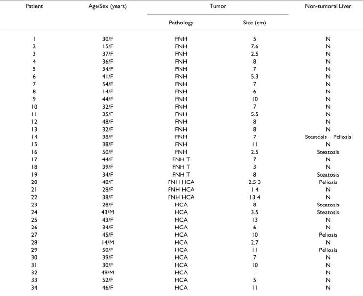
PatientsAll main clinical and pathological data are summarized in table 1. All patients with FNH were female, with a mean age of 36.4 years (15–54 years). The mean size of hepatic tumors was 6.45 cm (1–13 cm). Patients with hepatocel-lular adenoma were predominantly female (12/15) with a mean age of 38.6 years (14–52 years); hepatic tumors were of 7 cm mean size (2.7–13 cm), multiple in 2 cases (cases 27, 33) and/or associated with FNH in 3 cases (cases 20, 21, 22). All female patients were treated with oral contraceptive steroids. One male patient presented alcohol addiction and obesity (case 24), and one was treated with androgenic steroids for a Fanconi disease (case 28).
Histopathology and immunohistochemical analysis
Surrounding non tumoral liver tissue
The surrounding non-tumoral liver tissue (34 cases) showed a normal architecture and cytology. Five cases showed mild steatotic changes (<5%) in parenchyma sur-rounding hepatocellular adenoma (2 cases) or FNH (3 cases) and there were focal peliotic changes around ade-noma (3 cases) or FNH (1 case). Elastic fibers in walls of
portal and centrolobular vessels were clearly stained with orcein and no staining was observed in lobular zones. As previously described [5] (Figure 1), fibrillin-1 and elastin were both immunostained in the vessel walls and intersti-tial tissue in portal tracts; only fibrillin-1 was expressed, while elastin was absent close to the basement membrane of biliary ducts and at the interface with the hepatocytic limiting plate. Both fibrillin-1 and elastin were also present in the wall of centrolobular veins. In the lobules, only fibrillin-1 was detected as a thin, regular network in the space of Disse along sinusoids. There was marked alpha-SMA immunostaining in the vessel walls and in some interstitial cells of the portal tracts and only mild immunostaining in a few hepatic stellate cells of the parenchyma.
Focal nodular hyperplasia
Nineteen cases were histologically classified as a classical solid form of FNH, of which one exhibited steatosis (case 21), and 3 cases were telangiectatic FNH (cases 17, 18, 19).
All classical forms of FNH were similar and typical: they displayed numerous nodules of liver parenchyma sepa-rated by fibrous septa. Nodules were composed of nearly normal hepatocytes, with large areas of macrovesicular steatosis (>30%) in 1 case (case 21). A fibrous scar was observed in 16 cases and was inconspicuous in 3 cases. The fibrous septa between nodules contained thick-walled arteries with intimal thickening, sometimes partly
Table 1: Relevant clinical and pathological data
Patient Age/Sex (years) Tumor Non-tumoral Liver
Pathology Size (cm) 1 30/F FNH 5 N 2 15/F FNH 7.6 N 3 37/F FNH 2.5 N 4 36/F FNH 8 N 5 34/F FNH 7 N 6 41/F FNH 5.3 N 7 54/F FNH 7 N 8 14/F FNH 6 N 9 44/F FNH 10 N 10 32/F FNH 7 N 11 35/F FNH 5.5 N 12 48/F FNH 8 N 13 32/F FNH 8 N 14 38/F FNH 7 Steatosis – Peliosis 15 38/F FNH 11 N 16 50/F FNH 2.5 Steatosis 17 44/F FNH T 7 N 18 39/F FNH T 3 N 19 34/F FNH T 8 Steatosis 20 40/F FNH HCA 2.5 3 Peliosis 21 28/F FNH HCA 1 4 N 22 38/F FNH HCA 13 4 N 23 28/F HCA 8 Steatosis 24 43/M HCA 3.5 Steatosis 25 43/F HCA 13 N 26 34/F HCA 6 N 27 45/F HCA 10 Peliosis 28 14/M HCA 2.7 N 29 50/F HCA 11 Peliosis 30 39/F HCA 7 N 31 30/F HCA 10 N 32 49/M HCA - N 33 52/F HCA 5 N 34 46/F HCA 11 N
Comparative Hepatology 2004, 3 http://www.comparative-hepatology.com/content/3/S1/S57
thrombosed, veins, capillaries and numerous biliary ductules.
Orcein staining clearly showed the elastic lamina of arte-rial walls, which were sometimes disrupted, the delicate network of elastic fibers in the vein walls and some elastic fibers dispersed in the connective tissue of the septa; elas-tic fibers were however absent around capillaries. Sinu-soids in parenchymal nodules were not stained with orcein.
Immunohistochemical results were identical in all typical FNH (Figures 2,3,4,5):
i. Fibrillin-1 and elastin were both immunostained in the walls of arteries and veins, as well as in the fibrous tissue of septa and fibrous scars (Figures 2, 4, 5).
ii. Fibrillin-1 was expressed alone and more strongly close to the basement membrane of biliary ductules highlight-ing clearly the characteristic ductular reaction of FNH (Figures 4,5). There was no increase of the fibrillin-1 stain-ing at the interface between the fibrous septa and hepato-cytic nodules, as observed between portal tracts and the first hepatocytic plate in normal surrounding liver. iii. Within parenchymal nodules, only fibrillin-1 was expressed in the perisinusoidal spaces between hepato-cytic plates showing strong, continuous and regular Immunohistochemistry for fibrillin-1 in the surrounding
nor-mal liver Figure 1
Immunohistochemistry for fibrillin-1 in the surrounding nor-mal liver. In the lobule, fibrillin-1 is detected as a thin, regular network in the space of Disse all along sinusoids. In the por-tal tract, fibrillin-1 is detected in the vessel walls, in interstitial tissue, with an increase close to the basement membrane of biliary ducts (white arrow) and at the interface with the hepatocytic limiting plate (black arrow).
Immunohistochemistry for fibrillin-1 in a classical form of focal nodular hyperplasia
Figure 2
Immunohistochemistry for fibrillin-1 in a classical form of focal nodular hyperplasia. Fibrillin-1 is expressed in fibrous septa separating nodules of liver parenchyma and in between hepatocytic plates.
1
2
Immunohistochemistry for fibrillin-1 in focal nodular hyperplasia
Figure 3
Immunohistochemistry for fibrillin-1 in focal nodular hyper-plasia. Within parenchymal nodules, fibrillin-1 is expressed in the perisinusoidal spaces between hepatocytic plates showing a strong, continuous and regular staining.
staining, with a higher intensity compared with the surrounding normal liver (Figures 3, 4). Furthermore, there was an increase in the staining around sinusoids
close to the septa. Elastin staining was sometimes slightly expressed inside hepatocytic nodules, but only close to the fibrous septa.
There was a marked alpha-SMA immunostaining in the artery and vein walls and in some mesenchymal cells of the septa, as well as mild but regular immunostaining of the hepatic stellate cells within the parenchymal nodules, with an increase close to the septa.
The 3 cases of telangiectatic FNH exhibited similar fea-tures. They did not display nodules or fibrous scar. How-ever, a few short fibrous septa were found and showed numerous thick-walled arteries, a few veins and a mild degree or no bile ductular reaction. The hepatocytic plates were clearly separated by dilated sinusoids, sometimes alternating with areas of marked ectasia forming occasion-ally peliotic areas. Orcein stained the elastic lamina of arterial walls and the delicate network of elastic fibers in the vein walls.
Immunohistochemical results were nearly the same as in the classical form. Fibrillin-1 was expressed alone, with-out elastin in the perisinusoidal spaces between hepato-cytic plates, showing a relatively continuous and regular staining, but more accentuated around ectatic sinusoids. There was strong alpha-SMA immunostaining in the ves-sel walls and moderate but regular immunoreactivity in Immunohistochemistry for fibrillin-1 in focal nodular
hyperplasia Figure 4
Immunohistochemistry for fibrillin-1 in focal nodular hyper-plasia. Fibrillin-1 is expressed in the fibrous septa (right).
Immunohistochemistry for fibrillin-1 in focal nodular hyperplasia
Figure 5
Immunohistochemistry for fibrillin-1 in focal nodular hyper-plasia. Higher magnification of the area defined by a rectangle in the Figure 4. Fibrillin-1 staining is stronger close to the basement membrane of biliary ductules (white arrow) in the fibrous septa and around sinusoids near the septa (black arrow).
4
5
Immunohistochemistry for fibrillin-1 in hepatocellular adenoma
Figure 6
Immunohistochemistry for fibrillin-1 in hepatocellular ade-noma. Fibrillin-1 is detected in the numerous arterial sections (black arrow), whereas there is only a weak, discontinuous and irregular staining in the perisinusoidal spaces.
Comparative Hepatology 2004, 3 http://www.comparative-hepatology.com/content/3/S1/S57
the hepatic stellate cells of the parenchymal zones, pre-dominantly around dilated sinusoids.
Hepatocellular adenoma
All 15 cases of hepatocellular adenoma were typical. They displayed a loss of the normal lobular architecture with-out true portal tract and biliary ducts were always absent. The tumoral hepatocyte trabeculae interspersed with thin-walled vessels. Peliotic changes (10 cases), some fibrotic changes (5 cases) adjacent to large areas of necrosis and hemorrhage or massive steatosis (6 cases) were observed. Orcein staining showed the elastic lamina of vessel walls and sometimes a delicate network in the fibrotic changes. Fibrillin-1 and elastin were both immunostained in the vessel walls, especially in the numerous arteries (Figure 6). Within parenchymal zones, fibrillin-1 was sometimes irregularly expressed in the perisinusoidal spaces, show-ing a discontinuous and irregular stainshow-ing with lower intensity (8 cases) or with the same intensity (7 cases) compared with the surrounding liver (Figure 6), except in some areas of peliosis where the staining was more strong and continuous. It should be mentioned that, within the same case, there was often heterogeneity of fibrillin-1 staining in perisinusoidal spaces, with alternating stained and unstained zones. Elastin staining was always absent between tumoral hepatocytes. There was marked alpha-SMA cellular immunostaining in the vessel walls and fine and irregular immunostaining in some hepatic stellate cells inside the tumor, which were only clearly stained in the peliotic areas. When present, areas of fibrotic change exhibited strong alpha-SMA cellular immunostaining and strong staining for fibrillin-1 and elastin.
Discussion
In normal liver, immunohistochemistry is an effective tool for detection of the fibrillin-1 network around sinu-soids, since this glycoprotein is expressed alone in space of Disse, as a thin network, without elastin [5] and thus not stained with classical staining of elastic tissue such as orcein. The role of fibrillin-1, a component of elastic tis-sue, in space of Disse which is a strategic place for exchanges between hepatocytes and sinusoidal microcir-culation, remains however to be elucidated. The different patterns of fibrillin-1 expression in FNH and hepatocellu-lar adenoma, in comparison with normal liver, could be a reflection of the response.
In normal liver, sinusoids are supplied by portal vein blood which flows at a low pressure and by hepatic artery, representing respectively two thirds and one third of the total sinusoidal microcirculation [6].
It is well known that FNH are related to an increase in arte-rial flow due to primary or secondary artearte-rial abnormali-ties [7-10]. In these conditions, sinusoidal blood flow is
modified by an arteriohepatic hyperperfusion, as seen with radiologic imaging and illustrated after gelatin injec-tion [9-12]. It has been reported by Wanless [7,8] that in FNH, sinusoids were directly irrigated with inlet arterioles coming from fibrous septa. Injection of colored gelatin [9] indicated that the abnormal arteries were draining indi-rectly into sinusoids via capillaries in the fibrous septa. It could be postulated that these vascular conditions lead to an increase in sinusoidal blood pressure and could be an explanation for changing of the sinusoidal endothelial cell phenotype with neo-expression of CD34 and CD31 [9,13] and expression of alpha-SMA in hepatic stellate cells [14]. It is also well known that hepatic stellate cells are active in the adaptation to the changes in sinusoidal blood flow [15].
In comparison with normal surrounding liver, we showed in FNH, in addition to alpha-SMA positive hepatic stellate cells, increased immunostaining for fibrillin-1 in space of Disse, as a continuous and regular network with an enhancement near the fibrous septa. It is interesting to note that sinusoids adjacent to fibrous septa always express vascular endothelial markers such as CD34 [13,14,16].
As FNH represents a condition of arteriohepatic hyperper-fusion, it could be thought that the increased arterial blood flow may be a (causal) factor inducing the increase of fibrillin-1 microfibrillar network in space of Disse, as well as phenotypic changes with alpha-SMA neo-expres-sion in cells which secrete fibrillin-1 [5]. As elastic tissue is an important element in the regulation of the blood pres-sure in vessels, it can be assumed that the increased sinusoidal fibrillin-1 network, which is a component of elastic tissue, is involved in blood pressure regulation along the sinusoidal microvasculature between the hyper-plastic hepatocytic plates of FNH.
In telangiectatic FNH which lack the classical signs of FNH but exhibit characteristic features of sinusoidal dilatation, increase of fibrillin-1 network was more irregular, pre-dominating in zones of sinusoidal dilatation. This feature could reflect a predominant increase of arterial perfusion in these dilated areas.
In hepatocellular adenoma, another benign condition of hepatocytic proliferation associated with an irregular and abnormal vascularization, both elastin and fibrillin-1 immunostaining pointed to the great number of vascular sections distributed inside the tumor. The presence of fibrillin-1 alone along sinusoids was discontinuous, irreg-ular and some areas were completely unstained; on the contrary, peliotic zones showed a more intense staining than the non-peliotic ones, but this staining was irregu-larly distributed within the tumor. Unlike FNH where the
Publish with BioMed Central and every scientist can read your work free of charge
"BioMed Central will be the most significant development for disseminating the results of biomedical researc h in our lifetime."
Sir Paul Nurse, Cancer Research UK Your research papers will be:
available free of charge to the entire biomedical community peer reviewed and published immediately upon acceptance cited in PubMed and archived on PubMed Central yours — you keep the copyright
Submit your manuscript here:
http://www.biomedcentral.com/info/publishing_adv.asp
BioMedcentral
microfibrillar organization was correlated with the increase in blood pressure, the lack of a regular and con-tinuous network of microfibrils in the perisinusoidal space in hepatocellular adenoma could explain, at least in part, the frequency of peliosis and necrotico-hemorrhagic complications, characteristic of this tumor. Furthermore, the expression of alpha-SMA in hepatic stellate cells was more inconsistent, underlining again the heterogeneity of the lesion.
Conclusions
Based on our results, it can be hypothesized that in FNH, the increased microfibrillar network containing fibrillin-1 in the space of Disse could reflect an adaptation of the sinusoidal wall to variations of blood pressure related to an increased arterial blood flow in sinusoids. The different patterns of fibrillin-1 in hepatocellular adenoma could account for the heterogeneity of the arterial vasculariza-tion in this tumor characterized by frequent necrotico-hemorrhagic changes. These results could contribute to the understanding of the structural role of fibrillin-1 in space of Disse. Further studies are necessary to affirm whether the changes in fibrillin-1 network are the direct consequence of the liver microvascularisation abnormali-ties, and in particular of the increased arterial flow in FNH.
Acknowledgements
We are very grateful to D. Hartmann (Novotec, Lyon, France) for provid-ing the antibodies against elastin.
References
1. Raghunath M, Bächi T, Meuli M, Altermatt S, Gobet R, Bruckner-Tuderman L, Steinmann B: Fibrillin and elastin expression in skin
regenerating from cultured keratinocyte autografts: mor-phogenesis of microfibrils begins at the dermo-epidermal junction and precedes elastic fiber formation. J Invest Dermatol
1996, 106:1090-1095.
2. Sakai LY, Keene DR, Engvall E: Fibrillin, a new 350 kd
glycopro-tein, is a component of extracellular microfibrils. J Cell Biol
1986, 103:2499-2509.
3. Sakamoto H, Broekelmann T, Cheresh DA, Ramirez F, Rosenbloom J, Mecham RP: Cell-type specific recognition of RGD- and
non-RGD-containing cell binding domains in fibrillin-1. J Biol Chem
1996, 271:4916-4922.
4. Reinhardt DP, Sasaki T, Dzamba BJ, Keene DR, Chu M, Göhring W, Timpl R, Sakai LY: Fibrillin-1 and fibulin-2 interact and are
colo-calized in some tissues. J Biol Chem 1996, 271:19489-19496.
5. Dubuisson L, Lepreux S, Bioulac-Sage P, Balabaud C, Costa AMA, Rosenbaum J, Desmoulière A: Expression and cellular
localiza-tion of fibrillin-1 in normal and pathological human liver. J Hepatol 2001, 34:514-522.
6. Lautt WW, Greenway CV: Conceptual review of the hepatic
vascular bed. Hepatology 1987, 7:952-963.
7. Wanless IR, Mawdsley C, Adams R: On the pathogenesis of focal
nodular hyperplasia of the liver. Hepatology 1985, 5:1194-1200.
8. Wanless IR, Albrecht S, Bilbao J, Frei JV, Heathcote EJ, Roberts EA, Chiasson D: Multiple focal nodular hyperplasia of the liver
associated with vascular malformation of various organs and neoplasia of the brain: a new syndrome. Mod Pathol 1989, 2:456-462.
9. Fukukura Y, Nakashima O, Kusaba A, Kage A, Kojiro M:
Angioarchi-tecture and blood circulation in focal nodular hyperplasia of the liver. J Hepatol 1998, 29:470-475.
10. Gaiani S, Piscaglia F, Serra C, Bolondi L: Hemodynamics in focal
nodular hyperplasia. J Hepatol 1999, 31:576.
11. Carlson SK, Johnson CD, Bender CE, Welch TJ: CT of focal
nodu-lar hyperplasia of the liver. AJR Am J Roentgenol 2000, 174:705-712.
12. Bioulac-Sage P, Balabaud C, Wanless IR: Diagnosis of focal nodular
hyperplasia. Not so easy. Am J Surg Pathol 2001, 10:1322-1325.
13. Scoazec JY, Flejou JF, D'Errico A, Couvelard A, Kozyraki R, Fiorentino M, Grigioni WF, Feldmann G: Focal nodular hyperplasia of the
liver: composition of the extracellular matrix and expression of cell-cell and cell-matrix adhesion molecules. Hum Pathol
1995, 26:1114-1125.
14. Theuerkauf I, Zhou H, Fischer HP: Immunohistochemical
pat-terns of human liver sinusoids under different conditions of pathologic perfusion. Virchows Arch 2001, 438:498-504.
15. Mallat A: Hepatic stellate cells and intrahepatic modulation of
portal pressure. Digestion 1998, 59:416-419.
16. Fischer HP, Lankes G: Morphologic correlation between liver
epithelium and mesenchyme allows insight into histogenesis of focal nodular hyperplasia (FNH) of the liver. Virchows Arch B Cell Pathol Incl Mol Pathol 1991, 60:373-380.