965
Acute Human Immunodeficiency Virus Type 1 Disease as a Mononucleosis-Like
Illness: Is the Diagnosis Too Restrictive?
Philippe Vanhems, Robert Allard, David A. Cooper, Luc Perrin, Jeanette Vizzard, Bernard Hirschel, Sabine Kinloch-de Loes, Andrew Carr,
and Jean Lambert
From the Research Centre, Hotel-Dieu de Montreal, Building Cooper, and Departement de Medecine Sociale et Preventive, Universite de Montreal, Montreal, Quebec, Canada; the National Centre in HIV Epidemiology and Clinical Research, and the Department of Immunology/HIV Medicine, St. Vincent's Hospital, Sydney, Australia;
and the Division of Infectious Diseases, Geneva University Hospital, Geneva, Switzerland The purpose of this study was to describe the frequency and duration of clinical features at the
time of acute human immunodeficiency virus type 1 (HIV-1) disease in 218 patients with documented symptomatic primary HIV-1 infection. The mean duration of acute HIV-1 disease was 25.1 days (median, 20.0 days) and did not differ by gender, age, and risk factor. The frequency and mean duration of clinical features occurring in >50% of patients were as follows: fever, 77.1% and 16.9 days; lethargy, 65.6% and 23.7 days; cutaneous rash, 56.4% and 15 days; myalgia, 54.6% and 17.7 days; and headache, 50.9% and 25.8 days. Only 15.6% of patients presented with a typical mononucleosis-like illness (MU) defined as fever, pharyngitis or sore throat, and cervical adenopa-thy, and 10% had no features of an MLI. A meningitis-like syndrome occurred in 20 patients (9.2%). Acute HIV-1 disease is more diverse than previously reported, and the absence of fever or other MU features does not rule out acute HIV-1 disease.
Since the first reports of acute HIV type 1 (HIV-l) disease manifesting as an infectious mononucleosis-like illness (MLI) [1, 2], physicians have been alert to this diagnosis for popula-tions at risk. Nevertheless, many cases may go unnoticed be-cause this illness is not considered in the differential diagnosis, especially when signs and symptoms are atypical. Therefore, medical practitioners need to be aware of this illness in ambula-tory practice [3-6].
The timing of identification of acute HIV-1 disease is crucial because early treatment reduces the incidence of complications [7, 8], and individual counseling to prevent further transmission must be initiated as patients with acute HIV-1 infection may be particularly contagious [9]. Published studies on acute HIV-1 disease contain only short descriptions of clinical fea-tures based on small sample sizes [10-16]. The objective of our investigation was to describe the frequency and duration
Received 25 July 1996; revised 9 January 1997.
This work was presented in part at the 11th International Conference on AIDS held on 7-12 July 1996 in Vancouver, British Columbia, Canada.
Informed consent was obtained from the patients.
Financial support: This work was supported by the Swiss Foundation for Scientific Research (823B-033270), the University of New South Wales (Syd-ney, Australia), and the Canadian HIV Trials Network-Quebec Region. The National Centre in HlV Epidemiology and Clinical Research is supported by the Australian National Council on AIDS through the Commonwealth AIDS Research Grants Committee.
Reprints or correspondence: Dr. Philippe Vanhems, Research Center, Build-ing Cooper, Hotel-Dieu de Montreal, 3840 St-Urbain, Montreal, Quebec, H2W 1T8, Canada.
Clinical Infectious Diseases 1997; 24:965-70 ©1997 by The University of Chicago. All rights reserved, 1058-4838/97/2405 -0035$02.00
of clinical features reported during acute HIV-1 infection in a large cohort of patients.
Patients and Methods
Patients
The patient population consisted of 218 symptomatic sub-jects enrolled in four prospective studies of HIV-1 seroconver-sion between 1985 and 1994. One hundred twenty-four patients were included in two cohorts coordinated by the National Cen-tre in HIV Epidemiology and Clinical Research in Sydney (Australia): one cohort was from a prospective study of AIDS [17] that focused on homosexual men at risk of HIV serocon-version (67 patients), while the other was from a study that enrolled persons ofboth genders who were in all risk categories at the time of acute HIV-l disease (57 patients). Fourteen Australian patients came from a European-Australian con-trolled trial on the treatment of primary HIV-1 infection [7]. In addition, 80 European patients were included: 52 from the Geneva part of the Swiss HIV Cohort Study [18] and another 28 from the European-Australian controlled trial [7] mentioned above. Each prospective study was approved by the respective ethics committee.
Biological and Clinical Assessment
The biological inclusion criteria were as follows: (l) the presence of p24 antigen in blood with negative or indeterminate serology (135 patients [62%]); (2) two bands on a western blot, one of which corresponded to theenvgene (glycoprotein (gp) 160, gp 120, or gp 41), with negative or indeterminate serology
966 Vanhems et al.
ern
1997;24 (May)60
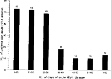
Figure 1. Distribution of the duration of acute HIV-1 disease in 218 patients.
pared by the Mann-Whitney Utest, Student's t-test, and one-way analysis of variance after logarithmic transformation. A two-tailed P value of <.05 was considered significant. The statistical analysis was performed with SPSS version 6.0 (SPSS, Chicago) for Windows (Microsoft, Redmond, WA).
>60 51-60
41-50 31-40
21-30
No. of days of acute HIV-1 disease
11-20 1-10 ~10 Q)
'"
11150'"
U (18 [8%]); or (3) a negative HIV-l screening test followed by a positive HIV-l test within 1 year (median, 140.0 days) (65 [30%]). The symptoms and signs analyzed were reported by the patients themselves and/or by their treating physicians at the time of documented acute HIV-1 disease (criterion 1 or 2) or within the previous months (median, 65.0 days) before the first positive HIV-l screening test (criterion 3). The clinical data were extracted from a review of standardized forms for data collection. We double-checked the data by examination of 190 available medical files (private and/or hospital).We compared the duration of acute HIV-1 disease according to infection with zidovudine-sensitive (wild-type) HIV-lor zidovudine-resistant (mutant) HIV-l. The most frequent geno-type related to zidovudine resistance is a strain with a mutation at codon 215 of the reverse transcriptase gene [19].It was detected by the method proposed by Larder et al. [19]. RNA was extracted and amplified by selective PCR analysis and then was reverse transcribed to complementary DNA with primer RTOI (5'-GTAGAATTCTGTTGAGTCAGATTGG-3'). The genotype determination was done on the first sample avail-able after the onset of acute HIV-1 disease (median, 25.0 days) in 99 patients.
Statistical Analysis
Table 1. Characteristics of 218 patients with documented acute HIV-1 disease.
Categorical variables are described in terms of frequency, and continuous variables are described as mean, median, and extreme values. The duration of acute HIV-1 disease was
com-Results
Table 2. Duration of acute HIV-1 disease stratifiedbyage for 218 patients. 23 (4-76) 17 (4-184) 19 (3-94) 21 (5-82) 20 (5-109) Median (range) Duration of acute HIV-1disease (d)*
26.5 (19.9) 22.2 (25.9) 25.1 (19.3) 24.8 (18.0) 28.7 (28.6) Mean (±SD)
The characteristics of the patients are listed in table 1, and the distribution of the duration of acute HIV-1 disease is shown in figure 1. The average duration of acute HIV-1 disease was 24.8 days for homosexual men and 26.0 days for other patients
(P
=
.34). There was no significant difference in the duration between men (24.6 days) and women (32.5 days) (P=
.13). The duration of acute HIV-1 disease stratified by age is reported in table 2; there was no difference according to age(P = .8). Forty-six patients (21%) were hospitalized. Tables 3 and 4 show the frequency and duration of clinical features.If we define the classical clinical triad of MLI to be fever (temperature of~38°C),sore throat or pharyngitis, and cervical
Age (y) at onset of acute HIV-1disease (no. of patients) ~24(49) 25-29 (51) 30-34 (66) 35-39 (25) ?o40 (27) Value 31.3 (7.8) 30.6 (18.5-64.8) 25.1 (22.2) 20.0 (3.0-184.0) 138 (63.3) 80 (36.7) 169 (77.5) 9(4.1) 25 (11.5) 15 (6.9) 204 (93.6) 14 (6.4) 62 (28.4) 54 (24.8) 102 (46.8) Characteristic
No. (%) of patients with categorical variable Study population Australian European Gender Male Female
Mode of infection (presumed) Homosexual contact Intravenous drug use Heterosexual contact Other or unknown
Year of onset of acute HIV -Idisease
~1987
1988 to 1991 1992 to 30 June 1994 Mean (±SD) for continuous variable
Age (y) Median (range) Duration* (d)
Median (range)
* The duration of acute HIV-1 disease was available for 205 patients.
* The difference in the mean duration according to age was not significant (one-way analysis of variance;P= .80).
cm
1997;24 (May) Clinical FeaturesofAcute HIV-l Disease 967Table3. Frequency and duration of signs and symptoms reported for~5%of218patients at the time of acute HIV-l disease.
Percentage of patients, sign or Median duration in d
symptom No. (%) of patients Average duration in d (no. of patients) (range)
>50% Fever (temperature of ;"38°C) 168 (77.1) 16.9 (162) 14.0 (3.0-184.0) Lethargy 143 (65.6) 23.7 (139) 18.0 (1.0-184.0) Cutaneous rash 123 (56.4) 15.0 (117) 11.0 (1.0-73.0) Myalgia 119 (54.6) 17.7 (112) 11.0 (2.0-184.0) Headache 111 (50.9) 25.8 (108) 13.0 (2.0-continuing*) >25%-50%
Pharyngitis or sore throat 96 (44.0) 12.2 (90) 8.0 (1.0-51.0)
Cervical adenopathy 85 (39.0) 15.1 (8) 12.0 (3.0-32.0)
Arthralgia 67 (30.7) 22.6 (64) 15.0 (3.0-184.0)
Oral ulcer 63 (28.9) 13.4 (63) 8.0 (1.0-85.0)
Odynophagia 61 (28.0) 16.3 (58) 14.0 (2.0-48.0)
5%-25%
Axillary adenopathy 53 (24.3) 164.1 (6) 13.5 (1.0-continuing*)
Weight loss 52 (23.9) 29.0 (49) 19.0 (3.0-continuing*)
Nausea 52 (23.9) 17.8 (50) 14.0 (2.0-109.0) Diarrhea 50 (22.9) 12.5 (47) 8.0 (1.0-39.0) Night sweats 48 (22.0) 14.8 (45) 10.0 (3.0-57.0) Cough 48 (22.0) 18:4 (48) 12.5 (2.0-184.0) Anorexia 46 (21.1) 14.6 (44) 10.0 (2.0-68.0) Inguinal adenopathy 44 (20.2) 8.5 (2) 8.5 (7.0-10.0) Abdominal pain 42 (19.3) 15.1 (40) 12.0 (1.0-73.0) Oral candidiasis 37 (17.0) 10.4 (34) 7.5 (1.0-34.0) Vomiting 27 (12.4) 9.8 (27) 10.0 (1.0-31.0) Photophobia 26 (11.9) 11.2 (25) 10.0 (2.0-39.0) Sore eyes 25 (11.5) 13.2 (24) 11.5 (3.0-36.0) Genital ulcer 15 (6.9) 13.5 (15) 12.0 (3.0-35.0) Tonsillitis 15 (6.9) 13.3 (13) 10.0 (1.0-41.0) Depression 14 (6.4) 22.8 (9) 7.0 (3.0-76.0) Dizziness 12 (5.5) 10.7 (10) 9.5 (2.0-26.0)
* The treating physician considered that the sign or symptom became a chronic feature.
adenopathy [20], 34 patients (15.6%) presented with this clini-cal picture. Only 26 patients (12%) had an MLI if lethargy was included in the definition. Twenty-one patients (10%) had none of these features.
Of the 60 patients with fever but no other MLI features, 31 (52%) had fever plus headache, 15 (25%) had fever plus oral ulcer, 15 (25%) had fever plus abdominal pain, and 10 (17%) had fever plus diarrhea. A meningitis-like syndrome (defined as fever, headache, and stiff neck) occurred in 20 patients (9.2%).
By contrast, fever was not described in 50 patients (23%). In these 50 patients, the most frequent clinical features were sore throat or pharyngitis (23 [46%]), lethargy (22 [44%]), myalgia (18 [36%)), cutaneous rash (17 [34%
D,
headache (17 [34%]), cervical adenopathy (16 [32%]), oral ulcer (10 [20%]), cough (10 [20%]), and arthralgia (10 [20%)).There were nine patients infected with zidovudine-resistant (mutant) HIV-l, and two patients were infected with a mixture of both wild-type and mutant strains; 88 patients were infected with a wild-type strain (total number of analyzed cases, 99). For the 11 patients infected only by a wild-type strain, the
mean duration of acute HIV-l disease was 21.0 days compared with 26.9 days for the 88 patients infected with a mutant or mutant and wild-type strains (P
=
.48). Infections with HIV-l variants (mutant strain or mutant and wild-type strains) occurred in 2 patients in 1990, in 3 in 1992, in 4 in 1993, and in 2 in 1994 (until 30 June). The frequency of clinical features was not significantly related to infection with zidovudine-resis-tant HIV-l (muzidovudine-resis-tant strain or muzidovudine-resis-tant and wild-type strains). The number of patients was too small to analyze further differences in acute HIV-1 disease between the 11 patients infected with a mutant strain or mutant and wild-type strains and the 88 infected only by a wild-type strain.Discussion
The purpose of this descriptive study was to investigate extensively the clinical features reported at the time of acute HIV-1 disease in a large cohort of patients. A potential limita-tion of the study was that the patients differed in terms of risk factors for HIV-1 infection: 91% of the patients in Australia were homosexual men compared with 54% of the patients in
968 Vanherns et at. em1997;24 (May)
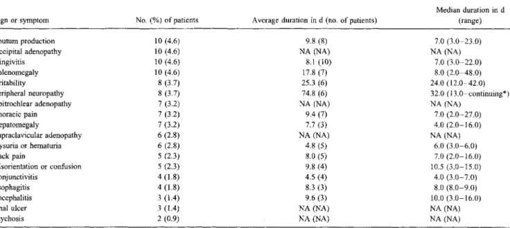
Table 4. Frequency and duration of signs and symptoms reported for <5% of 218 patients at the time of acute HIV-l disease.
Sign or symptom Sputum production Occipital adenopathy Gingivitis Splenomegaly Irritability Peripheral neuropathy Epitrochlear adenopathy Thoracic pain Hepatomegaly Supraclavicular adenopathy Dysuria or hematuria Back pain Disorientation or confusion Conjunctivitis Esophagitis Encephalitis Anal ulcer Psychosis No.(%) of patients 10 (4.6) 10 (4.6) 10 (4.6) 10 (4.6) 8 (3.7) 8(3.7) 7(3.2) 7 (3.2) 7 (3.2) 6 (2.8) 6 (2.8) 5 (2.3) 5 (2.3) 4 (1.8) 4 (1.8) 3 (1.4) 3 (1.4) 2(0.9)
Average duration in d (no. of patients) 9.8(8) NA (NA) 8.1 (10) 17.8 (7) 25.3 (6) 74.8 (6) NA (NA) 9.4 (7) 7.7(3) NA (NA) 4.8(5) 8.0(5) 9.8 (4) 4.5 (4) 8.3 (3) 9.6(3) NA(NA) NA(NA) Median duration in d (range) 7.0 (3.0-23.0) NA(NA) 7.0 (3.0-22.0) 8.0 (2.0-48.0) 24.0 (12.0-42.0) 32.0 (13.0-continuing*) NA (NA) 7.0 (2.0-27.0) 4.0 (2.0-16.0) NA (NA) 6.0 (3.0-6.0) 7.0 (2.0-16.0) 10.5 (3.0-15.0) 4.0 (3.0-7.0) 8.0 (8.0-9.0) 10.0 (3.0-16.0) NA(NA) NA (NA) NOTE. NA= not available.
*The treating physician considered that the sign or symptom became a chronic feature.
Europe (which also created an imbalance in the sex ratio). We decided to include patients of both sexes with different risk factors to facilitate the use of our results by physicians who work with populations in whom HIV-1 infection does not occur almost exclusively in one risk group. Until now, all previous studies except two [7, 15] have been limited to one risk popula-tion-homosexual and/or bisexual men. Physicians' experi-ence with acute HIV-1 disease may have affected the accuracy of the diagnosis, but this circumstance was probably of limited impact, because clinicians had reached similar levels of clinical expertise in Australia and Europe when the cohorts were started.
Some symptoms and signs overlaped (i.e., tonsillitis and pharyngitis or sore throat), and the reporting of diagnoses was not completely standardized. For example, in the case of tonsil-litis, we have to assume that physicians made this diagnosis when "true" tonsillitis rather than simple pharyngitis was pres-ent. In any case, tonsillitis and/or pharyngitis or sore throat occurred in 100 patients (46%), thus confirming the frequent involvement of the oral cavity during acute HIV-1 illness.
A description as complete as possible of acute HIV-1 disease gives physicians an understanding of the wide range of diagnos-tic clinical features that could lead to early therapeudiagnos-tic interven-tion [7, 8]. The mean and median durainterven-tions of acute HIV-l disease were similar to those previously reported [14-16]; for the first time, we report the absence of a difference in the duration by gender, age, andriskgroups. Because sicker pa-tients are more likely to be
enrolf~d
in the cohorts at the time of acute HIV-1 disease, the clinical description may have been biased toward more severe disease. Nevertheless, the durationof some signs and symptoms was only a few days, which suggests that mild acute HIV-1 disease was represented in our study.
A possible recall bias may be present for patients enrolled in the study according to biological inclusion criterion 3. To quantify this possible bias, we performed an additional fre-quency analysis restricted to 153 patients for whom the time relation between symptoms and acute HIV-1 illness was the strongest (criteria 1 and 2). Among this population, we ob-served an average increase of 3.7% in the frequency of each sign or symptom, a finding suggesting that the analysis per-formed on the whole sample could not have been very biased. Although MLI is the major clinical syndrome described in 50% to 70% of patients with acute HIV-l disease [5, 14, 16] and although MLI is emphasized in textbooks [21, 22], some atypical presentations have occurred, and they have been re-ported separately [23- 25]. The systematic description ap-pearing in this study yields a wide spectrum of clinical features of the disease and a relative frequency of an MLI-type presenta-tion that is low compared with that of less typical presentapresenta-tions. Thus, if the definition of acute HIV-1 disease as an MLI was helpful at the beginning of the AIDS epidemic and is still used [10, 12, 13], our results suggest that the definition of acute HIV-1 disease should be broadened.
When MLI is absent, fever associated with diarrhea, a cuta-neous rash, or headaches prompts the diagnosis, especially in the presence of mucosal ulcers. However, in nearly 25% of cases, fever is not reported, but a diagnosis may still be made by eliciting recent risk behavior followed by nonspecific clinical features of illness. Hence, when an acute infectious disease or
CID 1997;24 (May) Clinical Features of Acute HIV-l Disease 969
an unusual dermatologic condition is noted, practrtioners should consider the possibility of acute HIV-l infection. To do this rigorously, one needs to know how frequently the main signs and symptoms of acute HIV -1 disease occur in patients who do not have acute HIV -1 disease. Our study cannot provide this information, but clinical experience can nonetheless be useful.
The more severe features reported during acute HIV -1 dis-ease were neurological and gastrointestinal. The frequency of the meningitis-like syndrome, peripheral neuropathy, confu-sion, and encephalitis was similar to that reported in a French study [26] and confirmed the early tropism of HIV for the CNS [27]. Oral candidiasis was observed frequently, and esophagitis was seen more rarely; both conditions are probably related to transient depletion and dysfunction of CD4+ cells.
Analysis of each reported symptom or sign by using a stan-dard questionnaire had the advantage of a greater uniformity of data collection and perhaps a greater validity compared with analysis of pooled data from different clinical studies. For ex-ample, in a review of studies of pooled data, Niu et al. [5] reported that fever occurred in 96% of their patients (compared with 77.1% of our patients) and that headache was present in 32% of their patients (compared with 50.9% of our patients); oral ulcers were not reported (whereas they were present in 28.9% of our patients).
We did not find a difference in the duration of acute infection with a zidovudine-resistant strain, thus suggesting that primary infection with an HIV strain with a mutation at codon 215 does not necessarily itself induce a more aggressive initial infection. One previous report [28] cited the development of severe im-munodeficiency in a patient after deliberate injection of blood containing a mixture of HIV -1 strains. In this case, it is difficult to assess the relative contributions of inoculum size vs. viral characteristics to early immunodeficiency.
In conclusion, patients with acute HIV -1 disease present with a broad array of symptoms and signs, and <20% have the classical features of an MLI. A febrile syndrome, especially one associated with gastrointestinal or mucocutaneous features, occurring in a person at risk of HIV infection should alert physicians to the possibility of HIV -1 seroconversion. The rea-sons for the heterogeneous clinical presentation and the relation of those various clinical presentations to the pathogenesis of acute HIV -1 infection and to the response to early treatment remain unclear.
Acknowledgments
The authors are indebted to the patients and to the following physicians who participated in the study: B. Anderson, D. Baker, A. Beveridge, M. Bloch, N. Doong, C. Duncombe, R. Finlayson, V. Furner, B. Genn,1.Gold, J. Kidd, R. McFarlane, M. McMur-chie,A. McNulty, H. Michelmore,A. Pethebridge, D. Quan, and M. Robertson (medical practitioners, Sydney, Australia); the late B. Tindall (St. Vincent's Hospital, Sydney);1. Kaldor (National
Centre in HIV Epidemiology and Clinical Research, Sydney); J. F. Balavoine, A. Christina, C. Junet, and V. Sendersky (medical prac-titioners, Geneva); 1.Wintsch (Geneva University Hospital, Ge-neva); the investigators who participated in the controlled trial of zidovudine in primary HIV infection (see appendix of [7]);
A.Imrie (St. Vincent's Hospital) and S. Yerly (Geneva University Hospital) for determining HIV strains; and R. Gaudet (University of Montreal, Montreal) for technical assistance.
References
1. Anonymous. Needlestick transmission of HTLV-III from a patient infected in Africa [editorial]. Lancet 1984;2:1376-7.
2. Cooper DA, Gold J, MacLean P, et al. Acute AIDS retrovirus infection: definition of a clinical illness associated with seroconversion. Lancet 1985; 1:537-40.
3. Kassler WJ, Zenilman JM, Erickson B, Fox R, Peterman TA, Hook EW III. Seroconversion in patients attending sexually transmitted disease clinics. AIDS 1994;8:351-5.
4. Otten MW, Zaidi AA, Peterman TA, Rolfs RT, Witte Jl. High rate of HIV seroconversion among patients attending urban sexually transmit-ted disease clinics. AIDS 1994; 8:549-53.
5. Niu MT, Stein DS, Schnittman SM. Primary human immunodeficiency virus type 1 infection: review of pathogenesis and early treatment inter-vention in humans and animal retrovirus infections. J Infect Dis 1993;
168:1490-501.
6. Ku L, Sonenstein FL, Pleck JH. Young men's risk behaviors for HIV infection and sexually transmitted diseases, 1988 through 1991. Am J Public Health 1993;83:1609-15.
7. Kinloch-de Loes S, Hirschel BJ, Hoen B, et al. A controlled trial of zidovudine in primary human immunodeficiency virus infection. N Engl J Med 1995;333:408-13.
8. Ho DD. Time to hit HIV, early and hard. N Engl J Med 1995;333: 450-1.
9. Jacquez JA, Koopman JS, Simon CP, Longini 1M. Role of the primary infection in epidemics ofHIV infection in gay cohorts. J Acquir Immune Defic Syndr 1994;7:1169-84.
10. Lindback S, Brostrom C, Karlsson A, Gaines H. Does symptomatic pri-mary HIV-I infection accelerate the progression to CDC stage IV dis-ease, CD4 count below 200X106/1,
AIDS, and death from AIDS? BMJ 1994;309:1535-7.
11. Schechter MT, Craib KJP, Le TN, et al. Susceptibility to AIDS progression appears early in HIV infection. AIDS 1990;4:185-90.
12. Sinicco A, Fora R, Sciandra M, Lucchini A, Caramello P, Giannini P. Risk of developing AIDS after primary acute-HIV infection. J Acquir Immune Defic Syndr 1993;6:575-81.
13. Dorrucci M, Rezza G, Vlahov D, et al. Clinical characteristics and prognos-tic value of acute retroviral syndrome among injecting drug users. AIDS 1995; 9:597 -604.
14. Tindall B, Barker S, Donovan B, et al. Characterization of the acute clinical illness associated with human immunodeficiency virus infection. Arch Intern Med 1988; 148:945-9.
15. Kinloch-de Loes S, de Saussure P, Saurat JH, Stalder H, Hirschel B, Perrin LH. Symptomatic primary infection due to human immunodeficiency virus type I:review of 31 cases. Clin Infect Dis 1993; 17:59-65. 16. Pedersen C, Lindhart BO, Jensen BL, et al. Clinical course of primary
HIV infection: consequences for subsequent course of infection. BMJ 1989;299:154-7.
17. Sydney AIDS Study Group. The Sydney AIDS Project. Med J Aust 1984; 141:569-73.
18. Engel RR, Samuel MC, Rieder HL, Billo N, Somaini B. Completeness of AIDS reporting in Switzerland: a study based on deaths between Decem-ber 1987 and June 1990. AIDS 1992;6:1385-9.
970 Vanhems et al. CID 1997;24 (May)
19. Larder BA, Darby G, Richman DD. HIV with reduced sensitivity to zido-vudine (AZT) isolated during prolonged therapy. Science 1989;243: 1731--4.
20. Schooley RT. Epstein-Barr virus (infectious mononucleosis). In: Mandell GL, Bennett JE, Dolin R, eds. Mandell, Douglas, and Bennett's princi-ples and practice of infectious diseases. 4th ed. New York: Churchill Livingstone, 1995:1364-77.
21. Chamberland ME, Ward JW, Curran JW. Epidemiology and prevention of AIDS and HIV infection. In: Mandell GL, Bennett JE, Dolin R, eds. Mandell, Douglas, and Bennett's principles and practice of infectious diseases. 4th ed. New York: Churchill Livingstone, 1995: 1174-203. 22. Volberding PA. Clinical spectrum ofHIV disease. In: De Vita VT,
Hell-man S, Rosenberg SA, Curran J, Essex M, Fauci AS, eds. AIDS. Etiol-ogy, diagnosis, treatment and prevention. Philadelphia: JB Lippincott, 1992:123-40.
23. Tindall B, Hing M, Edwars P, Barnes T, Mackie A, Cooper DA. Severe manifestations of primary HIV infection. AIDS 1989;3:747-9.
24. del Rio C, Soffer0,Widell JL, Judd RL, Slade BA. Acute human immuno-deficiency virus infection temporally associated with rhabdomyolysis, acute renal failure, and nephrosis. Rev Infect Dis 1990; 12:282-5. 25. Calabresse LH, Proffitt MR, Levin KH, et al. Acute infection with the
human immunodeficiency virus (HIV) associated with acute brachial neuritis and exanthematous rash. Ann Intern Med 1987; 107:849-51. 26. Boufassa F, Bachmeyer C, Carre N, et al. Influence of neurologic
manifes-tations of primary human immunodeficiency virus infection on disease progression. J Infect Dis 1995; 171:1190-5.
27. Cheng-Mayer C, Levy JA. Human immunodeficiency virus infection of the CNS: characterization of "neurotropic" strains. Curr Top Microbiol Immunol 1990; 160:145-56.
28. Veenstra J, Schuurman R, Cornelissen M, et al. Transmission of zidovu-dine-resistant human immunodeficiency virus type I variants follow-ing deliberate injection of blood from a patient with AIDS: character-istics and natural history of the virus. Clin Infect Dis 1995; 21: 556-60.