334
CONCISE COMMUNICATION
Incubation Time of Acute Human Immunodeficiency Virus (HIV) Infection
and Duration of Acute HIV Infection Are Independent Prognostic Factors
of Progression to AIDS
Philippe Vanhems,1Bernard Hirschel,2 Andrew N. Phillips,3David A. Cooper,4
Jeanette Vizzard,4Joelle Brassard,5and Luc Perrin2
1Laboratory of Epidemiology and Public Health, INSERM U271,
Universite´ Claude Bernard, and Epidemiology Unit, Edouard Herriot Hospital, Lyon, France;2Division of Infectious Diseases, University
Hospital, Geneva, Switzerland;3Royal Free Centre for HIV Medicine
and Department of Primary Care and Population Sciences, Royal Free and University College Medical School, London, United Kingdom;
4National Centre in HIV Epidemiology and Clinical Research,
Sydney, Australia;5Institute of Social and Preventive Medicine,
University of Montreal, Montreal, Canada
The severity and the duration of acute human immunodeficiency virus (HIV) infection (AHI) are associated with a faster rate of progression to AIDS, but the prognostic value of the length of incubation time of AHI (IncAHI), defined as the time between HIV infection and AHI, on progression to AIDS has not been assessed. We explored this issue prospectively in 70 individuals with documented AHI and a known date of HIV infection. The median IncAHI was 21.5 days (range, 5–70 days), and the median duration of AHI was 15.5 days (range, 3–67 days). The adjusted relative hazard of progression to AIDS or to a CD4+
count ! 3/mL was 4.23 (95% confidence interval [CI], 1.40–12.73; ) for the patients
2003 10 P = .01
with an IncAHI!21.5 days, compared with those with longer IncAHI, and was 3.53 (95% CI, 1.09–11.36;P = .03) for the patients with a duration of AHI115.5 days, compared with those with shorter duration. Both IncAHI and duration of AHI were independent predictors of progression. This suggests that early pathogenic events before the onset of AHI influence the rate of HIV disease progression.
Acute human immunodeficiency virus type 1 (HIV-1) infec-tion (AHI) is a heterogeneous syndrome [1, 2]. Patients with more severe symptoms during AHI and an AHI lasting 115
days tend to progress faster toward AIDS [3, 4]. Data on the prognostic value of the incubation of AHI (IncAHI), defined as the time between HIV infection and AHI, are lacking.
The objective of this investigation was to determine whether the time from HIV infection until the onset of AHI is an
in-Received 10 November 1999; revised 18 April 2000; electronically pub-lished 19 June 2000.
Presented in part: 6th Conference on Retrovirus and Opportunistic Com-plications, Chicago, 31 January–4 February, 1999 (poster 73).
Financial support: Swiss Federal Office of Public Health through funding of the Swiss HIV Cohort Study (project 256) and Swiss National AIDS Program (grant 3200.56.059.98). The National Centre in HIV Epidemiology and Clinical Research is supported by the Australian National Council on AIDS through the Commonwealth AIDS Research Grants Committee.
Informed consent was obtained from patients.
Reprints or correspondence: Dr. Philippe Vanhems, Laboratoire d’Epi-de´miologie et de Sante´ Publique, Universite´ Claude Bernard Lyon 1, 8, av. Rockefeller, 69373 Lyon Cedex 08, France (philipva@lyon-sud.univ-lyon1.fr).
The Journal of Infectious Diseases 2000; 182:334–7
q 2000 by the Infectious Diseases Society of America. All rights reserved. 0022-1899/2000/18201-0046$02.00
dependent factor of progression to AIDS or toward a CD41 count!2003 103/mL after adjustment for duration of AHI.
Methods
This prospective study included a total of 70 patients with AHI for whom a date of HIV infection was known. These patients are a subgroup of a larger cohort of Swiss and Australian individuals described in detail elsewhere [1]. This cohort was initiated to study the clinical features of AHI and the comparison of the rates of disease progression by severity of AHI. The patients selected for this investigation were those with symptomatic AHI and with a date of HIV exposure reported in the data-collection form. The onset of AHI was before the end of 1987 in 6 patients, between 1988 and 1991 in 19 patients, and between 1992 and December 31, 1995 in 45 patients. Patients were asked about the date of infection at the time they presented with symptoms of AHI. The number of sexual partners within the last year was 1 for 57 patients (81.4%), 2 for 5 patients (7.1%), and12 for the remainder (11.4%). The
presumed route of HIV infection was self-reported; a total of 47 individuals (67%) reported infection by unprotected homosexual intercourse, 6 (8.5%) by unprotected receptive oral sex, 11 by het-erosexual intercourse (15.7%), 2 after syringe exchange (3%), 1 after blood transfusion (1.4%), and 3 by another route not reported. The
JID 2000;182 (July) Time From Infection to Acute Syndrome Predicts Progression to AIDS 335
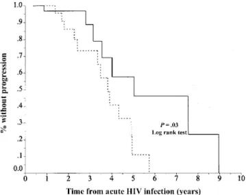
Figure 1. Probability of being free of clinical AIDS or not having a CD41cell count!2003 103/mL for individuals with incubation of acute human immunodeficiency virus (HIV) infection121.5 days (solid
line) or!21.5 days (dashed line).
serostatus of patients’ partners was available for 11 individuals. In 2 cases (3%), the partner had AIDS, and in 9 cases (13%) he was asymptomatic. The date of exposure was based on the patient’s best estimate. IncAHI was defined as the time elapsed between the date of HIV infection and the date of AHI onset. The clinical features of AHI were recorded on standardized collection forms in the 2 centers (in Geneva and Sydney) [1]. Clinical and laboratory follow-up procedures were similar in both centers [1].
The laboratory criteria of AHI were (1) the presence of p24 antigenemia (n = 58) in patients with negative or indeterminate EIA serology, (2) 2 bands on Western blots, one of which corresponded to the env gene (gp 160, gp 120, gp 41;n = 3) with a negative EIA serology, and (3) a negative HIV-1 EIA test followed by a positive HIV-1 EIA test within 6 months (median, 85 days; range, 4–188 days;n = 9). HIV infection was confirmed in all patients by Western blots. CD41cell counts were conducted in laboratories affiliated with either university center. The laboratories in Geneva and Syd-ney are national reference centers, and quality control testing is done regularly. T cell subsets were measured by flow cytometry in both locations.
Groups were compared by the x2
test or Fisher’s exact test for categorical variables and by Student’s t test for continuous vari-ables after logarthimic transformation, if necessary. Multiple linear regression was used for calculate the association between IncAHI and duration of AHI, after adjustment for confounders. The end-point was AIDS according to the 1993 definition by the Centers for Disease Control and Prevention or a first CD41 count
! 3/mL. Progression was calculated by the Kaplan-Meier 2003 10
method, with the onset of AHI as time 0 and censoring at the last visit or at June 30, 1995, to exclude the confounding effect of the use of highly active antiretroviral therapy. Comparison of survival distribution was based on the log-rank test. The Cox proportional hazard model was used to estimate the adjusted relative hazard (ARH) of disease progression. No patient was considered lost to follow-up, defined as 1 year without knowledge of vital status. P values!.05 were considered statistically significant. Analysis was
conducted with SPSS version 6.0 (SPSS, Chicago).
Results
The patients, 91% of whom were men, had a mean age of 30.6 years. Thirty-one patients were from Geneva and 39 from Sydney. The mean IncAHI was 25.8 days, with a median of 21.5 days (range, 5–70 days), and did not differ by center (P = .15). The mean duration of AHI was 16.8 days, with a median of 15.5 days (range, 3–67 days). The most common symptoms at AHI were fever (91%), lethargy (84%), skin rash (76%), headache (69%), myalgia (67%), arthralgia (41%), and diarrhea (27%). No difference in symptom frequency or the proportion of hospitalized patients (23%) was observed ac-cording to IncAHI greater than or less than 21.5 days.
Adjusted regression analysis did not identify independent variables associated with IncAHI (P = .77for duration of AHI;
for age; for gender; for route of
infec-P = .33 P = .50 P = .36
tion;P = .17for the center [Geneva vs. Sydney]).
The duration of follow up was 24.3 months for patients with IncAHI below the median (21.5 days; group 1) and 24.2 months for patients with IncAHI above the median (group 2;P = .9). Antiretroviral treatment was prescribed in a similar proportion in the 2 groups (P = 1.00) at AHI (48%) and during follow up (60% for group 1 and 51% for group 2;P = .47). The mean interval between AHI and the start of antiretroviral therapy was 6.2 months for group 1 and 5.5 months for group 2 (P = .84). The regimens prescribed at AHI for patients in group 1 was single nucleoside reverse transcriptase inhibitor for 15 patients (43%), a combination of 2 nucleoside reverse tran-scriptase inhibitors for 1 patient (6%), and a combination of nucleoside reverse transcriptase inhibitor and acyclovir for 1 patient (6%); the regimens prescribed at AHI in group 2 were a single nucleoside reverse transcriptase inhibitor for 15 patients (43%), a combination of nucleoside reverse transcriptase in-hibitor and acyclovir for 1 patient (6%), and a combination of nucleoside reverse transcriptase inhibitor and nonnucleoside re-trotranscriptase inhibitor for 1 patient (6%). The proportion of patients treated within 3 months after AHI was similar between the two centers (58% for Geneva and 41% for Sydney; P = ), and the total duration of antiretroviral was also similar .16
between the two centers (mean of 4.6 months for Geneva and mean of 4.1 months for Sydney;P = .5). The proportion of patients who received primary Pneumocystis carinii prophylaxis was similar in both groups (P = .32). A total of 13 patients in group 1 and 8 patients in group 2 reached the end point during the follow-up. Six AIDS events occurred in group 1 (2 chronic herpes simplex ulcerations 11 month, 2 cryptosporidiosis, 1 AIDS dementia complex, and 1 cytomegalovirus infection), and
336 Vanhems et al. JID 2000;182 (July)
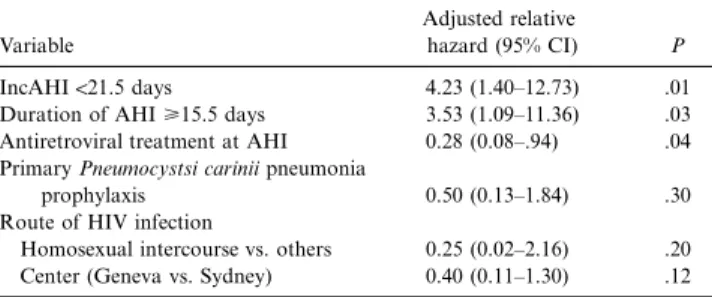
Table 1. Adjusted relative hazard of progression to AIDS or to a CD41cell count!2003 103/mL.
Variable
Adjusted relative
hazard (95% CI) P
IncAHI!21.5 days 4.23 (1.40–12.73) .01
Duration of AHI>15.5 days 3.53 (1.09–11.36) .03 Antiretroviral treatment at AHI 0.28 (0.08–.94) .04 Primary Pneumocystsi carinii pneumonia
prophylaxis 0.50 (0.13–1.84) .30
Route of HIV infection
Homosexual intercourse vs. others 0.25 (0.02–2.16) .20 Center (Geneva vs. Sydney) 0.40 (0.11–1.30) .12 NOTE. All the variables of the table were in the model equation. Age was not associated with progression (P = .69) and not included in the model. Model characteristics:22 log likelihood, 83.556; x2
, 24.513; 6 df;P = .0004. CI, confi-dence interval; IncAHI, incubation of acute human immunodeficiency virus (HIV) infection.
2 AIDS events occurred in group 2 (1 Mycobacterium avium complex infection and 1 chronic herpes simplex ulceration11
month). By use of the Kaplan-Meier analysis, the median of progression to AIDS or to a CD41cell count!2003 103/mL was 3.82 years (95% confidence interval [CI], 3.13–4.50) in group 1 and 5.05 years (95% CI, 1.80–8.28) in group 2 (P = ; figure 1). One patient received protease inhibitors 243 days .03
after AHI for a period of 92 days; this individual was in group 1 and did not have AIDS during follow-up. Survival analysis without this patient remained unchanged. By use of the Kaplan-Meier method, the median of progression to AIDS or to a CD41 cell count!2003 103/mL was 3.55 years (95% CI, 2.93–4.17) for individuals with a duration of AHI greater than the median (15.5 days) and 5.04 years (95% CI, 4.30–5.78) for those with a duration of AHI less than the median (P = .005).
We calculated the ARH of progression using the proportional hazard model after adjustment for the length of AHI and for potential confounders (table 1). The multivariate model iden-tified both IncAHI and duration of AHI as independent prog-nostic factors of disease progression. Similar analysis with IncAHI and AHI as continuous variables identified only a long IncAHI (AHR, 0.95; 95% CI, 0.91–0.99;P = .03) as protective effect on disease progression. Among patients free of antire-troviral treatment at AHI, the mean of CD41within 1 month after onset was 467/mL (SD, 271) for group 1 patients (n =
) and 604/mL (SD, 228) for group 2 patients ( ;
14 n = 21 P =
) and the mean of CD41at 2 months after onset was 637/ .12
mL (SD, 263) for group 1 patients (n = 19) and 695/mL (SD, 325) for group 2 patients (a;P = .52). The use of antiretroviral therapy at AHI had a protective effect in this observational cohort.
Discussion
The objective of this study was to identify the independent prognostic value of the IncAHI and duration of AHI. Previ-ously, early prognostic studies were done mostly at the onset of AHI and/or at the time of HIV seroconversion, but few
investigations of IncAHI have been conducted. The question of the validity of the reported date of HIV infection is central and was ascertained on 3 grounds. First, the information was collected prospectively, which reduced memory bias. Second, the duration between infection and the onset of AHI was similar to that in studies elsewhere [2, 5–8], in which IncAHI ranged from 1 to 8 weeks. For example, IncAHI lasted 11–28 days for Gaines et al. [5], 5–29 days (mean, 15 days) for Schacker et al. [2], 2–3 weeks for Quin [6], and 3–6 weeks for Clark et al. [7]. Third, our results are consistent with data obtained from health-care workers who developed AHI after needle-stick injuries [9]. In that case, the IncAHI reported was 14–28 days for Marcus et al. [10], 25 days for Oksenhendler et al. [11], and 16 days for Pratt et al. [12]. Although we cannot have total confidence in individual measures of the IncAHI, we think it unlikely that lack of precision would bias our results. For a bias to occur, patients destined to progress faster toward AIDS would have to systematically recall a presumed date of infection that was later than the real date of infection. The occurrence of such a systematic error does not seem plausible to us.
IncAHI was associated with progression toward AIDS in our cohort independently of duration of AHI. Because this result was obtained among individuals who reached a small number of outcomes, confirmatory studies are needed with larger co-horts. Pedersen et al. showed that duration of acute HIV in-fection was associated with a faster progression to AIDS [3]. The rapid rate of disease progression observed in our cohort was related to the absence of asymptomatic HIV seroconver-tors. Our study was conducted among patients who, except for 1, received what is now regarded as suboptimal therapy, because no protease inhibitors were administered until the end of follow-up. This provided the opportunity to study the natural history of disease progression without the confounding factor of highly active antiretroviral therapy.
The present investigation suggests that initial pathogenetic mechanisms could influence the rate of disease progression, even before the onset of AHI. The mechanisms influencing the length of IncAHI are not well understood. Because no specific anti-HIV immune response can be measured during that period, differences in the replicative capacities of HIV, in the interaction of HIV and mucosal surfaces or in the reaction of the innate immune system to HIV, could presumably play a role [13]. A short IncAHI could mean that the virus has faster access to the target cells. Investigations on the transition period between IncAHI and onset of AHI could yield essential information on the respective contribution of the viral replication [14] and im-munological response [15] that determine the prognosis. The impact of virus replication and its relationship to host genetic factors also need to be investigated.
This study suggests both IncAHI and duration of AHI were associated with the prognosis. These results confirm the key place of early events on late prognosis of HIV infection.
JID 2000;182 (July) Time From Infection to Acute Syndrome Predicts Progression to AIDS 337
Acknowledgments
We wish to thank B. Anderson, D. Baker, A. Beveridge, M. Bloch, N. Doong, C. Duncombe, R. Finlayson, V. Furner, B. Genn, J. Gold, J. Kidd, R. McFarlane, M. McMurchie, A. McNulty, H. Michelmore, A. Pethebridge, D. Quan, and M. Robertson (Sydney); J. Kaldor and the late B. Tindall, at the National Centre in HIV Epidemiology and Clinical Research (Sydney); and J. F. Balavoine, A. Christina, C. Junet, V. Sendersky, J. Wintsch (Geneva), and S. Kinloch-de Loe¨s (London). The members of the Swiss HIV Cohort Study are M. Bateguay (co-chair of the scientific board), E. Bernasconi, Ph. Bu¨rgisser, M. Egger, P. Erb (chairman of the “Laboratories” group), W. Fierz, M. Flepp (chairman of the “Clinics” group), P. Francioli (president of the Swiss HIV Cohort Study, Centre Hospitalier Universitaire Vaudois, CH-1011 Lausanne), H. J. Furrer, P. Grob, B. Hirschel (co-chair of the scientific board), L. Kaiser, B. Ledergerber, R. Lu¨thy, R. Malinverni, L. Matter, M. Opravil, F. Paccaud, G. Pantaleo, L. Perrin, W. Pichler, J.-C. Pif-faretti, M. Rickenbach, P. Sudre, J. Schupbach, A. Telenti, and P. Vernazza.
References
1. Vanhems P, Allard R, Cooper DA, et al. Acute human immunodeficiency virus type 1 disease as a mononucleosis-like illness: is the diagnosis too restrictive? Clin Infect Dis 1997; 24:965–70.
2. Schacker T, Collier AC, Hughes J, Shea T, Corey L. Clinical and epidemi-ologic features of primary HIV infection. Ann Intern Med 1996; 125: 257–64.
3. Pedersen C, Lindhardt BO, Jensen BJ, et al. Clinical course of primary HIV infection: consequences for subsequent course of infection. BMJ 1989; 299:154–7.
4. Vanhems P, Lambert J, Cooper DA, et al. Severity and prognosis of acute human immunodeficiency virus type 1 illness: a dose-response relationship. Clin Infect Dis 1998; 26:323–9.
5. Gaines H, von Sydow M, Pehrson P, Lundbergh P. Clinical picture of primary HIV infection presenting as a glandular-fever-like illness. BMJ 1988; 297: 1363–8.
6. Quin T. Acute primary HIV infection. JAMA 1997; 278:58–62.
7. Clark SJ, Saag MS, Don Decker W, et al. High titers of cytopathic virus in plasma of patients with symptomatic primary HIV infection. N Engl J Med 1991; 324:954–60.
8. Daar ES, Mougdil T, Meyer RD, Ho DD. Transient high levels of viremia in patients with primary human immunodeficiency virus type 1 infection. N Engl J Med 1991; 324:961–4.
9. Ippolito G, Puro V, Heptonstall J, Jagger J, De Carli G, Petrosillo N. Oc-cupational human immunodeficiency virus infection in health care work-ers: worldwide cases through September 1997. Clin Infect Dis 1999; 28: 365–83.
10. Marcus R, CDC Cooperative Needlestick Surveillance Group. Surveillance of health care workers exposed to blood from patients infected with the human immunodeficiency virus. N Engl J Med 1988; 319:1118–23. 11. Oksenhendler E, Harzic M, Le Roux JM, Rabian C, Clauvel JP. HIV infection
with seroconversion after superficial needlestick injury to the finger. N Engl J Med 1986; 315:582.
12. Pratt RD, Shapiro JF, McKinney N, Kwok S, Spector SA. Virologic char-acterization of primary human immunodeficiency virus type 1 infection in health care workers following needlestick injury. J Infect Dis 1995; 172: 851–4.
13. Phillips AN, McLean AR, Loveday C, et al. In vivo HIV-1 replicative capacity in early and advanced infection. AIDS 1999; 13:67–73.
14. Phillips AN. Reduction of HIV concentration during acute infection: inde-pendence from a specific immune response. Science 1996; 271:497–9. 15. Musey L, Hughes J, Schacker T, Shea T, Corey L, McElrath MJ. Cytotoxic
T-cell responses, viral load, and disease progression in early human im-munodeficiency virus type 1 infection. N Engl J Med 1997; 337:1267–74.