DEVELOPMENT OF THYMIC REGULA TORY T CELLS DURING HUMAN IMMUNODEFICIENCY VIRUS (HIV) INFECTION
THE SIS SUBMITTED
AS PARTIAL REQUIREMENT FOR MASTER'S IN BIOCHEMISTRY
BY
SHARADA SWAMINATHAN
DÉVELOPPEMENT DE LYMPHOCYTES T RÉGULATEURS THYMIQUES PENDANT UNE INFECTION PAR LE VIRUS DE L'IMMUNODÉFICIENCE
HUMAINE (VIH)
MÉMOIRE PRÉSENTÉ
COMME EXIGENCE PARTIELLE DE LA MAITRISE EN BIOCHIME
PAR
SHARADA SWAMINATHAN
Service des bibliothèques
Avertissement
La diffusion de ce mémoire se fait dans le respect des droits de son auteur, qui a signé le formulaire Autorisation de reproduire et de diffuser un travail de recherche de cycles supérieurs (SDU-522 - Rév.1 0-2015). Cette autorisation stipule que «conformément à · l'article 11 du Règlement no 8 des études de cycles supérieurs, [l'auteur] concède à l'Université du Québec à Montréal une licence non exclusive d'utilisation et de publication de la totalité ou d'une partie importante de [son] travail de recherche pour des fins pédagogiques et non commerciales. Plus précisément, [l'auteur] autorise l'Université du Québec à Montréal à reproduire, diffuser, prêter, distribuer ou vendre des copies de [son] travail de recherche à des fins non commerciales sur quelque support que ce soit, y compris l'Internet. Cette licence et cette autorisation n'entraînent pas une renonciation de [la] part [de l'auteur] à [ses] droits moraux ni à [ses] droits de propriété intellectuelle. Sauf entente contraire, [l'auteur] conserve la liberté de diffuser et de commercialiser ou non ce travail dont [il] possède un exemplaire.»
I extend my gratitude to my supervisor Dr. Mohammad-Ali Jenabian for giving me the opportunity to work in his laboratory, that has helped me build a strong foundation for my research career. His outright criticism and insightful comments have made me learn how to design and conduct a proj ect form scratch. He has taught me to become exceedingly self-reliant and self-confident. I am very thankful to Dr. Tatiana Scorza for agreeing to be my co-supervisor. My discussion sessions with her have always urged me think deeply about scientific problems, have enriched my critical thinking abilities and have rekindled my passion for science. With her humor and wit, she has been my pillar of support during many stressful and challenging situations. I thank Dr. Jonathan Angel and Dr. Gyaandeo Maharajh, for providing us with the precious thymus specimens. I am immensely thankful to the donors and their kind parents for their huge donation to the field of research. I acknowledge the support offered by our collaborators Dr. Cecilia T. Costiniuk, Dr. Petronela Ancuta, Dr. Nicolas Chomont and their teams. I thank Mr. Denis Flipo for helping me with cell sorting. I also appreciate the service offered by the members ofiRIC and Genome Quebec for the methylation sequencing. I extend my gratitude to Dr. Omar Famos, the former research associate in our lab who started working on this project before me and taught me how to work on the thymus specimens. This Master's journey would not have been possible without the physical, moral and intellectual support of my dearest labmates Alexis Y ero Diaz, Tao Shi, Syim Salahuddin, Celine Rothan, Pierre Gantner, Zehira Abdelhamid, Elaine Thomson and Oussama Meziane. I thank Dr. Francois Ouellet for helping me cope with many student problems. I thank the Faculty of Sciences, UQAM, for the scholarship awards that provided me with necessary financial support. I thank UQAM for providing me with
this wonderful opportunity to kick-start my research career. I am forever grateful to my family and friends for taking care of me all the time.
LIST OF FIGURES ... viii
LIST OF TABLES ... xi
LIST OF ABBREVIATIONS ... xii
RÉSUMÉ ... xvi
ABSTRACT ... xviii
CHAPTER I INTRODUCTION ... 1
1.1 T cell development in the thymus ... 1
1.1.1 Structure, organization and functions of the thymus ... 1
1.1.2 Hematopoiesis and lymphopoiesis ... 2
1.1.3 Importance ofNotch signaling in the thymus ... 3
1.1.4 T cell receptor rearrangement ... 4
1.1.5 Exisiting mo dels for CD4 T celllineage commitment ... 7
1.2 Regula tory T cells ... 9
1.2.1 Phenotype, functions and classification ofTregs ... 9
1.2.2 Mechanisms of Treg mediated immune suppression ... 11
1.2.3 Models ofthymic Treg development. ... 13
1.2.4 Epigenetic regulation of foxp3 expression ... 17
1.2.5 Role ofTGF-~ in Treg development. ... 21
1.2.6 Development of peripheral Tregs ... 26
1.3 HIV and AIDS ... 31
1.3.1 Viral family and discovery ... 31
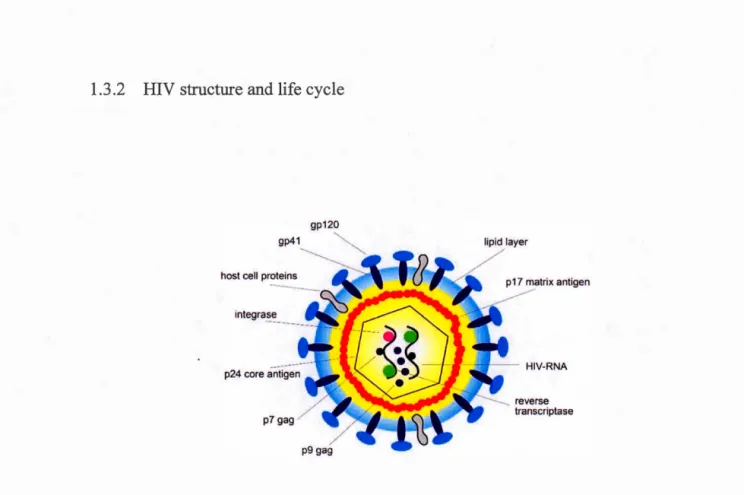
1.3.2 HIV structure and life cycle ... 32
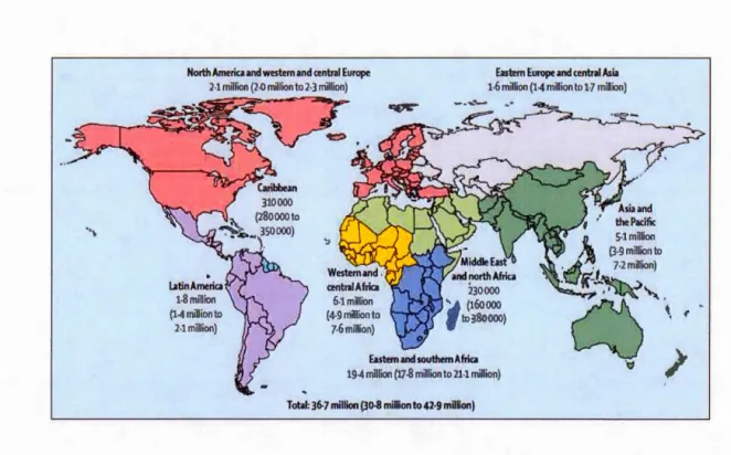
1.3 .3 Epidemiology of HIV infection ... 34
1.3.4 Pathophysiology ofHIV infection ... 36
1.3.5 Antiretroviral therapy (ART) and treatment challenges ... 38
1.4 HIV and Tregs ... 41
1.4.2 Dysregulation of Th17 /Treg balance during HIV /SIV infection ... 44
1.4.3 Dual role of Tregs during HIV infection ... 45
1.4.4 Deleterious role of Tregs in HIV infection ... 46
1.4.5 Role ofTGF-~ in HIV infection ... 48
1.4.6 Thymie fucntion during HIV infection in humans ... 50
CHAPTER II HYPOTHESIS AND OBJECTIVES ... 53
2.1 Rationale ... 53
2.2 Hypothesis ... 53
2.3 General Objectives ... 54
2.4 Specifie Objectives ... 54
CHAPTER III MATERIALS AND METHODS ... 55
3.1 Human thymus tissues and isolation ofthymocytes ... 55
3.2 HIV -1 viral stocks ... 56
3.3 In vitro assays with primary human thymocytes ...... 57
3.3.1 Co-culture model ... 57
3.3.2 In vitro TGF-~ treatment ... 58
3.3.3 HIV infection and TGF-~ treatment ... 58
3.4 Flow cytometry ... 60
3.5 Methylation sequencing ofjoxp3 ... 63
3 .5.1 Cell sorting ... 63
3.5.2 Genomic DNA extraction and bisulfite treatment ... 63
3.5.3 Nested PCR and MiSeq ... 64
3.6 qPCR ... 70
3. 7 Biostatistics ... 72
CHAPTER IV RE SUL TS ... 73
4.1 Ex vivo phenotypic characterization of human thymocytes using flow cytometry ... 73
4.2 Methylation status of foxp3 ... 89
4.3 Effect ofTGF-~ treatment on in vitro thymie Treg generation ... 92
4.4 In vitro HIV infection ofthymocytes ... 93
CHAPTER V DISCUSSION ... 100
CHAPTER VI CONCLUSIONS ... 111 REFERENCES ... 113
Figure Page
1.1 Somatic DNA rearrangement (VDJ recombination) process in TCR genes 05
1.2 Mechanisms of Treg mediated immune suppression... 13
1.3 Location of CNS elements on the foxp3 gene locus... 18
1.4 Role of TGF -~ in tT reg development ... ... 25
1.5 Shared developmental pathway of Th17 cells and Tregs in the peripheral blood ... 31
1.6 Structure of HIV ... 32
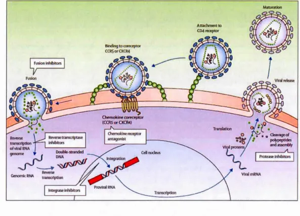
1. 7 HIV life cycle and drugs used in ART . . .. . . 34
1.8 HIV-infected people living worldwide, as of2016 ... 35
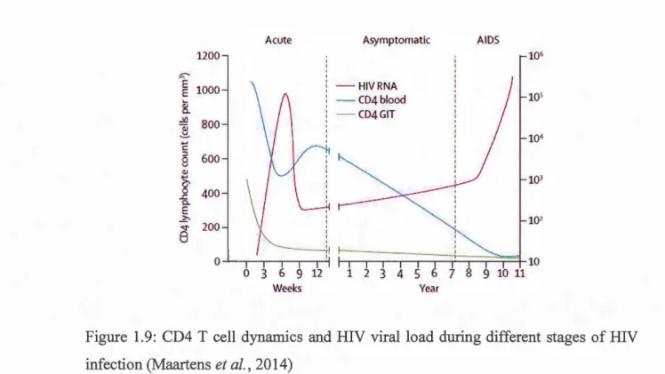
1.9 CD4 T cell dynamics and HIV viralload during different stages of HIV infection . . . 3 7 1.10 CD4 T cell dynamics and HIV viralload after ART... 39
1.11 Mechanisms accounting for increased Treg frequencies during HIV infection . . . 4 2 3.1 Steps involved in the ex vivo phenotypic characterization of human thymocytes... 56
3.2 Steps involved in the in vitro TGF -~ treatment and HIV infection ofhuman thymocytes... 59
3.3 Steps involved in the methylation sequencing ofjoxp3 ... ... 69
4.1 Relation between thymocyte maturation and CD3, CD4 and CD8 expression... 74
4.2 Phenotypic characterization of thymie Tregs ... 75
4.3 Mature thymocytes lose RORyt expression and do not co-express FoxP3 and RORyt ... ... ... 77
4.4 CD4+FoxP3+ thymocytes al ways express Helios... 79
4.5 Higher expression of CD45RO than CD45RA by thymie Tregs and Non Tregs ... 81
4.6 Discriminating Tregs of thymie origin from recirculating cells. ... 83
4. 7 Thymie Tregs and non-Tregs express gut ho ming markers ... 84
4.8 Thymie Tregs have higher expression ofHIV co-receptor CCR5 compared to Non Tregs. ... ... ... ... ... ... 86
4.9 Thymie Tregs and not non Tregs express HIV permissiveness marker CXCR3. ... 88
4.10 Methylation status of CNS elements of foxp3 in CD310w, CD3int, CD3high thymocytes ... ~... 90
4.11 Expression of DNA methyl transferases in CD310w, CD3int, CD3high thymocytes. . . . 91
4.12 Impact ofTGF-~ treatment on thymie Treg frequencies... 93
4.13 FoxP3+ thymocytes are not infected by HIV. ... 94
4.15 FoxP3 expression and thymie Treg generation are not altered by HIV infection and TGF -~ 1 treatment. . . ... .. . . .. . . .. . . . .. . . . .. . . ... . . . ... .. .. . . .. .. .. . . . .. . . .. 97
Tables Page
3.1 List of antibodies used for flow cytometry.... .. . . .. . . .. . ... . ... . . ... . .... .. . . . .. . .. . .. . 61
3.2 List of external prim ers used for first round of PCR... 65
3.3 List of intemal primers used for second round of PCR .. . . .. . ... .. . . . .. . . .. .. 66
3 .4 Constituents of PCR reaction . . . 67
ADP AIDS AIRE AMP AP-l APC ART ATP BMP CNS DC DN DNMT DP EAE Adenosine diphosphate
Acquired immunodeficiency syndrome
Autoimmune regulator gene
Adenosine monophosphate
Activator protein 1
Antigen presenting cell
Antiretroviral therapy
Adenosine triphosphate
Bone-morphogenetic protein
Conserved non-coding sequences
Dendritic Cell
Double negative
DNA methyl-tranferase
Double positive
ETP GALT GITR HAART
HCV
HIV
HSC
IDO IFN-y IL-10 IL-17 IL-2 IL-21 IL-22 IL-2R IL-35 IL-6Early T cell Progenitor
Gut-associated lymphoid tissue
Glucocorticoid-induced tumour necrosis factor TNF receptor
Highly active antiretroviral therapy
Hepatitis C virus
Human immunodeficiency
Hematopoietic stem cells
Indoleamine-2, 3 -dioxygenase Interferon-y Interleukin-1 0 Interleukin-17 Interleukin-2 Interleukin-21 Inter leukin-22 Interleukin-2 receptor Inter leukin-3 5 Interleukin-6
IL-7 Interleukin-7
iTreg Induced Treg
LAP Latency-associated peptide
LTR Long terminal repeat
MHC Major histocompatibility complex
mTEC Medullary thymie epithelial cell
Nrp1 Neuropilin-1
NFAT Nuclear factor of activated T cells
NI Non-infected
NICD N otch intracellular domain
PBMC Peripheral blood mononuclear cells
PD-1 Programmed death-1
PD-L1 Programmed death ligand-1
PFD Pirfenidone
pT reg Peripheral Treg
RTE Recent thymie emigrants
SIV Simian immunodeficiency virus
SMAD Sma and Mad -Mothers against decapentaplegic
SP Single positive
STAT-5 Signal transducer and activator of transcription 5
TCR T cell receptor
TGF-~ Transforming growth factor-~
Th1 T helper type 1
Th17 T helper type 17
TLR-4 Toll-like receptor-4
TNP Tumour necrosis factor
TREC T cell Receptor Excision Circles
Tregs Regulatory T cells
TSS Transcription start site
TSDR Treg specifie demethylated region
Contexte: Les lymphocytes T régulateurs (Treg) sont des cellules immunosuppressives ayant deux origines développementales: thymiques ou périphérique. FoxP3, le facteur de transcription principal des Tregs, contrôle leur différenciation et leurs fonctions suppressives. L'expression de FoxP3 par les cellules Tregs thymiques est en partie régulée par la cytokine anti-inflammatoire TGF-~, qui contribue également au développement des Tregs dans le sang périphérique et les tissus lymphoïdes. Les personnes vivant avec une infection chronique par le virus de l'immunodéficience humaine (VIH) ont une fréquence plus élevée de Tregs dans le sang périphérique et les tissus lymphoïdes. La fibrose des tissus lymphoïdes induite par le TGF-~ chez les individus infectés par le VIH est associée à la progression de la maladie. Malgré la corrélation entre TGF-~ et la pathologie associée au VIH, le rôle du TGF-~ dans l'induction et le maintien des Tregs dans le thymus pendant l'infection par le VIH demeure incertain.
Hypothèse: Le TGF-~ induit l'expression de FoxP3 et contribue ainsi à l'augmentation de la fréquence des Tregs thymiques au cours de l'infection par le VIH.
Objectif: Étudier les modifications sur l'expression de FoxP3 et la génération de Treg thymiques après traitement par le TGF -~ lors d'une infection in vitro de thymocytes humains par le VIH -1.
Méthodes: Des tissus thymiques humains frais ont été obtenus chez des patients pédiatriques subissant des chirurgies cardiaques correctives. La caractérisation phénotypique des thymocytes a été réalisée ex vivo par cytométrie en flux. L'évaluation ex vivo du statut épigénétique de foxp3 a été réalisée à l'aide de MiSeq. L'infection virale par les souches VIH-1 tropiques R5 et X4, ainsi que le traitement par le TGF-~ de thymocytes humains ont été réalisés dans un modèle de co-culture in vitro avec des lignées cellules OP9-DL1 exprimant le ligand de Notch.
Résultats: La caractérisation phénotypique ex vivo des thymocytes a révélé les faits suivants: 1) Quatre-vingt-dix-neuf% des thymocytes CD4+ FoxP3+ expriment Helios, un marqueur controversé des Tregs thymiques. 2) Le facteur de transcription maître des cellules Th17, RORyt, n'ést pas co-exprimé avec FoxP3 dans les cellules T CD4 thymiques, ce qui indique que l'expression dans le thymus est associée à la maturation
1 J
des thymocytes plutôt qu'à la différenciation de Th17. 3) Plus de 51% des Treg et des CD4+ FoxP3- non-Tregs dans le thymus expriment CD45RO, indiquant que ces cellules pourraient avoir reçu une forte stimulation du TCR par des auto-antigènes et ont probablement subi une sélection négative. 4) Environ 3% des Treg exprimaient le marqueur inflammatoire CCR6 et 2o/o les marqueurs de référence intestinaux Integrin-~7 et CCR9, suggérant leur repositionnement dans le thymus. 5) Plus de 87o/o des Tregs expriment le corécepteur du VIH CXCR4, et les Tregs expriment un niveau plus élevé
de CCRS (18,53%) par rapport aux non-Tregs (1,01 %), ce qui suggère que les Tregs
sont potentiellement plus susceptibles à l'infection par le VIH. 6) Les thymocytes CD3haut ont la plus haute expression de FoxP3 et une déméthylation de la région CNS 1 régulant foxp3. Comme prévu, le traitement des thymocytes par le TGF-~ induit l'expression de CD127 et entraîne une augmentation des fréquences de Tregs. L'infection des thymocytes par les souches VIH tropiques RS et X 4 a montré que les thymocytes FoxP3+ sont moins susceptibles à l'infection que les thymocytes FoxP3-. Le traitement par le TGF-~ n'a eu aucun effet sur le taux d'infection par le VIH. Lors de l'infection par le VIH et du traitement par le TGF-~, aucune augmentation de l'expression de FoxP3 ou des fréquences de Tregs n'a été observée.
Conclusions : La caractérisation phénotypique ex vivo des Tregs thymiques a fourni un aperçu complet des propriétés des Tregs thymiques. L'infection in vitro par le VIH et le traitement par le TGF-~ de thymocytes humains n'ont pas d'impact sur l'expression de F oxP3 et les fréquences de Tregs.
Mots-clés : Tregs, caractérisation phénotypique, TGF-~, l'infection par le VIH, statut épi génétique de foxp3
Background: Regulatory T cells (Tregs) are immunosuppressive cells with two developmental origins: thymie or peripheral. FoxP3, the master transcription factor of Tregs, controls their differentiation and suppressive functions. FoxP3 expression by thymie Tregs is partly regulated by the anti-inflammatory cytokine TGF-~, which also contributes to Treg development in the peripheral blood and lymphoid tissues. People living with chronic human immunodeficiency virus (HIV) infection have increased Treg frequencies in peripheral blood and lymphoid tissues. TGF-~-mediated fibrosis of lymphoid tissues in HIV infected individuals is associated with disease progression. However, the role of TGF-~ in the induction and maintenance of Tregs within the thymus during HIV infection remains unclear.
Hypothesis: TGF-~ induces FoxP3 expression and thus, contributes to increased thymie Treg frequencies during HIV infection.
Objectives: To investigate changes in FoxP3 expression and thymie Treg generation upon TGF -~ treatment during in vitro HIV infection of human thymocytes.
Methods: Fresh human thymie tissues were obtained from pediatrie patients undergoing corrective cardiac surgeries. Ex vivo phenotypic characterization of thymocytes was performed by flow cytometry. Ex vivo assessment ofjoxp3 epigenetic status was performed using MiSeq. HIV infection by both R5- and X4-tropic HIV -1 strains and TGF-~ treatment of human thymocytes was performed in an in vitro co-culture model with OP9-DL1 cells expressing Notch ligand.
Results: Ex vivo phenotypic characterization ofthymocytes revealed the following: 1) Ninety nine percent of CD4+ FoxP3+ thymocytes expressed Helios, a debated thymie Treg marker. 2) The master transcription factor of Th17 cells, RORyt, was not co-expressed with FoxP3 in the thymie CD4 T cells, indicating that RORyt expression by thymocytes is rather associated with their overall maturation than Th17 differentiation. 3) More than 51% of Tregs and CD4+ FoxP3- non-Tregs within thymus expressed CD45RO, which indicated that the cells might have received strong TCR stimulation by self-antigens and might be undergoing negative selection. 4) Only about 3% of Tregs expressed the inflammatory marker CCR6 and 2% expressed gut homing markers Integrin-~7 and CCR9, suggesting their re-localization into the thymus. 5) More than 87% of Tregs expressed the HIV co-receptor CXCR4 and Tregs expressed higher CCR5 (18.53%) compared to Non-Tregs (1.01 %), suggesting that Tregs are
more susceptible to HIV infection. 6) CD3high thymocytes had highest FoxP3 expression and demethylation ofthe CNSl region regulatingfoxp3. As expected,
TGF-~ treatment of thymocytes induced CD127 expression and resulted in increased Treg frequencies. Infection of thymocytes with R5 and X4-tropic HIV strains showed that FoxP3+ thymocytes were less prone to infection compared to FoxP3- thymocytes. TGF -~ treatment had no effect on the rate of HIV infection. Upon HIV infection and
TGF-~ treatment, an increase in FoxP3 expression or Treg frequencies were not observed.
Conclusions: The ex vivo phenotypic characterization provided a detalied overview of thymie Treg properties. The in vitro HIV infection and TGF -~ treatment of human thymocytes does not have an impact on FoxP3 expression and Treg frequencies. Key words: Tregs, phenotypic characterization, TGF-~, HIV infection, foxp3 epigenetic status
INTRODUCTION
1.1 T cell development in the thymus
1.1.1 Structure, organization and fun etions of the thymus
The thymus is a primary lymphoid organ of the immune system, and is morphologically similar in most mammals. It pla ys a central role in the immune system, as it is the site for maturation of T lymphocytes. The indispensable role of the thymus in establishing the immune system was demonstrated six decades ago: neonatal thymectomy made mice more prone to infections, and these animais developed primitive secondary lymphoid tissues and exhibited very poor immune responses (Miller, 2002). In addition to its immunological role, the thymus also functions as a glandular tissue and produces several hormones including thymosin alpha-1, thymulin and thymopoietin (Hadden, 1998), which regulate both developing thymocytes and mature T cells.
The thymus is located in the thoracic cavity above the heart. It has two lobes, both covered with a capsule of epithelial tissue and held together by connective tissue. Each thymie lobe has an outer cortex and inner medulla. Thymie epithelial cells,
macrophages and dendritic cells make the framework of the thymie cortex and medulla. These cells closely interact with developing thymocytes, thereby contributing to their
maturation. Thymie nurse cells have long membrane extensions, which ena ble efficient interaction with multiple thymocytes, forming multi-cellular complexes (Kindt, 2007). The Hassall's corpuscles are an additional histological identity of the thymus; these are clusters of thymie epithelial cells, mainly involved in the phagocytosis of apoptotic thymocytes (Blau, 1965) or in sustaining their development (Senelar et al., 1976). Hassall' s corpus cl es also contribute to thymie development of Tregs (Watanabe et al., 2005). Thymie stromal lymphopoietin (TSLP) produced by Hassall's corpuscles stimulate the expression of MHC-II, CD80 and CD86, thereby promoting the interactions between dendritic cells (DCs) and developing CD4 T cells, directing the differentiation of the latter to produce CD4+ CD25+ FoxP3+ Tregs (Watanabe et al., 2005).
Aging causes atrophy and reduces the immune functions of the thymus. In humans, thymie atrophy be gins after puberty. Age-associated thymie involution is characterized by significant changes in the thymie microenvironment, decrease in number of thymocytes and increase in the fat content of the tissue. These factors result in a net reduction in the weight of the thymus tissue (Kindt, 2007).
1.1.2 Hematopoiesis and lymphopoiesis
T cells are newly generated throughout the lifetime of an adult to replenish the T cell repertoire in various secondary lymphoid tissues. This is possible because the adult thymus constantly receives thymus-allotted progenitor cells from the borre marrow (Donskoy et Goldschneider, 1992) . Blood cells of all types arise from hematopoietic
stem cells (HSCs) in the bone marrow, in a process termed hematopoiesis. HSCs give rise to two fundamental cell lineages: the myeloid lineage (erythrocytes or red blood cells, megakaryocytes, granulocytes: eosinophils, basophils, neutrophils, and monocytes or macrophages) and the lymphoid lineage (T cell, B cells and NK cells ). To develop into a T lymphocyte, a lymphoid progenitor must exit the bone marrow, enter circulation and migrate to thymus (Petrie, 2003). There are several factors influencing the recruitment and migration of HSCs into the thymus. Different sets of markers are used to identify the migrating precursor cells to the fetal and adult thymus. HSCs with migratory potentiallack cell adhesion molecules like intergrins a4~1 (VLA-4) or a5~1 (Whetton et Graham, 1999) and express low levels ofCXCR4, which is the receptor for stromal cell-derived factor 1 (SDF-1) required to home and be retained in the bone marrow (Whetton et Graham, 1999). These precursors cells exit circulation and settle into the thymus by expressing CD44 (Wu et al., 1993) and L-selectin ( CD62L) (Perry et al., 2004). Once in the thymus, these settling precursor cells are called Early T cell progenitors (ETPs), and lack the capacity of self-renewal.
1.1.3 Importance ofNotch signaling in the thymus
Thymie development of T cells from ETPs involves complex signaling mechanisms. Two decades ago, severe impairment of thymocyte development was reported in mi ce lac king N otch-1 signaling (Radtke et al., 1999). N otch is a transmembrane receptor and its ligands are also transmembrane proteins of the Delta/Serrate/LAG-2 (DSL) family. Ligand binding stimulates Notch, following which the Notch intracellular domain (NICD) fragment enters the nucleus and activates gene transcription. T cell progenitors utilize Notch signaling after entering the thymie epithelial microenvironment (Harman et al., 2003). Notch signaling is essential forT celllineage
commitment, and T cell progenitors 1n the thymus of Notch-deficient m1ce differentiated into B cells (Wilson et al., 2001). Notch-1 signalling is also important to prevent ETPs from developing into other cell types like NK -cells, monocytes, or DCs (De Smedt et al., 2005). Experimentally, it was reported that upon interaction with a stromal cellline ectopically expressing Delta-like-1, HSCs develop into CD4+ CD8+ (double positive, DP) and single positive (SP) T cells in vitro (Schmitt, T. M. et Zuniga-Pflucker, 2002). Thus, Notch signalling is essential for thymie T cell development.
1.1. 4 T cell receptor rearrangement
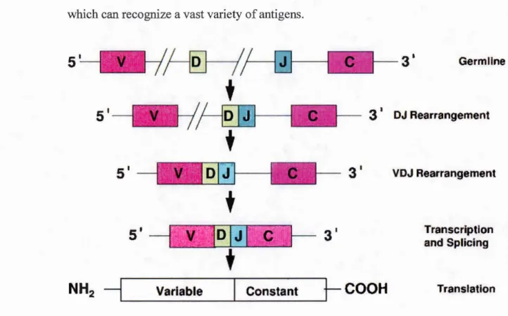
As for the immunoglobulin receptor on B cells, T cell receptor (TCR) genes also undergo somatic DNA rearrangement in order to generate a diverse repertoire of receptors, assuring recognition of a wide variety of antigens. ETPs entering the thymus do not express TCRs, and have germline TCR genes since they have not yet started the process of TCR gene rearrangement. These cells do not express CD3 or CD4 or CD8, and hence are called triple negative (CD3- CD4- CD8-) cells. The existence of a small proportion of immature CD410w cells, which are CD3-, has been reported, and it has been speculated that these cells later become triple negative cells (Malissen et al., 1999). Triple negative thymocytes do not express either CD4 or CD8 ( called double negative, DN cells) are further subdivided into four types based on CD44 and CD25 (IL-2Ra) expression: DN1 (CD44+ CD25-), DN2 (CD44+ CD25+ ), DN3 (CD44- CD25+) and DN4 (CD44-CD25-) (Godfrey et al., 1993); the numbering corresponds to the stage of maturation. DN2 cells possess a germline TCR gene, while DN4 thymocytes have a rearranged TCR gene (Godfrey et al., 1993).
Three regions in the TCR loci, namely, variable (V), diversity (D) and joining (J)
regions are subject to recombination, and therefore, the TCR gene rearrangement is also referred as the VDJ recombination process, which begins in the DN3 stage of thymocyte maturation. T cells can either express an a~-TCR or a yo-TCR, depending on which genes (Tcrb, Tcrg, Tcrd) are first successfully recombined. We will be
focusing here only on the development of a~ T cells. This somatic DNA rearrangement pro cess, shown in Figure 1.1, ens ures the generation a diverse repertoire of T cells, which can recognize a vast variety of antigens.
3 •
Germllne3 •
DJ Rearrangementt
s·
t
s'
N H2
-lL-__
v_ar_ia_b_le
_ _
...__c_o_n_s_ta_n_t_~t-
COOH
VDJ Rearrangement
Transcription and Splicing
Translation
Figure 1.1: Somatic DNA rearrangement (VDJ recombination) process in TCR genes, (Rezuke et al., 1997). The genes encoding the TCRs and immunoglobulin receptors on
B cells undergo DNA rearrangement thereby ensuring production of a diversity of receptors, which will enable recognition of a wide variety of antigens.
A crucial step in a~ T cell development is the ~-selection checkpoint. Thymocytes at the DN3 stage, which successfully complete TCR~ chain rearrangement, are provided with survival and proliferation signais, which enable them to proliferate and further differentiate. This step also signifies commitment to the a~ T cell phenotype. The rearrangement of Tcra gene locus follows the ~-selection process, resulting in a cell that expresses an a~-TCR. Signaling through the a~-TCR causes upregulation of the co-receptors CD4 and CD8 and the developing thymocytes enter the DP stage.
Two important regulatory steps in thymie T cell development are positive and negative selection. Positive selection is the process by which only thymocytes expressing a~ TCR that can recognize self-antigens presented along with MHC-I/II are selected; thymocytes which have not successfully rearranged their TCR loci are eliminated by apoptosis. DP thymocytes become either CD4SP if they interact with self-peptides presented by MHC-II molecules, or CD8SP if they interact with self-peptides presented by MHC-I molecules. Multitudes of factors influence CD4 or CD8 lineage commitment, sorne of them being addressed in the following section. The affinity with which TCRs recognize self-peptide-MHC complexes determine whether aT cell is eliminated or selected for survival. Negative selection is the process that discriminates highly self-reactive T cells and prevents autoimmunity by triggering their deletion (Spits, 2002).
1.1.5 Exisiting models for CD4 T celllineage commitment
The differentiation ofDP thymocytes into CD4SP or CD8SP T cells depends on which one of the two co-receptors is downregulated and transcriptionally silenced. Two theoretical models, the instructive and the stochastic models, attempt to explain the CD4-CD8 lineage commitment process in the thymus (Germain, 2002). According to the instructive model, interaction ofTCRs with MHC-I generates a downstream signal which is biochemically different from the signais generated upon interaction of TCRs with MHC-II, the former leading to CD8 differentiation and the latter leading to CD4 differentiation. This model is in line with the proposition that TCR affinity for self-peptides is one of the major factors influencing thymie T cell development. Negative selection, an important self-tolerance mechanism that elimina tes thymocytes with high-affinity TCRs, is also in line with the instructive model.
The stochastic model proposes that the DP thymocytes randomly express either one of the two receptors, which means that a DP thymocyte may initiate the expression of a co-receptor that is unrelated to the interaction of its TCR with either I or MHC-II. In such cases, if a mismatch between the MHC specificity of a clonai TCR and its co-receptor occurs, the developing thymocyte will not undergo the maturation process and will become apoptotic. Studies in mice validating both models have been conducted, and it was concluded that a combination ofboth models pro vides a complete and precise explanation for thymocyte development (Germain, 2002). A slight modification of the instruction model is the "duration of signal" model of CD4-CD8 lineage commitment, which proposes that the TCR signalling of longer duration results in down regulation of CD8 and favors CD4 differentiation, while TCR signais of shorter duration result in CD8 differentiation and downregulation of CD4 gene
expression (Singer et al., 2008). Signal durations were studied by changing the period for which TCR could engage with the ligand, i.e., by altering the TCR stimulation period during in vitro cultures. It was later demonstrated that the TCR signaling in all DP thymocytes is of the same duration and it brings about downregulation of CD8 expression. However, TCR signaling through interaction with MHC-I in CD4+ CD81ow thymocytes is of shorter duration than TCR signalling through interaction with MHC-II (Singer, 2002).
The latest, accurate and most widely accepted model of CD4/CD8 lineage differentiation is the kinetic model, which is grounded on crucial experimental evidences, which have defied all of the proposed classical theoretical models. Experimental observations showed that strong TCR signalling in DP thymocytes resulted in downregulation of CD8, leaving CD4 expression unaltered, and the resulting CD4+ CD810w cells remained uncommitted. Thus, the concept that CD4/CD8
lineage commitment is defined by the downregulation of either one of the co-receptors is incorrect. Taking into account all the mentioned factors conceming the duration of TCR signals, the kinetic signaling model proposes that CD4/CD8lineage commitment occurs not in DP thymocytes but in CD4+ CD810w thymocytes, which represent an intermediate developmental stage after the DP stage. These cells have the potential to differentiate into either CD4SP or CD8SP T cells. If TCR signals persist for a longer duration in CD4+ CD81ow thymocytes, they become CD4SP. If the signals last for a shorter duration, cells become CD8SP. The uniqueness of the kinetic signalling model is the explanation provided for CD8 lineage commitment- CD8 differentiation occurs when CD4+ CD810w thymocytes do not receive persistent TCR signal and hence reverse their co receptor expression (that is, they become CD8+ CD4-). More specifically, absence of long duration TCR signals are sensed through yc cytokines like IL-7, crucial for CD8 lineage commitment, which reverse co-receptor expression from CD4+
CD810w to CD4- CD8+ (Singer et al., 2008). The CD4/CD8 lineage choice is also regulated at the transcriptional and epigenetic levels. The gene expression of murine CD4 co-receptor is regulated by two important cis regulatory elements : a transcriptional enhancer and a transcriptional silencer. The transcriptional enhancer is called the cd4 proximal enhancer or E4p is a 300bp sequence located 13kb upstream of the transcription start site (TSS) and has been reported to be active in thymocytes in DN2 or DN3 stages of development (Sawada et Littman, 1991). The transcriptional silencer called S4, is located 1.6kbp downstream of the transcription start site and causes CD4 downregulation in CD8+ T cells (Sawada et al., 1994).
1.2 Regulatory T cells
1.2.1 Phenotype, functions and classification of Tregs
Studies about immune tolerance mechanisms started in the early 1970's (Gershon et Kondo, 1970), but untillate in the 1990's, the existence of immunosuppressive cells was much under debate (Green et Webb, 1993). Tregs were first described in 1995 by Sakaguchi et al. (Sakaguchi et al., 1995) as immunosuppressive cells, which limit the activity of other types of T cells. The normal physiological role of Tregs is to prevent autoimmunity (Kim, J. M. et al., 2007; Seddon, 2000) and maintain immune homeostasis. The transcription factor FoxP3 is considered the "master regulator" of Tregs since it is required for the expression of several genes responsible for the differentiation and maintenance of the Treg phenotype (CD3+ CD4+ CD25high CD12710w FoxP3high) (FontenotRasmussen, et al., 2005). From 2000 to 2015, 30,000 research articles referring to Tregs have been published (Zongyi et al., 2016), and the subject continues to be a hotspot in immunological research.
The existence of CD4+ T cells expressing IL-2R (CD25) responsible for immune suppression was first demonstrated in mice (Sakaguchi et al., 1995) and later in humans (Baecher-Allan et al., 2001). Initially, the regulatory phenotype was identified as CD4+ CD25+ T cells. Later, the role ofFoxP3 in human Tregs was demonstrated (Y agi et al., 2004), and the low expression ofiL-7 receptor (CD127) was shown to be another useful parameter to discriminate Tregs (Liu et al., 2006). Renee, a more complete way to define Tregs phenotypically is CD3+ CD4+ CD25high CD12710
w FoxP3high T cells (Jenabian et al., 2012). Tregs can be classified based on their origin as follows: 1) natural or thymie Tregs (tTregs) which originate within the thymus; 2) peripheral Tregs (pTregs) which are produced in the peripheral blood, i.e., are generated in vivo from CD4+ cells that differentiate into Tregs as they start expressing FoxP3 under inflammatory conditions (Curotto de Lafaille et Lafaille, 2009; Curotto de Lafaille et al., 2004). A third category of immunosuppressive cells are induced Tregs (iTregs), which are generated in vitro from CD4+ T cells, which start expressing F oxP3 when stimulated with TGF-~ (Chen, W. et al., 2003). However, the expression of FoxP3 alone, either induced by TGF-~ or expressed exogenously using transduction methods is insufficient to produce functionally active Tregs exhibiting suppressive capacity (Feuerer et al., 2010; Hill et al., 2007; Yadav et al., 2013). Renee, iTregs are not analogous ofpTregs (Yadav et al., 2013) but these names are used interchangeably in articles by different authors. A simplified nomenclature for Tregs has been suggested, which is to use tT regs instead of"natural Tregs"; pT regs to refer to peripherally derived Tregs and iTregs to refer to in vitro induced Tregs (Abbas et al., 2013). CD8+ cells expressing FoxP3, hence called CD8 Tregs are also known to originate within the thymus and in the peripheral blood. Although these cells have still not been clearly characterized, they are known to execute suppressive functions on non-Tregs, and secrete immunosuppressive cytokines like IL-10 and TGF- ~' similar to CD4+ Tregs (Yu et al., 2018).
1.2.2 Mechanisms of Treg mediated immune suppression
Tregs can execute their suppressive function only after being activated through their TCR (Takahashi et al., 1998; Thornton, A. M. et Shevach, 1998), which is triggered upon binding to a specifie antigenic peptide. Sustenance of TCR signal is vital for the maintenance of Treg function (Hoeppli et al., 20 16; Levine et al., 20 14; Vahl et al.,
2014). As CD4+ T cells, Tregs are also antigen-specific, since they contain a functional TCR resulting from somatic rearrangement of gene segments (Corthay, 2009). Based on their antigen specificity, Tregs can be classified as either self-antigen-specific Tregs, which help in preventing autoimmunity and maintain immune homeostasis or foreign-antigen-specific Tregs, such as tho se recognizing bacterial antigens (McGuirk et al.,
2002) or viral an ti gens (Zhao et al., 2014) and thereby controlling immune response to these pathogens. Several studies have tried to demonstrate the utility of "engineered" Tregs (autologous cells modified in vitro according to treatment needs) in immunotherapy approaches for the treatment of cancers (Geldres et al., 20 16), auto immune disorders (Stephens et al., 2009) and in overcoming graft rejections (Brennan et al., 2011 ).
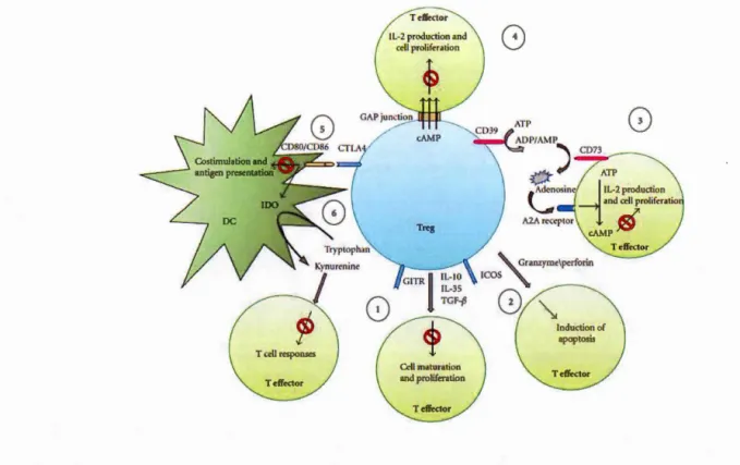
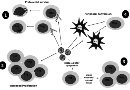
Tregs exert their immunosuppressive effects on effector T cells through several mechanisms. Figure 1.2 summarizes the different ways in which activated Tregs exert their immunosuppressive functions:
1. Anti-inflammatory cytokines secreted by Tregs such as IL-10 (Vieira et al., 2004), IL-35 (Castellani et al., 2010) and TGF-~ (Weiner, 2001) inhibit the proliferation of effector cells.
2. Tregs play a negative role in cancer by repressing anti-tumor immune responses via the granzyme-perforin pathway (Cao et al., 2007). Tregs also control inflammation in
response to viral infection (Loebbermann et al., 2012) using the same pathway. Granzymes are serine proteases while perforins are cell membrane toxins, which are often employed by NK cells and cytotoxic T cells, and are released by exocytosis to trigger apoptosis in target cells (Trapani et Smyth, 2002).
3. Tregs utilize two enzymes CD39 (ectonucleoside triphosphate diphosphorylase-1) and CD73 (ecto-5'-nucleotidase), which break down ATP into ADP, and subsequently, ADP into AMP or adenosine (Borsellino et al., 2007). Adenosine binds to A2A receptors on other immune cells, is converted into cAMP and in duces an ti-inflammatory effects - it restricts proliferation and cytokine production (Ohta et al., 2009; Ohta et Sitkovsky, 2001; Sitkovsky et al., 2004).
4. Tregs can transport cAMP to effector T cells through gap junctions, thereby inhibiting their proliferation and IL-2 production and preventing HIV replication in the infected T cell (Moreno-F emandez et al. , 2 0 11).
5. DCs are antigen presenting cells that express the co-receptors CD80 and CD86 which interact with CD28/CTLA-4 on T cells (McLellan et al., 1995). Tregs decrease expression of CD80/CD86 on DCs (Wing et al., 2008), via CLTA-4, which leads to downregulation of IDO (indoleamine-2, 3-dioxygenase), an enzyme that converts tryptophan to kynurenine (Hryniewicz et al., 2006).
Figure 1.2: Mechanisms of Treg-mediated immune suppression - 1)
Cytokine-mediated suppression, 2) Granzyme B-mediated suppression 3) CD39 and
CD73-mediated suppression 4) cAMP transport through gap-junctions 5) & 6) DC-mediated suppression (Jenabian et al., 2012).
1.2.3 Models of thymie Treg development
Development of thymie Tregs occurs in two steps: survival ofthymocytes bearing TCR with relatively high affinity to self-antigens and cytokine-dependent induction and maintenance ofFoxP3 expression (Lio et Hsieh, 2008).
Treg precursors are auto reactive CD4+ CD25+ thymocytes, which have TCRs that bind self-antigens with high affinity (Jordan et al., 2001). Developing thymocytes bearing TCRs with high affinity to self-antigens likely undergo negative selection, the process by which central tolerance is assured in the thymus. Therefore, for Tregs to develop in the thymus, certain autoreactive CD4+ CD25+ thymocytes must escape elimination. However, the exact strength ofbinding of the TCR with self-peptides exposed on MHC class II molecules that is required for Treg development has not yet been defined, and it is probable that other factors prevent their elimination. As proposed by Hsieh et al. (Hsieh et al., 20 12), Treg precursor thymocytes are expected to have TCRs that recognize self-antigens with a binding affinity not too high to trigger negative selection, but sufficient to assure survival through positive selection. The latter is a process by which cells with functional TCRs are selected for survival (Jameson et al., 1995). Expression of the cell surface glucocorticoid-induced tumour necrosis factor (TNF) receptor (GITR) is also critical to promote the Treg lineage. Expression ofGITR, OX40 and TNF-R2, which belong to the TNF receptor superfamily, corresponds to high TCR signal strength on developing Treg precursors in the thymus (Mahmud et al., 2014). Also, Tregs must respond to cytokines such as TGF-~ and IL-2, which promote FoxP3 expression and prevent apoptosis, as discussed in detaillater (section 1.2.5.4).
It is presently unclear at what stage developing thymocytes start expressing FoxP3 or whether this process initiates in the thymie cortex or medulla. CD25+ FoxP3+ Treg precursors have been shawn to appear in the DP stage (Cabarrocas et al., 2006). DP thymocytes are abundant in the thymie cortex, which is the site for positive selection (Bill et Palmer, 1989). Indeed, Liston et al. showed that the thymie cortex alone is sufficient to support initiation of FoxP3 expression- about one fourth of total FoxP3+ cells in thymie cortex are DP. In contrast to what was reported by Liston et al. (Liston et al., 2008), Lee et al. used more stringent gating strategies to eliminate doublets
( common within the pool of DP thymocytes) and showed that development of thymie Treg from FoxP3+ DP thymocytes is infrequent, close to 1% of total FoxP3+ cells (Lee et Hsieh, 2009). It should also be noted that a small percentage of DN T cells in the thymie cortex, express FoxP3, although not yet having a functional a~ TCR (Tuovinen et al., 2008). It is unknown whether these DN FoxP3+ thymocytes are precursors for functional Tregs.
More recently, it was proven that FoxP3 expression in the human and mouse thymus initiates before the DP stage but requires expression of a functional TCR (Nunes-Cabaco et al., 201 0). Interestingly, the thymie cortex hosts thymie cortical epithelial cells expressing MHC- II , which seem essential for generation of CD4+ CD25+
immunoregulatory T cells (Bensinger et al., 2001 ). All the aforementioned studies indicate that FoxP3 expression starts in the thymie cortex. However, it is to be noted that induction of F oxP3 expression al one is insufficient for the generation of functionally active thymie Tregs.
Several studies have highlighted the importance of the thymie medullary environment for the generation ofTregs. Negative selection occurs in the thymie medulla (Sprent et Kishimoto, 2002), and bypassing apoptosis triggered by negative selection is considered to be one of the major steps in thymie Treg generation. Treg differentiation in the thymus has been shown to be regulated by medullary thymie epithelial cells (mTECs) (Aschenbrenner et al., 2007) and thymie DCs (Watanabe et al., 2005) present in the thymie medulla, that present MHC class II-restricted self-antigens. Furthermore,
expression of autoimmune regulator gene (AIRE) in medullary thymie epithelial cells has been reported to be essential for generation of FoxP3+ thymie Tregs
(Aschenbreill1er et al., 2007). It has also been shown that migration of CCR 7+ developing thymocytes to the thymie medulla is crucial for induction of central immune tolerance (Kurobe et al., 2006). From the above information, it is clear that the expression of FoxP3 in potential Treg precursors depends on various factors including TCR activation, signaling through certain cytokine receptors as well as expression of cell surface molecules and chemokine receptors. In addition, contact with specifie types of thymie epithelial cell is crucial (Bettini et Vignali, 2010). In summary, although FoxP3 expression is initiated in developing thymocytes in the thymie cortex, the thymie medulla plays an important and indispensable role in the generation of functional tT regs capable of sustaining immune suppression.
As discussed more elaborately in the upcoming section, tT regs differ from iTregs with respect to the stability of FoxP3 expression (Floess et al., 2007). Several studies indicate that the transcription factor Helios, belonging to the Ikaros family of zinc finger proteins, might be crucial for maintenance of the Treg phenotype, involving stable FoxP3 expression and the ability to inhibit effector responses. For instance, Helios-deficient mice have CD4+ FoxP3+ cells with reduced inhibitory capacity compared to cells from wild type mice (Kim, H. J. et al., 2015), and siRNA mediated knockdown of Helios in CD4+ CD25+ Tregs downregulates FoxP3 expression (Getnet et al., 2010). Helios binds to the IL-2 gene promoter either by itself or together with FoxP3, thereby inducing its transcriptional silencing (Baine et al., 2013). It also plays an important role in maintaining self-tolerance mechanisms in the thymus: self-reactive thymocytes undergoing negative selection co-express Bim and Helios (Baldwin et Hogquist, 2007; Daley et al., 2013). The next section describes the epigenetic regulation of F oxP3 expression, which is crucial in the induction and maintenance of the Treg phenotype.
1.2.4 Epigenetic regulation ofjoxp3 expression
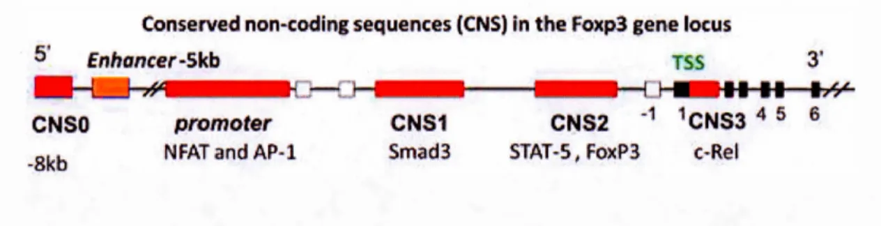
Treg development is not only determined by FoxP3 expression but also by the establishment of a Treg-specific CpG methylation pattern, which is also initiated upon TCR stimulation (Ohkura et al., 2012). Unique CpG methylation patterns, called Treg-specific demethylated regions (TSDRs), were found in Treg-Treg-specific genes including Foxp3, Ctla-4, Il2ra, Ikzf4 and Tnfrsf18 by genome wide CpG methylation analysis of mice CD4+ T cells versus Tregs (Ohkura et al., 2012). Interestingly, the establishment of this methylation profile seems independent of FoxP3 expression, as demonstrated by Okhura et al. in the same study (Ohkura et al., 2012). Tregs are known to contribute to immunosenescence, the process of graduai deterioration of the functioning of the immune system due to age (Jagger et al., 2014). Tregs from aged mice exhibit more demethylation at the foxp3 gene locus compared to younger mice, resulting in higher Treg mediated suppression of effector T cells in older mice (Garg et al., 2014). Lack of TSDR hypomethylation is also the reason behind impaired suppressive functions of iTregs, which in turn is concurrent to unstable FoxP3 expression (Floess et al., 2007). "Stability" and "plasticity" are two close terms, differing subtly from each other as accepted by researchers in the field of Treg biol ogy (Sakaguchi et al., 20 13). With respect to Tregs, stability refers to the establishment and maintenance of a molecular profile characteristic of Tregs, i.e., the stable expression of FoxP3 and other Treg-associated genes like CTLA-4 or IL-2R (Sakaguchi et al., 2013). The term plasticity is more often used to describe the functional characteristics of FoxP3+ T cells which might not necessarily have the transcriptional pro gram of Tregs (Sakaguchi et al., 2013). One of the major factors that determine Treg stability is the epigenetic regulation of the foxp3 gene. In the regula tory region of the foxp3 gene locus, six elements called the conserved non-coding sequences (CNS), have been identified (Iizuka-Koga et al., 2017). CNSO, enhancer, proximal promoter, CNS1, CNS2, and CNS3 are the six
regions and their positions on the foxp3 gene locus are shown in Figure 1.3 (Iizuka-Koga et al., 2017). We will now discuss briefly the role of each ofthese six regions in the order of their location on the foxp3 gene locus.
Conserved non·coding seq!uences (CNS) in the fo:xp3 gene locus
5 Enhancer-Skb 'IFSS ,J CNSOI -Bkb promo ter NFAT and AP-1. CNS1 Smad3 CNS2 -1 1CNS3 4 5 6 STAT~S, Fox:P3 C·Re~
Figure 1.3 Location of CNS elements on the foxp3 gene locus, adapted from Iizuka-Koga, M. et al. 2017. CNS elements serve as binding sites for specifie transcription factors as shown. Methylation status of the CNS elements control the stability ofFoxP3 expression on Tregs.
CNSO, also known as a "super-enhancer" element, is located about 8kb upstream of the transcription start site and serves as the binding site for Satb 1 (Kitagawa et al., 20 17). The major function of Sa tb 1 is to remo del the chromatin architecture to control transcriptional and epigenetic modifications, which in turn facilitates unique tissue-specifie gene expression profiles (Cai et al., 2003). Binding of Satb1 to CNSO triggers changes in chromatin structure, which alters its susceptibility to histone modifications (Iizuka-Koga et al., 20 17). Renee, Satb 1- CNSO interaction is a vital first step necessary for the functioning of other CNS elements and the induction of FoxP3 gene (Iizuka-Koga et al., 2017). It is important to note that CNSO has to be in a demethylated state to be able to support binding of Satb 1 (Kitagawa et al., 20 17).
The enhancer is a highly conserved sequence located about 5kb upstream of the transcription start site and its demethylation by DNA methyl transferase enzymes (DNMTs) is the key event that initiates FoxP3 expression (Lal et al., 2009). The proximal promoter is located relatively closer to the transcription start site and has binding sites for important transcription factors, such as NFAT and AP-l, STAT-5. Binding of AP- 1 and NF AT to the promoter region is associated with its trans-activation following the trans-activation of TCR. This region is T cell-specific and is responsible for induction ofFoxP3 after TCR stimulation (Mantelet al., 2006).
CNS 1 serves as the binding site for the transcription factors NF AT, AP-l, retinoic acid receptor and most importantly Smad2/3, the prote in complex downstream of TGF- ~
receptor-mediated signaling. As mentioned previously, TGF-~ plays an essential role in pT reg generation. It has been demonstrated that deletion of CNS 1 inhibits the differentiation of pTregs highlighting the importance of this region particularly in pT reg generation (Zheng et al., 201 0). The role of CNS 1 in tT reg generation remains unclear.
CNS2 is also called Treg specifie demethylated region (TSDR). As the name suggests, demethylation of this region is specifie to Tregs only- activated CD4+ T cells (in mice and in humans) that transiently express FoxP3 protein do not have demethylated CNS2 (Baron et al., 2007). It has binding sites for Stat5, NFAT, Runxl/Cbfb, CREB, and FoxP3 itself. For the stability of FoxP3 expression and hence the maintenance of the Treg phenotype, CNS2 region has to be maintained in a demethylated state. Studies in mice have shown that inheritance of the Treg phenotype is largely associated with stable FoxP3 expression, which is a consequence of demythalted CNS2 (Feng et al.,
2014). It has also been shown that tTregs, stably expressing FoxP3, have demethylated
CNS2 in contrast to iTregs in which FoxP3 protein expression (induced by TGF-~ in
vitro) is high but unstable with incomplete CNS2 demethylation (Floess et al., 2007). Now arises the question about CNS2 methylation in pTregs (referring to Tregs
generated in vivo, but not from the thymus). Two studies done in mice have addressed
this question- Weiss et al. and Yadav et al. distinguish pTregs from tTregs using
Neuropilin-1 (Nrp1, a receptor initially described in axon guidance, which also has a
role in Treg-DC interactions) have demonstrated that both Foxp3+ Nrp1 + (tTreg) and
Foxp3+ Nrp1- (pTreg) stably express FoxP3 and have demethylated CNS2 (Weiss, J.
M. et al., 2012; Yadav et al., 2012). However, it is controversial ifNrp1 can be used
to differentiate tT regs and pTregs. The numbers of Nrp 1-expressing Tregs are
comparable in genetically modified mice with impaired differentiation of tT regs, in
mice with defective pTreg differentiation, and in wild type mice, indicating that high or low expression ofNrp 1 is not an appropriate parameter to distinguish between tT regs
and pTregs (Yadav et al., 2012). Therefore, it remains unclear if pTregs have stable
FoxP3 expression and CNS2 demethylation, since distinguishing tTregs and pTregs
remains controversial.
CNS3 includes the binding site for the transcription factor c-Rel, a member of the
NF-KB family of pro teins. The NF -NF-KB signaling pathway is one of the many signaling cascades activated upon TCR stimulation and c-Rel binding to CNS3 has a direct
influence on FoxP3 expression after TCR stimulation (Long et al., 2009). TCR
stimulation is a vital step in the process of both pT reg and tT reg generation and hence
deletion of CNS3 in mi ce leads to the deficiency of both tT regs and pT regs (Zheng et
Having discussed the regions in the FoxP3 gene, which are rich in CpG sites, and act
as epigenetic switches regulating the gene and contributing to
development!maintenance of the Treg phenotype, the mechanisms responsible for controlling their methylation status will now be summarized. DNMTs are a class of enzymes controlling the epigenetic status ofjoxp3. Specifically, the enzymes DNMT1,
DNMT3b, MeCP2, and MBD2 are bound to the upstream enhancer ofjoxp3, thereby
maintaining it in the methylated state, in iTregs, activated CD4+ T cells, and naïve CD4+ T cells, all of which might unstably express FoxP3 protein and hence do not possess immune suppressive capacity (Lal et al., 2009).Tet enzymes are an additional class of methylcytosine dioxygenases controlling FoxP3 epigenetic status; these enzymes convert 5-methylcytosine (5mC) to 5- hydroxymethylcytosine (5hmC) in CpG islands. Yang et al. demonstrated that Tet-1 and Tet-2 enzymes are responsible
for the establishment of FoxP3 hypomethylation patterns associated with Treg phenotype and deletion of these enzymes resulted in FoxP3 hypermethylation and defective Treg generation, leading to auto immune diseases (Yang, R. et al., 20 15). Vitamin C also indirect! y pla ys a role in establishing Treg phenotype, since it regulates Tet family of enzymes, which in turn alter FoxP3 methylation patterns (Sasidharan Nair et al., 2016).
1.2.5 Role of TGF -~ in Treg development
1.2. 5.1 TG F-~: Classification, Isotypes, Structure and Function
Transforming Growth Factor Beta (TGF-~) is a cytokine belonging to the TGF-~
bone-morphogenetic proteins (BMPs), activins and inhibins. TGF-~ is involved in the process of cellular differentiation, proliferation, development and embryogenesis,
carcinogenesis, fibrosis, wound healing and angiogenesis (Blobe et al., 2000). In mammals, there are three different isoforms ofTGF-~ (TGF-~1, TGF-~2 and TGF-~3), each encoded by a different gene (Govinden et Bhoola, 2003). The TGF-~1 isoform plays an important role in immune regulation (Li, M. O. et al., 2006) and hence is of our interest.
1.2.5.2 TGF-~: Activation and Signalling
TGF-~ was initially identified as a 25kDa protein promoting anchorage-independent growth ofmouse fibroblasts (Moses et al., 1981; Roberts et al., 1981). It is produced as a precursor protein (prepro- TGF -~) and exists as a homodimer in its active form. The pre-region of prepro- TGF- ~1 contains a signal peptide at the N-terminal end,
which undergoes proteolytic cleavage in the Golgi apparatus. The pro-region consists of the latency associated peptide (LAP), which forms a homodimer with mature
TGF-~, called the smalllatent complex, which remains inactive. Once secreted into the extra-cellular matrix, activation of TGF -~ is triggered by proteolysis of LAP or by conformational changes in LAP which prevents its binding to TGF -~ (Armes et al., 2003). TGF-~ mainly signais thorough type I (TGF-~ R1) and type II (TGF-~ R2) receptors, which belong to the family of serine/threonine kinases. Until now, seven types ofTGF-~ Rl (Activin-like kinases 1-7) and five types ofTGF-~ R2 have been reported (Chang, H. et al., 2002). Active TGF-~ initially binds to a TGF-~ R2,
promoting conformational changes of this receptor and recruitment of TGF -~ R1 through phosphorylation and subsequent activation (Heldin et al., 1997). The activated
TGF-~ R1 phosphorylates Smads (Sma and Mad -Mothers against decapentaplegic), specifically, Smad-2 (Nakao et al., 1997) and 3 which then associate with Smad-4 and translocate into the nucleus (Inman et al., 2002), leading to the activation of target genes in the nucleus (Heldin et al., 1997).
1.2.5.2 Effect ofTGF-~ on early stages ofthymocyte development
TGF-~ influences thymocyte development at several stages. Newly arrived ETPs proliferate extensively in response to IL-2 and IL-7. These cells secrete TGF -~ 1, which pro vides auto erine regulation of proliferation (Mossalayi et al., 1995). Both TGF- ~ 1 and TGF-~2 restrict proliferation of IL-1,-2, -4, -6, -7 and TNF-a-stimulated thymocytes (Chantry et al., 1989; Ellingsworth et al., 1988; Licona-Limon et Soldevila, 2007; Wahl et al., 1988). Early studies with murine fetal thymie cultures report that TGF- ~ 1 inhibits the differentiation of DN T cells (Plum et al., 1995). In mi ce, thymie epithelial cells expressing TGF-~1 assist the conversion of CD4-CD810w thymocytes into CD4+ CD8+ thymocytes (Takahama et al., 1994). In vivo Cre/lox system-based models ofTGF-~ RH-deficient mice report increased numbers ofproliferating CD8+ T cells, suggesting its importance in thymie T cell development (Leveen et al., 2005).
1.2.5.3 Implications ofTGF-~ in tTreg development
Studies in mice have shown that tTregs develop from CD4+ CD25+ cells bearing relatively high affinity TCRs, which are however insufficient to cause their elimination
through negative selection (Fontenot et al., 2003). TGF-~1 is anatomically confined to the thymie medulla, and it was initially reported that Treg development most likely occurs in the thymie medulla (Fontenot Dooley, et al., 2005). The role of the thymie
cortex and the thymie medulla in Treg development has been discussed in detail (section 1.2.3). The development of tT regs seems to first require the activation of the TCR, followed by cytokine-driven induction and maintenance of FoxP3 expression. The promoter region of the FoxP3 gene contains cis-regulatory elements, called conserved non-coding sequences (CNS), which are crucial for the establishment and sustained maintenance of Treg phenotype (described in detail in section 1.2.4). Particularly, the CNS 1 region of mo use and human FoxP3 gene contain a unique Smad-3 binding site (Chakraborty et al., 2017; Li, X. et Zheng, 2015). Smad-3 is one of the primary downstream mediators of the TGF-~ signalling pathway and in response to
TGF-~1, its binding to CNS1 is crucial for sustained expression of FoxP3 and maintenance ofTreg phenotype (Li, X. et Zheng, 2015).
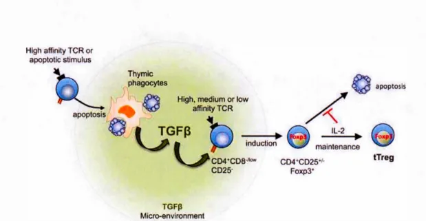
Ouyang et al. reported that TGF-~ safeguards thymocytes from negative selection i.e., pro-apoptotic proteins are upregulated in TGF-~ RH-deficient tTreg cells (Ouyang, W. et al., 2010). Renee, TGF-~ promotes Treg generation in the thymus independently of FoxP3 as well. The anti-apoptotic effects of TGF-~ in thymocytes was demonstrated in two other earlier studies (Chen, W. et al., 2001; Wahl et al., 2000). However, it is still unclear whether this protective effect is only restricted to tT regs, or may also affect developing thymocytes. Konkel et al. reported that apoptosis of developing thymocytes is crucial for tTreg development due to the fact that apoptotic cells and macrophages, which phagocytose apoptotic cells are sources ofTGF-~, which in turn induces FoxP3 expression in Treg precursors (Konkel et al., 2014).
Based on these events, a new model for tTreg development has been proposed,
described as the "apoptosis-TGF-~-FoxP3 axis" (Figure 1.4) (Chen, W. et Konkel,
2015). This model, proposed by Chen et al. summarizes the process of Treg
development as follows: a) Developing thymocytes undergo negative selection, which
results in high numbers of apoptotic thymocytes and maintenance of elevated TGF -~
levels in situ. High concentrations ofTGF-~ favor the development oftTreg cells from
precursors, provided that signais are transmitted through their TCR, and protection
against apoptosis is assured. b) These precursors then initiate and maintain FoxP3
expression. As noted by Chen et al. several questions still remain unanswered. First,
the potential populations of tT reg precursor cells have not been clearly discriminated.
Second, the threshold concentrations ofTGF-~ required for inducing FoxP3 expression
in these precursors are unknown. Third, the mechanisms and signaling pathways by
which TGF -~ stimula tes F oxP3 expression in tT regs require investigation.
Hlgh affinity TCR or apoptotic stimulus Thymie phagocytes Il
0
apoptosis·o Hlgh, mediu~ or low afflnityTCR TGFJl Micro-envlronmentÂ
0
apoptosls IL-2 - - - - . . Ollp maintenance CD4 CD2S·I· Foxp3• tTregFigure 1.4: Role ofTGF-~ in tTreg development, shown here is the apoptosis-TGF-~
1.2.6 Development of peripheral Tregs
1.2.6.1 Physiological context of pT reg generation
The existence of extra-thymically generated suppressor cells in mice was described
since the early 1990's (Taguchi et al., 1994). Not all the Tregs arise from the thymus
since not all Tregs encounter their cognate antigen in the thymus. Tregs which are foreign antigen-specific develop extra-thymically. Differentiation of pTregs occurs
more frequently in mucosal surfaces compared to other tissues (Coombes et al., 2007;
Sun et al., 2007; Yadav et al., 2013), since they serve as sites for direct host-pathogen
interaction, and often there are heightened immune responses, at these sites, which have
to be downregulated. Peripherally induced Tregs play a very crucial role in establishing
immune privilege (tolerance to the presence of antigens, to prevent inflammatory
damage which might have serious consequences), in or gans such as the eyes (Sugita,
2009), and the testis (Li, N. et al., 2012). Another peripheral tolerance mechanism
mediated by Tregs is the maternai tolerance towards patemal allo-antigens expressed
on the fetus, which is crucial for preventing miscarriages (La Rocca et al., 2014).
Development of peripheral antigen-specific Tregs in certain tissues is crucial for preventing immune disorders. For instance, accelerated immune response to gut-resident commensal bacteria could result in the development of inflammatory bowel
disease (Elson et Cong, 2012; Jostins, 2012), and one of the mechanisms that helps in
tolerance towards gut microbiota is the presence of Tregs (Belkaid et Hand, 2014;
be induced by one of the most common intestinal bacterial strains belonging to the genus Clostridium - higher amounts of TGF-~ and a higher number of FoxP3 expressing T cells were detected in the colon of mice colonized with Clostridia (of 46 different strains) compared to wildtype mi ce, suggesting the tole of TGF -~ in promoting FoxP3 induction (Atarashi et al., 2011). Polysaccharide A of Bacillus fragilis, another species belonging to the intestinal microbiome, also mediates
generation of functionally active, IL-l 0 secreting,FoxP3+ pT regs from CD4+ T cells (Round et Mazmanian, 201 0). Y et, another mechanism of pT reg generation occurs through the interaction of CD4+ T cells with specifie APCs. For instance, lung resident macrophages, expressing TGF -~ and retinoic ac id, were shown to induce antigen-specific FoxP3+ Tregs from CD4+ T cells (Soroosh et al., 2013). DCs also play an
indispensable role in pT reg generation- depletion ofDCs leads to lower Treg numbers (Darrasse-Jeze et al., 2009). Certain classes of DCs, such as migratory DCs, which
move from tissues to lymph nodes, carrying self-antigenic peptides from the tissues, are more efficient in generating Tregs compared to tissue resident DCs (Idoyaga et al.,
2013). Plasmacytoid DCs, which are specialized DCs that secrete type-1 interferons are also known to induce FoxP3+ Tregs in the lungs to maintain immune tolerance (Lombardi et al., 2012).
1.2.6.2 Role ofTGF-~ in pTreg generation
TGF-~, along with TCR co-stimulation, triggers the expression of FoxP3 in murine naïve CD4+ CD25- T cells, and hence, mediates their differentiation to Tregs having immune suppressive functions (Chen, W. et al., 2003). TGF-~ is crucial for the induction of FoxP3 in human CD4+ T cells as well (Amamath, Shoba et al., 2007;