Université de Montréal
Molecular and Functional studies of human immunodeficiency virus type 1 accessory protein Vpr
Par YongXiao
Programme de virologie et immunologie Faculté des études supérieures
Thèse présentée à la Faculté des études supérieures en vue de l’obtention du grade de
Philosophie Doctoral (Ph.D) En virologie et immunologie
Dec, 2006 © Yong Xiao, 2006
Université
de Montréal
Direction des bibliothèques
AVIS
L’auteur a autorisé l’Université de Montréal à reproduite et diffuser, en totalité ou en partie, par quelque moyen que ce soit et sur quelque support que ce soit, et exclusivement à des fins non lucratives d’enseignement et de recherche, des copies de ce mémoire ou de cette thèse.
L’auteur et les coauteurs le cas échéant conservent la propriété du droit d’auteur et des droits moraux qui protègent ce document. Ni la thèse ou le mémoire, ni des extraits substantiels de ce document, ne doivent être imprimés ou autrement reproduits sans l’autorisation de l’auteur.
Afin de se conformer à la Loi canadienne sur la protection des renseignements personnels, quelques formulaires secondaires, coordonnées ou signatures intégrées au texte ont pu être enlevés de ce document. Bien que cela ait pu affecter fa pagination, il n’y a aucun contenu manquant. NOTICE
The author of this thesis or dissertation has granted a nonexclusive license aflowing Université de Montréal to reproduce and publish the document, in part or in whole, and in any format, solely for noncommercial educational and research purposes.
The author and co-authors if applicable retain copyright ownership and moral rights in this document. Neither the whole thesis or dissertation, nor substantial extracts from it, may be printed or otherwise reproduced without the author’s permission.
In compliance with the Canadian Ptïvacy Act some supporting forms, contact information or signatures may have been removed from the document. While this may affect the document page court, it does rot represent any loss of
o
Université de Montréal Faculté des études supérieuresCette thèse intitulée
Molecular and Functïoual studies of human immunodeficiency virus type 1 accessory protein Vpr
présenté par Yong Xiao
A été évaluée par un jury composé des personnes suivantes
Dr Guy Lemay: President-rapporteur
Dr Eric A. Cohen: Diretuer de recherché
Dr Ah Ahmad: Member du jury
Dr Benoit Barbeau Examinateur externe
C
ABSTRACTThe 96-amino acid Vpr protein encoded by HW-l performs multiple functions during the retroviral life cycle, including the enhancement of viral replication in macrophages at early stage of viral replication, the induction of G2 celi cycle arrest in proliferating T lymphocytes, and the modulation ofHIY-l-induced apoptosis. In addition, extracellular full-length and processed forms of Vpr have been previously detected in the sera and cerebralspinal fluids of RW-infected patients. However, the mechanism underlying this processing and its implication for HW-l pathogenesis remain unknown.
The first goal of this thesis was to investigate the mechanismof Vpr release and processing during HIV-l infection. Herein, we report that fiill-length and several cleaved species ofVpr could be detected in the culture media of HIV-l expressing ceils, independently of Vpr virion incorporation. Tmncated forms of Vpr were abundant in the extracellular medium from
HIV
producing celis but flot from ceils expressing Vpr alone. Moreover a small portion of cleaved Vpr was found to be associated with the extemal ceil surface of HIV-producing ceils through binding with celi surface heparin suiphate proteoglycans. Mutagenesis and mass spectrometry analyses indicated that Vpr was processed at its C-terminus afterthe highly conserved R85QRR88 motif a putative pair-basic proprotein convertase (PC) cleavage site. Consistently, the PC peptide inhibitor dec-RVKR-cmk and the serine protease inhibitors (Œl-PDX and Spn4A) specifically inhibited extracellular Vpr processing. Transient expression of proprotein convertases PC5A and PACE4 and to a lower extent fiirin increased extracellular Vprprocessing, strongly suggesting that Vpr is processed by proprotein convertases. We provide evidence suggesting that Vpr was processed in the extracellular medium through PCs that are celi surface associated. Finally, the tmncated Vpr protein was defective for the induction of celi cycle arrest and apoptosis, suggesting that Vpr— proteolytic processing might 5e a cellular mechanism to control
the level of functionally active extracellular Vpr during HW- 1 infection.
The second goal of this thesis was to investigate Vpr-interacting proteins within HIV- 1 vii-ion particles and their fiinctional relevance. Vpr early firnctions are closely related to its specific virion incorporation. Vpr localization within the virion core and its association wiffi the pre-integration complex suggest a role for Vpr in the early phases of HIV infection. However, littie is known about Vpr interactions with other viral components in the virion particles and their functional relevance. To address this question, we constructed an infectious molecular clone of
HIV-l expressing HA-tagged Vpr and we isolated purifiedvirions containing HA-Vpr. Analysis of anti-HA co-immunoprecipitated protein complexes by proteomic or western blot approaches revealed that Vpr could form a complex with the matrix protein (MA) within viral particles produced from various human celi lines. Furthermore, the MA-Vpr interaction was shown to occur independently of the presence of RT and IN and could be detected by in vitro GST pulldown experiments using recombinant Vpr and puHfied GST-fused MA proteins. These resuits indicate that the Vpr-MA association involves a direct interaction. The respective interacting domains were mapped by in vitro binding assays. We found that the fifth alpha helix of MA (residues 97-10$), and the arginine-rich C-terminal domain of Vpr (residues $6-96) were implicated in the Vpr-MA interaction. Since Vpr and MA are karyophilic proteins, and are both components of the pre-integration complex (PIC), theirinteraction might have a synergistic effect in the nuclear targeting of PIC and could contribute to the efficiency of viral infection during the early stages of HIV- 1 infection.
it is important to investigate how host and viral factors interact to establish HIV-1 infection in human ceils. Vpr has been shown to contribute to HIV-1 infection in human ceils when it is present as an extracellular species as well as a virion-associated species. Here, we identified a cellular protease that regulates extracellular Vpr activity and characterized Vpr
interacting-proteins within virion particles. The present study might contribute to a better understanding of Vpr early functions during HW- 1 viraI replication and might provide new targets for therapeutic intervention.
Key words: extracellular Vpr; proteolytic processing; proprotein convertase; 11EV-1; virion; matrixprotein; HA-tagged Vpr provirus
C
RÉSUMÉ
La protéine Vpr codée par le rétrovirus VIH-1 est une protéine de 96 acides aminés qui remplit de multiples fonctions au cours du cycle réplicatif du virus, comme l’augmentation de la réplication virale dans les macrophages primaires aux stades précoces de l’infection, l’arrêt du cycle cellulaire en phase G2
dans
les lymphocytes T en division ou encore la régulation de l’apoptose induite par le VIH-1. En plus de la forme sauvage, des formes tronquées de Vpr avaient été précédemment détectées dans le sérum et le fluide cérébro-spinal de patients infectés par le VIH. Toutefois, le mécanisme de ce clivage ainsi que son rôle dans la pathogènèse du VIH1 restent inconnus.
Le but premier de cette étude était de caractériser le mécanisme par lequel Vpr est modifié et relâché dans le milieu extracellulaire au cours d’une infection par le VIH-1. Nous rapportons ici que la protéine sauvage ainsi que plusieurs formes tronquées de Vpr ont pu être détectées dans le milieu de culture des cellules exprimant VIII-1 indépendamment de l’incorporation de Vpr dans les particules virales. Les formes tronquées de Vpr étaient abondantes exclusivement dans le milieu extracellulaire des cellules exprimant VIH-1 et non de celles exprimant Vpr seul. De plus, une faible fraction de Vpr clivée s’associe à la surface extracellulaire de la membrane plasmique, grâce à la présence de protéoglycanes contenant des chaînes d’héparine sulfate, des cellules exprimant le VIH-1. Des études de mutagènèse dirigée et de spectrométrie de masse ont montré que Vpr est clivé à l’extrémité C-terminale, en aval du motif hautement conservé
R85QRR88,
un site de clivage putatif spécifique des proprotéine convertases (PC). Le peptide dec-RVKR-cmk, un inhibiteurpeptidique des protéine convertases, ainsi que les inhibiteurs des sérine protéases Œ1-PDX et Spn4A inhibent spécifiquement le clivage extracellulaire de Vpr. L’expression transitoire des proprotéines convertases PC5A etPACE4 et dans une moindre mesure de la furine favorise le clivage extracellulaire de Vpr, suggérant ainsi fortement que Vpr est clivé par les proprotéines convertases. Nous avons apporté la preuve que Vpr était clivé dans le milieu extracellulaire par des proprotéines convertases associées à la surface cellulaire. Enfin, les formes tronquées de Vpr se sont révélées déficientes pour l’arrêt du cycle cellulaire et l’induction de l’apoptose, suggérant ainsi que le clivage protéolytique de Vpr peut être un mécanisme cellulaire destiné à réguler le niveau des formes
extracellulaires fonctionnelles de Vpr au cours d’une infection par le VIH-1.
Le second objectif de cette thèse est de caractériser les protéines interagissant avec Vpr au sein des particules virales ainsi que leur fonctionnalité. Le rôle fonctionnel de Vpr est étroitement relié à son incorporation spécifique au sein des particules virales. Sa localisation dans le core viral et son association au complexe de préintégration (PIC) suggèrent unrôle de Vpr dans les étapes précoces de l’infection par le VIH. Toutefois, peu de données sont disponibles sur le mécanisme d’interaction de Vpr avec d’ autres partenaires viraux au sein des particules virales ainsi que sur la signification fonctionnelle de telles interactions. Afin de résoudre ces questions, nous avons construit un clone moléculaire infectieux de VIH- I exprimant Vpr en fusion avec le peptide HA (HA-Vpr) et avons purifié les virions contenant la protéine HA-Vpr. L’analyse par protéomique ou Western blot des complexes protéiques co-immunoprécipités grâce à l’anticorps anti-HA a permis de révéler que Vpr formait un complexe avec la protéine de matrice (MA) au sein de particules virales produites à partir de différentes lignées cellulaires humaines. De plus, l’interaction Vpr-MA est indépendante de la présence de la transcriptase inverse (RT) et de l’intégrase (IN) et a pu être détectée par des expériences de GST pulldownin vitro à l’aide de Vpr recombinant et de la protéine GST-MA purifiée. Ces résultats indiquent que Vpr et MA s’associent grâce à une interaction directe. Grâce à des expériences de liaison peptidique invitro, les domaines responsables de l’interaction entre MA et Vpr ont été cartographiés,respectivement, au niveau de la 5 hélice LI (résidus 97 à 10$) et du domaine C-terminal riche en arginines. Étant donné les propriétés karyophiles de MA et de Vpr et qu’elles sont toutes deux membres du
PIC, leur interaction pourrait avoir un effet synergique dans le transport nucléaire du PIC et ainsi optimiser l’efficacité des étapes précoces d’une infection parle VIH- I.
L’ étude de l’interaction des facteurs cellulaires et viraux impliqués dans l’infection des cellules humaines par le VIH-1 est d’une importance majeure. Dans ce contexte, iI a été démontré que la protéine Vpr contribue à l’infection virale lorsque présente sous sa forme extracellulaire ainsi que lorsque présente à l’intérieur du virion. Dans la présente étude, nous avons identifié une protéase cellulaire qui contrôle l’activité extracellulaire Vpr et avons caractérisé des protéines interagissant avec Vpr à l’intérieur des virions. Ces données pourraient contribuer à une meilleure compréhension des fonctionsprécoces de la protéine Vpr dans le cycle de réplication viral et pourraient ainsi mener à l’identification de nouvelles cibles thérapeutiques.
Mots-clés : Vpr extracellulaire; clivage protéolytique, proprotéine convertase; VIH- 1; virion; protéine de matrice; provims; HA-Vpr.
DEDICATION
ACKNOWLEDGEMENTS
I would like to express my sincerest gratitude and appreciation to my supervisor Dr. Fric A.Cohen for lis excellent guidance and support throughout my graduate studies, especially for teaching me how to be vigorous with my experiments, to be a critical thinker, and to be persistent for the sake of producing beller science and knowledge of the field. I would like to thank Dr. Ghisiaine Duisit andXiaoj jan Yao for taking the time to help me and to answer my questions and for their advice and encouragement when experiments did notaiways go the way that we predicted.
I want to thank Dr. Nabil $eidah for the valuable discussions and for providing ail the PCs
expressor plasmids. I also would iike to thank my committee—Dr. Guy Lemay, Ah Ahmad, and Benoit Barbeau—for taking such an active role in the progressionand completion ofmy thesis.
I thank the Cohen lab members, in particular, Dr. GhislaineDuisit, Jean Phillippe Belize, and Alex Caillaud for their critical review of the manuscript; I thank Nicole Rougeau for her excellent technical support for the Vpr project during the late stage of my PhD work; I thank Nicole Rougeau, Johanne Mercier, andMelanie Welman for their friendship and support; I thank Drs. Jacques Thibodeau, Alexandre Brunet, and Andres Finzi for their collaboration on the MHC II molecuie effect on the HIV-l assembly, and Dr. Andrew Mouland for his collaboration on the hnRNP A2 project.
Finally, I would like to thank Dr. Hayami Masanori and Shida Hisatoshi for introducing me to this field. I also wouhd like thank my family for beingextremely supportive and encouraging me in everything I do; without my wife Xiuying, I wouldflot have made it to the end, and I cannot begin to thank for her for ail ber encouragement and confidence in me, for her love and support—
PREFACE
This Ph.D. thesis was written in accordance with the Guild Concerning Thesïs Preparation from the Faculté des etudes supérieures at Université deMontréal. The structure and contents of the thesis conforms to the option, subject to the approval of the department, of including, as part of the thesis, copies of the text of paper(s) submifted for publication, or the clearly-duplicated text of published paper(s). The thesis includes, as separate chapters or sections: (1) a table of contents, (2) a general abstract in English and French, (3) an introduction which clearly states the rationale and objectives of the study, (4) a comprehensivegeneral review of the background literature of the subject of the study, when the review is appropriate, and (5) a final overail conclusion and /or summary.
I have included, as a chapter of this thesis, two original manuscripts that have been submitted for publication. The papers presented in the thesis arethe following:
1) Yong Xiao, Gang Chen, Nicole Rougeau, Hongshan Li, Nabil G. Seidah,
Éric
A. CohenCeli-surface Processing of Extracellular Human Immunodeficiency Virus Type 1 Vpr by Proprotein Convertases. J Virology. Submitted Mar, 2007.
2) Yong Xiao, Piene-Alexandre Bonicard, Xiao-Jian Yao, Ghislaine duisit and Éric A. Cohen. Human Retrovirology Unit, IRCM
Direct Association of HIV-1 Vpr and Matrix (MA) Proteins in Virions: Implication for Early Events ofHIV-1 Infection. Manuscript will be submitted to Retrovirology.
TABLES 0F CONTENTS ABSTRACT ffi RÉSUMÉ w DEDICATION lx ACKNOWLEDGEMENTS X PREFACE XI
TABLES 0F CONTENTS XII
LIST 0F FIGURES XV
LIST 0F TABLES XVII
LIST 0FABBREWAllONS XVIII
CHAPTER 1: LITERATIJRE REVIEW 1
1.1. THE ACQUIRED IMMUNE DEFICIENCY SYNDROME 2
1.1.1. Discovery of111V-1virus 2
1.1.2. Emergence ofHIV-1 disease 3
1.1.3. fflV/AIDS global pandemic: current status 4
1.2.111VVIRAL STRUCTURE AND GENOMIC ORGANIZATION 5
1.2.1. fflV-1 genomic RNA 5
1.2.2. The viral proteins 6
1.2.3. 111V virion structure 10
1.3. fflV-1 LIFECYCLE 11
1.3.2. Uncoting .14
1.3.3. Reverstrscription .15
1.3.4. Nuclear transiocation of the PIC 17
1.3.5. lutegration 1$
1.3.6. HIV-1 gene expression 19
1.3.7. Assembly and release of 111V-1 21
1.4. BI0LOGICAL FUNCTIONS 0F VPR 23
1.4.1. Structure ofthe protein 23
1.4.2. Subcellular localization 27
1.4.3. Viral incorporation ofVpr 2$
1.4.4. Effects ofVpr on 111V-1 replication and pathogenesis in vivo 28
1.4.5. Biological functions of the 111V-1 Vpr protein 30
1.4.6. Vpr induced G2 arrest 33
1.5. PR0PR0TEIN CONVERTASE ANDfflV-1INFECTION 35 1.5.1. Tissue distribution and subcellular Iocalization of proprotein convertases 35
1.5.2. PC expression in T lymphocytes celis 38
1.5.3. Proprotein convertase Inhibitors 39
1.5.4. 111V-1 env and tat are processed by PCs 40
1.6. VPR AND111V-1CYTOTOXICITY 42
1.6.1. IIIV-1-induced cytopathic effect 42
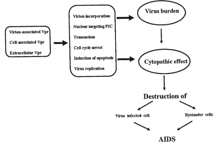
1.6.3. Extracellular Vpr and its biological funcon .45
1.7. VPR MODULATION 0F 111E NUCLEARTR4NSLOCATION 0F fflV-1 PIC 48
1.7.1. Viral components involved in PIC nuclear import 48
1.7.2. Interactions of Vpr with other components of the virus particle 54
1.8. THE OVERALL OBJECTIVE 0F THE STUDY 56
Objective #1: Characterization of the release of extracellular Vpr 57
Objective #2: Determination of Vpr partners within the core 58
Authors contribution in article 1 60
Chapter 2 (article 1): CelI-surface Processing of ExtracellularHuman Immunodeficiency
Virus Type 1 Vpr by Proprotein Convertase 61
Introduction 63
Materials & methods 65
Resuits 70
Discussion 78
References $4
Authors contribution in article 2 106
CIIAPTER 3 (Article 2): Direct Association offflV-1 Vprand Matrix (MA) Proteins in Virions: Implication for Early Events of 111V-1 Infection 107
Introduction 109
Materials & methods 110
Resuits 114
Discussion 118
CHAPTE R 4: GENERAL DISCUSSION. 134
CHAPTER 5: CONCLUSIONS Error! Bookrnark flot defined.
REFERENCES 150
APPENDIX 1 200
Publication I: Andrés fiuzil,3, Alexandre Br,,uet2,3+, Youg Xicto 1,3+,Jacques Thibodeau2,3
findÉric A. (‘ohenl,3.MHC-II molectiles euh once HIV-1 ctsseniblv and budding to lote endoso,;ial/[VItt!tivesiculctr bodies compartments. J Virol. 2006 Oct;80(19):9 789-97 200
Pttblication II: Beriatt!t V, (7enzeitt If, Levesqite K, Lebel C, Xiao Y, Chabot B, Cohen EA,
(ochraue A W, Rigby Wf, MoutandAJ. A lote rote for the association ofhnRNP A2 with the
HIV-1 hnRNP A2 response elemeuts ingenomic RNA, Gag, and Vpr localization. J Bio! Chem.
2004 Oct 15;2 79 (42) :44141-53 200
LIST 0f FIGURES
Page CHAPTER 1
Figure 1.1. Organization of MIV genome 6
Figure 1.2. HIV virion structure 11
Figure 1.3. Schematic representation ofHIV-1 replication cycle 14
Figure 1.4. Schernatic representation ofHIV-1 LTR 20
Figure 1.5. The prirnaty Vpr arnino acid sequence 23
Figure 1.6. 3D sttucture of Vpr protein 25
Figure 1.8. Schematic representation of PIC nuclear targeting during the earlystage of FIIV
infection 48
Figure 1.9. HIV-l MA protein domains 50
CHAPTER 2.
Figure 2.1. 111V-I Vpr and cleaved products are found in the extracellutarmedium ofHW-Ï
producing ceils 96
Figure 2.2. Vpr and C-terminalcleaved products are detected in theextracellular medium of
HTV-l infected Jurkat T ceils 97
Figure 2.3. Vpris cleavedat PC processing site located withinthe arginine-ricli C-terminal
domain 99
Figure 2.4. Proprotein convertases mediate HW-l Vpr processing 101
Figure2.5. Vpr is processed extracellularly by a PC that is ceil surface-associated 102
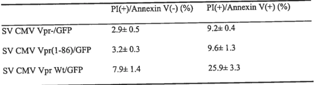
Figure 2.6. Effect of processing on Vpr-mediated ceil-cycle arrest 103
Supplemeutal Figure S2.1. Anatysis ofFllV-I Env gpl6O processing in presence ofPCs
inhibitors Œl-PDX and Spn4A 104
Supplemental Figure S2.2. Extracellular Vpr is associated with heparansulfate
proteoglycans 105
CHAPTER 3:
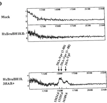
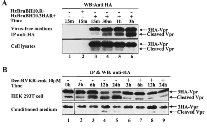
Figure 3.1. Construction of isogenic provirus expressing HA-tagged Vpr 126
Figure 3.2. Detection ofVpr interacting proteinswithin virion particles 127
Figure 3.4. The 5th helix of MA protein is involved in the interaction with Vpr 130
Figure 3.5. Effect of Matrix mutants on Vpr interaction 131
Figure 3.6. Direct interaction of Vpr with MA in GST puIl-down assays 132
Figure 3.7. Effect of Vprtruncationon the MA interaction 133
LIST 0f TkBLES
CHÀPTER 2
o
LIST 0F ARBREVIATIONS
ADCC : antibody-dependent cellular cytoxicity AIDS :acquired immunodeficiency syndrome ALSV :Avian Leukosis Sarcoma Virus
APOBEC3G :apolipoprotein B mRNA-editing enzyme catalytic poypeptide-1ike 3G ARV :AIDS-associated retrovims
AT :ataxia telangiectasia
ATR :Ataxia-Telangiectasia mutated and Rad3-reated
BFA :brefeldin A
BLV :Bovine Leukemia Virus
CA :capsid protein
CAEV :Caprine Arthritis-Encephalituis Virus CAP :CBP-associated factor
CBP :CREB-binding protein
Cdk9 :cyclin-dependent kinase 9
CMV :cytomegalovims
CPE :cytopathic effect
CRD :cysteine-rich domain
CREB :cAMP response element-binding protein CRM1 :chromosomal region maintenance Ï
CsA :cyclosporin A
CTL :Cytotoxic T-lymphocyte.
CyPs :cyclophilins
()
EJAV :Equine Infectious Anemia VirusER :endoplasmic reticulum
ESCRT :endosomal sorting complex required for transport FGf :fibroblast growth factor
FW :Feline Immunodeficiency Virus Env :envelope glycoprotein
Gag :group-specific antigen GfP :green fluorescent protein GR :glucocorticoid receptor
HA :hemagglutinin
HIV-1 :Human Irnmunodeficiency Virus-1 HSPG :heparan sulfate proteoglycans
HTLV :Human T-cell Leukaemia Virus
IN ;integrase
ICAM :intracellular adhension molecule JNK :c-Jun NH(2)-terminal kinase
LAS :lymphadenopathy syndrome
LAV :Lymphadenopathy-Associated Virus LTNP :long term non-progressor
LTR :long terminal repeat
MA :matrix protein
MHC :major histocompatibility complex MVBs :multivesicular bodies
NC :nucleocapsid protein
Nef :negative factor
NF-icB :nuclear factor-Kappa B
NK :natural killer celi
NLS :nuclear Iocalization signal
NPC :nuclear pore complexes
Nt :nucleotide
PACE4 :basic amino-acid-cleaving enzyme 4 PAK-2 :p2-activated protein kinase 2 PBL :peripheral blood lymphocytes
PC :proprotein convertases
PCD :programmed ceil death
PI :propidium iodine
PIC :pre-integration complex
PHA :phytohemagglutinin
Rev :regulator of viral protein expression
RSL :reactive site loop
RT :reverse transcriptase
RTC :reverse transcription complex PTD :protein transduction domains
RRE :Rev responsive element
SELDI-TOF-MS :surface-enhanced laser desorption]ionization time-of-fliglit mass spectrometry Serpin :serine protease inhibitor
$1V :Simian Immunodeficiency Virus TAR :transactivation response element Tat :Transactivator of transcription
TCR :T ceil receptor
TSGÏO1 :human tumor susceptibility gene 101 UNMDS ;United Nation program on HIV/MD$
VMV :Visna-Meadi Virus
Vpr :viral protein R
Vpu :viral protein U
C
CHAPTER 1: LITERATUREThe Retroviridae famlly bas 7 subfamily, composed of the foïlowing virus genera: C-type,B-type, D-type, HTLV-BLV, Spumavirus, Avian Leukosis $arcoma (ALSV), and Lentivirus. Lentiviruses are further broken down into tbree categories: human primate lentivims, nonhuman primate lentivirus, and non-primate lentivinis. Human primate lentivinises consist of the HumanImmunodeficiency Virus-1 and the Human Imrnunodeflciency Virus-2 (HIV- 1 and HIV-2). The former is highlypathogenic and the later demonstrates a similar but less pathogenic etiology. Non-human primatelentiviruses form several distinct lineages named the Simian Immunodeficiency Virus (SIV). They are endemic to their hosts and do not usually cause immune suppression as seen in humans. Simian lmmunodeficiency Viruses are distinct primate groups that include strains from mandrills (SIVmnd), mangabees (SIVrcp and SIVsm), l’hoest ($1V l’hoest), and sabaeus (SIVsab) monkeys among others (282). The non-primate lentiviruses include Visna-Maedi Virus (VMV), Caprine Arthritis-Encephalituis Virus (CAEV), Equine Infectious Anemia Virus (EIAV), Bovine Immunodeficiency Virus (BIV), and FelineImmunodeficiency Virus (FIV) (196).
Like other retroviruses, HIV-l produces Gag, Pol, and Env proteins. HIV-1 produces six additional proteins: Tat, Rev, Nef, Vif, Vpr, and Vpu. While Tatand Rev are required for viral replication, Nef, Vif, Vpr, and Vpu usually are dispensable for viral growth inmany in vitro systems (112) and hence
are known as accessoiy proteins. However, these proteins are often necessary for viral replication and pathogenesis in vivo, and they carry out many essential fiinctions during the viral life cycle. Consequently, the presence or absence ofthese accessory proteins can significantly change the course and severity ofthe viral infection (112).
Viral protein R (Vpr) is a small, highly conserved accessory protein that serves many functions in HIV-1 life cycle. These functions include cytoplasmic-nuclear shuttling (146), induction ofthe ceil cycle G2 arrest (144)fland celi killing (337). These three Vpr-specific activities are shown to be ftinctionally
independent of each other (95, 252) and have been demonstrated in a wide variety of eukaryotic celis ranging from human to yeast, indicating that Vpr most likely affects highly conservedcellular processes.
in this review, I descHbe the background of the HIV virus, the current understanding of Vpr firnctions in HIV- 1 viral infection, and the potential roles of Vpr activities in viral pathogenesis and disease progression.
Li. Tire acquired immune deflciency syndrome 1.11. Discovery of 11W-1 virus
In the early 1980s, physicïans in United States noticed an increasing number of young male homosexuals with a range of opportunistic infections and malignancieswhich had not been seen before in this age group (133, 156, 331). The most frequently observed symptoms were pneumocystis carinii pneumonia, oesophageal candidiasis toxoplasmosis of the brain, Kaposi sarcoma, and non-Hodgkins lymphoma, as well as other unusual complications. In 1981, Gottlieb et al. described these patients as anergic, lymphopenic, later more appropriately renamed acquired immunodeficiency syndrome (AIDS). At first, physicians thought that the origin of these diseases was a possible viral infection from the cytomegalovirus (356). Based on the epidemiological observations, it was proposed that MD$ is caused by an infectious agent transmifted by sex, contaminated blood products, and from mothers to chiidren (277).
When it became apparent that the disease is transmissible, a wide range of known microorganisms was suggested as the agent. The french group from the Pasteur Institute in Paris first reported the isolation of a new human virus as the causative agent for AIDS, which was named LAV for the lymphadenopathy-associated virus. This virus had distinct properties from HTLV-1 because it does flot establish a transformed state in CD4+ T helper ceils but caused ceil death afier high level of replication (23). The same French team made the most significant observation that the virus is cytopathic to the helper 14 (CD4) ce!!, providing for the first time, an explanation ofhow and why AIDS develops (184).
Several other research groups also were searching for the virus that might be responsible for MDS. Afier this description of LAV by Luc Montagnier’ s laboratory, two other groups reported the isolation of retroviruses from AIDS patients. In a series of papers published in 1984, Gallo and colleagues later reported the discovery of an MDS-associated retrovinis, which appears distinct from HTLV, named HTLV-III (121, 138). Levy and a coworker isolated a retrovirus from a San Francisco subject and named it AIDS-associated retrovirus (ARV). The discovery of ARV in asymptomatic individuals indicated, for the first time, a carrier state for the AIDSvirus(201).
Subsequent research established that the different AIDS virus nucleic acid sequences are extremely polymorphic, making it feasible unequivocally to identify the precise origin of different HIV isolates. This first revealed that HTLV-III is in fact LAV because of its unique identity (280). Actually, the virus had been supplied to Dr Robert GaHo’s lab at various times by the French team (233).
The genetic organization and the proteins encoded by HIV-1 were shown to be distinct from those of HTLV; these retroviruses were recognized as members of the same group of retrovinises-Lentivirinae (280). In 1986, the International Committee on the Nomenclature of Virus renamed the AIDS virus the Human Immunodeficiency Virus (HIV) (61). Shortly afier the identification of HJV, another human retrovirus was recovered from a West Africa patient with AIDS (59). It was noted to be sufficiently different genetically from HIV-l (by up to 40%) and was named HIV-2.
1.1.2. Emergence of 111V-1 dïsease
The origin of HIV-1 has been linked to a virus found in chimpanzees (SIVcpz), originating in the species Pan troglodyte troglodytes andpan troglodytes schweinfurthii from Central and Western Africa (123). Previous data from the earliest documented case of AIDS in 1959 places HIV-l within this geographic area, and phylogenetically proximal to the sequences of ancestral origin (412). The mode of transfer of the SWcpz virus to humans is a continuous debate, but recently theories have been disproved, which suggested that SIV was transfened either by contaminated polio vaccines given to tens of thousands of African workers by French colonialists in the early 1930s, or by reusing needles for
vaccination (387). Direct human contact with primate bush meat during slaughter is stili a possibility (139). The appearance of the less pathogenic HIV-2 strain in humans isthought to be caused by multiple transfers of 51V from sooty mangabees, since the sequence similaritiesbetween these three viruses group appears to be inseparable by phylogenetic analysis (124).
1.1.3. HW/AIDS global pandemic: current status
It is well accepted that MDS began in Africa (172). Not only is the disease widely spread in sub Saharan Africa but also in Africa, monkey species are naturally infectedwith lentiviruses related to HIV, as first shown by the Dr. Hayami research team (255). No evidence can be shown for the existence of HIV in Europe, America, or Arabia during the past century or even the first haif of twentieth century, strongly suggesting that the widespread HIV infection in Africa is a recent event. The epidemiological evidence thus points to the spread of HIV infection from Africa afier the Second World War. The proliferation seems to coincide with the widespread use of syringes and needles from the West, at the time vaccination programs were being promoted and introduced. It also coincides with a post-war period of
greatly improved transportation and the extensive migration ofAfricanpeople.
Since the first diagnosis was made in the early 19$Os, AIDS has spread through the world, affecting ail cultures and ethnic groups. At the end of 1984, the number ofAIDS cases in the United States was 7,699 (with 3,665 deaths resulting from AIDS) and in the UnitedKingdom, 764 cases were reported (50). According to the 2005 AID$ epidemic update report from the Joint United Nations Program on HIV/AID$ (UNAIDS) and the World Health Organization (WHO),the estimated number of people living with AIDS in North America in 2005 is between 650,000 and 1.8 million, and the estimated world population ofpeople living with AIDS is approximately 40.3 million, with 3.1 million death due to HIV 1/AID$. Seventy percent of alI individuals with HIV are living in sub-Saharan Africa. Despite education, testing, and awareness of the disease, the number of newly acquired cases bas increased. In the United States, the spread of HIV bas disproportionately increased in minorities where AIDS is the leading cause of death among African American women between the ages of25 to 34. High-risk behaviors, such as
heterosexual ami homosexual unprotected sex and IV drug use, account for the increased numbers. The spread of HP! has been most problematic in underdeveloped countries where both treatment and education have flot been logistically and financially achievable for these govemments. Organizations such as UNAIDS ami the WHO have implemented programs involving the support ofmany Western countries to deliver affordable and simplified treatments.
1.2. HW viral structure and genornic organization 1.2.1. 111V-1 genomic RNA.
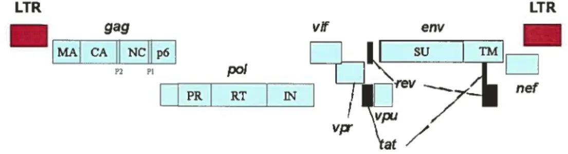
In general, two copies of viral genomic RNA are incorporated into virionparticles. The genomic size ofHIV is about 9.8 kb with open reading frames coding for at least nine viral proteins (see figure 1.1).
Like the oncoretroviruses, HIV-l genomic RNA contains cis-acting elements required for the reverse-transcription in double-stranded DNA, the provirus integration into the host ceil genome, the transcription of the different viral RNAs species, and the incorporation of newly synthesized genomic RNAs into nascent particles. HP! expression is flanked by long terminal repeats (LTR). The 5’LTR binds specific and general transcription factors that regulate the initiation of mRNA synthesis, whereas the 3’LTR signais the point at which mRNA synthesis should end, with the addition of a poly A tail. The LTR sequence has three functionally distinct domains (U3, R, and U5) and contains transcriptional promoter elements that regulate basal and inducible transcription firnctions. U3—the first promoter region—contains a moduiatory enhancer region, which is the core promoter region for regulating transcription. The core promoter region is where RNA polymerase II and TATA-box binding proteins form the multiprotein complex that is responsible for initiating transcription and for providing the binding sites for these proteins, as well as three sites for Spi (344). The upstream modulatory enhancer region binds Nf-icB and the NF-AT transcription factor, and has binding sites for various cellular proteins thought to be important for specificity of replication in ceil types such as macrophages (299). These transcription factors include NF-IL-6, the cAMP response element-bindingprotein (CREB), and nuclear hormone receptors (287). In contrast to the oncoretroviruses, HIV-I rnRNA contains a 5’UTR eÏement
called transactivation response element (TAR), that prevents RNA polymerase II processing in absence of the viral Tat protein (see below) (170). Located between the PJU5 regions in 3’LTR is the site for retroviral 3’ end processing and polyadenylation signais. U5—the last prornoter region—when located at the 5’ end, contains a GT-rich exon that ail viral transcripts contain, and encodes putative control elements for 3’ end processing when it is located at the 3’ LTR. Celluiar machinery caps the 5’ end ofthe viral transcript by the addition of methylated G nucieotide, it involves condensation of the trphosphate group of molecule of GT with a diphosphate left at the 5’ end of initial transcript. The 5’ end cap is necessary for the proper function of mRNAs in protein synthesis; it also seems to protect the growing RNA transcriptfrom degradation.
LTR LTR
_____
gag MA CA NCHp6 P2 PI p0! PR RTE
Figure 1.1. Organization of HIV-1 genome. The relative location ofthe HIV-1 open readingfraine gag, p01, env, vf vpr, pu, nef tat, and rev are indicated.
1.2.2. The viral proteins.
Like other retroviruses, HIV encodes for Gag, Pol, and Env virion structural and enzymatic proteins. In addition, the lentivirus encodes for two regulatory proteins, Tat and Rev, important for HIV-1 protein expression, and four accessory proteins Vif, Vpr, Vpu, and Nef dispensable for productive infection of transformed T lymphocytes, but oflen necessary for viral replication and pathogenesis in vivo. l-11V-1 Tat, Rev and Nef proteins are expressed early during infection from fuiiy spiiced messenger RNA. In contrast, Gag-Pol polyproteins, as weii as Env, Vpr, Vpu, and Vif are expressed inthe late phase of infection from intron-containing viral mRNAs.
HIV- I Gag precursor p55 gives rise, by proteolytic cleavage, to the smaller proteins, including the capsid protein CA (p24), matrix protein MA (p17), the nucleocapsid protein NC (p9), and the p6 protein (147). Polis initially made as a Gag-Pol polyprotein A ribosomal frame shift of—1 in gag allows a ribosomal reading through of gag and the production ofPro-Pol at the ratio of 1:20 within the celi (257). The Pol precursor protein is cleaved into products consisting of the reverse transcriptase (RI), protease
(PR), and integrase (IN) proteins, which are essential for viral replication (357).
Env gene encodes a glycoprotein precursor gpl6O. This precursor is cleaved in the tran-Golgi network (TGN) by furin-like convertase into a gpl2O extemal surface (SU) envelope protein and gp4l transmembrane (1M) protein, which constitute the membrane glycoprotein of the virus (140).
Tat (transactivator of transcription) is a small fransactivating protein (101 amino acids in most clinical HIV-1 isolates, $6 amino acids in the laboratory HIV-1 HXB2 strain). Tat, along with other cellular proteins, interacts with an RNA loop structure formed in the 3’ portion ofthe viral long terminal repeat (LTR) called TAR. Tat potently transactivates LTR-driven transcription, which resuits in a remarkable increase of viral gene expression ($4).
Rev (regulator of viral protein expression) is a 116 amino acid sequence-specific RNA binding phosphoprotein that is expressed during the early stages of HIV-1 replication (216). The protein is necessary for the expression of intron-containing RNAs. In its absence, only the fully spliced class of HIV-1 mRNAs is present in the cytoplasm, while intron-containing RNAs remain nuclear (103). Rev multimers interact with a cis-acting RNA ioop structure called the Rev responsive elements (RRE), located within the Env gene (301). This interaction permits the nuclear export ofunspliced or partially spliced RNAs (14). The transition from multiple spliced mRNA to unspliced mRNAinto the cytoplasm is the marker between early and late stages ofthe viral replication cycle (388). The export ofRev and single spliced transcript to the cytoplasm is mediated by the cellular transport receptor, CRMI (Chromosomal region maintenance 1), where the recycling of Rev to the nucleus is mediated by importin ci (2, 116).
Vif (viral infectivity factor) gene encodes a 23-kDa protein. Vif is essential for the reproduction of HIV-1 in peripheral blood lymphocytes, macrophages, and certain ceil unes (340). Vif suppresses the
antiviral activity of the cellular protein APOBEC 3G found in T cells (143, 323). APOBEC3G (apolipoprotein B mRNA editing enzyme catalytic polypeptide-like 3G) is a member of the cytidine deaminase family, which prevents viral cDNA synthesis through deaminating deoxycytidines (dC) in the minus-strand retroviral cDNA replication intermediate (143, 401). Vif binds directly to APOBEC3G and counteracts its anti-HIV activity by promoting its degradation. Vif-mediated APOBEC3G degradation involves the recmitment of a specific E3 ligase complex, which Jeads to polyubiquitylation and protease mediated degradation (324, 395).
Vpu (viral protein U) is an accessory protein found only in HW-1 and SWcpz strains, and no analogous proteins are present in HIV-2 and other SWs (64, 222). Vpu is a small (9 kDa) membrane protein that enhances the release of progeny virion from infected celis and inducesthe degradation of the CD4 protein (34). Vpu expressed in the ER interacts with a membrane-proximal domain of the cytoplasmic tau of CD4 and links it to h-I3TrCP (221), a member of the F-box protein family first
characterized as components of the ubiquitin-ligase complex (183). The CD4-Vpu-3TrCP ternary complex then recruits SKP 1, another member of the ubiquitinated machinery (381). CD4 is ubiquitinated and targeted to proteasomes for degradation after recruiting SKPI. Initially, the ability of Vpu to increase viral release from infected celis had been attributed to ion conductive membrane pore formation characteristic to celis over-expressing Vpu (34). However, a recent reportshows that the requirement for Vpu is host ceil-dependent, suggesting that Vpu may counteract an inhibitory factor expressed in some, but flot in other ceils (363). TASK-l, a widely expressed acid-sensitive K+ channel, is structurally homologous to Vpu, suggesting oligomerization as a possible mechanism of inactivation of the ion channel activity of these proteins (154). However, the mechanism by which TASK-1 inhibits virion release is still unclear.
Vpr is a 96-amino-acid, 14-kDa protein which is expressed at the late stage ofviral replication and is virion incorporated (62). Vpr is much conserved among the primate lentiviruses HIV-l, HIV-2, and the Simian Immunodeficiency Virus, suggesting that it may play an important role in the viral life cycle in vivo. AÏthough Vpr is dispensable in vitro viral replication in T ceil unes, it plays an important role in
macrophages infection and HIV- 1 pathogenesisin vivo (65, 131). Details regarding the Vpr protein amiits biological function are discussed below.
Nef protein is a 27 kDa myristoylated protein that is abundantly produced during the early phase of viral infection. It is highly conserved in ail primate lentivimses, suggesting that its function is essential for the survival of these pathogens. 51V with Nef mutations quickly reverted to a wildtype Nef in infected monkeys or did not progress to AIDS-like symptoms (175). This suggests that Nef is essential for viral pathogenesis in vivo. The role of Nef in HIV-1 replication and disease pathogenesis is determined by at least four independent activities of this protein. First, Nef down regulated the ceil surface CD4 (125) and the major histocompatibility complex class I (MHC-I) protein (313). Nef-induced CD4 down-regulation bas been shown to be the result of rapid intemalization ami degradation of ifie CD4 receptor (125, 295). Down-regulation of MHC I protects HIV-1 infected ceils from host CTL response, where as down regulation of CD4 probably limits the adhesion of an expressing T ceil to the antigen-presenting celiand prevents the interaction between CD4 and the envelope of a newly produced virion. Second, Nef
expression interferes with the cellular signal transduction pathway. Nef myristoylation and its proline-rich SH3-binding domain mediate Nef association with lipid rafi, the cholesterol-rich membrane microdomains that concentrate potent signaling mediators (373). Nef was found to complex with and activate the serine/threonine protein kinase PAK-2 (p21-activated protein kinase 2) (283), which may contribute to the activation of infected celis. Third, Nef enhances virion infectivity and viral replication. This effect is mediated by the presence of Nef in the HIV-l virion and is due, at least in part, to the ability of Nef to induce actin remodeling and to facilitate the movement of the viral core to pass the potentially obstructive cortical actin barrier (48). fourth, Nef regulates cholesterol trafficking in HIV-infected ceils. Cholesterol plays an important role in the HIV life cycle, since HIV assembly and budding—as well as the infection of targeted ceÏÏs—depends on plasma membrane cholesterol. Nef has been shown to bind cholesterol via a cholesterol-recognition motif at its carboxy-terminus andtotransportnewly synthesized cholesterol to the site of viral budding (409).
C
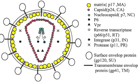
1.2.3. 111V virion structureThe typical mature HIV-1 vii-ion is about lOOnm in diameter. It is enveloped by alipid bilayer that is derived from the membrane of a host celi. The exposed surface glycoprotein SU, (gpl2O) anchors to the virus via interaction with the fransmembrane TM protein (gp4l). The lipid bilayer also contains several cellular membrane proteins from the host celi, including the major histocompatibility complex antigen, actin, and ubiquitin (15); integral membrane-spanning intracellular adhesion molecules (ICAMs) such as ICAM-1 is also incorporated witbin the envelopes of HIV-1 virion and can dramatically enhances infectivity ofHIV-1 virion (349). A matrix sheil comprising the matrix protein (MA, p17) limes the inner surface of viral membrane, and a conica capsid core particle comprising the capsid protein (CA, p24) is located in the center of the virus.
The capsid particle encapsidates two copies of the unspliced dimerized viral RNA genome, which is stabilized as a ribonucleoprotein complex with 2000 copies of the nucleoprotein (NC, p7) and also contains three essential virally encoded enzymes—PR, RT, [N (see Figurel.2). MA has been found in HIV-1 core preparations (187, 380). It is believed that phosphorylated MA locates in the viral core by interacting with integrase (119, 120).
Virus particles also package the accessory protein Vpr via its interaction with p6 in the Gag p55 precursor (186, 266) Vpr is localized within the 111V-1 viral coi-e (1, 380). Mulleret al. have reported that HIV-l virus particles contain a lesser amount of Vpr in comparison to Gag (7:1 ratio of Gag to Vpr), i.e.
Q
matrix( p17,MA)
Capsid(p24, CA) o Nucleocapsid( p7, NC) • P6 À Vpr Reverse transcriptase (p66/p51, RI) • Integrase (p31, IN) X Protease (pli, PR)O
Surface envelop protein(gpl2O, SU)
Iransmembrane envelope protein (gp4 1, 1M)
Figure 1.2. HIV-1 virion structure. Positions of major viral proteins, the Ïtpid bilayer, and the genomic RIVA are indicated.
Recent data shows the association ofNefwith the I-11V-1 core (187). Vif, another accessory HIV-1 protein, was also found to be associated with the HIV-1 core structure (207). However, whether Vif is a genuine virion component or a contaminant remains controversial because other researchers have reported that Vif is essentially absent from the highly purifled HIV-1 particles (81). Three additional accessory proteins Rev, Tat, and Vpu that function in the host ce!! do not appear to be packaged (353).
1.3. HIV-1 Lfe cycle
HIV rep!ication cycle c!ose!y resembles that of other retroviruses (f igure 1 .3). However there are anumber of unique aspects of HIV replication such as HIV target receptor and coreceptors distinct from other retroviruses. Lentivirus like HIV encodes a number of regu!atoiy and accessoty proteins not encoded by the genome of the prototypical simple retroviruses. Lentivirus like HIV has the ability to
productively infect certain type of nondividing ceils. The basic HW-1 replication process wiii be introduced in the following.
1.3.1. Virus entry
Infected host ceits. HP’!- 1 infects CD4+ T helper celis and macrophages ofthe immune system. Celis of macrophage lineage are among the first ceil types to become infected during the process of HIV transmission (379). The ability of primate lentiviruses to infect nondividing ceils was first observed in macrophages but now include microglia, mucosal dendritic celis, and epidermal Langerhans ceils, ail of which are important for establishing a productive infection (127, 124, 193, 281). The successful infection of nondividing ceils is attributed to the ability of thevirusto transport its virai genome to the nucleus for integration into a host celi. Retrovinises such as MLV require the breakdown of the nuclei envelope for efficient nuclear import and integration of the viral cDNA (297). HIV-1 can replicate in nondividing celis, such as macrophage, which relies on the active transport of the virai PIC through the nuclei pore complex (44). Other ceii types, including PBMCs and the activated CD4+ T celi, have also been found to benefit from this function, as mitosis is only a smali fraction of the entire cell cycle, and active nuclear importin these ceils can enhance their infection (137, 294).
HIV-] receptors and co-receptors. The primary binding receptor for the HW-1 envelope is CD4, whichis found on lymphocytes and macrophages. Numerous celi types throughout the body are infectable with HP’! in the absence of the CD4 receptor. They range from celis in the brain, intestine, and skin to ceils in the heart, kidney, as well as other organs (200). Subsequent studies have shown that the CD4 receptor aione is flot sufficient, nor is it the only way for HIV to enter ceiis. Chemokine receptors are found to act as coreceptors for the entry of HIV into ceils. The CXCR4 acts as a coreceptor for the HIV-1 T ceil-celi tropic strains. Subsequently, other molecules named CCR-5, CCR3, and CCR-2b were found to act as coreceptors for the macrophage tropic HIV-1 strains (29, 58). The CXCR4 coreceptor is expressed on
virtually ail lymphocyte subsets, aibeit at vaiying levels. CCR5 is predominantly expressed on pnmary T ceils, macrophages, dendritic ceils and microgiia (234). Disease progression conelates with coreceptor usage, as vii-uses from early in the course of infection predominantly use CCR5 for their mode of entry, and later evoive to include the use of CXCR4 (66, 78). It is unciear why these vimses evoive, as celis expressing
CCR5
are stiil present at the iate stages of infection. One theory is that the CD4+ T celi is present in larger numbers, and in more tissues, aliowing the evolving virus to infect more target celis. The switch from CCR5 to CXCR4 using strains ultimateiy leads to the increased infection of CD4+ T celis, and the progression to AIDS (66)Virus entiy. HIV-1 replication cycle begins with the attachment of the virus to the target celi. The envelope glycoprotein subunit gpl2O initially binds to CD4 (70, 184). The initial contact of CD4 and the envelope leads to conformational changes that expose the surface required for coreceptor binding. A subsequent interaction between gp 120 and the corereceptor triggers new confonriationai shifis in the envelope glycoprotein (190). These sequentiai conformationai changes finaily lead to the dissociation of gpl2O from gp4l, and the transition of gp4l to its fiisogenic conformation. The entiy of the virions into the ceil is achieved by insertion of the gp41 fusion peptide into the target membrane, which resuits in the fusion of the viral and cellular membranes and the release of the viral core in the cytoplasm (122). HIV-i entry does flot depend on the pH (224).
C
1.3.2. Uncoating.
Afler the membrane fusion, the viral core is released into the cytoplasm and further rearranged. Ibis process called uncoating is flot fully understood. Current evidence suggests that both viral and cellular proteins are involved in the process of uncoating. Ihe infectivity defect observed with delta nef vimses occurs after entry and before the completion of reverse transcription, suggesting that nef has a role in the uncoating (312). The second viral accessory protein Vif that may be important for uncoating was described in Section 1.2.2. The most promising cellular factor identified that may contribute to viral uncoating is cyclophulin A (CypA). Cyclophulins are a family of proteinsthat bind the immunosuppressant cyclosporin A, possess peptidyl-prolyl cis-trans isomerase activity, and assist in the folding of proteins. Human cyclophilins A and B are host ceil proteins that bind specifically to the HIV-l capsid protein proline-rich ioop in the virion and are critical for HIV replication in human cells (110). Tripartite motif protein 5cx (TRJM5Œ), a cellular restriction factor for HIV-1 replication, inhibits HIV-1 replication at the Figure 1.3. Schematic representatïon of IUV-1 replication cycle. Themajor steps in rhe earÏyandlate stages ofreplication cycle are indicated (described in detail in the text)
step before reverse transcription by interacting capsid with its CypA domain in owi monkey ceil (253). HW-1 cores undergo a progressive uncoating that leads to the generation of sequential nucleoprotein complexes now referred as reverse transcription complex (RTC) andpre-integration comptex (PIC) (44). HIV- 1 proteins RT, IN, NC, MA, Vpr and Nef but not CA remain associated with the viral genome.
1.3.3. Reverse-transcription.
Sequentiat dsDNA conversion. Reverse-transcription is initiated within the virion and continues shortly afier uncoating (402). It should be noted that the distinction between RTCs and PICs is somewhat arbitrary, since uncoating is believed to occur progressively. However, PICs are usually defined as the integration-competent complex, whereas reverse-transcription is incomplete in RTCs.
The biologically relevant fonu of HIV-1 and HIV-2 RI is an heterodimer consisting of two polypeptides of molecular mass of 66 kDa (p66) and 51 kDa (p51); p51 is derived from p66 by proteolytic cleavage of its C-terminal domain (83). The p51 subunit lacks the RNase H domain. Ihe heterodimer form of the enzyme is found in the infectious virion and represents the biologicallyrelevant and active form of the enzyme, since the isolated subunit is functionally inactive (292). A recent study showed that the interaction between the thumb domain of p51 and the RNase-H domain of p66 plays a major role in an essential conformational change that is required for the proper folding of the primer/template and the tRNA-binding site (for maturation and for activation of heterodimer reverse transcriptase) (237). In addition to providing a strong structural support to the p66 subunit, the functional role 0fp51 may involve the facilitation ofthe binding oftemplate-primer to the p66 subunit (142). HIV-1
uses human transfer RNA specific for lysine (tRNAs) to prime negative-strand DNA synthesis. DNA
synthesis proceeds to the 5’ end ofthe RNA molecule generating a DNA!RNA hybrid. The RNA portion
0f this hybrid is degraded by the RNase H activity that is an inherent part of the RI holoenzyme, generating a DNA fragment known as the minus-strand strong stop DNA. By using short regions of homology (the so- called “R” regions), the minus strand strong stop DNA “jump” from the 5’ to the 3’
the 3’ end of the minus-strand strong stop DNA as a primer. Plus-strand synthesis occurs, using as primers fragments of RNA remaining from minus-strand synthesis. The primaiy site of priming for retroviruses takes place at a purine-rich sequence known as the polypurine tract (PPT). for HIV, priming also occurs efficiently from another site, known as the central PPT. The tRNA bound to the primer binding site is removed by RNase H, thereby allowing second-strand transfer to take place. Plus-strand synthesis proceeds to the end of the minus strand. for MW, an additional termination site, referred to as the central termination signal (CTS), is located near the center of the genome. Approximately 100 nucleotides of plus-strand DNA is displaced, resulting in the formation of a DNA “flap” (357). Once synthesized linear viral DNA migrate from the cytoplasm to the nucleus of infected celis, where it eau integrate in host genome or circularize. The circular DNA is found exclusively in the nucleusas 1-LTR or 2-LTR molecule as a marker of nuclear import detected early after MIV infection.
Fidelity of the RT. It is well-established that MIV mutates or evolves during replication, which allows the virus to escape from both the cellular and hormonal immune response and to develop drug resistance against ail licensed anti-retroviral medications. This critical iack of fidelity of MIV bas been attributed at least in part to the reverse transcriptase because the enzyme is lacking Y- to 5’-exonucleolytic proofreading activity and it has been shown to be error-prone in cell-free systems. Multiple factors may also influence HIV fidelity, including cellular DNA deaminases (notably APOBEC3G) and uracil DNA glycosylase 2 (UNG2). The nuclear form of Uracil DNA glycosylase (UNG2) is an enzyme involved in the base excision repair pathway that specifically removes the RNA base uracil from DNA. Uracil can occur in DNA either by misincorporation of dUTP or by cytosine deamination. Interestingly, HIV-l encodes different proteins able to bind these enzymes. Vif counteracts the mutagenic effect of APOBEC3G by reducing its stability and by incorporating it into the progeny virion (323). Vpr and N promote UNG2 viral incorporation (32, 384). The interaction of Vpr with uracil DNA glycosylase modulates the Human Immunodeficiency Virus type 1 in vivo mutation rate (219) and decrease the mutation rate in ceIl (166), Priet et al. recently showed that RNA interference knockdown of UNG in
macrophages blocked HW-1 replication (27$). They further showed that in vitro, on model substrates, UNG and reverse transcriptase acted in concert to remove uradils that had been misincorporated during reverse transcription (27$). Another research group had opposite point of opinion, they showed that Vpr interacts with UNG-2 induces LLNG-2 proteasomal degradation; their data suggested that removal ofUNG by Vpr did not appear to interfere with viral infectivity (310). It is stili possible that smallamount of UNG are required for HIV- 1 replication and these UNG are not removed by Vpr. The exact role of HIV-1 Vpr regulating UNG activity and its impact on RT fidelity during HIV-1 replication need to be confirmed. Although Vpr interacts with another DNA repair protein the human homologue of yeast RAD23 protein (HHR23A), the Vpr-HHR23A interaction does flot influence the HIV- 1 in vivo mutation rate or the Vpr G2 celJ cycle arrest function (21$).
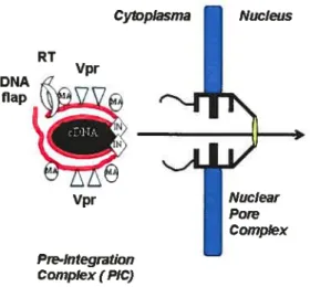
1.3.4. Nuctear transtocation of the PIC.
Nucleoprotein complexe utilize cytoskeletal components to reach the nucleus (41). PICs are composed of the double-stranded linear DNA associated with the viral proteins MA,RT, IN, and Vpr. It lias an estimated stoke diameter of 56nm (230). Since the central channel of the nuclear pore lias a maximum diameter of 25nm and the pore is known to be able to transport macromoleculesup to 39 nm (262), HIV has developed a strategy to pass tbrough these structures. Unlike the oncoretroviruses, lentiviruses including HIV-1 have the ability to infect non-dividing ceils without the breakdown of the nuclei envelope and mitosis for viral replication, a feature important to HIV-1 in its establishment of a long-lived infection of the host (379) (44). This property is shared with other lentiviruses and reflects the existence of determinants that govem the active transport of the viral preintegration complex through the nucleopore (44).
Nuclear pore complexes (NPCs) are large supramolecular protein structures that span the nuclear membrane and protrude into both cytoplasm and nucleoplasm. Signal-mediated nuclear import involves the interaction of nuclear localization signaIs (NLSs) in proteins with nucleocytoplasmic shuttiing receptors, belonging to the karypherin 3 family, also known as importins. NLSs are typically short
stretches of amino acids, the best studied of which are basic amino acid-rich sequences that interact with the adapter importin Π(115). Importin 3 interacts with other classes of NLS by using differentadapters,
including snurportin, RIP (Rev interacting protein), and importin 7. Recently, importin 7 has been proposed as playing a key role in the nuclear import of HW-1 PICs in primarymacrophages (101). It is believed that multiple factors are involved in the nuclear targeting of the HIV-1 preintegration complex (PIC) in non-dividing celis, such as matrix protein (MA), karyophilic Vpr, integrase protein (LN), and DNA flap (136, 275). Their respective involvement will be discussed later.
1.3.5. Integration
Integrase prepares the viral cDNAs for integration by cleaving their 3’ ends. An integrase mediated hydroxyl group oriented attacking occurs on the host ceil DNA and thishydroxyl group forms new bonds with the 3’ ends of the viral cDNA (309). HIV- 1 integration preferentially occurs in genes highly transcribed by the RNA Pol 11(309). Several cellular factors have been described to interact with integrase and may therefore constitute good candidates for directing the PIC to its target site. The integrase interactor
(mii,
also called hSNF5), a subunit of the SWI/SNf chromatin-remodeling complex, was initially isolated by yeast twohybrid
screen for liuman proteins interacting with the IN and was proposed to stimulate the in vitro DNA-joining activity of the IN and to target the viral genome to active genes in an as yet undetermined manner (167). Equally, high mobllity group protein HMG-I(Y), which lias been proposed to be important for integration (9$), appears to be required for efficient integration in vitro, but their respective role in directing the PIC to precise sites ofhostgenome was not evaluated. Two other IN-binding partners were isolated which seem to be critical for directing the PIC to the host chromatin. This is the case for the EED protein which is encoded by thehuman homologue of the mouse ernbiyonic ectoderrn deveÏopment (eed) gene product and of the Drosphila esc gene, arid which also interacts with the matrix protein of HIV-1 (269, 367) . These genes belong to the family of widelyconserved polycomb genes, involved in the maintenance of the silent state ofchromatin and reduction of DNA accessibility. An interaction occurring between EED and the viral proteins MA and IN might not
oniy direct the PIC to the host chromatin but also trigger transcriptional activation (367). finally,the lens epithelium-derived growth factor (LEDGf/p75), a protein implicated in the regulation of geneexpression and in the cellular stress response was found to interact with HIV- 1 iN (54). Interestingly,this interaction is flot essential for nuclear accumulation ofHIV-1 IN, but seems to be absolutely requiredto dock the PIC to the host chromatin (214).
1.3.6. HI”/-l gene expression
ReguÏation of the transcriptional activity. After the viral DNA has successfiully integrated into the host ceil DNA, the process of viral gene expression begins. The cis-regulatory sequences in the LTR promoter allow RNA polymerase II, together with other cellular factors, to bindand initiate transcription (see Figure 1.4). The translation produces the basal amount of Tat, Rev, and Nef(165).
After a sufficient amount of Tat bas been produced, Tat controls the transcription ofthe HIV-1 gene. Tat increases the efficiency of transcription by enhancing the elongation capacity ofthe RNA Polymerase II complex by 10,000 fold. This increase is accomplished by the ability ofTat to recruit cyclin-dependent kinase 9 (Cdk9) to the HIV-1 LTR by interacting with Cyclin Ti and binding it to the TAR elements, a stem ioop structure found at the S’end of viral transcripts (378). Cyclin T is part of a family ofproteins involved in celi cycle regulation that forms a complex with cyclin-dependent kinases (Cdk9)
F
-454
R U5
+60 +181
TAR
— figure.1.4. Schematic representation of 11W-1 LTR The position of binding sites for
host factors (LBF-1, NF-KB,LEfEts, USF-1, and NFAT-J) are shown at the 5’ ofthe transcription startsite. The TAR stem/loop structure, with its butge, is represented at the 5’ end ofa nascent mRNA. Numberingbelow the boxed region is relative to the transcription start site nucleotide +J(here using the HIV-1 HXb2 sequences; accession no. K03 459).
to phosphorylate the C-terminal domain (CTD) of RNA polymerase II and initiate transcription. The binding ofboth Cyclin T and Tat to the TAR element provides a high affinity complexthat binds to the P TEFb (positive elongation factor-b) complex. Cdk9 phosphoiylates the C-terminus ofRNA polymerase II and allows efficient elongation to occur. Tat has been found to interact with Spi and require Spi and NF
wB sites for its function (341, 342). Tat also recruits the chromatin remodeling enzymes (CBP/p300 and
pCAF) to the site of transcription to unravel the histone/chromatin structure ofthe integrated viral DNA, and enhances the elongation process of RNA pol II, resulting in a several hundred fold increase in transcription (260). HIV-1 Vpr interacts with HIV-l tat which causes synergic effect of Tat transactivation (307). The detailed mechanism of Vpr role on its transactivation LTR is described in section 4.5. Early reports showed that HIV-1 nef had a negative effect on LTR activity (5). The nuclear factor of activated T ceils (NfAT) is an important transcription factor in regulation of gene expression in T cell. Together with the activator (AP-1) it promotes transcription of several celiular genes involved in T ccli activation, such as interleukin-2. Increased production of IL-2 is a critical step in T ccli activation. Since the activation of T ceils strongly correlates with the ability of fflV- 1 to infect and replicate in these ceils. It has been reported that NFAT can also directly bind to and activate HIV-1 LTR (182). Vpr can potentiate Nef-induced activation of nuclear factor of activated T ceils (NfAT)-dependent transcription (191). Unlike Nef, which stimulates calcium signaling to activate NFAT, Vpr functions farther downstream via distinct mechanism to cooperate with Nef in NFAT-directed gene expression and promote transactivation by CREB (191).
tE
Post-transcripfionat reg-utation. Regulation of the expression of a large numberof structure and regulatory genes within a relatively small genome highlights the complexity of HIV gene regulation compared to other simple retroviruses. This regulation implicates a complex arrangement of genes encoded in overlapping reading ftames and the expression ofthese genes through elaborate spiicing ofthe single mRNA precursor. Through transcription and spiicing, HW4 produces three classes ofmRNA: the multiply spliced 2kb mRNA species which encode the viral regulatory protein Tat, Rev, and Nef, the unspliced (rn-9kb) and single spliced (-4kb) transcripts which encode the structure protein (Gag polyprotein precursor, Gag-Pol polyprotein precursor, and Env glycoprotein precursor) and some of the accessory proteins (Vif Vpr, and Vpu). The expression of unspliced and singlespliced mRNA species is tightly controlled by the HIV-1 regulatory protein Rev as described in section 2.2. Full-length unspliced transcripts are exported by Rev and are recmited to the assembling virionby an interaction with the psi packaging element located 3’ ofthe 5’ LTR.
1.3.7. Assembly and release ofHTV-1
Env trafficking. The env gene is translated into the precursor protein gp160, which is glycosylated within the endoplasmic reticulum and fransported to the plasma membrane via the secretory pathway to areas of high lipid content (sphingolipids and cholesterol), known as lipid rafts. gpl6O is cleaved into gp4l and gpl2O by the host protease furin during its transport through the Golgi apparatus (140). After translation, the Env proteins migrate and insert into the plasma membrane.
Gag trafficking. The synthesis ofthe HIV Pr55gag and Pr160 gag-pol precursor polyproteins ofHIV occurs on cytosolic polysomes. Unspliced viral RNA is translated by ribosome scanning from the first AUG (+789). At least 90% of ail translation events terminate at the UAAstopcoden (+2289) and result in the synthesis of the gag polyprotein precursor. An infrequent —1 ribosomal frame shift (approximately 1-5% of gag translation) at astretch of uridine bases (+2023 to ±2089) resuits in readtbrough to the latter UAA stop code and the synthesis ofthe protein ofthese precursors (159). Gag and Gag-Pol polyproteins also migrate to the cellular membrane and start to assemble, directed by a series of basic residues and a
C
myristoylation sequence present at the N-terminus of MA of the Gag polyprotein(308). At the C-terminalend of Gag, NC recruits two copies of unspliced RNAs, and the p6 late domain mediates the process of budding and detachment by its interaction with the host cellular proteinTsg 101.
HIV-1 budding occurs mostly at the plasma membrane of infected T lymphocytes. In macrophage, however, this virus accumulates in an intracellular, vacuole-like compartment (256). Recent ultiastructural studies identified this compartment as late endosome/multivesticularbodies (MVBs) (284). HIV-l p6 PTAP late domain recruits the cellular protein tumor susceptibility gene 101 (TsglOl) to facilitate virus budding (126, 365). In uninfected celis, TsglOl functions in the biogenesis of the multivicular body (MVB) (173). Reduction of TSG1O1 levels by siRNA treatment or introduction of a dominant-negative TsglOl mutant blocks viral budding and produces tethered structures at the plasma membrane that resemble the phenotype of late-domain (PTAP) mutants (77, 126). AIl these results suggest that HIV may bind TsglOl in order to gain access to the downstream machineiy that catalyzes MVB vesicle budding. Studies in yeast show that TsglOI is part of a 350-kDa intracellular complex known as endosomal sorting complex required for transport-I (ESCRT-I), which along with ESCRT-II and ESCRT-IIL directs monoubiquitinated endocytosed cargo to the MVB (19, 184). Wills et ai. first noted the topological similarity between budding of vesicles into the lumen of the MVB and budding of viral particles into the extracellular milieu-both processes irivolve budding away from the cytosol (265). Although ESCRT components are required for HIV-l particle formation, it is flot known if they perform this function at the MVB, with other compartment, at the plasmamembrane, or at ail three locations.
Particte release and maturation. The process of viral buddingor release triggers the activation of the PR that autocatalytically cleaves the Gag and Gag-Pol polyprotein, which releases the structural proteins and enzymes MA (p17), CA (p24), NC (3)9), p6, PR (pi0), the reverse transcriptase (RT), and integrase (p32) (416). The individual proteins undergo fiirther interaction, with CA and NC forming the conic nucleocapsid, MA remaining associated to the viral envelope (39, 383).
1.4. Biotogicalfunctions of Vpr.
Vpr is the only accessory protein found within the virion in substantial amounts.In in vitro culture systems, Vpr is not required for viral replication in transformed ceil unes and primary T lymphocytes; however, it plays an important role in the productive infection of ceils, such as macrophages and monocytes (65, 146, 368). HIV-1 replication in macrophages is extremely important for the pathogenesis in vivo because terrninally differentiated macrophages are natural ceil targets for HIV and provide a reservoir of viral production during the asymptomatic stages of disease (20) The most convincing evidence that Vpr plays an important role in vivo cornes from experirnents showing that rhesus monkeys infected with Vpr deleted SIV virus have a low viral burden and slow disease progression cornpared to
those infected with the wild-type virus (157, 192).
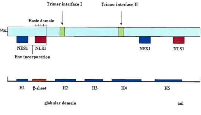
1.4.1. Structure of the protein.
Priniary seqilence. The Vpr gene of HIV- I encodes a 96-amino-acid Ï 4-kDa protein that is produced late in the viral life cycle (62). Tue prirnary Vpr sequence is shown in Fig 1 .5. Vpr sequence is well consen’ed arnong the primate lentiviruses HIV-l, HIV-2, and SIV (350).
10 20 30 40
MEQAPEDQGP QREPHNEWTL ELLEELKNEA VRHFPRJWLH
50 60 80
GLGQHIYETY GDTWAGVEAI IRILQQLLFI HFRIGCHSR
1GVTRQRRAR NGASRS96
Figure 1.5. The primary Vpr amino acid sequence. The negative amino acids are shown inpuiple, positive charge amino acids in redandphospho,yÏation site ingreen.
3D-structure. Vpr shows welI-characterized helical domains with amphipathic properties and y -turns throughout the protein. NMR analysis of a soluble full length Vpr (1-96) polypeptide was recently performed, and revealed the tertiaty structure of the protein, confirming the arnphipathic nature of the