Neutralizing Antibody Responses to Human Immunodeficiency Virus Type 1 in
Primary Infection and Long-Term – Nonprogressive Infection
Alice K. Pilgrim, Giuseppe Pantaleo, Oren J. Cohen, Department of Surgery, Duke University Medical Center, Durham, North Carolina; Laboratory of AIDS Pathogenesis, Hoˆpital de Lisa M. Fink, Ji Ying Zhou, Jin Tao Zhou,
Beaumont, Lausanne, Switzerland; Laboratory of Immunoregulation, Dani P. Bolognesi, Anthony S. Fauci,
National Institute of Allergy and Infectious Diseases, National Institutes
and David C. Montefiori of Health, Bethesda, Maryland
The role of neutralizing antibodies in human immunodeficiency virus type 1 (HIV-1) infection is poorly understood and was assessed by evaluating responses at different stages of infection. Undi-luted sera from long-term nonprogressors (LTNP) had broad neutralizing antibodies against heterol-ogous primary isolates and were more likely to neutralize the contemporaneous autolheterol-ogous isolate than were sera from short-term nonprogressors and progressors. In primary infection, envelope-specific IgG was detected before the initial decline in plasma viremia, but neutralizing antibodies developed more slowly. Here, neutralizing antibodies against strains SF-2 and MN were sometimes the first to be detected, but titers were low for at least 17 weeks from onset of symptoms. Neutralizing antibodies against the early autologous isolate were detected for 4 patients by 5 – 40 weeks but were undetectable in 2 additional patients for 27 – 45 weeks. The results indicate that neutralizing antibody responses are slow to develop during primary infection and are uniquely broad in LTNP.
Infection with human immunodeficiency virus type 1 (HIV- Although most infected persons progress to AIDS within a 1) is accompanied by cellular and humoral immune responses median time of 10 years from initial seroconversion, Ç5% – of various magnitudes and specificities that are thought to 10% tolerate infection without immune suppression or other down-regulate plasma viremia during primary infection [1, 2] clinical manifestations for longer periods of time [13 – 15]. This and to establish a period of clinical latency that follows [3]. latter group of infected persons is referred to as long-term Despite the benefit provided by these responses early in infec- nonprogressors (LTNP). LTNP generally have low virus loads tion, nearly all HIV-1 – infected persons progress to AIDS. Spe- in their plasma, peripheral blood mononuclear cells (PBMC), cific responses that are at least partially beneficial during the and lymph nodes and mount robust cellular and humoral im-course of infection are poorly understood. Detection of HIV- mune responses [16 – 21]. Some cases of long-term nonprogres-1 – specific cytotoxic T lymphocytes (CTL) correlates well with sion are associated with infection by attenuated virus [22] or the initial down-regulation of plasma viremia during primary with a heterozygous defect in the CCR5 co-receptor [23]. Many infection [4 – 7] and could be a dominant antiviral immune other LTNP are thought to benefit from the overall quality of response. Another correlate during primary infection is the their immune responses.
production of HIV-1 – specific IgM [8] and possibly IgG [2, Previous studies showed that neutralizing antibody responses 9], but it is not clear whether these early antibodies are effective in LTNP are greater in magnitude and breadth than they are against the virus. For example, neutralizing antibody responses in other HIV-1 – infected persons. For example, the average against the early autologous virus have been shown to be de- titer of neutralizing antibodies against HIV-1
IIIBand HIV-1MN
layed relative to when plasma viremia is first down-regulated was significantly higher in sera from LTNP than from persons [5 – 7, 10]. Similar delays in neutralizing antibody production who had been infected for õ7 years and had no signs of have been reported for simian immunodeficiency virus (SIV) immune suppression [17]. In addition, we [17] and others [18] infection in macaques [11, 12]. This slow development of neu- determined that sera from LTNP neutralize heterologous pri-tralizing antibodies during primary infection might be one of mary isolates more effectively than do sera from progressors. the reasons the virus is able to establish persistent infection. Importantly, it was not clear from these studies whether the greater capacity of sera from LTNP to neutralize primary iso-lates was merely a reflection of diminished response in immu-Received 10 December 1996; revised 28 April 1997.
nologically suppressed persons. Informed consent, as approved by local institutional review boards and
biosafety committees, was obtained from all patients and controls. The study The present study aimed to determine whether the broad followed human experimentation guidelines of the US Department of Health neutralizing antibody responses seen in LTNP are unique com-and Human Services.
pared with responses in other HIV-1 – infected persons, includ-Financial support: NIH (AI-45218, AI-28662).
Reprints or correspondence: Dr. David C. Montefiori, Dept. of Surgery, Box ing short-term nonprogressors (STNP) who had been infected 2926, Duke University Medical Center, Durham, NC 27710.
for 2 – 7 years. We took advantage of the fact that virus neutral-The Journal of Infectious Diseases 1997; 176:924 – 32 ization was more readily detected with undiluted serum
sam-q 1997 by The University of Chicago. All rights reserved.
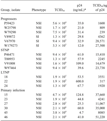
Table 1. Characteristics of primary isolates from HIV-1 – infected from LTNP, progressors, recent seroconverters, and STNP
subjects. were evaluated with autologous and heterologous primary
iso-lates. We also determined whether low-level neutralizing
anti-p24 TCID50/ng
bodies might be present during primary infection that had pre- Group, isolate Phenotype TCID
50 (ng/mL)* of p24
viously gone undetected. Sequential serum samples obtained
Progressors during and immediately following primary infection were
as-P59423 NSI 5.61 104 35.0 1600
sessed for neutralizing antibodies against the early autologous
W25798 NSI 1.71 104 21.0 809
virus and T cell line – adapted (TCLA) variants of HIV-1,
in-W79290 NSI 7.51 103 31.4 239
cluding IIIB, MN, and SF-2. V89872 SI 1.31 105 29.8 4362
V67970 SI 9.41 105 32.9 28,571 W179273 SI 3.31 105 12.0 27,500 STNP Methods P46471 NSI 9.41 105 61.0 15,410 T00953 NSI 1.31 105 57.9 2245
Study participants. HIV-1 – infected LTNP and progressors
V91008 NSI 1.61 106
109.0 14,679
were described previously [16, 17]. LTNP had well-documented
W97464 NSI 9.41 105
39.6 23,738
infection for§10 years, were asymptomatic, and had ú600 CD4
LTNP
T lymphocytes/mm3in their peripheral blood; these levels were
18 NSI 1.91 105
53.5 3551
stable over time. With the exception of 1 LTNP (no. 17), no 22 NSI 1.91 105
600.0 317
LTNP received antiretroviral therapy. All progressors had signs 24 NSI 1.31 105
67.7 1920
of immunosuppression as evidenced by low CD4 T lymphocyte Primary infection
15 NSI 4.71 106
124.8 37,660
counts (range, 10 – 371/mm3
). A group of STNP, defined as having
23 NSI 1.91 105
44.8 4241
documented infection for 2 – 7 years but remaining healthy, with
27 NSI 2.81 105
25.3 11,067
ú600 CD4 T lymphocytes/mm3
, were recruited from the Durham,
30 NSI 2.11 106
60.0 35,000
North Carolina, area. Information about antiretroviral therapy for
45 NSI 3.81 104 4.7 8085
progressors and STNP was unavailable.
46 NSI 2.11 106 41.0 51,220
Six persons with primary HIV-1 infection were referred for
study by their private physicians. Each of them had developed NOTE. STNP, short-term nonprogressors; LTNP, long-term nonpro-transient mild clinical symptoms, which brought them to the atten- gressors; SI, syncytium-inducing; NSI, non – syncytium-inducing.
* Concentration of p24 at time of harvest from initial cocultivation.
tion of their physicians. Four of them began antiretroviral therapy within a few days of entry into study. Exceptions were patient 45, who received no antiretroviral therapy during the course of study,
and patient 46, who began antiretroviral therapyÇ25 weeks from of cells (51 104
/well). Cultures were then incubated for the mini-mum length of time that allowed for extensive syncytium forma-the onset of symptoms. Healthy, HIV-1 – negative persons were
recruited from local research laboratories. tion to occur, as observed microscopically (3 – 4 days). Cell viabil-ity was quantified by neutral red staining as described [25]. Primary isolates. Viruses were isolated by coculture of PBMC
from infected persons with phytohemagglutinin-stimulated PBMC Percentage of protection was calculated by the difference in ab-sorbance at 540 nm between test wells (cells, serum sample, and (PHA-PBMC) from healthy, noninfected persons in the presence
of interleukin-2 (IL-2) as described [17]. Primary isolates from virus) and virus control wells (cells and virus) divided by the difference in absorbance between cell control wells (cells only) acute seroconverters were obtained at the time of their first serum
collection. Virus stocks were made cell-free by low-speed centrifu- and virus control wells. Neutralization titers were defined as the reciprocal of the serum dilution required to protect at least 50% gation and passage through 0.5-mm cellulose acetate filters before
being stored in aliquots at0707C. Some stocks were later ex- of cells from virus-induced killing. Assays were standardized by including positive and negative control sera that had been assayed panded a single time in PHA-PBMC. TCID50was determined by
the method of Reed and Muench [24] after 14 days of incubation multiple times and for which the average titer was well-character-ized. Virus stocks for these assays were produced in H9 cells. with PHA-PBMC. Viral antigen concentration was measured by
p24 immunoassay (DuPont, Wilmington, DE). Syncytium-induc- Antibody-mediated neutralization of primary isolates was mea-sured in PHA-PBMC as described [17] by use of a p24 immunoas-ing (SI) and non-SI (NSI) phenotypes were determined in MT-2
cells and PHA-PBMC as described [17]. The TCID50, p24 concen- say to quantify virus production. For assays with undiluted serum,
20 mL of cell-free virus was incubated with 80 mL of serum for tration, and phenotype of each virus stock are given in table 1.
Additional information on isolates obtained from LTNP and prog- 1 h at 377C, and 25 mL of this mixture was added to triplicate wells containing PHA-PBMC (41 105cells in 175 mL added per
ressors has been published [17]. Primary isolates were used as
either first- or second-passage stocks produced in PHA-PBMC. well). For measurement of titer, 50 mL of virus was incubated in triplicate as above with an equal volume of serum samples that Serologic assays. Neutralizing antibodies against TCLA
strains of HIV-1 were measured in MT-2 cells (strains IIIB and were undiluted and diluted 1:2, 1:4, 1:16, and 1:32 (dilutions were made in IL-2 growth medium). Next, 25 mL was transfered to MN) or CEMx174 cells (strain SF-2) in 96-well plates by a
reduc-tion in virus-induced cell killing as described previously [25]. In corresponding wells of U-bottom plates containing PHA-PBMC. Cells were incubated with the virus-serum mixtures for 3 h at 377C brief, cell-free virus (1000 TCID50) and serial dilutions of test sera
remove the virus inoculum and antibodies. Washed cells were Statistical analyses. The significance of the frequency by which sera from paired groups of infected persons neutralized suspended in 250 mL of IL-2 growth medium and incubated in
96-well flat-bottom plates for the duration. Culture supernatants primary isolates in a panel of 6 isolates was evaluated by x2
analysis with 1 df. This analysis assumed that the ability of a (25 mL) were collected on a daily basis and mixed with 225 mL
of 0.5% Triton X-100 for the quantification of p24. This volume particular serum sample to neutralize one primary isolate does not predict its ability to neutralize other primary isolates. Yates’s was replaced with 25 mL of fresh IL-2 growth medium at each
collection. correction factor was used to give a more conservative estimate
of P. Neutralization sensitivity of NSI and SI viruses was also Viral p24 produced in the absence of test sera (virus control)
was determined for each daily sampling and was used to construct compared by x2
analysis, but here sera from all infected persons were grouped as one. The small number of NSI and SI isolates (3 a viral replication curve. Neutralization was measured at a time
when the concentration of p24 in virus control wells exceeded 5 each) did not permit a comparison of neutralization sensitivity between sera from different subgroups of persons.
ng/mL (limit of detection was 0.1 ng/mL) but had not reached peak production (4 – 6 days). Serum samples were considered posi-tive for primary isolate neutralization if they causedú80%
reduc-Results
tion in p24 relative to a negative control serum (D01). This cutoff
corresponds to a minimum 1 log reduction in infectious virus yield. Autologous and heterologous neutralizing antibody re-Standard deviations for a given serum sample were divided by the
sponses at different stages of HIV-1 infection. Serum samples average p24 value of the virus control and then multiplied by 100
obtained at different stages of HIV-1 infection were assessed for percentage. An equal volume of undiluted virus stock was used
for neutralization breadth with a panel of 6 primary isolates. for all assays. While the infectious dose contained within this
Sera from 6 recent seroconverters (sera obtained 13 – 53 weeks volume was not the same for all isolates, each serum sample was
from onset of symptoms), 5 progressors, 6 STNP, 6 LTNP, and tested against an equal dose on a per-virus basis. Sera were
heat-5 healthy HIV-1 – negative persons were used in this evaluation inactivated at 567C for 1 h before use.
Antibodies that bound to HIV-1 antigens were assessed by HIV- (table 2). Primary isolates were derived from the progressors 1IIIB Western immunoblot (Cambridge Biotech, Worcester, MA) listed in table 2. These same primary isolates and sera from
and by HIV-1MN gp160 ELISA. Baculovirus-derived HIV-1MN progressors and LTNP were used in a previous study in which
gp160 was obtained from Quality Biologicals (Gaithersburg, MD). the primary isolates were rarely neutralized by 1:16-diluted ELISAs were done as described previously [26] with alkaline phos- serum samples [17]. Undiluted serum was used in the present phatase – conjugated goat anti-human IgG (whole molecule; Sigma,
study to improve the detection of neutralizing antibodies. St. Louis) and p-nitrophenylphosphate disodium hexahydrate
No neutralization of primary isolates was detected with sera (Sigma; catalog no. N-9389) used for detection and color
develop-from HIV-1 – negative persons. In some cases, mild enhance-ment, respectively. Titers are reported as the reciprocal of the
ment of infection was observed with these sera, but the enhanc-highest serum dilution to produce an average absorbance reading
ing effect was never more than a doubling of p24 production, of at least twice that of a negative control serum (D01).
which would not be considered true enhancement [12, 17]. Removal of IgG. IgG was removed from serum samples by
absorption with protein G – sepharose (GammaBind Plus Sepha- Neutralization of each virus was detected sporadically with rose; Pharmacia, Piscataway, NJ). Protein G – sepharose was ob- serum samples from HIV-1 – infected persons. Positive neutral-tained by the supplier as a suspension in PBS containing thimerosal ization was detected most often with sera from LTNP. Of the as a preservative. The resin was washed five times with PBS and LTNP sera evaluated, 20 (56%) of 36 virus-serum combina-collected by low-speed centrifugation as premeasured aliquots after tions tested positive. Neutralization was detected less fre-the last wash. Supernatants were removed and replaced with a
quently with sera from STNP (25% of combinations testing volume of undiluted serum equal to the volume of packed resin.
positive), recent seroconverters (17% of combinations testing Suspensions were incubated at room temperature for 1 h with
positive), and progressors (17% of combinations testing posi-constant gentle mixing. Protein G – sepharose was then separated
tive). The breadth of neutralization was significantly greater from the serum by low-speed centrifugation, and the serum was
for sera from LTNP compared with sera from all other groups collected by pipette. Serum samples were subjected to two
addi-(PÅ .0014 compared with recent seroconverters, P Å .0028 tional absorption steps to ensure the removal of all IgG. Complete
removal of HIV-1 – specific IgG was confirmed by Western immu- compared with progressors, and P Å .0163 compared with
noblot analysis. STNP; 95% confidence level). The breadth of neutralization
Measurements of plasma viremia. HIV-1 RNA in plasma sam- with sera from STNP was no greater than with sera from recent ples was quantified by the branched DNA method as described seroconverters or progressors (P ú .05). There was also no
previously [27]. significant difference in neutralization sensitivity between NSI
Chemokine assays. Quantification of the chemokines
and SI viruses when sera from all infected persons were RANTES and MIP-1b in serum samples was done with
commer-grouped as one for the analysis (PÅ .124). cially available ELISA kits as described by the supplier (R&D
Undiluted sera from LTNP neutralized the autologous virus Systems, Minneapolis). Levels of RANTES and MIP-1b were
obtained at the time of serum collection in all 3 cases examined quantified when sufficient sample volumes were available. Levels
(table 3). These same sera had failed to neutralize the autolo-of MIP-1a were not measured because autolo-of a limited volume autolo-of
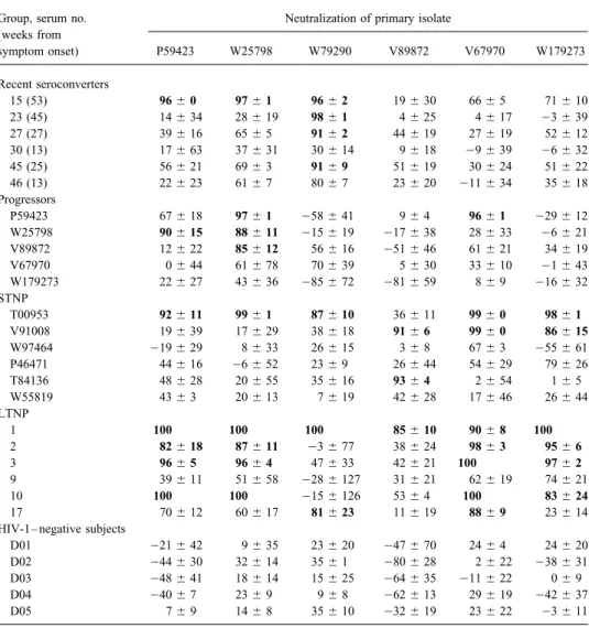
Table 2. Neutralization of primary isolates by undiluted sera obtained at different stages of HIV-1 infection.
Group, serum no. Neutralization of primary isolate (weeks from symptom onset) P59423 W25798 W79290 V89872 V67970 W179273 Recent seroconverters 15 (53) 96{ 0 97{ 1 96{ 2 19{ 30 66{ 5 71{ 10 23 (45) 14{ 34 28{ 19 98{ 1 4{ 25 4{ 17 03 { 39 27 (27) 39{ 16 65{ 5 91{ 2 44{ 19 27{ 19 52{ 12 30 (13) 17{ 63 37{ 31 30{ 14 9{ 18 09 { 39 06 { 32 45 (25) 56{ 21 69{ 3 91{ 9 51{ 19 30{ 24 51{ 22 46 (13) 22{ 23 61{ 7 80{ 7 23{ 20 011 { 34 35{ 18 Progressors P59423 67{ 18 97{ 1 058 { 41 9{ 4 96{ 1 029 { 12 W25798 90{ 15 88{ 11 015 { 19 017 { 38 28{ 33 06 { 21 V89872 12{ 22 85{ 12 56{ 16 051 { 46 61{ 21 34{ 19 V67970 0{ 44 61{ 78 70{ 39 5{ 30 33{ 10 01 { 43 W179273 22{ 27 43{ 36 085 { 72 081 { 59 8{ 9 016 { 32 STNP T00953 92{ 11 99{ 1 87{ 10 36{ 11 99{ 0 98{ 1 V91008 19{ 39 17{ 29 38{ 18 91{ 6 99{ 0 86{ 15 W97464 019 { 29 8{ 33 26{ 15 3{ 8 67{ 3 055 { 61 P46471 44{ 16 06 { 52 23{ 9 26{ 44 54{ 29 79{ 26 T84136 48{ 28 20{ 55 35{ 16 93{ 4 2{ 54 1{ 5 W55819 43{ 3 20{ 13 7{ 19 42{ 28 17{ 46 26{ 44 LTNP 1 100 100 100 85{ 10 90{ 8 100 2 82{ 18 87{ 11 03 { 77 38{ 24 98{ 3 95{ 6 3 96{ 5 96{ 4 47{ 33 42{ 21 100 97{ 2 9 39{ 11 51{ 58 028 { 127 31{ 21 62{ 19 74{ 21 10 100 100 015 { 126 53{ 4 100 83{ 24 17 70{ 12 60{ 17 81{ 23 11{ 19 88{ 9 23{ 14
HIV-1 – negative subjects
D01 021 { 42 9{ 35 23{ 20 047 { 70 24{ 4 24{ 20
D02 044 { 30 32{ 14 35{ 1 080 { 28 2{ 22 038 { 31
D03 048 { 41 18{ 14 15{ 25 064 { 35 011 { 22 0{ 9
D04 040 { 7 23{ 9 9{ 8 062 { 13 29{ 19 042 { 37
D05 7{ 9 14{ 8 35{ 10 032 { 19 23{ 22 03 { 11
NOTE. Virus neutralization was measured in phytohemagglutinin-stimulated peripheral blood mononuclear cells by using undiluted serum. Values are % reduction in p24 production relative to virus control (no test serum) in HIV-1 – negative subjects and relative to p24 production in presence of serum D0HIV-1 for all others. Results are given as average of triplicate wells{ SD. Boldface type indicates ú80% reduction (positive for neutralization). STNP, short-term nonprogressors; LTNP, long-short-term nonprogressors.
progressors were neutralized by undiluted autologous serum in sepharose no longer had neutralizing activity. Loss of neutraliz-ing activity could not be attributed to sample dilution durneutraliz-ing only 1 of 5 cases examined (table 2), whereas isolates from
STNP were neutralized by undiluted autologous serum in 0 of absorption, since the volume of processed samples never ex-ceeded the original preabsorption volumes. These results 4 cases examined (table 3).
Virus neutralization by undiluted serum is antibody-medi- strongly indicate that virus neutralization was mediated by IgG. Antibody-independent neutralization was further assessed by
ated. We examined whether high concentrations of antiviral
cytokines or chemokines in undiluted serum were at least par- quantifying the concentration of the anti – HIV-1 chemokines, RANTES and MIP-1b in serum samples (table 5). Many in-tially responsible for neutralizing activity. Protein G – sepharose
was used to remove the total IgG from 5 serum samples that fected persons had moderately elevated levels of RANTES in their serum compared with levels in serum from healthy, had previously neutralized 1 or more primary isolates. Samples
were negative for anti – HIV-1 antibodies by Western blot anal- noninfected persons (ú14.8 ng/mL). Elevated levels were mostly seen in sera from progressors, and these particular se-ysis after absorption (data not shown). Serum samples were
reevaluated for virus neutralization before and after absorption. rum samples had poor neutralizing activity against primary isolates. Only 1 infected subject had a level of MIP-1b that As can be seen in table 4, samples absorbed with protein G –
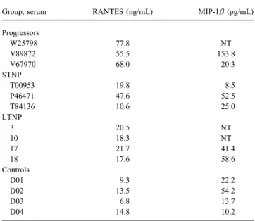
Table 3. Neutralization of primary isolates from long-term and Table 5. Analysis of serum chemokine concentrations at different stages of HIV-1 infection.
short-term nonprogressors (LTNP and STNP) by autologous serum.
Subject Status Neutralization of autologous virus Group, serum RANTES (ng/mL) MIP-1b (pg/mL)
Progressors 18 LTNP 99{ 0 22 LTNP 94{ 3 W25798 77.8 NT V89872 55.5 153.8 24 LTNP 99{ 0 P46471 STNP 30{ 18 V67970 68.0 20.3 STNP T00953 STNP 45{ 18 V91008 STNP 52{ 13 T00953 19.8 8.5 P46471 47.6 52.5 W97464 STNP 32{ 9 T84136 10.6 25.0
NOTE. Undiluted serum from each subject was evaluated for neutralizing LTNP
antibodies against autologous isolate obtained at time of serum collection. 3 20.5 NT Values are % reduction in p24 production in phytohemagglutinin-stimulated 10 18.3 NT peripheral blood monocuclear cells and are given as average of triplicate wells
17 21.7 41.4 { SD. 18 17.6 58.6 Controls D01 9.3 22.2 D02 13.5 54.2
was highly elevated relative to all others. This subject (V89872) D03 6.8 13.7
was a progressor whose serum rarely neutralized primary iso- D04 14.8 10.2
lates. Sera with the most potent neutralizing activity (i.e., serum
NOTE. LTNP, long-term nonprogressors; STNP, short-term nonpro-samples 3, 10, and T00953) had lower concentrations of these
gressors; NT, not tested. chemokines than did the remaining sera that had poor or no
neutralizing activity. We conclude that virus neutralization
us-ing undiluted serum was unrelated to the concentration of anti – levels of plasma viremia ofú105RNA copies/mL at the earliest
time point examined. These levels subsequently declined and re-HIV-1 chemokines.
CD4 T cells, plasma virus loads, and serum antibody responses mained persistently low in 3 of them (patients 15, 23, and 30), all of whom began antiretroviral therapy shortly after the first
during primary infection. CD4 T cells, plasma virus, and serum
antibodies were quantified for HIV-1–infected persons starting at time point. The fourth patient with high levels of plasma virus on entry continued to have high virus loads for 20 weeks of 3–10 weeks from onset of symptoms and continuing for an
addi-tional 20–57 weeks thereafter (table 6). Four of 6 patients had follow-up (patient 45); this patient received no antiretroviral
ther-Table 4. Neutralization of primary isolates after removing IgG from serum samples. Neutralization of primary isolate
Serum, IgG P59423 W25798 W79290 V89872 V67970 W179273 18 15 (53 weeks) / 82{ 4 95{ 2 94{ 4 NT NT NT NT 0 26{ 16 11{ 16 59{ 48 NT NT NT NT T00953 / NT NT NT NT 99{ 1 97{ 3 NT 0 NT NT NT NT 0{ 22 15{ 52 NT V91008 / NT NT NT 89{ 4 100 NT NT 0 NT NT NT 0{ 39 0{ 15 NT NT 10 / NT 100 NT NT 100 NT NT 0 NT 0{ 37 NT NT 0{ 19 NT NT 18 / NT NT NT NT NT NT 87{ 7 0 NT NT NT NT NT NT 14{ 45
NOTE. Undiluted sera were evaluated for neutralizing antibiotics against indicated primary isolate. Values are % reduction in p24 production in phytohemagglutinin-stimulated peripheral blood mononuclear cells and are given as average of triplicate wells{ SD. NT, not tested. Samples were evaluated before (/) and after (0) IgG was removed by absorption with protein G – sepharose.
Table 6. CD4 T cells, plasma virus concentration, and antibody titers in primary HIV-1 infection. Neutralizing antibody titer§
Patient,
weeks from CD4 Plasma ELISA Autologous
symptom onset* T cells/mm3
virus† titer‡ isolate IIIB MN SF-2 Patient 15 4 704 744,000 150 õ2 õ10 õ10 õ10 20 638 13,000 4050 4 õ10 24 393 25 542 19,000 4050 8 26 55 490 29 502 11,000 12,150 32 82 125 830 35 655 12,000 36,450 32 67 156 713 53 644 õ10,000 36,450 ú64 72 149 1932 Patient 23 10 756 171,000 450 õ2 õ10 23 14 18 523 137,000 1350 õ2 õ10 78 136 25 484 19,000 4050 õ2 õ10 157 520 29 385 31,000 4050 õ2 12 200 665 42 363 41,000 4050 õ2 õ10 342 661 45 391 58,000 12,150 õ2 õ10 199 1039 Patient 27 4 441 40,000 150 õ2 õ10 õ10 õ10 5 NT NT 450 õ2 õ10 õ10 õ10 6 313 õ10,000 450 õ2 22 29 õ10 11 395 õ10,000 1350 õ2 õ10 72 20 17 359 õ10,000 1350 õ2 14 24 840 27 357 15,000 4050 õ2 24 72 552 Patient 30 3 558 1,224,000 1350 õ2 õ10 õ10 õ10 7 679 37,000 1350 õ2 24 16 õ10 13 665 õ10,000 1350 õ2 32 23 226 18 655 12,000 1350 õ2 29 71 778 40 525 õ10,000 4050 2 48 144 2014 Patient 45 5 502 594,000 450 2 õ10 õ10 õ10 12 613 377,000 1350 2 õ10 õ10 õ10 20 651 297,000 1350 4 23 200 14 25 722 230,000 4050 16 43 137 20 Patient 46 5 848 43,000 4050 õ2 49 235 46 13 498 70,000 4050 2 120 236 143 30 436 õ10,000 1350 8 308 685 2335 51 400 õ500 450 32 227 544 762 62 488 õ500 1350 32 279 1317 2066
NOTE. NT, not tested.
* Virus was isolated from each patient at first time point indicated.
†
Quantified by branched DNA assay. Values are copies of viral RNA/mL of plasma.
‡Envelope-specific IgG was quantified by ELISA using HIV-1
MNgp160 as antigen for detection. §
Neutralizing antibodies were measured in human peripheral blood mononuclear cells against early autologous isolate obtainedÇ1 month from onset of symptoms. Neutralizing antibodies to HIV-1IIIBand HIV-1MNwere measured
in MT-2 cell – killing assay, whereas neutralizing antibodies to HIV-1SF-2were measured in CEMx174 cell – killing
assay. Values are reciprocal of highest serum dilution to reduce production of p24 byú80% relative to negative control serum D01 (primary isolates) or to result in 50% reduction in virus-induced cell killing (strains IIIB, MN, and SF-2).
apy. Two remaining patients (27 and 46) had moderate levels of Envelope-specific IgG to HIV-1MNgp160 was detected by
ELISA at the earliest time point for all 6 patients (table 6). plasma virus at the earliest time point examined; these levels were
undetectable by weeks 5 and 30 of follow-up, respectively, and Seroconversion to the viral envelope glycoproteins at these early time points was confirmed by HIV-1IIIB Western blot
remained low or undetectable thereafter. The last 2 patients began
antiretroviral therapy shortly before plasma viremia declined to analysis (data not shown). Despite early seroconversion, devel-opment of neutralizing antibodies to the early autologous iso-undetectable levels.
late was delayed in most patients. For example, autologous ous autologous isolate than were sera from STNP or prog-ressors.
neutralizing antibodies in sera from patients 23, 27, and 30
were undetectable for 45, 27, and 18 weeks from onset of Broadly cross-reactive neutralizing antibodies in sera from LTNP might be explained by responses against highly con-symptoms, respectively. Neutralizing antibodies in sera from
patient 15 were undetectable at 4 weeks but were present at a served epitopes, such as those recognized by the human mono-clonal antibodies 2G12, IgG1b12, and 2F5 (reviewed in [28]) titer of 1:4 by 20 weeks and rose to a titerú1:64 by 53 weeks.
Patient 45 developed low-titer (1:2) autologous neutralizing or by multiple responses to isolate-specific epitopes that accu-mulated over time [29]. The sporadic neutralization of our antibodies by week 5, and the titer increased to 1:16 by week
25. This patient received no antiretroviral therapy and main- primary isolates by many sera suggests that variable, isolate-specific epitopes dominate the neutralizing antibody response. tained high levels of plasma HIV-1 RNA of 594,000 – 230,000
copies/mL during this period of time. Patient 46 developed It is reasonable to assume that the virus regularly mutates to escape contemporaneous neutralizing antibodies against these detectable neutralizing antibodies to his early autologous
iso-late by week 13 (titer of 1:2), and the titer steadily rose to 1:32 variable epitopes. Consistent with this, Delwart et al. [30] ob-served a high and constantly changing complexity of viral qua-by week 51 and was maintained at week 62. This latter patient
began antiretroviral therapy at about week 25, which was fol- sispecies in LTNP over time. This could create a wide range of neutralization epitopes for antigen presentation and eventu-lowed by a drop in plasma HIV-1 RNA to undetectable levels
while titers of autologous neutralizing antibodies continued to ally lead to broadly cross-reactive neutralizing antibodies after long-term nonprogressive infection.
increase. Neutralizing antibodies to the early autologous isolate
were undetectable in sera from patients 23 and 27 throughout Early neutralizing antibody responses would have to be long-lived to become cumulative over time as an explanation for the the entire period of follow-up (i.e., 45 and 27 weeks from onset
of symptoms, respectively). broad responses in LTNP. Our results showed that neutralizing
antibodies against early isolates continue to rise in titer for at Neutralizing antibodies were detected within 5 – 20 weeks
from onset of symptoms when TCLA strains of HIV-1 were least 1 year (patients 15 and 46, table 6). Others have shown that isolate-specific neutralizing antibody responses may be used for detection (table 6). These neutralizing antibodies were
most effective against strain SF-2, followed by MN and, to a maintained for at least 2 – 3 years [29, 31 – 33]. Whether these responses can last for even longer periods of time has not been much lesser degree, IIIB. Titers of neutralizing antibodies were
relatively low before 17 weeks for SF-2 and for a considerably addressed. Isolate-specific neutralizing antibody responses would be maintained if the autologous virus persisted either as longer period of time for MN and IIIB. For example, average
titers of neutralizing antibodies in sera from asymptomatic a minor population of replicating virus or as immune complexes on follicular dendritic cells. Additional studies are needed to HIV-1 – infected persons infected for 2 – 7 years are 1:345 and
1:680 for IIIB and MN, respectively [17], and 1:2259 for SF- determine more precisely the longevity of isolate-specific neu-tralizing antibody responses in HIV-1 – infected persons and 2 (data not shown) in the same assay as that used above. On
the basis of these average titers and the data presented in table whether neutralization-escape variants arise periodically in LTNP.
6, the development of high-titer neutralizing antibodies to IIIB
and MN usually requiresú25 – 53 weeks of infection. It should be noted that the titers of neutralizing antibodies against primary isolates were low. Specifically, neutralizing Finally, 3 of 6 seroconverters showed signs of immune
sup-pression within 1 year from the onset of symptoms, as evi- antibodies detected in undiluted sera from progressors and LTNP were much broader than those detected previously [17] denced by a drop in CD4 T cells below 500/mm3(patients 23,
27, and 46). This immune suppression showed no obvious when the same serum samples were evaluated at a 1:16 dilution against this panel of primary isolates. The earlier study also correlation with plasma virus loads or neutralizing antibody
responses. determined that autologous neutralizing antibodies against
vi-ruses from 3 of 3 LTNP tested were undetectable at a 1:16 serum dilution, whereas here they were detected for all 3 LTNP Discussion
when undiluted serum was used. Although the titers of these neutralizing antibodies were low, they may still be significant Our results show that long-term nonprogressive HIV-1
infec-tion is associated with broad neutralizing antibody responses in the host, where they are undiluted.
Our detection of virus neutralization with undiluted serum against primary isolates. Similar results have been reported
[17, 18], but those studies did not determine whether broad raised the possibility that antiviral cytokines, such as the inter-ferons [25], or antiviral chemokines, such as RANTES, MIP-responses were unique to LTNP. Our results showed that
neu-tralizing antibodies in sera from LTNP are reactive against a 1a, MIP-1b [34], and SDF-1 [35, 36], were partially responsi-ble. Clearly this was not the case for sera from HIV-1 – negative broader spectrum of primary isolates than are sera from
prog-ressors and non – immune-suppressed asymptomatic persons persons, since no neutralization was detected. It remained pos-sible that the levels of antiviral cytokines and chemokines in who had been infected forõ7 years (STNP). In addition, sera
possibility and found that RANTES and MIP-1b were not infection could allow the virus to establish persistent infection. In this regard, any means of accelerating the neutralizing anti-elevated in serum samples with broad neutralization activity.
Others have shown that serum chemokine levels are similar body response, such as by vaccination, might provide a clinical benefit that is not realized by the natural response to infection. for LTNP and progressors and that these levels are below those
reported to block HIV-1 infection in vitro [37]. Additional Neutralization epitopes recognized by sera from LTNP would be interesting to pursue for HIV-1 vaccine development. We evidence that neutralization was antibody-mediated came from
lack of neutralization when IgG was removed from serum sam- also wish to emphasize that envelope-specific IgG was detected very early during primary infection. Similar results have been ples. The virus neutralization detected with undiluted serum
samples was therefore most likely antibody-mediated. reported previously [2], and although these antibodies are non-neutralizing, they might still provide a benefit by participating Our improved detection of primary isolate neutralization
with undiluted serum led us to investigate how soon after pri- in antibody-dependent cellular cytotoxicity [7, 42] or in com-plement-mediated clearance [43, 44]. Finally, it may be possi-mary infection autologous neutralizing antibodies could be
de-tected. Most studies agree that neutralizing antibodies are slow ble that continuous and high production of circulating virus [45] will sometimes remove a significant portion of neutralizing to develop and do not correlate with the initial down-regulation
of plasma viremia during primary infection [5 – 7, 10]. The antibodies and that these antibodies are not replenished at a pace to keep up with their removal. Our studies do not eliminate initial down-regulation in plasma viremia in the patients
fol-lowed here was probably due to a combination of their immune this as a possible explanation for cases in which neutralizing antibody titers were low or undetectable.
response and antiretroviral therapy. Our results agree that autol-ogous neutralizing antibodies against the early isolate are slow to develop in many persons, even when measured with a 1:2
Acknowledgments serum dilution. Possible exceptions were found with 3 patients
who developed detectable autologous neutralizing antibodies
We thank the HIV-1 – infected persons who participated in this within 5 – 20 weeks from onset of symptoms (patients 15, 45,
study. We also thank all of the investigators associated with the and 46, table 6). These antibodies might have been present
Multicenter AIDS Cohort Study, particularly Roger Detels, John sooner, but appropriate samples were not available for analysis. Phair, Charles Rinaldo, and Alfred Saah; the San Francisco City Of particular interest was patient 45; this patient received no Clinic Cohort Study, particularly Susan Buchbinder; the NIAID antiretroviral therapy and had high levels of plasma viremia lymph node study, particularly Mauro Vaccarezza; and their staffs, (ú230,000 viral RNA copies/mL) at all time points even who cooperated in recruiting study subjects.
though neutralizing antibodies against the early isolate were detected within 5 weeks from onset of symptoms.
Neutraliza-tion-escape variants might have rapidly emerged in this person. References
In addition to autologous primary isolates, several neutraliza- 1. Daar ES, Moudgil T, Meyers RD, Ho DD. Transient high levels of viremia tion-sensitive TCLA strains of HIV-1 were used to assess neu- in patients with primary human immunodeficiency virus type 1
infec-tion. N Engl J Med1991; 324:961 – 4.
tralizing antibodies following seroconversion. Interestingly,
2. Clark SJ, Saag MS, Decker WD, et al. High titers of cytopathic virus in neutralizing antibodies against strains MN and SF-2 were
de-plasma of patients with symptomatic primary HIV-1 infection. N Engl tected well before the detection of neutralizing antibodies
J Med1991; 324:954 – 60.
against the autologous virus in 3 of 6 infected persons. A 3. Mellors JW, Kingsley LA, Rinaldo CR Jr, et al. Quantitation of HIV-1 similar phenomenon has been described for neutralization-re- RNA in plasma predicts outcome after seroconversion. Ann Intern Med
1995; 122:573 – 9.
sistant variants of SIV in macaques [12, 38]. One possible
4. Pantaleo G, Demarest JF, Soudeyns H, et al. Major expansion of CD8/
explanation is that primary isolates contain neutralization
epi-T cells with a predominant Vb usage during the primary immune re-topes that are shared with TCLA virus and are immunogenic
sponse to HIV. Nature1994; 370:463 – 7.
but either are poorly exposed on primary isolate envelope gly- 5. Koup RA, Safrit JT, Cao Y, et al. Temporal association of cellular immune coproteins or are not needed for primary isolate infectivity. In response with the initial control of viremia in primary human
immuno-deficiency virus type 1 syndrome. J Virol1994; 68:4650 – 5.
the case of poorly exposed epitopes, neutralizing antibodies
6. Connick E, Marr DG, Zhang XQ, et al. HIV-specific cellular and humoral that are specific for TCLA viruses could be induced when
immune responses in primary HIV infection. AIDS Res Hum Retrovi-monomeric gp120 and gp160 are released into circulation by
ruses1996; 12:1129 – 40.
infected cells or when infected cells are killed by cytotoxic T 7. D’Souza MP, Mathieson BJ. Early phases of HIV-1 infection. AIDS Res lymphocytes. One region that appears to contain neutralization Hum Retroviruses1996; 12:1 – 9.
8. Gallarda JL, Henrard DR, Liu D, et al. Early detection of antibody to epitopes that are hidden in native, oligomeric gp120 is the V3
human immunodeficiency virus type 1 by using antigen conjugate im-loop [39], which also is an important region for co-receptor
munoassay correlates with the presence of immunoglobulin M antibody. interactions [40, 41].
J Clin Microbiol1992; 30:2379 – 84.
We conclude that neutralizing antibodies are slow to develop 9. Moore JP, Cao Y, Ho DD, Koup RA. Development of the anti-gp120 during primary infection and are uniquely broad in LTNP. The antibody response during seroconversion to human immunodeficiency
virus type 1. J Virol1994; 68:5142 – 55.
10. Ariyoshi K, Harwood E, Chiengsong-Popov R, Weber J. Is clearance of 28. Burton DR, Montefiori DC. The antibody response in HIV-1 infection. HIV-1 viremia at seroconversion mediated by neutralising antibodies? AIDS1997; 11(suppl A):S87 – 98.
Lancet1993; 340:1257 – 8. 29. Wrin T, Crawford L, Sawyer L, Weber P, Sheppard HW, Hanson CV. 11. Reimann KA, Tenner-Racz K, Racz P, et al. Immunopathogenic events Neutralizing antibody responses to autologous and heterologous isolates in the acute infection of rhesus monkeys with simian immunodeficiency of human immunodeficiency virus. J Acquir Immune Defic Syndr1994;
virus of macaques. J Virol1994; 68:2362 – 70. 7:211 – 9.
12. Montefiori DC, Baba TW, Li A, Bilska M, Ruprecht RM. Neutralizing 30. Delwart EL, Sheppard HW, Walker BD, Goudsmit J, Mullins JI. Human and infection-enhancing antibody responses do not correlate with the immunodeficiency virus type 1 evolution in vivo tracked by DNA het-differential pathogenicity of SIVmac239D3 in adult and infant rhesus eroduplex mobility assays. J Virol1994; 68:6672 – 83.
monkeys. J Immunol1996; 157:5528 – 35. 31. Albert J, Abrahamsson B, Nagy K, et al. Rapid development of
isolate-13. Buchbinder SP, Katz MH, Hessol NA, O’Malley PM, Holmberg SD. Long- specific neutralizing antibodies after primary HIV-1 infection and conse-term HIV-1 infection without immunological progression. AIDS1994; quent emergence of virus variants which resist neutralization by
autolo-8:1123 – 8. gous sera. AIDS1990; 4:107 – 12.
14. Sheppard HW, Lang W, Ascher MS, Vittinghoff E, Winkelstein W. The 32. Tremblay M, Wainberg MA. Neutralization of multiple HIV-1 isolates characterization of non-progressors: long-term HIV-1 infection with from a single subject by autologous sequential sera. J Infect Dis1990; stable CD4/T-cell levels. AIDS1993; 7:1159 – 66.
162:735 – 7. 15. Munoz A, Kirby AJ, He YD, Margolick JB, Visscher BR, Rinaldo CR,
33. Arendrup M, Nielsen C, Stig Hanson JE, Pedersen C, Mathiesen L, Kaslow RA, Phair JP. Long-term survivors with HIV-1 infection:
incu-Nielsen JO. Autologous HIV-1 neutralizing antibodies: emergence bation period and longitudinal patterns of CD4/lymphocytes. J Acquir
of neutralization-resistant escape virus and subsequent development Immune Defic Syndr1995; 8:496 – 505.
of escape virus neutralizing antibodies. J Acquir Immune Defic 16. Pantaleo G, Menzo S, Vaccarezza M, et al. Studies in subjects with
long-Syndr1992; 5:303 – 7.
term nonprogressive human immunodeficiency virus infection. N Engl
34. Cocchi F, DeVico AL, Garzino-Demo A, Arya SK, Gallo RC, Lusso P. J Med1995; 332:209 – 16.
Identification of RANTES, MIP-1a, and MIP-1b as the major HIV-17. Montefiori DC, Pantaleo G, Fink LM, et al. Neutralizing and
infection-suppressive factors produced by CD8/ T cells. Science 1995; 270:
enhancing antibody responses to human immunodeficiency virus type
1811 – 5. 1 in long-term nonprogressors. J Infect Dis1996; 173:60 – 7.
35. Bleul CC, Farzan M, Choe H, et al. The lymphocyte chemoattractant SDF-18. Cao Y, Qin L, Zhang L, Safrit J, Ho DD. Virologic and immunologic
1 is a ligand for LESTR/fusin and blocks HIV-1 entry. Nature1996;
characterization of long-term survivors of human immunodeficiency
382:829 – 32. virus type 1 infection. N Engl J Med1995; 332:201 – 8.
36. Oberlin E, Amara A, Bachelerie F, et al. The CXC chemokine SDF-1 is 19. Klein MR, van Baalen CA. Kinetics of Gag-specific cytotoxic T
lympho-the ligand for LESTR/fusin and prevents infection by T-cell-line – cyte responses during the clinical course of HIV-1 infection: a
longitudi-adapted HIV-1. Nature1996; 382:833 – 5.
nal analysis of rapid progressors and long-term asymptomatics. J Exp
37. McKenzie SW, Dallalio G, North M, Frame P, Means RT Jr. Serum Med1995; 181:1356 – 72.
chemokine levels in patients with non-progressing HIV infection. AIDS 20. Rinaldo C, Huang XL, Fan Z, et al. High levels of anti – human
immunode-1996; 10:F29 – 33.
ficiency virus type 1 (HIV-1) memory cytotoxic T-lymphocyte activity
38. Hirsch V, Adger-Johnson D, Campbell B, et al. A molecularly cloned, and low viral load are associated with lack of disease in HIV-1 – infected
pathogenic, neutralization-resistant simian immunodeficiency virus, long-term nonprogressors. J Virol1995; 69:5838 – 42.
21. Harrer T, Harrer E, Kalams SA, et al. Strong cytotoxic T cell and weak SIVsmE543-3. J Virol1997; 71:1608 – 20.
neutralizing antibody responses in a subset of persons with stable non- 39. Bou-Habib DC, Roderiquez G, Oravecz T, Berman PW, Lusso P, Norcross progressing HIV type 1 infection. AIDS Res Hum Retroviruses1996; MA. Cryptic nature of envelope V3 region epitopes protects primary
12:585 – 92. monocytotropic human immunodeficiency virus type 1 from antibody
22. Kirchoff F, Greenough TC, Brettler DB, Sullivan JL, Desrosiers RC. Ab- neutralization. J Virol1994; 68:6006 – 13.
sence of intact nef sequences in a long-term survivor with nonprogres- 40. Choe H, Farzan M, Sun Y, et al. The b-chemokine receptors CCR3 and sive HIV-1 infection. N Engl J Med1995; 332:228 – 32. CCR5 facilitate infection by primary HIV-1 isolates. Cell 1996; 85:
23. Dean M, Carrington M, Winkler C, et al. Genetic restriction of HIV-1 1135 – 48.
infection and progression to AIDS by a deletion allele of the CKR5 41. Oravecz T, Marina P, Norcross MA. b-chemokine inhibition of monocyto-structural gene. Science1996; 273:1856 – 62.
tropic HIV-1 infection. J Immunol1996; 157:1329 – 32.
24. Johnson VA, Byington RE. Infectivity assay (virus yield assay). In: Aldovini
42. Connick E, Marr DG, Zhang XQ, et al. HIV-specific cellular and humoral A, Walker BD, eds. Techniques in HIV research. New York: Stockton
immune responses in primary HIV infection. AIDS Res Hum Retrovi-Press,1990:71–6.
ruses1996; 12:1129 – 40.
25. Montefiori DC, Robinson WE Jr, Schuffman SS, Mitchell WM. Evaluation
43. Zhou JT, Montefiori DC. Complement-activating antibodies that target of antiviral drugs and neutralizing antibodies against human
immunode-human immunodeficiency virus type 1 to complement receptor type 1 ficiency virus by a rapid and sensitive microtiter infection assay. J Clin
(CR1/CD35). Virology1996; 226:13 – 21.
Microbiol1988; 26:231 – 7.
44. Montefiori DC, Graham BS, Zhou JY, Zhou JT, Ahearn JM. Binding of 26. Montefiori DC, Reimann KA, Letvin NL, Zhou J, Hu SL. Studies of
human immunodeficiency virus type 1 to the C3b/C4b receptor, CR1 complement-activating antibodies in the SIV/macaque model of acute
(CD35), and red blood cells in the presence of envelope-specific antibod-primary infection and vaccine protection. AIDS Res Hum Retroviruses
ies and complement. J Infect Dis1994; 170:429 – 32. 1995; 11:963 – 70.
45. Perelson AS, Neumann AU, Markowitz M, Leonard JM, Ho DD. HIV-1 27. Dewart R, Highbarger H, Sarmiento M, et al. Application of branched
dynamics in vivo: virion clearance rate, infected cell life-span, and viral DNA signal amplification to monitor human immunodeficiency virus