224
Association of CD4 Cell Depletion and Elevated Blood and Seminal Plasma
Human Immunodeficiency Virus Type 1 (HIV-1) RNA Concentrations with
Genital Ulcer Disease in HIV-1 – Infected Men in Malawi
John R. Dyer,* Joseph J. Eron, Irving F. Hoffman, Departments of Medicine and of Microbiology and AIDS Control and Prevention Project, University of North Carolina at Chapel Hill, Chapel Peter Kazembe, Pietro L. Vernazza, Eniffa Nkata,
Hill, North Carolina; Lilongwe Central Hospital, Lilongwe, Malawi; Celine Costello Daly, Susan A. Fiscus,
Department of Medicine, Kantonsspital, St. Gallen, Switzerland; Malawi
and Myron S. Cohen Support To AIDS and Family Health Project, John Snow Inc.,
Boston, Massachusetts CD4 cell counts and blood plasma and seminal plasma human immunodeficiency virus type 1
(HIV-1) concentrations were compared in HIV-1 RNA – seropositive men with urethritis and with or without genital ulcer disease (GUD). GUD was associated with lower CD4 cell counts (median, 258 vs. 348/mL) and increased blood plasma HIV-1 RNA (median, 240 1 103
vs. 79.41 103
copies/ mL). Men with nongonococcal urethritis and GUD shed significantly greater quantities of HIV-1 in semen (median, 195 1 103
vs. 4.01 103
copies/mL) than men with nongonococcal urethritis without GUD. These levels decreasedÇ4-fold following antibiotic therapy. The results indicate an
association between GUD and increased blood HIV-1 RNA levels. Increased HIV-1 in semen was demonstrated in some men with GUD; such an increase could lead to increased transmission, thus complicating interpretation of the role of the genital ulcer itself in the infectiousness of HIV. Reasons for increased HIV RNA in semen in men with GUD remain to be determined.
There is epidemiologic evidence that sexually transmitted tration of HIV in semen [5]. To examine the effect of GUD on the shedding of HIV-1 in semen, we studied HIV-1 – infected diseases (STDs), particularly those associated with genital
ul-ceration, significantly increase the risk of acquiring human men attending an STD clinic in Lilongwe, Malawi [5]. immunodeficiency virus type 1 (HIV-1) infection [1 – 3]. The
high prevalence of such ulcerative sexually transmitted diseases
Methods (STDs) may contribute to the rapid expansion of the HIV-1
epidemic in sub-Saharan Africa. Ghys et al. [4] recently
re-The study was conducted between January and March 1996 at ported an independent association between genital ulcer disease
the STD clinic of the Lilongwe Central Hospital. A major aim of (GUD) and the degree of immunosuppression (CD4 cell count) the study was to examine the effect of symptomatic urethritis on in HIV-infected female sex workers from Abidjan, Ivory Coast. HIV-1 shedding in semen; the results have been reported elsewhere They postulated that this association could result in increased [5]. Men attending the STD clinic with symptoms of urethritis transmission of HIV from women with GUD to their partners. and ú4 white blood cells per high-power field on microscopic examination of a urethral smear were enrolled in the study. HIV-Our group has been interested in factors that affect the
concen-1 serostatus was evaluated using two EIAs (HIV-concen-1/HIV-2 EIA; Genetics Systems, Redmond, WA; and HIV-1/ 2; Murex Diag-nostics Dartford, UK). If EIA results were equivocal, serostatus was evaluated by Western blot (Organon-Teknika, Durham, NC).
Received 24 February 1997; revised 10 July 1997.
Semen was obtained and processed as previously described [6],
Informed consent was obtained from all patients participating in this study.
The research was conducted according to the human experimentation guide- without dilution. HIV-1 RNA concentrations in blood and seminal
lines of the US Department of Health and Human Services. The protocol was plasma were quantified by use of a nucleic acid sequence – based approved by the University of North Carolina Committee on the Protection of
amplification assay (NASBA; Organon-Teknika, Boxtel,
Nether-Human Rights and the Malawi Health Sciences Research Committee.
lands) [7], which has been shown to reliably detect a wide variety
The results of this study do not necessarily reflect the views or policies of
US Agency for International Development (USAID). of HIV-1 clades, including those prevalent in sub-Saharan Africa
Financial support: USAID (as part of Family Health International’s AIDS [8]. CD4 cell proportions were measured by fluorescence-activated Control and Prevention Project, 623-02380A-00-4031-00); WHO (SD1/94/ cell sorter analysis of thawed cryopreserved peripheral blood 009); NIH (UO-31496, RO-149381); NIH Office of AIDS Research and Swiss
mononuclear cells. The total number of CD4 cells was determined
Federal Office of Public Health (32-38818.93 and 316.93.7159); Pfizer, Inc.
by multiplying the CD4 cell percent by the total lymphocyte count.
Reprints or correspondence: Dr. Joseph J. Eron Jr., Dept. of Medicine, CB#
7030, 547 Burnett-Womack Bldg., University of North Carolina, Chapel Hill, Rapid plasma reagin tests were done in Malawi using the
Macro-NC 27599-7030. Vue Card test (Becton Dickinson Microbiology Systems,
Cockeys-* Current affiliation: Infectious Diseases Unit, Townsville General Hospital,
ville, MD].
Queensland, Australia.
All men were treated for gonorrhea with one dose of gentamicin The Journal of Infectious Diseases 1998; 177:224 – 7
(240 mg intramuscularly); men with Gram’s-stained urethral q 1998 by The University of Chicago. All rights reserved.
225 JID 1998; 177 (January) Concise Communications
oral azithromycin. Men with genital ulcerations were treated with ding was significantly increased compared with that for men one dose of benzathine penicillin and 1 g oral azithromycin [9]. with NGU alone (PÅ .039; table 1). In general, in individual Continuous data were analyzed using nonparametric tests, the men with GUD and NGU, the seminal HIV-1 RNA level was Mann-Whitney U test for unpaired data, and the Wilcoxon signed close to the blood plasma HIV-1 RNA level, whereas in men rank test for paired comparisons. Categorical variables were
com-with NGU alone, the blood plasma level was typically much pared by x2
analysis. The predictive value of multiple independent
higher. The median ratio of individual blood plasma concentra-variables, including CD4 cell count, blood plasma HIV-1 load,
tion to seminal plasma HIV-1 RNA concentration approached and the presence or absence of gonorrhea and GUD, on HIV-1
unity in men with NGU and GUD, while the median ratio RNA concentrations in semen was analyzed by multiple linear
wasú14 in men with NGU alone (table 1). Two weeks after regression with hierarchical addition of variables. Reported R
val-antibiotic therapy there was a significant decrease in seminal ues are independent regression coefficients in each case. The level
of significance for all analyses was set at .05. plasma HIV-1 RNA levels in men with NGU and GUD but not in men with NGU alone (PÅ .017 for comparison of week-2 and baseline seminal plasma HIV-1 RNA concentrations in Results
GUD group; PÅ .51 in non-GUD group; table 1). By contrast, blood plasma HIV-1 RNA concentrations did not change Two hundred six consecutive male STD clinic attendees with
urethritis were studied; 113 of the men (55%) were confirmed significantly in any group of patients after antibiotic therapy (table 1).
to be HIV-1 seropositive. Thirty-five of the HIV-1 seropositive
subjects (31%) had active GUD, compared with 18 of the In a multiple linear regression model, gonococcal infection and blood plasma HIV-1 RNA concentration independently seronegative men (19%; P Å .058). Syphilis serology (rapid
plasma reagin test RPR only) was reactive in 6 HIV-1 – sero- predicted baseline seminal plasma HIV-1 levels in the group as a whole (R Å .57, P õ .001, and R Å.40, P Å .001, negative (6.5%) and 6 HIV-1 – infected (5.3%) men, only 1 of
whom had active genital ulceration: this individual did not respectively), while GUD and CD4 cell count did not. For this model, overall R2was 0.33, PÅ.0003. GUD did not have a
donate semen or blood for HIV-1 RNA quantitation. The
me-dian peripheral blood CD4 lymphocyte count in 1 – sero- significant independent effect on baseline seminal plasma HIV-1 levels in patients with NGU when blood plasma HIV-HIV-1 RNA positive patients with GUD (available for 23/35 men) was 258
cells/mL compared with 348/mL in those without GUD (cell levels and CD4 cell counts were entered in the same linear regression model (RÅ .23; P Å .19).
counts available for 51/78) (PÅ .069).
Eighty-six HIV-1 – seropositive men, 24 (28%) of whom had GUD, donated a semen sample at their initial visit. A baseline
Discussion blood plasma sample was available for analysis for 69 of these
86 subjects. Differences between CD4 cell counts for men with In this study, CD4 cell counts and HIV-1 RNA levels in blood and semen of HIV-1 – infected men with GUD and ure-or without GUD in the group that donated semen reflected
differences in the study group as a whole (PÅ .10; table 1). thritis were compared with those for HIV-1 infected men with urethritis alone. The causes of GUD were not defined. How-Overall, in the 24 men with urethritis and GUD, GUD was
associated with increased blood plasma HIV-1 RNA concentra- ever, our group recently demonstrated that among Malawi men with GUD and with or without HIV, 26% had chancroid, 29% tions (PÅ .005; table 1). In the subset of men with
nongono-coccal urethritis (NGU) and GUD, seminal HIV-1 RNA shed- had syphilis, and among those with nonhealing ulcers had who
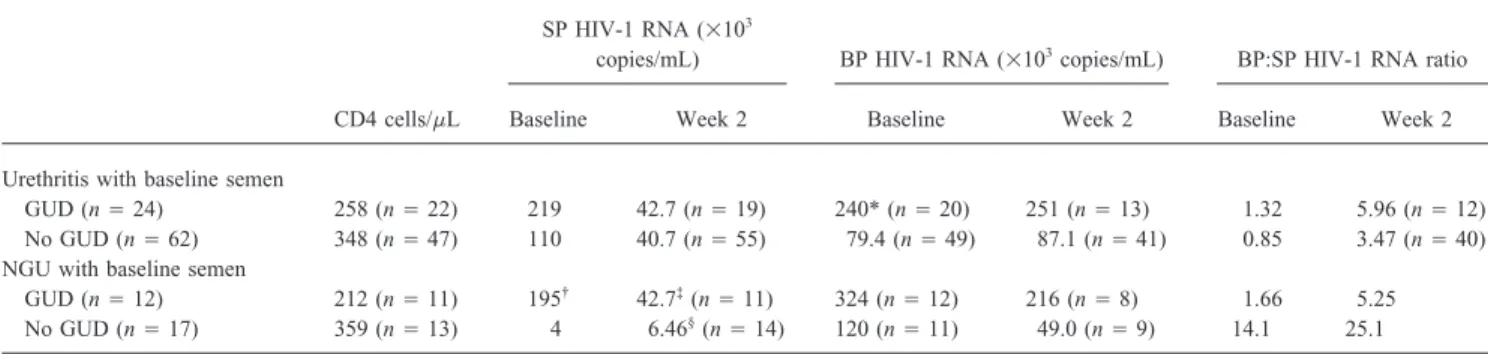
Table 1. CD4 cell counts and seminal plasma (SP) and blood plasma (BP) HIV-1 RNA concentrations for patients with or without genital ulcer disease (GUD) who provided a baseline semen sample and for the subset of patients with nongonococcal urethritis (NGU).
SP HIV-1 RNA (1103
copies/mL) BP HIV-1 RNA (1103copies/mL) BP:SP HIV-1 RNA ratio
CD4 cells/mL Baseline Week 2 Baseline Week 2 Baseline Week 2
Urethritis with baseline semen
GUD (nÅ 24) 258 (n Å 22) 219 42.7 (n Å 19) 240* (n Å 20) 251 (n Å 13) 1.32 5.96 (n Å 12) No GUD (nÅ 62) 348 (n Å 47) 110 40.7 (n Å 55) 79.4 (n Å 49) 87.1 (n Å 41) 0.85 3.47 (n Å 40) NGU with baseline semen
GUD (nÅ 12) 212 (n Å 11) 195†
42.7‡
(nÅ 11) 324 (n Å 12) 216 (n Å 8) 1.66 5.25 No GUD (nÅ 17) 359 (n Å 13) 4 6.46§
(nÅ 14) 120 (n Å 11) 49.0 (n Å 9) 14.1 25.1
NOTE. Data are medians. PÅ *.005 or†
.039, compared with values for men with urethritis but not GUD. PŇ
.017 or§
226 Concise Communications JID 1998; 177 (January)
were tested, 23% herpes simplex virus [9]. Only 1 man in the rectly related to the concentration of virus in genital secretions. Enhanced HIV-1 transmission in the setting of mucosal STDs current study had GUD and a positive rapid plasma reagin test,
although some men may have had primary syphilis in an early could be due to increased levels of viral shedding in semen [5]. GUD increases the risk of sexual acquisition of HIV-1, stage prior to a positive serology.
HIV RNA levels in blood were significantly higher in HIV- presumably by directly exposing susceptible submucosal cells to infectious virus in genital secretions [1, 2]. Similarly, direct 1 – infected men with coexisting GUD and urethritis compared
with levels in men with urethritis alone. The higher blood virus contact of HIV-1 – producing cells with the mucosal surface of uninfected sex partners could increase the infectiousness of burden in the presence of GUD may be because this group of
men had more advanced HIV disease or had systemic immune patients with HIV-1 and GUD [1]. Independent of mucosal disruption, the increase of HIV in semen, which was observed activation caused by GUD itself, as has been demonstrated for
other infection (e.g., herpes simplex virus) [10]. CD4 cell in some men with GUD, could also affect transmission and complicate interpretation of studies focused on the effects of counts were lower in the men with GUD, as has been observed
in female sex workers [4]. We believe many of the ulcers in genital ulcers. Given the high prevalence of genital ulceration in sub-Saharan Africa, our findings strongly support HIV pre-this study were chancroid, which appears to develop more
readily and be cured less easily in the presence of HIV-related vention programs that provide for prompt recognition and ef-fective treatment of STDs, including GUD.
immunodeficiency [11]. Genital herpes simplex virus infection would also be expected to be reactivated more frequently in more immunosuppressed persons. Consistent with these
obser-Acknowledgments vations, we found GUD more frequently in HIV-1 –
seroposi-tive men than seronegaseroposi-tive subjects in this population. We thank Jerry Russell and Peter Killick of John Snow Inc./ Seminal plasma HIV-1 RNA levels were also higher in men Support To AIDS and Family Health Project and F. L. Musisi of with GUD and urethritis than in men with urethritis alone. This the Tick-borne Diseases Vaccination Production Centre, Lilongwe, and Terrie Taylor of the Malaria Project, Blantyre, Malawi, for difference was most marked in men with NGU. Our laboratory
administrative and logistical support; Jodie Schock, John Schmitz, previously demonstrated that the presence of gonorrhea and its
Michelle Fiordisi, Elaine Doherty-Leach, and Lance Nkana for attendant inflammation resulted in high levels of HIV RNA in
technical and laboratory support; and Carol Porter of the Sheps seminal plasma (158 1 103 copies/mL) [5]. In the current
Center, Chapel Hill, North Carolina, for assistance with data man-analysis, the presence of GUD did not significantly increase
agement. HIV-1 RNA levels in the semen of men with gonorrhea (data
not shown). We have also reported that treatment of NGU
significantly reduced HIV-1 RNA levels in seminal plasma [5]. References Our current analysis demonstrates that this effect can be
as-1. Wasserheit JN. Epidemiological synergy: interrelationships between hu-cribed to changes observed in men with NGU and GUD whose man immunodeficiency virus infection and other sexually transmitted levels of HIV RNA in semen are comparable to levels in men diseases. Sex Transm Dis1992; 19:61 – 77.
2. Dickerson MC, Johnston J, Delea TE, White A, Andrews E. The causal with gonococcal urethritis.
role for genital ulcer disease as a risk factor for transmission of human Increased HIV-1 RNA shedding in semen in men with GUD
immunodeficiency virus. An application of the Bradford Hill criteria. could be explained in part by their higher blood plasma
HIV-Sex Transm Dis1996; 23:429 – 40.
1 levels, as suggested by the multivariate analysis and by our 3. Plummer FA, Simonsen NJ, Cameron DW, et al. Cofactors in male-female previous work in HIV-1 – infected men in North America and transmission of human immunodeficiency virus type 1. J Infect Dis
1991; 163:233 – 9. Europe [12]. The fact that blood HIV-1 RNA levels in men
4. Ghys PD, Diallo MO, Ettie`gne-Traore´ V, et al. Genital ulcers associated with GUD and NGU did not change with treatment (while
with human immunodeficiency virus – related immunosuppression in fe-semen concentrations were reduced) supports the hypothesis
male sex workers in Abidjan, Ivory Coast. J Infect Dis 1995; 172: that GUD may cause compartmentalized enhancement of HIV- 1371 – 4.
1 shedding. The multivariate analysis, which was of limited 5. Cohen MS, Hoffman IF, Royce RA, et al. Reduction of concentration of HIV-1 in semen after treatment of urethritis: implications for prevention power, did not conclusively demonstrate an effect of GUD on
of sexual transmission of HIV-1. Lancet1997; 349:1868 – 73. seminal virus level independent of blood plasma virus level.
6. Vernazza PL, Eron JJ, Fiscus SA. Sensitive method for the detection of Our conclusions are limited somewhat by the relatively small
infectious HIV in semen of seropositive individuals. J Virol Methods number of patients with GUD, the confounding effects of coex- 1996; 56:33 – 40.
isting urethritis, and by incomplete data for CD4 cell and 7. Dyer JR, Gilliam BL, Eron JJ, Grosso L, Cohen MS, Fiscus SA. Quantita-tion of human immunodeficiency virus type 1 RNA in cell free seminal plasma virus load measures. Further studies of the effects of
plasma: comparison of NASBA with Amplicor reverse transcription – GUD on HIV-1 shedding in genital secretions in larger numbers
PCR amplification and correlation with quantitative culture. J Virol of patients with and without urethritis are warranted.
Methods1996; 60:161 – 70.
The risk of HIV-1 transmission has been linked with the 8. Gobbers E, Hilgers P, Fransen K, van de Wiel P, van der Groen G. size of the virus inoculum in a variety of settings [13 – 15]. Sensitivity and amplification efficiency of the NASBA HIV-1 RNA amplification system with regard to different HIV-1 genotypes [abstract Sexual transmission from an infected male is likely to be
di-227 JID 1998; 177 (January) Concise Communications
We.B.3167]. In: Program and abstracts: XI International Conference on 12. Vernazza PL, Gilliam BL, Dyer JR, et al. Quantification of HIV in semen: correlation with antiviral treatment and immune status. AIDS1997; 11: AIDS (Vancouver, Canada, 7 – 12 July 1996). Vol 2. Vancouver: XI
International Conference on AIDS Society,1996. 987 – 93.
13. Lee TH, Sakahara N, Fiebig E, Busch MP, O’Brien TR, Herman SA. 9. Behets FMT, Liomba G, Lule G, et al. Sexually transmitted diseases and
human immunodeficiency virus control in Malawi: a field study of Correlation of HIV-1 RNA levels in plasma and heterosexual transmis-sion of HIV-1 from infected transfutransmis-sion recipients [letter]. J Acquir genital ulcer disease. J Infect Dis1995; 171:451 – 5.
10. Mole L, Ripich S, Margolis D, Holodniy M. Plasma HIV RNA levels are Immune Defic Syndr Hum Retrovirol1996; 12:427 – 8.
14. Fang G, Burger H, Grimson R, et al. Maternal plasma human immunodefi-increased during active herpes simplex virus infection. In: Program and
abstracts: 2nd Conference on Retroviruses and Opportunistic Infections ciency virus type 1 RNA level: a determinant and projected threshold for mother-to-child transmission. Proc Natl Acad Sci USA1995; 92: (Washington, DC). Alexandria, VA: Infectious Disease Society of
America,1995:98. 12,100 – 4.
15. Busch MP, Operskalski EA, Mosley JW, et al. Factors influencing human 11. Tyndall M, Malisa M, Plummer FA, Ombetti J, Ndinya-Achola JO, Ronald
AR. Ceftriaxone no longer predictably cures chancroid in Kenya. J immunodeficiency virus type 1 transmission by blood transfusion. J Infect Dis1996; 174:26 – 33.
Infect Dis1993; 167:469 – 71.
Horizontal and Vertical Transmission of Human Immunodeficiency Virus Type 1
Dual Infections Caused by Viruses of Subtypes B and C
Luiz M. Janini, Amilcar Tanuri, Mauro Schechter, Division of AIDS, STD, TB, Laboratory Research, and Division of Parasitic Disease, Centers for Disease Control and Prevention, Atlanta, Jose M. Peralta, Ana C. P. Vicente, Nick Dela Torre,
Georgia; Instituto de Microbiologia and Biologia, and Programa SIDA/ Norman J. Pieniazek, Chi-cheng Luo, Artur Ramos,
AIDS, Hospital Clementino Fraga Filho, Universidade Federal do Rio Vincent Soriano, Gerald Schochetman, Mark A. Rayfield, de Janeiro, and Instituto Oswaldo Cruz, Rio de Janeiro, Brazil; Instituto
and Danuta Pieniazek de Salud Carlos III, Madrid, Spain
This article describes a case of horizontal (heterosexual) and subsequent vertical (mother to infant) transmission of 2 human immunodeficiency viruses type 1 (HIV-1) subtypes. Dual infection in a husband, his wife, and their child was initially detected by use of a restriction fragment length polymorphism assay of the proviral protease in peripheral blood mononuclear cells. The simultane-ous presence of highly similar sets of HIV-1 subtypes B and C infecting the 3 family members was confirmed by DNA sequence analysis of pol, gag, and env genes. These data, together with available epidemiologic information, may indicate that the husband’s high-risk sexual behavior was the source of dual infections. Because his wife did not report such activities, it was likely that he passed HIV-1 strains to his spouse, who subsequently transmitted them to their child.
The human immunodeficiency virus type 1 (HIV-1) pan- are considered responsible for the spreading of diverse HIV-1 subtypes into regions previously affected by HIV-1 strains of demic is now recognized as consisting of many separate
epi-demics caused by 10 viral subtypes [1]. With an estimated 15 1 subtype. As a consequence, the potential for HIV-1 mixed infections and genetic recombination involving strains of dis-million HIV-1 – infected persons, the geographic distribution of
viral clades is becoming more dispersed, and the simultaneous tinct subtypes in an infected individual has increased [2, 3]. The development of specific genetic methods has enabled iden-presence of multiple subtypes in certain regions has become
tification of dual HIV-1 infections caused by subtypes B and more common. Population migration and international travel
E in Thailand [4], as well as by subtypes D and F, and subtypes B and F in Brazil [5].
Detection of naturally occurring HIV-1 multiple infections has important implications for vaccine development because it suggests Received 10 April 1997; revised 7 August 1997.
that infection with 1 HIV-1 subtype may not fully protect against Presented in part: 4th Conference on Retroviruses and Opportunistic
Infec-tions, Washington DC, 22 – 27 January 1997 (abstract 29). subsequent superinfection with distinct HIV-1 strains. Although Informed consent was obtained from human subjects.
dual HIV-1 infections caused by viruses of distinct subtypes have Sequences have been submitted to GenBank under accession nos. U19432,
been documented, the transmission of 2 HIV-1 subtypes from 1 U19433, U19437, U19438, U19450, U19451, and U83689-U83700.
Reprints or correspondence: Dr. Danuta Pieniazek, HIV/Retrovirus Diseases dually infected person to another has not been reported. Branch, Division of AIDS, STD, TB, Laboratory Research, CDC, 1600 Clifton
Rd., Mail Stop G19, Atlanta, GA 30333. Materials and Methods
The Journal of Infectious Diseases 1998; 177:227 – 31
Specimens. Two Brazilian specimens were from a married
q 1998 by The University of Chicago. All rights reserved.