TRANSPORTERS
Recycling of aromatic amino acids via TAT1 allows efflux
of neutral amino acids via LAT2-4F2hc exchanger
Tamara Ramadan&Simone M. R. Camargo&
Brigitte Herzog&Mauro Bordin&Klaas M. Pos&
Francois Verrey
Received: 3 November 2006 / Accepted: 8 January 2007 / Published online: 2 February 2007 # Springer-Verlag 2007
Abstract The rate of amino acid efflux from individual cells needs to be adapted to cellular demands and plays a central role for the control of extracellular amino acid homeostasis. A particular example of such an outward amino acid transport is the basolateral efflux from trans-porting epithelial cells located in the small intestine and kidney proximal tubule. Because LAT2-4F2hc (Slc7a8– Slc3a2), the best known basolateral neutral amino acid transporter of these epithelial cells, functions as an obligatory exchanger, we tested whether TAT1 (Slc16a10), the aromatic amino-acid facilitated diffusion transporter, might allow amino acid efflux via this exchanger by recycling its influx substrates. In this study, we show by immunofluorescence that TAT1 and LAT2 indeed colo-calize in the early kidney proximal tubule. Using the Xenopus laevis oocytes expression system, we show thatL
-glutamine is released from oocytes into an amino-acid-free medium only when both transporters are coexpressed. High-performance liquid chromatography analysis reveals that several other neutral amino acids are released as well. The transport function of both TAT1 and LAT2-4F2hc is necessary for this efflux, as coexpression of functionally inactive but surface-expressed mutants is ineffective. Based
on negative results of coimmunoprecipitation and cross-linking experiments, the physical interaction of these transporters does not appear to be required. Furthermore, replacement of TAT1 or LAT2-4F2hc by the facilitated diffusion transporter LAT4 or the obligatory exchanger LAT1, respectively, supports similar functional cooperation. Taken together, the results suggest that the aromatic amino acid diffusion pathway TAT1 can control neutral amino acid efflux via neighboring exchanger LAT2-4F2hc, by recycling its aromatic influx substrates.
Keywords Basolateral efflux . Slc16a10 . Slc7a8 . Aromatic amino acid transporter . Glycoprotein-associated amino acid transporters . Xenopus oocytes . Kidney
Introduction
Transcellular absorption of neutral amino acids from the lumen of the small intestine and reabsorption from the primary urinary filtrate of the kidney proximal tubule involves transport across two sequential membranes. Apical uptake from the lumen into epithelial cells against a con-centration gradient is mediated by secondary and tertiary active transporters, whereas basolateral efflux into the extracellular space is thought to be driven by the amino acid concentration gradient [28]. The major player for the luminal neutral amino acid uptake is B0AT1 (Slc6a19), which catalyzes the Na+-dependent secondary active trans-port of most neutral amino acids into epithelial cells [4,5,
15]. The efflux of neutral amino acids across the basolateral membrane into the extracellular space could potentially take place entirely via facilitated diffusion pathways. However, the abundant basolateral amino acid transporters LAT2-4F2hc and y+LAT1-4F2hc have been shown to
T. Ramadan
:
S. M. R. Camargo:
B. Herzog:
M. Bordin:
K. M. Pos:
F. VerreyZurich Centre for Integrative Human Physiology (ZIHP), University of Zürich,
Winterthurerstrasse 190, 8057 Zurich, Switzerland
T. Ramadan
:
S. M. R. Camargo:
B. Herzog:
M. Bordin:
K. M. Pos:
F. Verrey (*)Institute of Physiology, University of Zurich, Winterthurerstrasse 190,
8057 Zurich, Switzerland e-mail: [email protected]
function as obligatory exchangers [9, 17, 20, 24]. For that reason, these heterodimeric amino acid transporters cannot perform net overall amino acid efflux but only net efflux of specific amino acids provided that others are taken up. It has been proposed that (a) parallel amino acid transporter(s), mediating the unidirectional efflux of some amino acids, may provide heterodimeric exchangers with influx substrates that would allow them to efflux other intracellular amino acids [18,27]. Thus, the heterodimeric exchangers could be envisaged as modules extending the efflux selectivity across the membrane, whereas the overall efflux function would be controlled by the parallel trans-porters potentially functioning unidirectionally.
For the following several reasons, we considered that TAT1 (SLC16A10) is a plausible candidate for such a role as a substrate recycling pathway that may control the efflux function of basolateral heterodimeric amino acid trans-porters in small intestine and proximal tubule epithelial cells. First, the function of TAT1, a member of the Slc16 family of monocarboxylate transporters, was identified as that of a facilitated diffusion pathway for aromatic amino acids [13,14]. In particular, it was shown that it may efflux
L-phenylalanine independent of the presence of
extracellu-lar amino acids, thus being able to perform a unidirectional transport [13, 22]. Second, we recently showed that TAT1 displays symmetrically low apparent affinities for its substrates with Km’s in the millimolar range. This implies
that TAT1 can kinetically adapt its transport rate to varying amino acid concentrations [22]. Third, TAT1 (SLC16A10) was previously shown to be expressed in the small intestine and proximal kidney tubule and its expression to be restricted to the basolateral membrane of these epithelia, where it colocalizes with 4F2hc and thus probably with LAT2 [22].
To test the possibility that TAT1 may function as a recycling pathway to allow and actually control the function of LAT2-4F2hc and thus to better understand its physiological role, we first verified its coexpression with LAT2-4F2hc in the mouse kidney proximal tubule by immunofluorescence and then addressed the question of its possible functional cooperation with LAT2-4F2hc and the potential physical interaction of these transporters using Xenopus oocytes as an expression system.
Materials and methods
Site-directed mutagenesis of mTAT1 and mLAT2
Functionally inactive mutants of mTAT1 (R340A) and mLAT2 (E257Q) were created by site-directed mutagenesis as described previously [10]. Oligonucleotide primer pairs used were as follows: mLAT2E257Q_for: 5′-CTTAATT
AT G T G A C T G A G C A G C T G G T G G AT C C T TA C A A GAACC-3′, mLAT2E257Q_rev: 5′-GGTTCTTGTAAGG ATCCACCAGCTGCTCAGTCACATAATTA AG-3′; m TAT 1 R 3 4 0 A _ f o r : 5′ - C A C T T C A G G A G T T G GAGCGCTTCTCTTTGGCCGC-3′, mTAT1R340A_rev: 5′-GCGGCCAAAGAGAAGCGCTCCAACTCCT GAAGTG-3′. Each reaction consisted of indicated primer pairs, plasmid templates mLAT2 or pSDeasy-mTAT1, respectively, and 2.5% formamide as an additive [6, 11]. The amplification mixture was treated with DpnI for 2 h at 37°C, and digested samples were used to transform Escherichia coli MACH1-T1 (Invitrogen).
Xenopus laevis oocytes and cRNA synthesis
Oocytes were treated with collagenase A1 for 2–3 h at room temperature (RT) in a Ca2+-free buffer containing 10 mM HEPES, pH 7.4, 82.5 mM NaCl, 2 mM KCl, and 1 mM MgCl2. Oocytes were kept at 16°C in a ND96
solution (96 mM NaCl, 2 mM KCl, 1 mM MgCl2, 1.8 mM
CaCl2, and 5 mM HEPES/Tris, pH 7.4), supplemented with
50 mg/l of tetracycline. Mouse TAT1 cDNA, R340A TAT1 mutant cDNA, mouse LAT2 cDNA, and E257Q LAT2 mutant cDNA, all in pSDeasy Xenopus oocyte expression vector, were used for synthesis of cRNA transcripts as described before [22]. Briefly, plasmids containing the described cDNAs were linearized using a unique BglII restriction site. Likewise, the plasmid containing mouse LAT4 cDNA (in vector pTLN, kindly provided by Palacin) was linearized using the MluI restriction site, and the plasmid containing human LAT1 (vector pcDNA/Amp-pSP64T) was linearized using the EcoRV restriction site. cRNA was synthesized with SP6 RNA polymerase using the MEGAscript high-yield transcription kit (Ambion, Austin, TX) according to the manufacturer’s protocol. The plasmid containing human 4F2hc cDNA (vector pSport) was linearized using a unique HindIII restriction site, and the corresponding cRNA was synthesized using T7 RNA polymerase.
Measurement of amino acids released by oocytes
Amino acid efflux from oocytes expressing LAT2-4F2hc and/or TAT1 was measured in the extracellular ND96 buffer after 24 h incubation. Samples of 200 μl were collected from a total number of 20 oocytes for each group and analyzed by high-performance liquid chromatography (HPLC). One microliter was injected for precolumn derivatization with ortho-phthaldialdehyde and analyzed on an Amino Quant amino acid analyzer (Agilent Technol-ogies GmbH, Deutschland) at the Protein Analysis Group (Functional Genomics Center Zürich). The absolute amounts of amino acids measured in the extracellular
buffer are expressed as concentration [μM] of free amino acid detected in the analyzed sample. Results were analyzed using the GraphPad Prism 4 Software (San Diego, CA).
Tracer flux studies using X. laevis oocytes
Treatment and injection of oocytes was performed as described previously [17]. Briefly, after injection of 5–
25 ng of cRNA (for wild-type or mutant transporter, respectively), oocytes were incubated 2–3 days at 16°C in ND96 solution. For efflux studies, oocytes were micro-injected with [14C]-radiolabeled amino acid (∼10 nCi per oocyte; Hartmann Analytic, Braunschweig, Germany). After a brief wash with uptake buffer at RT, single oocytes were incubated in 200 μl of uptake solution (10 mM HEPES, pH 7.4, 100 mM NaCl, 2 mM KCl, 1 mM MgCl2,
and 1 mM CaCl2). Aliquots were collected at indicated
times, and transport of amino acids was terminated by washing the individual oocytes five times with 2 ml of ice-cold uptake buffer. To determine the retained radioactivity, the oocytes were separately dissolved in 2% sodium dodecyl sulfate (SDS, 250 μl) and shaken for 60 min. Upon addition of scintillation cocktail (3 ml, Emulsifier-Safe™), radioactivity was measured using a liquid scintil-lation counter (TRI-CARB 2900TR, Packard Instrument). The values were corrected for the volume of the incu-bation solution and divided by the total amount of radio-activity injected into the oocyte. This injected amount (corresponding to 100%) was calculated for each oocyte separately by adding the volume-corrected radioactivity measured in the extracellular sample at the last time point, radioactivity measured in the previous sample (60 min, not corrected for volume), and the radioactivity remaining in the oocyte. For graphical representation, raw data sets were analyzed using the computer software GraphPad Prism 4 (GraphPad Prism version 4.02, GraphPad soft-ware). Statistical analysis was done using the two-tailed paired t test or one-way analysis of variance followed by Bonferroni’s multiple comparison post test, using GraphPad Prism 4.
Antibodies
Affinity purified rabbit anti-mTAT1 antibody was used at a dilution of 1:500 for Western blot analysis and 1:200 for immunohistochemistry, as described before [22]. The other antibodies used were previously characterized: rabbit anti-mLAT2 antibody, serum SZ560 (dilution 1:1000 used for Western blotting and 1:250 for immunohistochemistry) [19], goat anti-m4F2hc (Santa Cruz Biotech, Santa Cruz, CA, 1:500 dilution used for immunohistochemistry), and mouse anti-h4F2hc (1 μg used per IP reaction) [24]. Secondary antibodies used for immunohistochemistry were
Alexa Fluor® 594 donkey anti-rabbit IgG (1:1000; Molec-ular Probes, Portland, OR) and Alexa Fluor® 488 donkey– anti-goat IgG (1:500; Molecular Probes), whereas, for Western blot analysis, a goat anti-rabbit IgG conjugated to Horseradish peroxidase was used as secondary antibody (1:10,000; BD Transduction Laboratories, Lexington, KY).
Immunohistochemistry
Serial sections (4μm) of perfused and frozen mouse kidney were collected on polylysine-coated slides (O. Kindler GmbH, Freiburg, Germany). Animals were killed according to the Swiss Animal Welfare Laws and as approved by the local Veterinary Authority (Kantonales Veterinäramt Zürich). Immunostaining of tissue sections was carried out as described previously [29]. Briefly, sections were rehy-drated in a phosphate buffer solution (PBS) for 15 min at RT, washed three times with PBS, and incubated with PBS containing 1% bovine serum albumin for 15 min before addition of the primary antibodies. Affinity-purified prima-ry rabbit anti-mTAT1 antibody or rabbit anti-mLAT2 serum together with affinity purified goat anti-m4F2hc antibody were applied to the sections and incubated overnight at 4°C. Sections were then washed twice by immersion for 5 min in hypertonic PBS (PBS containing 2.7% NaCl) and once in PBS before incubation with the secondary antibody (Alexa Fluor® 594 donkey anti-rabbit IgG, dilution 1:1000 and Alexa Fluor® 488 donkey–anti-goat IgG, dilution 1:500) for 1 h at RT. Sections were washed twice with hypertonic PBS, followed by a washing step with PBS before mounting them with VectaMount (Vector Laboratories, Burlingame, CA). Sections were examined using a LEICA SP1 UV CLSM confocal micro-scope. Pictures were processed and assembled using the Imaris® 5.0.1 and Adobe Photoshop® 7.0 software. Incubation of sections with only secondary antibodies did not result in detection of signal and signal specificity of anti-mTAT1 antibody was further confirmed by addition of antigen peptides to primary antibodies before application (data not shown).
Western blotting on oocyte protein lysates
Total membrane lysates of oocytes were prepared by homogenization in an oocyte lysis buffer (250 mM sucrose, 0.5 mM ethylenediamine tetraacetic acid [EDTA], 5 mM Tris–HCl, pH 6.9, 1 mM phenylmethylsulfonyl fluoride (PMSF), and 1 μg/ml of leupept). Lysates were twice centrifugated at 100g for 10 min, and the supernatants were subjected to SDS-polyacrylamide gel electrophoresis (SDS-PAGE). Proteins were subsequently transferred electropho-retically from unstained gels to a polyvinylidene difluoride (PVDF) membrane (Immobilon-P, Millipore, Bedford,
MA), blocked with 2% Top BLOCK™ powder (Juro, Lucerne, Switzerland) and probed with indicated anti-bodies as described. Chemiluminescence was detected with a DIANA III camera (Raytest Schweiz, Dietikon, Switzerland).
Immunoprecipitation, SDS-PAGE, and fluorography
Oocytes injected with indicated cRNAs were biosyntheti-cally labeled with 1 mCi/mlL-[35S]-methionine for 2 days
of expression. After extensive washing with ND96, oocytes were lysed by addition of 20μl of EBC buffer (120 mM NaCl, 50 mM Tris–HCl, pH 8.0, 0.5% NP-40, 1 mM PMSF, and 10 μl/ml protease inhibitor cocktail [Sigma]) per oocyte and centrifuged at 12,000 rpm at 4°C for 10 min, and supernatants were separated and precipitated with trichloroacetic acid for determination of incorporation of radioactivity. For the immunoprecipitation assay, 1 μg of mouse monoclonal anti-h4F2hc antibody per reaction was coupled to 30 μl of ImmunoPure® Immobilized Protein A/G (Pierce, Rockford, IL). Protein lysates were precleared by mixing with protein A/G agarose beads and rotating at 4°C for 1 h. Precleared protein lysates, containing equal amounts of incorporated L-[35S]-methionine, were after-wards mixed with anti-h4F2hc-coupled beads in EBC buffer (500 μl) and incubated overnight at 4°C with continuous rotation. Immunoprecipitated samples were afterwards briefly centrifuged to pellet the beads, washed six times with NET-N buffer (containing 20 mM Tris–HCl, pH 8.0, 100 mM NaCl, 1 mM EDTA, and 0.5% NP40), and resuspended in SDS-PAGE-loading buffer containing β-mercaptoethanol. The samples were subsequently boiled for 5 min at 95°C and separated by SDS-PAGE. After separation, proteins were either electrophoretically trans-ferred to PVDF membrane or the gels were fixed and stained with Coomassie Brilliant Blue R250, subsequently treated with Amplify Fluorographic Reagent (Amersham Biosciencies, Little Chalfont, Bucks, UK) for 30 min, dried, and exposed to Fuji Super RX medical X-ray film at −80°C.
Cell surface biotinylation and streptavidin precipitation
Cell-surface proteins of oocytes were labeled with cell-membrane-impermeable and primary-amines-reactive sulfo-NHS–LC–biotin (Pierce), according to the manufacturer’s protocol. Briefly, oocytes were injected with indicated cRNAs and kept at 16°C for 2–3 days. Subsequent steps were done on ice, except mentioned otherwise. The oocytes were washed in groups three times with ice-cold PBS, pH 8.0, and afterwards incubated on ice for 30 min in biotinylation solution containing 2 mM sulfo-NHS–LC– biotin. The reaction was finally quenched by addition of
10 mM Tris–glycine, for 15 min. After washing six times with ice-cold PBS, the oocytes were lysed as described above. After a centrifugation step of 12,000 rpm for 10 min at 4°C, supernatants containing total oocyte proteins were transferred to new tubes and kept at−80°C. For streptavidin precipitation, 40μl of ImmunoPure® Immobilized Strepta-vidin agarose beads were washed three times with TBS-TWEEN20 (0.1%), mixed with 100 μl of total protein oocyte lysates (approx. 4μg/μl), and afterwards rotated for 4 h at 4°C. Samples were then washed six times with TBS-TWEEN20, with a 1 min centrifugation step (3,000 rpm) between washes. After the final wash, 4× SDS-PAGE sample buffer was added, and samples were heated to 65°C for 15 min and subjected to SDS-PAGE analysis. Protein concentrations were determined according to the modified method of Lowry (DC Protein Assay, Bio-Rad Laborato-ries, California).
Crosslinking experiments
The crosslinking reagent 1,4-bis-maleimidobutane (BMB; Pierce) has homobifunctional maleimide ends reactive towards the sulfhydryl groups on proteins and peptides, in a distance of 10.9 Å given by the noncleavable spacer arm. The lyophilized crosslinker was dissolved in dimethyl sulfoxide to 2.5 mM final concentration, before the reaction setup. Optimization of reaction included titration of the crosslinker concentration, as well as time course of incubation (data not shown). The final conditions used for the reaction mixture included 50 μg of total membranes extracted from oocytes and 100μM BMB crosslinker. The reaction buffer used was PBS, pH 7.4. The samples were treated with BMB for 1, 10, or 30 min at RT or left untreated. Samples were subjected to SDS-PAGE analysis using 6% acrylamide gel, electrophoretically transferred to a PVDF membrane, and afterwards probed with anti-LAT2 or anti-TAT1 antibodies.
Results
TAT1 and LAT2-4F2hc are colocalized in the proximal kidney tubule
Transepithelial transport of amino acids requires an active uptake of amino acids at the luminal side of the epithelial cell and their extrusion at the basolateral side. In the kidney proximal tubule, expression of LAT2 that heterodimerizes with 4F2hc was shown to be basolateral and to progres-sively decrease towards the late segments of the proximal tubule [2,24]. Similarly, TAT1 was shown to be expressed at the basolateral membrane of the kidney proximal tubule epithelial cells in the S1 and S2 segments [22]. In this
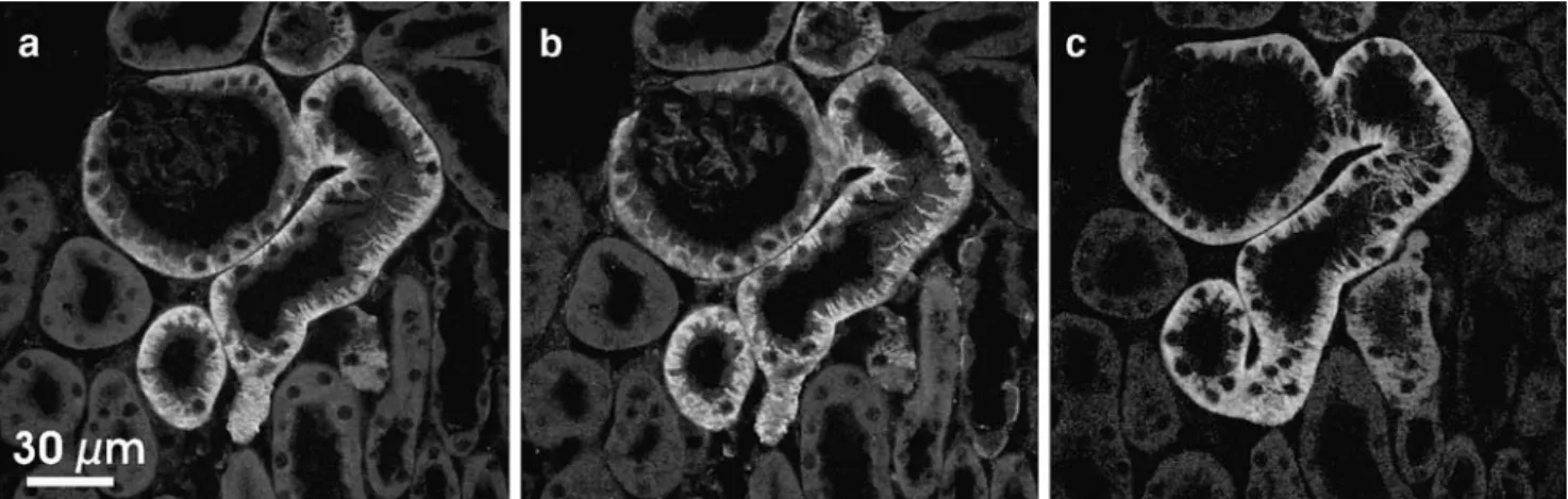
study, we show on consecutive sections of mouse kidney that, as expected, TAT1 and LAT2-4F2hc proteins colo-calize their signals starting from the glomerular capsule and extending into the S1 and S2 segments of the proximal tubule (Fig.1).
Coexpression of TAT1 with the exchanger LAT2-4F2hc induces efflux ofL-glutamine and of other neutral amino acids
The aromatic amino acid transporter TAT1 is known to function as a facilitated diffusion pathway, whereas the neutral amino acid transporter LAT2-4F2hc functions as an obligatory exchanger [2, 17, 22]. To test the potential functional cooperation of these transporters, we expressed them individually and together in Xenopus oocytes. We measured the efflux of radiolabeled L-Gln, which is a
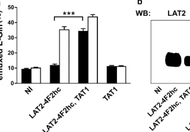
known efflux substrate of LAT2 4F2hc but not of TAT1. Figure2a shows that oocytes preinjected withL-glutamine
released this amino acid into the amino-acid-free medium (black bars) when both transporters were simultaneously expressed. This observation is in line with the hypothesis that TAT1 provides extracellular aromatic amino acids to LAT2-4F2hc and that this latter exchanger transports them back into the oocyte in exchange for effluxingL-glutamine.
The fact that LAT2-4F2hc was functionally expressed also in the absence of TAT1 and could releaseL-glutamine in the presence of sufficient influx substrate was demonstrated by the rapid L-glutamine efflux from LAT2-4F2hc-expressing
oocytes upon addition of 2.5 mM L-phenylalanine to the medium for 10 min at the end of the experiment (white bars). The expression of both transporters was verified by Western blotting (Fig.2b).
The spectrum of amino acids released by Xenopus oocytes expressing both TAT1 and LAT2-4F2hc was tested by HPLC quantification of the amino acids accumulated in the extracellular amino-acid-free medium within 24 h
(Fig. 3). As previously shown, the expression of TAT1 alone mediated an efflux of the three aromatic amino acids independent of the presence of LAT2-4F2hc, as expected for a facilitated diffusion pathway selective for aromatic amino acids [22]. In contrast, as expected for a (near) obligatory exchanger, the expression of LAT2-4F2hc alone did not lead to a significant leak of any amino acid into the media [17]. However, as shown in this paper, the co-expression of these transport proteins led to an efflux of
L-glutamine, L-serine, L-asparagine, and L-alanine into the
medium that was statistically significant compared to that from both oocytes expressing TAT1 or LAT2-4F2hc alone. The additional efflux ofL-histidine,L-threonine, andL
-iso-leucine did not reach a significant level. These results show that all neutral amino acids that significantly leak into the extracellular medium belong to the good efflux substrates of LAT2-4F2hc and that they are the more concentrated ones in oocytes [17,26].
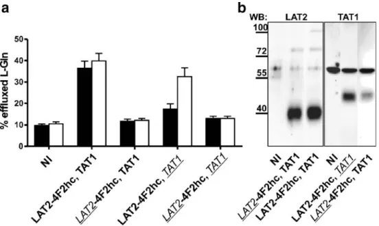
To test whether the additional efflux of neutral amino acids from TAT1- and LAT2-4F2hc-coexpressing oocytes is mediated by the functional cooperation of the two trans-porters or, alternatively, is mediated by a modified transport function of one of the two proteins because of the physical presence of the other, we produced mutant transporters that are defective in amino acid transport function but nonethe-less are normally expressed at the cell surface. As shown in Fig. 4a, the expression of either combination of functional and mutant transporters did not induce the efflux of L
-glutamine, although Western blot analysis of surface-biotinylated proteins (Fig.4b) showed that both transporters were expressed at the oocyte surface. Taken together, these data strongly suggest that it is the combination of the functions of TAT1 and LAT2-4F2hc that provides the efflux pathway for L-glutamine.
Fig. 1 Basolateral colocalization of TAT1 and LAT2-4F2hc in the epithelial cells of the mouse proximal kidney tubule. Expression and subcellular localization of TAT1, LAT2, and 4F2hc was determined by
immunofluorescence microscopy. Two consecutive sections of frozen mouse kidney were costained with anti-4F2hc (a) and anti-LAT2 (b) or with anti-TAT1 (c) antibody, respectively
TAT1 and LAT2-4F2hc appear not to interact physically
The functional cooperation of TAT1 with LAT2-4F2hc suggested the possibility of a physical interaction of these two transporters that would favor substrate channeling from TAT1 to LAT2. To address this possibility, we tested whether TAT1 could be coimmunoprecipitated with LAT2-4F2hc. Lane 2 of Fig. 5 shows the proteins immunoprecipitated with an anti-4F2hc antibody from a lysate of oocytes expressing LAT2-4F2hc and TAT1 (fluorography of SDS-PAGE). 4F2hc appears as two bands, one at ∼66 kDa corresponding to the core-glycosylated form and around 85 kDa, the terminally glycosylated form,
whereas a single other band of ∼42 kDa is visible that presumably corresponds to LAT2 [24]. Western blot analysis of the same lysate used for immunoprecipitation showed that both LAT2 (lane 4) and TAT1 (lane 8) were present. In the 4F2hc immunoprecipitate, the catalytic subunit of the heterodimer LAT2 was detected (lane 6) but not TAT1 (lane 10).
To test the possibility that the physical proximity/ association of TAT1 with LAT2-4F2hc was missed by the coprecipitation experiment, we submitted membranes of coexpressing oocytes to crosslinking using a series of different crosslinking agents, in particular glutaraldehyde, DTSSP, DSP, DFDNB, BM(PEO)3, and BMB and
ana-Fig. 3 Coexpression of TAT1 and LAT2-4F2hc in Xenopus oocytes induces efflux ofL-Gln and other neutral amino acids. Free amino acids, accumulated during 24 h in the extracellular buffer, were separated by HPLC after derivatization and quantified as described in “Materials and methods.” The mean values were obtained from five independent groups of each 20 oocytes. NI stands for oocytes not
injected with cRNA. Error bars represent the SEM (*P<0.05 efflux with 4F2hc and TAT1 significantly higher than with LAT2-4F2hc or TAT1 alone; #P<0.05 efflux with LAT2-4F2hc and TAT1 significantly higher that with LAT2-4F2hc alone but not than with TAT1)
Fig. 2 Coexpression of LAT2-4F2hc and TAT1 in Xenopus oocytes induces efflux ofL-Gln. a Oocytes injected with tracer amounts of radiolabeledL-Gln were incubated in an uptake solution containing no amino acids for 60 min (black bars). Transtimulation was induced for additional 10 min by the addition of 2.5 mML-Phe to the extracellular buffer (white bars). Each bar represents the mean of 50–67 oocytes,
pooled from 12 different batches of oocytes. Error bars represent the standard error of the mean (SEM; ***P<0.001) b Western blot analysis of total protein lysates from a representative batch of oocytes. PVDF membranes were probed with anti-LAT2 and anti-TAT1 antibodies, respectively. Molecular weight of marker proteins is indicated in kilodaltons
lyzed the proteins precipitated with anti-4F2hc antibody on SDS-PAGE. As shown in Fig.6, for the case of BMB, none of the these agents crosslinked TAT1 to LAT2-4F2hc.
Taken together, the negative results of the coimmuno-precipitation and crosslinking experiments suggest that there is no close physical interaction between LAT2-4F2hc and TAT1.
Functional cooperation of TAT1 or LAT2-4F2hc with other transporters
We therefore tested whether the demonstrated function of TAT1 to allowing L-glutamine efflux via LAT2-4F2hc
could be fulfilled by another facilitated diffusion amino acid transporter. We chose to use for the substitution assay LAT4 (Slc43a2). This L-type transporter is expressed, unlike LAT2-4F2hc, in the distal nephron and does not belong to the same structural family. It was shown to transport L-phenylalanine, L-leucine, L-isoleucine, and L -methionine and to mediateL-phenylalanine efflux
indepen-dent of the presence of extracellular amino acids [3]. Interestingly, upon coexpression of LAT4 with LAT2-4F2hc, an efflux of L-glutamine was induced (Fig. 7a,
Fig. 6 Chemical crosslinking indicates no physical interaction between LAT2-4F2hc and TAT1. Total membrane lysates extracted from oocytes injected with LAT2-4F2hc, TAT1 (LT) cRNAs or not injected (NI), were subjected to BMB crosslinking and separated by SDS-PAGE. Duration of BMB crosslinking is given in minutes. LAT2 and TAT1 antibodies do not label the same bands. The main band labeled by LAT2 antibody corresponds presumably to a heterotetramer (2 LAT2-4F2hc heterodimers). The nature of the 130-kDa band labeled by TAT1 antibody is not known and could represent an oligomer
Fig. 5 TAT1 is not physically associated with 4F2hc or LAT2. Coimmunoprecipitation was performed on lysates of biosynthetically labeled oocytes coinjected with 4F2hc, LAT2, and TAT1 cRNAs (LT) or not injected oocytes (NI). Precipitations with the anti-4F2hc antibody were carried out in nonreducing conditions and separated by SDS-PAGE. RX autoradiograph, WB Western blot on total lysate, IP-4F2hc WB on immunoprecipitate
Fig. 4 Functionally inactive mutants of LAT2 (E257Q) and TAT1 (R340A) are expressed at the surface of Xenopus oocytes but do not support L-Gln efflux. a Oocytes expressing wild-type (bold) and/or mutant cRNA (italic, underlined) were injected with tracer amounts of radiolabeled L-Gln and incubated as described previously (black bars
represent 60 min of L-Gln efflux; white bars represent 10 min of subsequent transtimulation with 2.5 mML-Phe). Each bar represents the mean of 15–25 oocytes, pooled from five different batches. Error bars represent the SEM. b Oocytes were surface biotinylated, lysates separated by SDS-PAGE and probed with anti-LAT2 or anti-TAT1 antibodies
black bars) that was slightly but not significantly higher than the sum of the effluxes observed with each transporter expressed separately.
We also replaced the other component of the functional couple, namely, the catalytic subunit of the heterodimeric exhanger LAT2-4F2hc, with another catalytic subunit, LAT1. BecauseL-glutamine is not a good efflux substrate
for this transporter, we measured the efflux of L-leucine instead. Interestingly, this transporter combination was also quite efficient and released, in the absence of extracellular amino acids, significantly more L-leucine than either transporter alone (Fig. 7b, black bars). Together with the apparent lack of physical interaction of TAT1 with LAT2-4F2hc, these results suggest that the coexpression of an amino acid efflux pathway (facilitated diffusion transporters TAT1 or LAT4) with an obligatory amino acid exchanger (LAT2- or LAT1-4F2hc) can lead to a functional coopera-tion independent of physical interaccoopera-tion.
Discussion
The present study shows that the expression of the aromatic amino acid transporter TAT1 in the same membrane as the obligatory exchanger of neutral amino acids LAT2-4F2hc leads to an efflux of several neutral amino acids into the amino-acid-free medium. It is suggested that the efflux pathway used by these amino acids is the obligatory exchanger LAT2-4F2hc. This hypothesis is strongly sup-ported by the fact that the function of both surface-expressed transporters is necessary and that the amino
acids released additionally to the aromatic TAT1 substrates are all known LAT2-4F2hc efflux substrates [17]. An additional argument is that upon replacing the obligatory exchanger LAT2-4F2hc by the other neutral amino acid obligatory exchanger LAT1-4F2hc, we observe a more efficient release of the typical LAT1-4F2hc efflux substrate
L-leucine than of L-glutamine, which is not a good
LAT1-4F2hc efflux substrate (Fig.7b, data now shown, and [17]). Together, these experiments indeed very strongly suggest that it is the functional cooperation of TAT1 with LAT2-4F2hc that leads to this new efflux function. Aromatic amino acids released extracellulary by TAT1 would function as an influx substrate for LAT2-4F2hc and thereby allow the efflux of other amino acids via this obligatory exchanger. In the following paragraphs, we will discuss a few questions that arise from this suggested functional cooperation of TAT1 with LAT2-4F2hc.
The first question is why, from the efflux substrates of LAT2-4F2hc, it is in particular L-glutamine, L-asparagine, L-serine, and L-alanine that accumulate over 24 h in the
extracellular medium of oocytes expressing LAT2-4F2hc and TAT1. We suggest that the major reason that favors a high long-term efflux rate of specifically these four amino acids is the fact that they are nonessential and can be produced in the oocytes from abundant precursors. In contrast, all other LAT2-4F2hc efflux substrates that do not significantly accumulate outside of the oocytes are essential amino acids present in limited amounts within oocytes, with the exception of glycine [26].
The second question arising from the functional cooper-ation of LAT2-4F2hc with TAT1 is why it is specifically an
Fig. 7 Substitution assays indicate that other combinations of an exchanger with a facilitated diffusion transporter can lead to functional cooperation. a Oocytes expressing LAT4 instead of TAT1, with LAT2-4F2hc, were injected with radiolabeledL-Gln, incubated for 60 min in amino-acid-free medium (black bars) and subsequently transtimulated byL-Phe (white bars). Each bar represents the mean of 17–25 oocytes
pooled from three to five different experiments. b Following the same protocol, efflux of radiolabeledL-Leu by oocytes coexpressing LAT1-4F2hc, instead of LAT2-LAT1-4F2hc, together with TAT1 was measured. Each bar represents the mean of eight to ten oocytes pooled from two different experiments. Error bars represent the SEM (ns, not significant, P>0.05; ***P<0.001)
aromatic amino acid transporter that controls the efflux via the exchanger LAT2-4F2hc. We suggest here two non-mutually exclusive possibilities. First, from the point of view of organism nutrition and extracellular amino acid homeostasis, it appears meaningful that the amino acid efflux function of (transporting) cells be regulated accord-ing to essential amino acid levels, because they depend on the protein nutritional state [7,8]. Second, it might be that physico-chemical properties of aromatic amino acids, in particular, low affinity binding to proteins and/or mem-branes [25], might be an advantage for their recycling function. It can be envisaged that this type of interaction might favor the maintenance of higher local aromatic amino acid concentrations that would facilitate substrate channel-ing through the cell-surface-unstirred layer to neighborchannel-ing transporters.
A third question of importance regarding the proposed functional cooperation of the TAT1 with LAT2-4F2hc is the fact that the concentrations of substrate amino acids necessary to substantially activate LAT2-4F2hc from the outside is clearly much higher than the extracellular amino acid concentrations measured in the present experiments [17]. This discrepancy suggested to us the possibility of a physical interaction between TAT1 and LAT2-4F2hc. However, none of the coimmunoprecipitation and cross-linking experiments that we performed (Figs.5,6, and data not shown) supported such a hypothesis. Thus, we conclude that the mere proximity of the transporters within the same membrane domain must be sufficient. In the case of the Xenopus oocytes, as in basolateral membranes of epithelial cells, the membrane surface is not flat, and there is a surface layer of extracellular fluid that forms an unstirred layer in which the concentration of solutes effluxing from the cells can be much higher than in the bulk solution [12]. Further-more, as mentioned below, it is possible that solute-specific physico-chemical properties might favor this type of con-centration difference between bulk fluid and the membrane-contiguous unstirred layer [21].
A fourth question arising from the proposed functional cooperation of TAT1 with LAT2-4F2hc is why evolution has favored for the basolateral efflux of neutral and cationic amino acids, as well as in many other cases of transmem-brane amino acid transports, the use of obligatory exchang-ers such as the 4F2hc-associated transportexchang-ers LAT2 and y+LAT1. Plausible reasons could be that these exchangers are, on the one hand, optimal to maintain similar relative concentrations of the different substrate amino acids on both sides of membranes even in the presence of large absolute concentration differences and, on the other hand, that their function in the context of net transport can be controlled easily by one (or a few) unidirectionally functioning transporter(s) expressed in the same membrane. We suggest that TAT1, as a facilitated diffusion pathway for
aromatic amino acids, can play such a regulatory role, as its net transport function depends on the transmembrane aromatic amino acid concentration difference.
A fifth question, relating to the suggested role of TAT1 with amino acid exchangers in controlling the amino acid efflux via LAT2-4F2hc and possibly also via the cationic amino acid efflux pathway y+LAT1-4F2hc, is whether TAT1 needs to be highly regulated at the level of its function and/or expression. The first answer to this question is that substrate-dependent regulation is provided by the kinetic behavior of TAT1 as a facilitated diffusion pathway with relatively low apparent affinity for its substrates. However, its expression level certainly has a major impact on the velocity and extent of the regulatory reaction toward transmembranous concentration difference in aromatic amino acids. Thus, besides the certainly important kinetic aspect of the TAT1 regulatory function, it is likely that this transporter is also tightly regulated at its expression level. In that respect, it is interesting to notice that compared to most other member of the Slc16 family, TAT1 has a long NH2-terminal cytoplasmic tail that contains so-called PEST (proline, glutamic acid, serine, or threonine) sequences. This amino acid sequence pattern is indicative of a possible rapid and eventually regulated ubiquitin-mediated turnover [1,16, 23]. Thus, further experiments are required to test this possibility.
Taken together, our data strongly suggest that by allowing aromatic amino acids to efflux, TAT1 provides the obligatory exchanger LAT2-4F2hc with influx sub-strates and thereby controls the net efflux of other neutral amino acids.
Acknowledgment This work was supported by grants from the Swiss National Science Foundation (grant number 31-59141.99/02 to FV) and EUGINDAT (The European FP6 Project).
References
1. Barriere H, Nemes C, Lechardeur D, Khan-Mohammad M, Fruh K, Lukacs GL (2006) Molecular basis of oligoubiquitin-dependent internalization of membrane proteins in mammalian cells. Traffic 7:282–297
2. Bauch C, Forster N, Loffing-Cueni D, Summa V, Verrey F (2003) Functional cooperation of epithelial heteromeric amino acid transporters expressed in Madin–Darby canine kidney cells. J Biol Chem 278:1316–1322
3. Bodoy S, Martin L, Zorzano A, Palacin M, Estevez R, Bertran J (2005) Identification of LAT4, a novel amino acid transporter with system L activity. J Biol Chem 280:12002–12011
4. Broer A, Klingel K, Kowalczuk S, Rasko JE, Cavanaugh J, Broer S (2004) Molecular cloning of mouse amino acid transport system B0, a neutral amino acid transporter related to Hartnup disorder. J Biol Chem 279:24467–24476
5. Camargo SM, Makrides V, Virkki LV, Forster IC, Verrey F (2005) Steady-state kinetic characterization of the mouse B(0)AT1
sodium-dependent neutral amino acid transporter. Pflugers Arch 451:338–348
6. Chen SY, Bhargava A, Mastroberardino L, Meijer OC, Wang J, Buse P, Firestone GL, Verrey F, Pearce D (1999) Epithelial sodium channel regulated by aldosterone-induced protein sgk. Proc Natl Acad Sci USA 96:2514–2519
7. Cynober LA (2002) Plasma amino acid levels with a note on membrane transport: characteristics, regulation, and metabolic significance. Nutrition 18:761–766
8. Divino Filho J, Bergström J, Stehle P, Fürst P (1997) Simulta-neous measurements of free amino acid patterns of plasma, muscle and erythrocytes in healthy human subjects. Clin Nutr 16(6):299–305
9. Fernandez E, Torrents D, Chillaron J, Martin Del Rio R, Zorzano A, Palacin M (2003) Basolateral LAT-2 has a major role in the transepithelial flux of L-cystine in the renal proximal tubule cell line OK. J Am Soc Nephrol 14:837–847
10. Fischer N, Setif P, Rochaix JD (1997) Targeted mutations in the psaC gene of Chlamydomonas reinhardtii: preferential reduction of FB at low temperature is not accompanied by altered electron flow from photosystem I to ferredoxin. Biochemistry 36:93–102 11. Furriols M, Chillaron J, Mora C, Castello A, Bertran J, Camps M,
Testar X, Vilaro S, Zorzano A, Palacin M (1993) rBAT, related to L-cysteine transport, is localized to the microvilli of proximal straight tubules, and its expression is regulated in kidney by development. J Biol Chem 268:27060–27068
12. Hill WG, Southern NM, MacIver B, Potter E, Apodaca G, Smith CP, Zeidel ML (2005) Isolation and characterization of the Xenopus oocyte plasma membrane: a new method for studying activity of water and solute transporters. Am J Physiol Renal Physiol 289:F217–F224
13. Kim DK, Kanai Y, Chairoungdua A, Matsuo H, Cha SH, Endou H (2001) Expression cloning of a Na+-independent aromatic amino acid transporter with structural similarity to H+/monocarboxylate transporters. J Biol Chem 276:17221–17228
14. Kim do K, Kanai Y, Matsuo H, Kim JY, Chairoungdua A, Kobayashi Y, Enomoto A, Cha SH, Goya T, Endou H (2002) The human T-type amino acid transporter-1: characterization, gene organization, and chromosomal location. Genomics 79:95–103 15. Kleta R, Romeo E, Ristic Z, Ohura T, Stuart C, Arcos-Burgos M,
Dave MH, Wagner CA, Camargo SR, Inoue S, Matsuura N, Helip-Wooley A, Bockenhauer D, Warth R, Bernardini I, Visser G, Eggermann T, Lee P, Chairoungdua A, Jutabha P, Babu E, Nilwarangkoon S, Anzai N, Kanai Y, Verrey F, Gahl WA, Koizumi A (2004) Mutations in SLC6A19, encoding B(0)AT1, cause Hartnup disorder. Nat Genet 36:999–1002
16. Medintz I, Wang X, Hradek T, Michels CA (2000) A PEST-like sequence in the N-terminal cytoplasmic domain of Saccharomyces
maltose permease is required for glucose-induced proteolysis and rapid inactivation of transport activity. Biochemistry 39:4518– 4526
17. Meier C, Ristic Z, Klauser S, Verrey F (2002) Activation of system L heterodimeric amino acid exchangers by intracellular substrates. EMBO J 21:580–589
18. Palacin M, Nunes V, Font-Llitjos M, Jimenez-Vidal M, Fort J, Gasol E, Pineda M, Feliubadalo L, Chillaron J, Zorzano A (2005) The genetics of heteromeric amino acid transporters. Physiology (Bethesda) 20:112–124
19. Pfeiffer R, Loffing J, Rossier G, Bauch C, Meier C, Eggermann T, Loffing-Cueni D, Kuhn LC, Verrey F (1999) Luminal hetero-dimeric amino acid transporter defective in cystinuria. Mol Biol Cell 10:4135–4147
20. Pfeiffer R, Rossier G, Spindler B, Meier C, Kuhn L, Verrey F (1999) Amino acid transport of y+L-type by heterodimers of 4F2hc/CD98 and members of the glycoprotein-associated amino acid transporter family. EMBO J 18:49–57
21. Pohl P, Saparov SM, Antonenko YN (1998) The size of the unstirred layer as a function of the solute diffusion coefficient. Biophys J 75:1403–1409
22. Ramadan T, Camargo SM, Summa V, Hunziker P, Chesnov S, Pos KM, Verrey F (2006) Basolateral aromatic amino acid transporter TAT1 (Slc16a10) functions as an efflux pathway. J Cell Physiol 206:771–779
23. Rechsteiner M, Rogers SW (1996) PEST sequences and regulation by proteolysis. Trends Biochem Sci 21:267–271
24. Rossier G, Meier C, Bauch C, Summa V, Sordat B, Verrey F, Kuhn LC (1999) LAT2, a new basolateral 4F2hc/CD98-associated amino acid transporter of kidney and intestine. J Biol Chem 274:34948–34954
25. Takada A, Grdisa M, Diksic M, Gjedde A, Yamamoto YL (1993) Rapid steady-state analysis of blood–brain transfer of L-Trp in rat, with special reference to the plasma protein binding. Neurochem Int 23:351–359
26. Taylor PM, Kaur S, Mackenzie B, Peter GJ (1996) Amino-acid-dependent modulation of amino acid transport in Xenopus laevis oocytes. J Exp Biol 199:923–931
27. Verrey F (2003) System L: heteromeric exchangers of large, neutral amino acids involved in directional transport. Pflugers Arch 445:529–533
28. Verrey F, Ristic Z, Romeo E, Ramadan T, Makrides V, Dave MH, Wagner CA, Camargo SM (2005) Novel renal amino acid transporters. Annu Rev Physiol 67:557–572
29. Wagner CA, Finberg KE, Stehberger PA, Lifton RP, Giebisch GH, Aronson PS, Geibel JP (2002) Regulation of the expression of the Cl−/anion exchanger pendrin in mouse kidney by acid–base status. Kidney Int 62:2109–2117