Discovery and Characterization of
Super-Enhancer-Associated Dependencies in Diffuse Large B Cell Lymphoma
The MIT Faculty has made this article openly available.
Please share
how this access benefits you. Your story matters.
Citation
Chapuy, Bjoern, Michael R. McKeown, Charles Y. Lin, Stefano Monti,
Margaretha G.M. Roemer, Jun Qi, Peter B. Rahl, Heather H. Sun,
Kelly T. Yeda, John G. Doench, Elaine Reichert, Andrew L. Kung,
Scott J. Rodig, Richard A. Young, Margaret A. Shipp, and James
E. Bradner. "Discovery and Characterization of
Super-Enhancer-Associated Dependencies in Diffuse Large B Cell Lymphoma."
Cancer Cell 24:6 (December 2013), pp. 777-790.
As Published
http://dx.doi.org/10.1016/j.ccr.2013.11.003
Publisher
Elsevier
Version
Author's final manuscript
Citable link
http://hdl.handle.net/1721.1/103897
Terms of Use
Creative Commons Attribution-NonCommercial-NoDerivs License
Discovery and Characterization of Super-Enhancer Associated
Dependencies in Diffuse Large B-Cell Lymphoma
Bjoern Chapuy1,7, Michael R. McKeown1,7, Charles Y. Lin1, Stefano Monti2, Margaretha
G.M. Roemer1, Jun Qi1, Peter B. Rahl3,9, Heather H. Sun4, Kelly T. Yeda1, John G Doench5, Elaine Reichert1, Andrew L. Kung6,10, Scott J. Rodig4, Richard A. Young3, Margaret A.
Shipp1,8,11, and James E. Bradner1,8,11
1Department of Medical Oncology, Dana-Farber Cancer Institute, Boston, MA
2Section of Computational Biomedicine, Boston University School of Medicine, Boston, MA 3Whitehead Institute of Genome Research, Massachusetts Institute of Technology, Cambridge,
MA
4Department of Pathology, Brigham & Women’s Hospital, Boston, MA 5Broad Institute, Cambridge, MA
6Department of Pediatric Oncology, Dana-Farber Cancer Institute, Boston, MA
Summary
Diffuse Large B-Cell Lymphoma (DLBCL) is a biologically heterogeneous and clinically aggressive disease. Here, we explore the role of BET bromodomain proteins in DLBCL, using integrative chemical genetics and functional epigenomics. We observe highly asymmetric loading of BRD4 at enhancers, with approximately 33% of all BRD4 localizing to enhancers at 1.6% of occupied genes. These super-enhancers prove particularly sensitive to bromodomain inhibition, explaining the selective effect of BET inhibitors on oncogenic and lineage-specific transcriptional circuits. Functional study of genes marked by super-enhancers identifies DLBCLs dependent on OCA-B and suggests a strategy for discovering unrecognized cancer dependencies. Translational studies performed on a comprehensive panel of DLBCLs establish a therapeutic rationale for evaluating BET inhibitors in this disease.
© 2013 Elsevier Inc. All rights reserved.
11Correspondence: James E. Bradner, james_bradner@dfci.harvard.edu. Margaret A. Shipp, Margaret_Shipp@dfci.harvard.edu.
7These authors contributed equally to this work. 8These authors contributed equally to this work. 9Present address: Syros Pharmaceuticals, Watertown, MA
10Present address: Department of Pediatrics, Columbia University, New York, NY
Publisher's Disclaimer: This is a PDF file of an unedited manuscript that has been accepted for publication. As a service to our
customers we are providing this early version of the manuscript. The manuscript will undergo copyediting, typesetting, and review of
NIH Public Access
Author Manuscript
Cancer Cell. Author manuscript; available in PMC 2014 December 09.
Published in final edited form as:
Cancer Cell. 2013 December 9; 24(6): 777–790. doi:10.1016/j.ccr.2013.11.003.
NIH-PA Author Manuscript
NIH-PA Author Manuscript
Introduction
Diffuse large B-cell lymphoma (DLBCL) is the most common form of non-Hodgkin lymphoma in adults. The majority of DLBCLs arise from antigen-exposed B-cells during the germinal center (GC) reaction, a process that optimizes the affinity of antibodies for
antigens (Klein and Dalla-Favera, 2008). Despite significant advances in the biological understanding of DLBCL pathogenesis, current treatment regimens include empiric combination immuno/chemotherapy at induction and relapse. Mechanistic insights guiding the development of targeted therapeutic agents are urgently needed, as relapsed and refractory disease comprise significant unmet medical needs (Gisselbrecht et al., 2010). DLBCL exhibits significant biological heterogeneity. Gene expression profiling has allowed functional classification of tumors into distinct subgroups. Presently, DLBCL is described using two transcriptional classifications, commonly referred to as the cell of origin (COO) and the consensus clustering classification (CCC). The COO classification relates subsets of DLBCL to specific stages of normal B-cell development, assigning tumors to either a germinal center-B (GCB) or activated B-cell (ABC) subtype (Lenz and Staudt, 2010). The CCC classification defines 3 groups of DLBCLs on the basis of transcriptional heterogeneity solely within tumors. Here, DLBCL subtypes rely on B-cell receptor (BCR) survival signals and glycolysis (BCR), BCR-independent fuel utilization and oxidative phosphorylation (OxPhos), or exhibit an increased inflammatory and immune cell infiltrate (host response; HR) (Caro et al., 2012; Chen et al., 2013; Monti et al., 2005). Both classifications provide insights into disease pathogenesis and suggest potential tumor cell dependencies and rational therapeutic targets.
Several genome sequencing studies of DLBCL defining the mutational landscape have revealed substantial genetic heterogeneity (Lohr et al., 2012; Morin et al., 2011; Pasqualucci et al., 2011; Zhang et al., 2013). In contrast to Burkitt lymphoma, another germinal center-derived tumor characterized by a hallmark t(8;14) translocation of MYC into the
immunoglobulin heavy or light chain enhancer region, DLBCLs have high genotypic diversity. These tumors exhibit multiple low frequency copy number alterations (CNAs), additional chromosomal translocations and over 50 recurrent somatic mutations (Lohr et al., 2012; Monti et al., 2012; Morin et al., 2011; Pasqualucci et al., 2011). In DLBCL, the underlying biological and genetic heterogeneity are associated with highly variable clinical outcomes, ranging from long-term overall survival (“cure”) to rapidly progressive disease (Monti et al., 2012).
Mechanistically, the transcriptional heterogeneity of DLBCL is conferred, in part, by pathologic activation or inactivation of lineage-specific and growth-associated master regulatory transcription factors (TF), including NFκB (Lenz and Staudt, 2010), BCL6 (Basso and Dalla-Favera, 2012), MYC (Slack and Gascoyne, 2011) and p53 (Monti et al., 2012), and also through upstream pathway deregulation. Recently, we demonstrated that multiple, low-frequency CNAs converge functionally to deregulate p53 and cell cycle resulting in increased proliferation and enhanced signaling from the master regulatory transcription factor, E2F1 (Monti et al., 2012). In this study, deregulated cell cycle and increased expression of E2F1 target genes were associated with inferior outcome (Monti et
NIH-PA Author Manuscript
NIH-PA Author Manuscript
al., 2012). In recent studies, a newly defined subset of “double-hit” DLBCLs which overexpress MYC in association with BCL2 also have an unfavorable outcome (Hu et al., 2013; Johnson et al., 2012). Together, these findings underscore the centrality of master regulatory TFs in DLBCL.
TFs control cancer cell state by binding proximal (promoter) and distal (enhancer) regulatory elements (Lee and Young, 2013). The subsequent recruitment of multiprotein complexes leads to local remodeling of chromatin, which establishes mitotic memory, and transmission of transcriptional signals to RNA polymerase II poised at genes associated with growth and survival (Fuda et al., 2009; Schreiber and Bernstein, 2002). Chromatin
associated with TF binding sites is markedly enriched in histone proteins post-translationally modified by lysine side-chain acetylation (Marushige, 1976). This mark biophysically facilitates opening of chromatin and recruits an emerging class of co-activators which recognize ε-acetyl lysine through a specialized recognition motif or bromodomain (Owen et al., 2000).
Among the 46 known bromodomain-containing proteins (Filippakopoulos et al., 2012), the sub family of bromodomain and extra-terminal domain (BET) co-activators (BRD2, BRD3 and BRD4) are particularly appealing targets in DLBCL. Structurally, BET proteins possess twin amino-terminal bromodomains that facilitate binding to hyperacetylated promoter/ enhancer regions (Filippakopoulos et al., 2012; Zhang et al., 2012), as well as a distal carboxy-terminal binding site for the positive transcription elongation factor (P-TEFb) (Bisgrove et al., 2007). In cancer, BET bromodomains promote M to G1 cell cycle
progression (Yang et al., 2008) and contribute to mitotic memory (Dey et al., 2003; Zhao et al., 2011). Collaborative research from our group and others has recently identified a role for BET bromodomains in supporting the transcription of known DLBCL oncogenes (MYC and BCL2) in studies of acute leukemia, multiple myeloma and Burkitt lymphoma (Dawson et al., 2011; Delmore et al., 2011; Mertz et al., 2011; Ott et al., 2012; Zuber et al., 2011). Interestingly, overexpression of BRD2 from an engineered immunoglobulin heavy-chain promoter-enhancer construct caused an aggressive B-cell neoplasm resembling DLBCL in mice (Greenwald et al., 2004). Together, the findings establish a compelling hypothesis that BET bromodomains serve as chromatin-associated modulators of major gene regulatory pathways in DLBCL.
In an effort to study the role of BET bromodomains in cancer, we recently developed specific inhibitors of bromodomain and extra-terminal domain (BET) transcriptional co-activator proteins (Filippakopoulos et al., 2010) including a prototypical triazolo-diazepine inhibitor of the acetyl-lysine binding site, JQ1. Here, we explore the role of BET
bromodomains in oncogenic transcription by master regulatory TFs and assess BRD4 as a therapeutic target in DLBCL.
NIH-PA Author Manuscript
NIH-PA Author Manuscript
Results
BET bromodomain inhibition exerts pan-subtype growth arrest in DLBCL and in Burkitt lymphoma (BL)
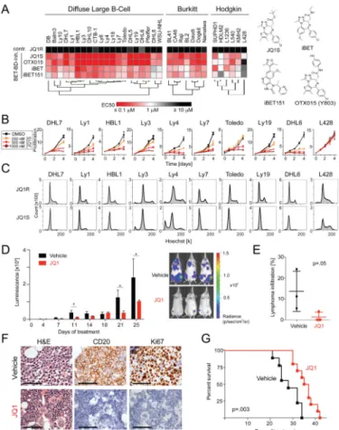
To assess the role of BET bromodomains as cancer cell dependencies in DLBCL, we first studied the effects of four structurally dissimilar BET bromodomain inhibitors on a comprehensive panel of 34 human lymphoma cell lines (21 DLBCL, capturing all transcriptionally defined subtypes, 6 BL and 7 Hodgkin Lymphoma [HL], Table S1), in comparative high-throughput format. In addition to the prototypical BET inhibitor, JQ1 (JQ1S) (Filippakopoulos et al., 2010), we resynthesized, characterized and tested an analogous thienodiazepine from Mitsubishi-Tanabe Pharmaceutical Corportation (Y803, OTX015; Oncoethix SA) which was developed for inflammatory bowel disease, (Figure S1A (Miyoshi et al., 2009)), a benzodiazepine inhibitor (iBET) (Nicodeme et al., 2010) and a dimethylisoxazole inhibitor (iBET-151) from GlaxoSmithKline (Dawson et al., 2011). Analyses of cellular proliferation at 72 hr revealed a potent class effect of BET
bromodomain inhibitors on the DLBCL and BL cell lines irrespective of subtype and the lack of effect of an inactive enantiomer, (JQ1R; Figure 1A). The HL cell lines were comparatively less sensitive to BET inhibition and one HL line, L428, was resistant to all 4 compounds.
The effects of BET inhibition on growth over time, cell cycle progression, and apoptosis were then studied in a representative panel of nine DLBCL cell lines, using the L428 HL line as a negative control (Figure 1B–C; S1B–C). BET inhibition with JQ1 significantly attenuated growth in a dose-responsive manner in all tested DLBCL cell lines (Figure 1B). In three representative DLBCL cell lines, genetic depletion of BRD2 or BRD4 similarly decreased DLBCL proliferation, consistent with an on-target effect of JQ1 (Figure S1D–G). We observed a profound S phase and G2 peak reduction following JQ1 treatment consistent with a G1 cell cycle arrest (Figure 1C, S1B). BET inhibition (500 nM) did not induce apoptosis in most cell lines studied, evidenced by low AnnexinV/7AAD staining (Figure S1C) and absence of a sub-G1 peak (Figure 1C). Neither enantiomeric (JQ1R) nor vehicle (DMSO) controls affected DLBCL proliferation or survival (Figure 1C; S1B–C). Treatment with 500 nM JQ1 was similarly cytostatic in Burkitt lymphoma cell lines (Figure S1C). The L428 HL cell line was resistant to BET inhibition in all tested assays.
Efficacy of BET inhibition in DLBCL xenograft models
We next explored the therapeutic potential of BET inhibition in two independent DLBCL xenotransplantation models (Figure 1D–G; S1H–K). First, the human DLBCL cell line Ly1 was engineered to ectopically express firefly luciferase and mCherry allowing surrogate measurement of tumor growth in vivo. NOD SCID Il2rγnull (NSG) mice xenotransplanted with Ly1-Luc-mCherry cells had a statistically significant reduction in tumor burden when treated with JQ1 (30 mg BID by intraperitoneal (IP) injection; Figure 1D). A representative cohort of animals was sacrificed on day 13 of treatment for full hematological analysis. JQ1-treated animals had significantly decreased lymphoma infiltration of the bone marrow (BM) as measured by flow cytometric assessment of mCherry+ cells (Figure 1E). Morphological
and immunohistochemical analyses revealed that the highly proliferative (Ki67+) human
NIH-PA Author Manuscript
NIH-PA Author Manuscript
CD20+ lymphoma cell infiltrate (Figure 1F, upper panel) was markedly reduced in animals treated with the BET bromodomain inhibitor (Figure 1F, lower panel). In the remainder of the Ly1 xenograft cohort, the JQ1-treated mice had a significant median overall survival advantage of 9 days (p=.003) (Figure 1G).
To confirm the pharmacodynamic findings, a second xenotransplantation model was established using the Toledo DLBCL cell line. NSG mice with established tumors had delayed tumor progression when treated with JQ1 (Figure S1H). Full hematological analysis revealed that JQ1-treated animals had lower spleen weights (Figure S1I) and decreased lymphomatous infiltration of bone marrow and spleen (Figure 1J). Morphological and immunohistochemical analysis of the BM revealed significantly reduced the infiltration of CD20+/Ki67+ human lymphoma cells following BET inhibition (Figure S1K).
BET inhibition down-regulates oncogenic transcriptional pathways
To gain insights into the transcriptional pathways regulated by BET bromodomain co-activator proteins, we performed kinetic transcriptional profiling of vehicle- and JQ1-treated DLBCL cell lines. Five human DLBCL cell lines which captured the recognized
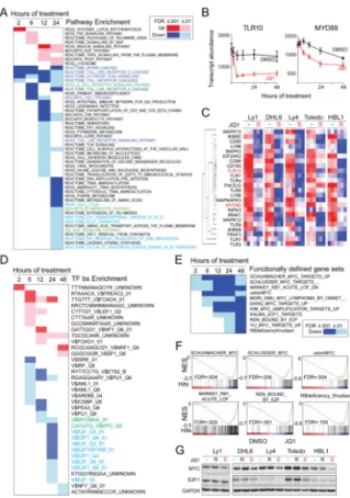
transcriptional heterogeneity (Ly1, BCR/GCB; DHL6, BCR/GCB; Ly4, OxPhos/unclass.; Toledo, OxPhos/unclass; and HBL1, BCR/ABC) were treated with JQ1 (500 nM) or vehicle control for 0, 2, 6, 12, 24, 48 hr. At each timepoint, differential analysis was performed between the vehicle and JQ1-treated samples (24 hr comparison, fold change (FC) >1.3, false discovery rate (FDR) <.01, Figure S2A). Consistent with prior studies of BET bromodomain function and inhibition, HEXIM1 was markedly up-regulated by JQ1 in all DLBCL cell lines (Figure S2B) (Bartholomeeusen et al., 2012; Puissant et al., 2013). The most differentially expressed genes were assessed for pathway enrichment using a comprehensive pathway compendium (C2, CP; MSigDB 3.0) and each timepoint was ranked by FDR and visualized as a color-coded matrix (Figure 2A, full list Table S2). We observed the early down-regulation of MYD88/toll-like receptor (TLR) pathway
components following JQ1 treatment, including TLR10 and MYD88 (Figure 2A–C; S2C). These data are consistent with previous studies in which the anti-inflammatory effect of BET inhibition in normal B cells was attributed to TLR pathway down-regulation (Nicodeme et al., 2010). In the JQ1-treated DLBCL cell lines, we also observed down-regulation of multiple components of the BCR signaling pathway, E2F transcriptional targets and additional cell cycle transition gene sets (Figure 2A; Figure S2D). Similar results were obtained when GCB and ABC DLBCL cell lines were analyzed separately (Figure S2E–F).
BET inhibition modulates MYC and E2F target gene transcription
Cell state transitions are influenced by the function of specific regulatory TFs. To identify candidate TFs associated with BET bromodomain co-activators, we assessed the effects of JQ1 on sets of genes with common TF binding motifs (C3, MSigDB 3.0). The differentially expressed genes in vehicle- vs. JQ1-treated DLBCL cell lines were tested for enrichment of candidate TF targets at 2 to 48 hr. Results at each timepoint were ranked by FDR and visualized as a color-coded matrix (Figure 2D and Table S2).
NIH-PA Author Manuscript
NIH-PA Author Manuscript
Of interest, gene sets with MYC and E2F binding motifs were significantly down-regulated following JQ1 treatment (Figure 2D). To further evaluate the effects of BET bromodomain inhibition on MYC and E2F transcriptional programs, we used multiple functionally validated MYC and E2F target gene sets to perform directed pathway analyses. Following JQ1 treatment, there was highly significant early down-regulation of well defined and functionally validated MYC and E2F target gene sets (Figures 2E–F; S2G–H). In complementary studies, we performed gene set enrichment analyses (GSEA) of multiple independent MYC and E2F target gene sets in vehicle- versus JQ1-treated samples and found that MYC and E2F targets were significantly less abundant in JQ1-treated cells (Figure 2F; S2I–J).
BET bromodomain proteins may function as co-activators of the MYC and E2F proteins and/or direct modulators of MYC and E2F expression. To distinguish between these possibilities, we assessed the transcript abundance and protein levels of MYC and E2F in vehicle- and JQ1-treated DLBCLs. BET bromodomain inhibition resulted in an apparent decrease in MYC transcripts and protein in each of the DLBCL cell lines (Figure 2G; S2K– L) suggesting that BET bromodomains directly modulate MYC transcription. The
consequences of MYC down-regulation following BET inhibition have been characterized by our group and others in hematologic malignancies (Dawson et al., 2011; Delmore et al., 2011; Mertz et al., 2011; Zuber et al., 2011). Notably, ectopic expression of MYC in DLBCL cell lines failed to rescue the anti-proliferative effects of JQ1 (Figure S2M–O), suggesting a model where BET bromodomains function as co-activators of MYC target gene transcription. In contrast to effects on MYC expression, in four of five cell lines, JQ1 treatment did not measurably alter E2F1 transcript or protein abundance over 24 hr (Figure 2G; S2P–Q). These data suggest that BET bromodomains may function at regulatory elements at E2F1 target genes, rather than by influencing the abundance of E2F1 itself.
BET bromodomains as promoter-bound co-activators of E2F1-dependent transcription
To explore the role of BETs as co-activators of oncogenic E2F1 transcriptional signaling, we performed chromatin immunoprecipitation with next-generation DNA sequencing (ChIP-Seq), using a chemical genetic approach. We selected Ly1 cells for mechanistic consideration owing to the robust down-regulation of E2F target genes in the transcriptional profiling (Figure S3A) and the lack of effect of JQ1 on E2F1 protein expression (Figure 2G). Changes in BET localization, chromatin structure and RNA polymerase function were studied in Ly1 cells treated with JQ1 (500 nM) or vehicle control.
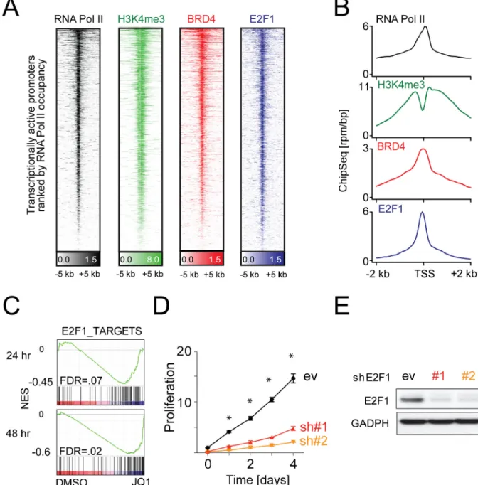
First, we established a chromatin landscape for Ly1 using H3K4me3 to identify promoters, H3K27ac to reveal enhancers and H3K27me3 to define repressive regions of the genome. Then, we assessed the genome-wide localization of E2F1 and the representative BET protein, BRD4, also by ChIP-Seq using the respective antibodies. Rank-ordering of all transcriptionally active promoters based on H3K4me3 enrichment and RNA Pol II occupancy identifies pervasive binding of BRD4 and E2F1 to active promoter elements (Figure 3A). Analysis of enrichment data as a metagene of all active promoters centered on the transcription start site reveals spatial colocalization of E2F1 and BRD4 at all
transcriptionally active promoters (Figure 3B).
NIH-PA Author Manuscript
NIH-PA Author Manuscript
ChIP-Seq for E2F1 allowed the annotation of an E2F1 target gene set, based on the top promoter-bound genes in Ly1 cells (Table S3). Using a gene set of the top 100 ChIP-Seq defined E2F1 targets (Table S3), we performed GSEA in Ly1 and additional DLBCL cell lines, DHL6, Ly4, Toledo, and HBL1. JQ1 treatment significantly decreased the transcript abundance of our functionally defined E2F1 targets at 24 hr and 48 hr in all cell lines studied (Figure 3C; S3A). The E2F1 dependency of these DLBCL cell lines as previously reported (Monti et al., 2012) was validated herein by genetic depletion in three representative cell lines (Figure 3D–E; S3B–C). Together, these data mechanistically establish BET bromodomains as E2F1 co-activators in DLBCL.
Disproportionate binding of BRD4 to overloaded enhancers
At the time this research was initiated, studies of BET bromodomains mainly focused on effects at promoter regions of the genome. Our research in multiple myeloma (MM) identified a role for BRD4 in enforcing MYC transcription from the translocated IgH enhancer locus (Delmore et al., 2011), where massive accumulation of BRD4 was identified by ChIP (approximately 200-fold enrichment). As oncogenic TFs may signal to RNA Pol II through distal enhancer elements, we sought to characterize the genome-wide localization of BRD4 to enhancers in DLBCL.
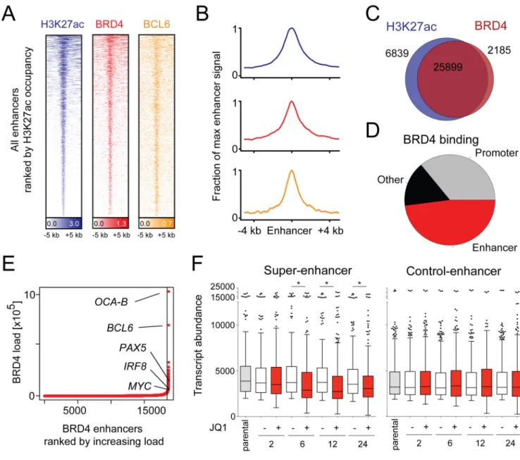
Rank-ordering of enhancer regions by H3K27ac enrichment reveals that BRD4 binds to the vast majority of active enhancers in the Ly1 DLBCL genome (Figure 4A, blue and red tracks). Given the established role of BCL6 in the pathogenesis of DLBCL, we also documented genome-wide co-localization of BRD4 and BCL6 at H3K27ac-defined enhancers (Figure 4A, orange tracks). A metagene for active enhancers illustrates focal, superimposable enrichment for BRD4 with H3K27ac (Figure 4B). The correlation between BRD4 occupancy and H3K27 acetylation is extremely strong genome-wide, with 79.1% of H3K27ac regions containing BRD4 and 92.2% of all chromatin-bound BRD4 occurring in regions marked by H3K27ac (Figure 4C). Genome-wide binding data for BRD4 reveal that BRD4 is most commonly associated with enhancer regions, defined by the presence of H3K27ac and absence of H3K4me3 (Figure 4D).
As predicted, BRD4 load is asymmetrically distributed throughout the genome at enhancer sites. Completely unexpected is the magnitude by which BRD4 load varies among active enhancer regions (Figure 4E). Only a small subset of BRD-loaded enhancers, 285/18330 (1.6%), account for 32% of all of the BRD4 enhancer binding in the cell (Figure 4E, Table S4). The BRD4-loaded enhancers in the Ly1 DLBCL cell line are considerably larger than typical enhancer elements, resembling the super-enhancers we recently described with Richard Young (Loven et al., 2013).
Notably, the top two gene loci with BRD4-loaded enhancers, POU2AF1 (which encodes the OCA-B transcriptional co-activator protein) and BCL6, and additional genes with
disproportionally BRD4-loaded enhancers such as PAX5 and IRF8 (Figure 4E), are essential for B-cell fate determination and germinal center formation (Basso and Dalla-Favera, 2012; Klein and Dalla-Favera, 2008; Teitell, 2003; Wang et al., 2008). In fact, mice with genetic ablation of POU2AF1, BCL6, PAX5 or IRF8 lack the ability to form germinal centers, the physiological structures from which most DLBCLs arise (Cobaleda et al., 2007; Nutt et al.,
NIH-PA Author Manuscript
NIH-PA Author Manuscript
2011; Teitell, 2003; Wang et al., 2008; Ye et al., 1997). Additionally, BRD4-superloaded enhancers are found adjacent to known oncogenes relevant to DLBCL biology, such as CD79B and MYC (Figure S4A–B).
These data indicate that BRD4 is predominantly an enhancer-associated factor, which distributes throughout DLBCL euchromatin in a highly asymmetric manner, adjacent to known oncogenes and lineage-specific transcription factors (Figure 4E). BET bromodomain inhibition selectively decreased the transcript abundance of the 285 genes with the most BRD4-loaded super-enhancers, in contrast to the 285 genes with the least BRD4-loaded enhancers (Figure 4F). Similar results were obtained using H3K27ac as surrogate enhancer mark (Figure S4C) Taken together, these data suggest that BRD4 loading of select DLBCL enhancers underlies the pathway-specific transcriptional consequences of BET inhibition.
JQ1 targets the POU2AF1 super-enhancer and decreases OCA-B expression and activity
The POU2AF1 locus emerged as the most BRD4-overloaded enhancer in the Ly1 DLBCL cell line (Figure 4E), prompting further analysis of the effect of BET inhibition on OCA-B expression and function. OCA-B is a gene regulatory factor that interacts with the OCT1 and OCT2 TFs at octamer motifs and regulates B-cell development, maturation and GC
formation (Teitell, 2003). Although OCA-B is expressed throughout B-cell development, it is most abundant in normal GC B cells and GC-derived tumors including DLBCL (Greiner et al., 2000).
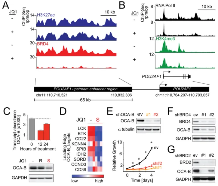
POU2AF1 has a large H3K27ac-defined enhancer with strong BRD4 binding that is abolished by JQ1 treatment (Figure 5A). Consistent with this observation, BET inhibition reduced RNA Pol II elongation of POU2AF1, with a relative increase in promoter-paused RNA Pol II near the transcriptional start site (TSS; Figure 5B). Notably, intronic enrichment for H3K4me3 and RNA Pol II was observed in POU2AF1 consistent with an alternate promoter element, which was also affected by JQ1 treatment. JQ1 treatment (500 nM) decreased OCA-B transcript abundance and protein expression in Ly1 (Figure 5C), as well as additional DLBCL cell lines (Figure S5A).
We next assessed the consequences of JQ1 treatment on the OCA-B transcriptional program by performing GSEA with a well-defined series of OCA-B target genes (Table S5, modified from (Teitell, 2003)). OCA-B targets were down-regulated in JQ1-treated DLBCLs as illustrated in Ly1 (Figure 5D) and recapitulated in the full DLBCL panel (Figure S5B–C). OCA-B depletion with two independent shRNAs significantly decreased the proliferation of Ly1 (Figure 5E), and enforced expression of OCA-B partially rescued the JQ1-mediated anti-proliferative effects (Figure S5D–E). Genetic depletion of either BRD2 or BRD4 phenocopied the JQ1-mediated reduction of OCA-B (Figure 5F–G; S5F–G). Together, these data underscore the importance of OCA-B to DLBCL growth and illustrate the use of super-enhancers to identify cancer dependencies.
NIH-PA Author Manuscript
NIH-PA Author Manuscript
JQ1 targets super-enhancers of additional critical B-cell TFs and modulates the GC program
Three of the master regulatory TFs with BRD4-loaded super-enhancers (BCL6, IRF8 and PAX5), promote and maintain the B-cell gene expression program and limit differentiation into antibody-secreting plasma cells (Nutt et al., 2011). For these reasons, we further assessed the functional consequences of BET inhibition on the critical B-cell TFs with BRD4-loaded super-enhancers.
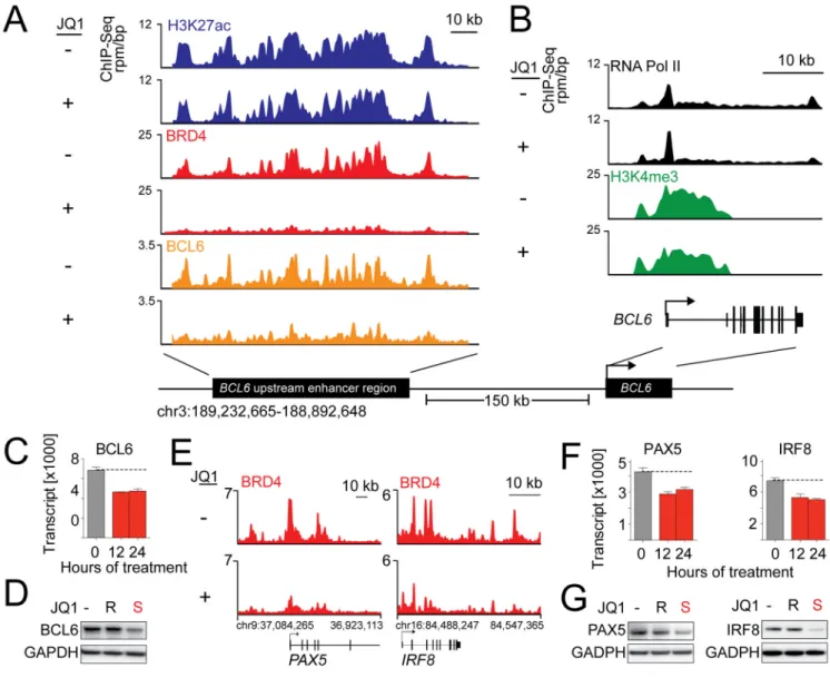
Given the known oncogenic function of deregulated BCL6 in GC B-cells (Basso and Dalla-Favera, 2012) and the sensitivity of certain DLBCLs to BCL6 depletion (Polo et al., 2007), we first evaluated the consequences of BET inhibition on BCL6 expression and function. The BCL6 locus includes a large previously defined upstream enhancer (Ramachandrareddy et al., 2010) that is severely depleted of BRD4 upon JQ1 treatment (Figure 6A). Consistent with depletion of BRD4 from the BCL6 enhancer, the promoter reveals a suggestion of increased RNA Pol II pausing and reduced elongating RNA Pol II (Figure 6B). JQ1 treatment markedly decreased BCL6 transcript abundance and protein expression in Ly1 (Figure 6C–D), and additional DLBCL lines (Figure S6A). Interestingly, the observed broad localization of BCL6 to its enhancer region was reduced by JQ1 (Figure 6A–B), a finding we confirmed by ChIP-qPCR (Figure S6B). This may reflect an influence of BET inhibition on TF binding or a consequence of down-regulation of BCL6 by JQ1. Similarly, PAX5 and IRF8 also have BRD4-loaded super-enhancers that are severely depleted of BRD4 following JQ1 treatment (Figure 6E). BET inhibition also decreased PAX5 and IRF8 transcript abundance and protein expression (Figure 6F–G).
Super-enhancer clustering identifies transcriptional subtypes of DLBCL
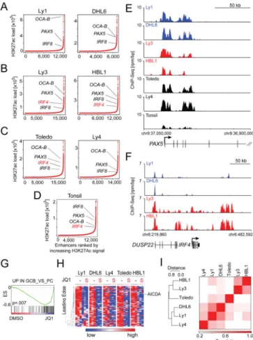
Using the robust H3K27ac mark to identify and discriminate super-enhancers (SE), we conducted ChIP-Seq SE analysis on five additional human DLBCL lines (DHL6, BCR/ GCB; HBL1 and Ly3, BCR/ABC; Toledo and Ly4, OxPhos/Unclass, Table S7) and a normal lymphoid sample (tonsil, Table S7). Asymmetric enhancer loading was detected in all of the DLBCL cell lines (Figure 7A–C) and the normal tonsil (Figure 7D), confirming the ubiquitous nature of this epigenomic structural element.
In all of the DLBCL cell lines and normal tonsil, large SEs were identified adjacent to genes encoding master TFs such as PAX5, OCA-B, and IRF8 (Figure 7A–D; 7E–F, tracks and Table S7) that maintain the B-cell program and limit plasma cell differentiation. Given the critical role of these master regulatory TFs in maintaining GC integrity and limiting GC exit (Nutt et al., 2011), we functionally assessed the consequences of BET inhibition on the GC program in all DLBCL cell lines. To that end, we used a publicly available compendium of GEPs of highly purified human B-cell subsets and defined a set of genes that are
significantly more abundant in GC centrocytes and centroblasts than in post-GC plasma cells (UP_IN_GCB_VS_PC). Using GSEA, we confirmed that this common GC developmental program was down-regulated in Ly1 and in all five DLBCL cell lines following JQ1 treatment (Figure 7G–H and S7B).
NIH-PA Author Manuscript
NIH-PA Author Manuscript
In a subset of the DLBCL cell lines, SEs were identified adjacent to differentially expressed genes validated as discriminating DLBCL subtypes by the COO classification (Figure S7C; Table S7). In the ABC DLBCL cell lines, but not the GCB lines, the subtype-specific TF locus, IRF4, had an adjacent SE (Figure 7A, GCB; Figure 7B, ABC and Figure 7F, tracks; Table S7). The IRF4 SE was also detected in normal tonsil (Figure 7D) suggesting that it represented a developmental epigenetic mark rather than a tumor-specific feature. Additional genes associated with the developmental ABC signature, including PIM1 and CCND2, featured adjacent SEs in ABC, but not GCB, cell lines (Figure S7C, Table S7). Observing lineage-specifying genes flanked by SEs, we explored whether SE analysis could independently discriminate DLBCL subtypes. Indeed, unsupervised bidirectional
hierarchical clustering of DLBCL cell lines by SEs distinguished ABC from GCB cell lines (Figure 7I and S7C).
To evaluate the clinical significance of these findings, we performed SE analysis on primary patient samples by genome-wide ChIP-Seq for H3K27ac on four primary DLBCLs that were previously subtyped as either GCB or ABC (Monti et al., 2012). All primary DLBCLs exhibited the same characteristic asymmetry in H3K27ac enrichment, with readily identified regulatory regions consistent with SEs (Figure 8A–B, Table S8). Again, SEs were found adjacent to lineage-specifying TFs, such as PAX5, and subtype-associated TFs, such as IRF4 (Figure 8A–B, tracks in Figure 8C–D, Table S8). Importantly, aggregate unsupervised hierarchical clustering of all SE data principally segregated cell lines from primary samples (malignant or non-malignant), while isolated unsupervised clustering of primary tissue segregated DLBCL samples in agreement with transcriptional developmental distinctions (Figure 8E, S8, Table S8).
DISCUSSION
Here, we provide mechanistic evidence of BET bromodomains as transcriptional co-activators at large enhancers and E2F1-driven promoters and contribute data supporting the study of BET inhibitors in all recognized subtypes of DLBCL. BET inhibition caused a profound G1 cell cycle arrest in a panel of DLBCL cell lines representing all
transcriptionally defined subtypes and significantly delayed tumor growth in two
independent DLBCL xenograft models. Gene expression profiling of multiple JQ1-treated DLBCL cell lines revealed down-regulation of MYD88/TLR and BCR signaling
components, important for certain subtypes of DLBCL (Chen et al., 2013; Ngo et al., 2011; Nicodeme et al., 2010). More broadly relevant for all subtypes of DLBCLs, we identified highly significant transcriptional down-regulation of MYC and E2F1 target genes and the selective depletion of BRD4-loaded promoters and enhancers.
BET inhibition decreased the abundance of multiple functionally defined E2F target genes but did not measurably alter E2F1 protein levels. Epigenomic analyses confirmed the co-localization of BRD4 and E2F1 at active promoters. The selective decrease in BRD4 loading of E2F1-driven genes following JQ1 treatment is consistent with studies suggesting a role of BET proteins in E2F1-mediated transcription (Peng et al., 2007; Sinha et al., 2005).
Genome-wide assessment of effects of BRD4 on transcriptional elongation at E2F1 target genes were statistically significant but subtle on an individual gene level (data not shown),
NIH-PA Author Manuscript
NIH-PA Author Manuscript
leaving open the possibility that BRD4-mediated effects on elongation are most apparent on super-enhancer associated genes. Given the recently identified structural genetic signature of deregulated cell cycle and increased E2F activity in poor prognosis DLBCLs (Monti et al., 2012), BET inhibition may represent a promising targeted treatment strategy.
Studies from our lab and others have highlighted the important role of BRD4 as a co-activator of MYC mediated transcription (Delmore et al., 2011; Mertz et al., 2011; Ott et al., 2012). In multiple myeloma cell lines with Ig/MYC translocations, BRD4 was postulated to function via long-range interactions with the distal IgH enhancer (Delmore et al., 2011). However, emerging data indicate that BET inhibitors suppress MYC transcription in the context of translocated, amplified or wild-type MYC alleles and BRD4 localizes to MYC promoter and enhancer elements (Delmore et al., 2011; Loven et al., 2013; Mertz et al., 2011; Ott et al., 2012). In the current studies, we identify a BRD4-loaded MYC enhancer and find that BET inhibition decreases MYC transcription and expression in DLBCL cell lines with translocated, amplified or wild-type MYC alleles. In this extensive DLBCL cell line panel, the functional consequences of BET inhibition – cell cycle arrest and decreased cellular proliferation – were largely comparable. Although JQ1 treatment broadly down-regulated the transcriptional targets of MYC and E2F, we sought additional bases for the effect of BET inhibition across multiple DLBCL subtypes.
We observed that a small subset of genes had a disproportionately high BRD4 load at their proximal enhancers. These unusual regulatory elements were approximately 12-fold larger than typical enhancer regions. Integrated epigenomic and transcriptional studies established that such super enhancer-marked genes were particularly sensitive to BET inhibition. As SEs were found adjacent to genes encoding known lineage factors and DLBCL
oncoproteins, we surmised that SE analysis may identify previously unrecognized tumor dependencies. The functional exploration of OCA-B, encoded by SE-marked POU2AF1, validates this factor as a cancer dependency in DLBCL. Mechanistic research has established OCA-B as a co-activator protein which binds into the OCT1-OCT2 transcriptional complex, enhancing IgH promoter-enhancer communication (Luo and Roeder, 1995; Ren et al., 2011). Mice lacking OCA-B expression due to germline knock-out of POU2AF1 are developmentally normal, even capable of early transcription from
immunoglobulin promoters, however they lack an apparent germinal center reaction to antigen (Kim et al., 1996; Teitell, 2003). Collectively, the earlier studies and the current research support a putative therapeutic window to targeting OCA-B, potentially by protein-protein inhibition via the POU domain. More generally, these studies establish a rationale to systematically explore SEs for unrecognized tumor dependencies, and potentially use SEs as biomarkers for targeted therapeutic development.
In this comparative epigenomic analysis of human DLBCL cell lines, primary tumor specimens and normal lymphoid samples, we analyzed patterns of H3K27ac enrichment to understand the relevance of enhancer variation and function. These studies reveal SEs as characteristic features of human lymphoid tissues, both benign and malignant. Preservation of tissue-specific SEs is observed comparing non-malignant nodal tissue to primary DLBCL samples, as well as in patient-derived human DLBCL cell lines.
NIH-PA Author Manuscript
NIH-PA Author Manuscript
In summary, our data suggest that the BET inhibition limits the growth of DLBCLs by at least two complementary activities: a specific effect on genes that define a given cell type by high BRD4 loading at enhancers and a more general suppression of transcription at E2F-and MYC-driven target genes. Thus, an E2F/MYC pathway effect is combined with massive depletion of proteins driven by BRD4 overloaded enhancers preventing cell cycle
progression and leading to growth arrest. The majority of DLBCLs have a structural basis for increased E2F1-mediated cell cycle progression; however, these tumors may differ in BRD4 super-loading of cell fate-determining enhancers, including MYC, depending on their molecular context. This framework of BET inhibition explains its broad subclass- and tumor-type independent mechanism of action and reconciles the apparent pleiotropic effects and cell type-specific outcomes. Importantly, these data provide a compelling rationale for further human clinical investigation.
Experimental procedures
High throughput screening of BET bromodomain inhibitors in B-cell lymphoma cell line panel
Using a semi-automated screen, we tested the indicated compounds in 34 human lymphoma lines in a 384-well format. Cell viability at 72 hr was evaluated using ATPlite (Perkin Elmer). The mean of absolute EC50s from two independent screens were visualized and clustered using GENE (http://www.broadinstitute.org/cancer/software/GENE-E/index.html).
Human Samples
Frozen biopsy specimens of newly diagnosed, previously untreated primary DLBCLs with >80% tumor involvement and known transcriptional subtyping (Monti et al., 2012) were obtained according to Institutional Review Board (IRB)-approved protocols (Brigham & Women Hospital, and Dana-Farber Cancer Institute). A waiver to obtain informed consent was granted by the local IRBs because only coded, de-identified, discarded tissue was used.
Animal Studies
All animal studies were performed according to Dana-Farber Cancer Institute Institutional Animal Care and Use Committee (IACUC)-approved protocols as previously described (Monti et al., 2012) and Supplemental Experimental Procedures.
Analyses of cellular proliferation and apoptosis, immunohistochemistry, immunoblotting, transcriptional profiling, GSEA and lentiviral mediated shRNA, chromatin
immunoprecipitation with massively parallel sequencing (Seq) and analysis of ChIP-Seq data
A full description of these methods is listed in Supplemental Experimental Procedures.
Accession Numbers
The Gene Expression Omnibus accession numbers for the gene expression and ChIP-Seq data reported in this paper is GSE45630 and GSE46663, respectively.
NIH-PA Author Manuscript
NIH-PA Author Manuscript
Supplementary Material
Refer to Web version on PubMed Central for supplementary material.
Acknowledgments
The authors acknowledge support from the National Institutes of Health and the Leukemia & Lymphoma Society (M.A.S. and J.E.B.), the Damon-Runyon Cancer Research Foundation, the Broad New Idea Award, the William Lawrence and Blanche Hughes Foundation and the American Society of Hematology (J.E.B.), the Department of Defense (C.Y.L., CDMRP CA120184), the American Cancer Society (P.B.R., PF-11-042-01-DMC), and the German Research Foundation (B.C., DFG Ch 735/1-1).
References
Bartholomeeusen K, Xiang Y, Fujinaga K, Peterlin BM. Bromodomain and Extra-terminal (BET) Bromodomain Inhibition Activate Transcription via Transient Release of Positive Transcription Elongation Factor b (P-TEFb) from 7SK Small Nuclear Ribonucleoprotein. The Journal of Biological Chemistry. 2012; 287:36609–36616. [PubMed: 22952229]
Basso K, Dalla-Favera R. Roles of BCL6 in normal and transformed germinal center B cells. Immunological Reviews. 2012; 247:172–183. [PubMed: 22500840]
Bisgrove DA, Mahmoudi T, Henklein P, Verdin E. Conserved P-TEFb-interacting domain of BRD4 inhibits HIV transcription. Proc Natl Acad Sci U S A. 2007; 104:13690–13695. [PubMed: 17690245]
Caro P, Kishan AU, Norberg E, Stanley IA, Chapuy B, Ficarro SB, Polak K, Tondera D, Gounarides J, Yin H, et al. Metabolic signatures uncover distinct targets in molecular subsets of diffuse large B cell lymphoma. Cancer Cell. 2012; 22:547–560. [PubMed: 23079663]
Chen L, Monti S, Juszczynski P, Ouyang J, Chapuy B, Neuberg D, Doench JG, Bogusz AM,
Habermann TM, Dogan A, et al. SYK inhibition modulates distinct PI3K/AKT- dependent survival pathways and cholesterol biosynthesis in diffuse large B cell lymphomas. Cancer Cell. 2013; 23:826–838. [PubMed: 23764004]
Cobaleda C, Schebesta A, Delogu A, Busslinger M. Pax5: the guardian of B cell identity and function. Nat Immunol. 2007; 8:463–470. [PubMed: 17440452]
Dawson MA, Prinjha RK, Dittmann A, Giotopoulos G, Bantscheff M, Chan WI, Robson SC, Chung CW, Hopf C, Savitski MM, et al. Inhibition of BET recruitment to chromatin as an effective treatment for MLL-fusion leukaemia. Nature. 2011; 478:529–533. [PubMed: 21964340]
Delmore JE, Issa GC, Lemieux ME, Rahl PB, Shi J, Jacobs HM, Kastritis E, Gilpatrick T, Paranal RM, Qi J, et al. BET bromodomain inhibition as a therapeutic strategy to target c-Myc. Cell. 2011; 146:904–917. [PubMed: 21889194]
Dey A, Chitsaz F, Abbasi A, Misteli T, Ozato K. The double bromodomain protein Brd4 binds to acetylated chromatin during interphase and mitosis. Proc Natl Acad Sci U S A. 2003; 100:8758– 8763. [PubMed: 12840145]
Filippakopoulos P, Picaud S, Mangos M, Keates T, Lambert JP, Barsyte-Lovejoy D, Felletar I, Volkmer R, Muller S, Pawson T, et al. Histone recognition and large-scale structural analysis of the human bromodomain family. Cell. 2012; 149:214–231. [PubMed: 22464331]
Filippakopoulos P, Qi J, Picaud S, Shen Y, Smith WB, Fedorov O, Morse EM, Keates T, Hickman TT, Felletar I, et al. Selective inhibition of BET bromodomains. Nature. 2010; 468:1067–1073. [PubMed: 20871596]
Fuda NJ, Ardehali MB, Lis JT. Defining mechanisms that regulate RNA polymerase II transcription in vivo. Nature. 2009; 461:186–192. [PubMed: 19741698]
Gisselbrecht C, Glass B, Mounier N, Singh Gill D, Linch DC, Trneny M, Bosly A, Ketterer N, Shpilberg O, Hagberg H, et al. Salvage regimens with autologous transplantation for relapsed large B-cell lymphoma in the rituximab era. J Clin Oncol. 2010; 28:4184–4190. [PubMed: 20660832]
NIH-PA Author Manuscript
NIH-PA Author Manuscript
Greenwald RJ, Tumang JR, Sinha A, Currier N, Cardiff RD, Rothstein TL, Faller DV, Denis GV. E mu-BRD2 transgenic mice develop B-cell lymphoma and leukemia. Blood. 2004; 103:1475–1484. [PubMed: 14563639]
Greiner A, Muller KB, Hess J, Pfeffer K, Muller-Hermelink HK, Wirth T. Up-regulation of BOB.1/ OBF.1 expression in normal germinal center B cells and germinal center-derived lymphomas. Am J Pathol. 2000; 156:501–507. [PubMed: 10666379]
Hu S, Xu-Monette ZY, Tzankov A, Green T, Wu L, Balasubramanyam A, Liu WM, Visco C, Li Y, Miranda RN, et al. MYC/BCL2 protein coexpression contributes to the inferior survival of activated B-cell subtype of diffuse large B-cell lymphoma and demonstrates high-risk gene expression signatures: a report from The International DLBCL Rituximab-CHOP Consortium Program. Blood. 2013; 121:4021–4031. quiz 4250. [PubMed: 23449635]
Johnson NA, Slack GW, Savage KJ, Connors JM, Ben-Neriah S, Rogic S, Scott DW, Tan KL, Steidl C, Sehn LH, et al. Concurrent expression of MYC and BCL2 in diffuse large B-cell lymphoma treated with rituximab plus cyclophosphamide, doxorubicin, vincristine, and prednisone. J Clin Oncol. 2012; 30:3452–3459. [PubMed: 22851565]
Kim U, Qin XF, Gong S, Stevens S, Luo Y, Nussenzweig M, Roeder RG. The B-cell-specific transcription coactivator OCA-B/OBF-1/Bob-1 is essential for normal production of immunoglobulin isotypes. Nature. 1996; 383:542–547. [PubMed: 8849728]
Klein U, Dalla-Favera R. Germinal centres: role in B-cell physiology and malignancy. Nat Rev Immunol. 2008; 8:22–33. [PubMed: 18097447]
Lee TI, Young RA. Transcriptional regulation and its misregulation in disease. Cell. 2013; 152:1237– 1251. [PubMed: 23498934]
Lenz G, Staudt LM. Aggressive lymphomas. N Engl J Med. 2010; 362:1417–1429. [PubMed: 20393178]
Lohr JG, Stojanov P, Lawrence MS, Auclair D, Chapuy B, Sougnez C, Cruz-Gordillo P, Knoechel B, Asmann YW, Slager SL, et al. Discovery and prioritization of somatic mutations in diffuse large B-cell lymphoma (DLBCL) by whole-exome sequencing. Proc Natl Acad Sci U S A. 2012; 109:3879–3884. [PubMed: 22343534]
Loven J, Hoke HA, Lin CY, Lau A, Orlando DA, Vakoc CR, Bradner JE, Lee TI, Young RA. Selective inhibition of tumor oncogenes by disruption of super-enhancers. Cell. 2013; 153:320– 334. [PubMed: 23582323]
Luo Y, Roeder RG. Cloning, functional characterization, and mechanism of action of the B-cell-specific transcriptional coactivator OCA-B. Molecular and Cellular Biology. 1995; 15:4115–4124. [PubMed: 7623806]
Marushige K. Activation of chromatin by acetylation of histone side chains. Proc Natl Acad Sci U S A. 1976; 73:3937–3941. [PubMed: 1069278]
Mertz JA, Conery AR, Bryant BM, Sandy P, Balasubramanian S, Mele DA, Bergeron L, Sims RJ 3rd . Targeting MYC dependence in cancer by inhibiting BET bromodomains. Proc Natl Acad Sci U S A. 2011; 108:16669–16674. [PubMed: 21949397]
Miyoshi, S.; Ooike, S.; Iwata, K.; Hikawa, H.; Sugaraha, K. Antitumor agent. International Patent No. PCT/JP2008/073864 (WO/2009/084693). 2009.
Monti S, Chapuy B, Takeyama K, Rodig SJ, Hao Y, Yeda KT, Inguilizian H, Mermel C, Currie T, Dogan A, et al. Integrative Analysis Reveals an Outcome-Associated and Targetable Pattern of p53 and Cell Cycle Deregulation in Diffuse Large B Cell Lymphoma. Cancer Cell. 2012; 22:359– 372. [PubMed: 22975378]
Monti S, Savage KJ, Kutok JL, Feuerhake F, Kurtin P, Mihm M, Wu B, Pasqualucci L, Neuberg D, Aguiar RC, et al. Molecular profiling of diffuse large B-cell lymphoma identifies robust subtypes including one characterized by host inflammatory response. Blood. 2005; 105:1851–1861. [PubMed: 15550490]
Morin RD, Mendez-Lago M, Mungall AJ, Goya R, Mungall KL, Corbett RD, Johnson NA, Severson TM, Chiu R, Field M, et al. Frequent mutation of histone-modifying genes in non-Hodgkin lymphoma. Nature. 2011; 476:298–303. [PubMed: 21796119]
NIH-PA Author Manuscript
NIH-PA Author Manuscript
Ngo VN, Young RM, Schmitz R, Jhavar S, Xiao W, Lim KH, Kohlhammer H, Xu W, Yang Y, Zhao H, et al. Oncogenically active MYD88 mutations in human lymphoma. Nature. 2011; 470:115– 119. [PubMed: 21179087]
Nicodeme E, Jeffrey KL, Schaefer U, Beinke S, Dewell S, Chung CW, Chandwani R, Marazzi I, Wilson P, Coste H, et al. Suppression of inflammation by a synthetic histone mimic. Nature. 2010; 468:1119–1123. [PubMed: 21068722]
Nutt SL, Taubenheim N, Hasbold J, Corcoran LM, Hodgkin PD. The genetic network controlling plasma cell differentiation. Seminars in Immunology. 2011; 23:341–349. [PubMed: 21924923] Ott CJ, Kopp N, Bird L, Paranal RM, Qi J, Bowman T, Rodig SJ, Kung AL, Bradner JE, Weinstock
DM. BET bromodomain inhibition targets both c-Myc and IL7R in high-risk acute lymphoblastic leukemia. Blood. 2012; 120:2843–2852. [PubMed: 22904298]
Owen DJ, Ornaghi P, Yang JC, Lowe N, Evans PR, Ballario P, Neuhaus D, Filetici P, Travers AA. The structural basis for the recognition of acetylated histone H4 by the bromodomain of histone acetyltransferase gcn5p. EMBO J. 2000; 19:6141–6149. [PubMed: 11080160]
Pasqualucci L, Trifonov V, Fabbri G, Ma J, Rossi D, Chiarenza A, Wells VA, Grunn A, Messina M, Elliot O, et al. Analysis of the coding genome of diffuse large B-cell lymphoma. Nat Genet. 2011; 43:830–837. [PubMed: 21804550]
Peng J, Dong W, Chen L, Zou T, Qi Y, Liu Y. Brd2 is a TBP-associated protein and recruits TBP into E2F-1 transcriptional complex in response to serum stimulation. Molecular and Cellular
Biochemistry. 2007; 294:45–54. [PubMed: 17111193]
Polo JM, Juszczynski P, Monti S, Cerchietti L, Ye K, Greally JM, Shipp M, Melnick A.
Transcriptional signature with differential expression of BCL6 target genes accurately identifies BCL6-dependent diffuse large B cell lymphomas. Proc Natl Acad Sci U S A. 2007; 104:3207– 3212. [PubMed: 17360630]
Puissant A, Frumm SM, Alexe G, Bassil CF, Qi J, Chanthery YH, Nekritz EA, Zeid R, Gustafson WC, Greninger P, et al. Targeting MYCN in neuroblastoma by BET bromodomain inhibition. Cancer Discovery. 2013; 3:308–323. [PubMed: 23430699]
Ramachandrareddy H, Bouska A, Shen Y, Ji M, Rizzino A, Chan WC, McKeithan TW. BCL6 promoter interacts with far upstream sequences with greatly enhanced activating histone modifications in germinal center B cells. Proc Natl Acad Sci U S A. 2010; 107:11930–11935. [PubMed: 20547840]
Ren X, Siegel R, Kim U, Roeder RG. Direct interactions of OCA-B and TFII-I regulate
immunoglobulin heavy-chain gene transcription by facilitating enhancer-promoter communication. Mol Cell. 2011; 42:342–355. [PubMed: 21549311]
Schreiber SL, Bernstein BE. Signaling network model of chromatin. Cell. 2002; 111:771–778. [PubMed: 12526804]
Sinha A, Faller DV, Denis GV. Bromodomain analysis of Brd2-dependent transcriptional activation of cyclin A. The Biochemical Journal. 2005; 387:257–269. [PubMed: 15548137]
Slack GW, Gascoyne RD. MYC and aggressive B-cell lymphomas. Advances in Anatomic Pathology. 2011; 18:219–228. [PubMed: 21490439]
Teitell MA. OCA-B regulation of B-cell development and function. Trends in Immunology. 2003; 24:546–553. [PubMed: 14552839]
Wang H, Lee CH, Qi C, Tailor P, Feng J, Abbasi S, Atsumi T, Morse HC 3rd . IRF8 regulates B-cell lineage specification, commitment, and differentiation. Blood. 2008; 112:4028–4038. [PubMed: 18799728]
Yang Z, He N, Zhou Q. Brd4 recruits P-TEFb to chromosomes at late mitosis to promote G1 gene expression and cell cycle progression. Mol Cell Biol. 2008; 28:967–976. [PubMed: 18039861] Ye BH, Cattoretti G, Shen Q, Zhang J, Hawe N, de Waard R, Leung C, Nouri-Shirazi M, Orazi A, Chaganti RS, et al. The BCL-6 proto-oncogene controls germinal-centre formation and Th2-type inflammation. Nat Genet. 1997; 16:161–170. [PubMed: 9171827]
Zhang J, Grubor V, Love CL, Banerjee A, Richards KL, Mieczkowski PA, Dunphy C, Choi W, Au WY, Srivastava G, et al. Genetic heterogeneity of diffuse large B-cell lymphoma. Proc Natl Acad Sci U S A. 2013; 110:1398–1403. [PubMed: 23292937]
NIH-PA Author Manuscript
NIH-PA Author Manuscript
Zhang W, Prakash C, Sum C, Gong Y, Li Y, Kwok JJ, Thiessen N, Pettersson S, Jones SJ, Knapp S, et al. Bromodomain-containing protein 4 (BRD4) regulates RNA polymerase II serine 2
phosphorylation in human CD4+ T cells. J Biol Chem. 2012; 287:43137–43155. [PubMed: 23086925]
Zhao R, Nakamura T, Fu Y, Lazar Z, Spector DL. Gene bookmarking accelerates the kinetics of post-mitotic transcriptional re-activation. Nat Cell Biol. 2011; 13:1295–1304. [PubMed: 21983563] Zuber J, Shi J, Wang E, Rappaport AR, Herrmann H, Sison EA, Magoon D, Qi J, Blatt K, Wunderlich
M, et al. RNAi screen identifies Brd4 as a therapeutic target in acute myeloid leukaemia. Nature. 2011; 478:524–528. [PubMed: 21814200]
NIH-PA Author Manuscript
NIH-PA Author Manuscript
Significance
Although oncogenic transcription factors underlie the pathophysiology and biological heterogeneity of DLBCL, studies of transcriptional co-activator proteins are limited in this disease. In this chemical genetic study, we demonstrate the efficacy of BET inhibition and characterize the broad function of BET bromodomains in supporting the transcriptional growth program in all subclasses of DLBCL, including co-activation of E2F and MYC target genes. We define an asymmetry in the localization of BRD4 to enhancer regions nearby oncogenic and master regulatory genes and expand the finding to a representative panel of cell lines and primary samples. This finding likely explains the specific transcriptional effect of BET inhibition which modulates the expression of master transcription factors that control B-cell fate and germinal center formation.
NIH-PA Author Manuscript
NIH-PA Author Manuscript
Highlights
1. BET inhibition exhibits antitumor efficacy in vitro and in vivo.
2. BRD4 localizes in an asymmetric manner to massively overloaded enhancer regions.
3. Genes with adjacent BRD4-loaded super-enhancers are sensitive to BET inhibition.
4. Cancer dependencies are found among super-enhancer marked genes.
NIH-PA Author Manuscript
NIH-PA Author Manuscript
Figure 1. In vitro analyses of BET Bromodomain inhibition in various B-cell lymphomas
(A) Hierarchical clustering of mean EC50s of the four BET inhibitors (72 hr treatment) in the indicated panel of B-cell lymphoma cell lines. EC50 values in a colorimetric scale: very sensitive (≤1 μM) in red; sensitive (=1 μM) in white; to resistant (≥10 μM) in black. Corresponding structures are shown. (B) Proliferation of the indicated DLBCL and HL cell lines treated with vehicle or 250–1000 nM JQ1 for 1–4 days. (C) Cell cycle analysis following 72 hr treatment with JQ1 (500 nM) or inactive enantiomer JQ1R (500 nM). Error bars represent the SD of triplicates. (D) Bioluminescence of JQ1 (30 mg/kg IP BID) or vehicle-treated NSG mice xenotransplanted with luciferized mCherry+ Ly1 cells. Asterisks indicate p <.05 using a one-sided t-test. Error bars represent SEM. (E) Lymphoma
infiltration of bone marrow in a representative set of animals was assessed by flow cytometric analysis of mCherry+ cells and visualized as scatter plots (median, line; whiskers, SEM). The p values were obtained with a one-sided Mann-Whitney U test. (F) Immunohistochemical analysis of lymphoma (Ly1) BM infiltration in JQ1- and vehicle-treated mice: H&E; anti-human CD20, and anti-Ki67 immunostaining. Scale bar represents 100 μm. (G) Kaplan-Meier plot of the remainder of the Ly1 cohort (n = 21) treated with either vehicle or JQ1 30 mg/kg bid. The p value was obtained by Log-rank test. See also Figure S1 and Table S1.
NIH-PA Author Manuscript
NIH-PA Author Manuscript
Figure 2. Transcriptional response to BET inhibition in representative DLBCL cell lines
(A) Hyperenrichment analysis of differentially expressed genes in all five lines (FDR<.01, FC>1.3) following 24 hr of treatment with 500 nM JQ1 or vehicle was performed using a pathway compendium (MSigDB, C2.CP). Results at each timepoint were ranked by FDR and visualized as a color-coded matrix. Up-regulated motifs in red, down-regulated motifs in blue. Intensity of color correlates with FDR significance levels. Highlighted pathways include: Toll-like receptor MYD88, blue; BCR signaling, green; and Cell cycle/E2F, cyan. (B) Mean transcript abundance of TLR10 (left) and MYD88 (right) in all five lines. Error bars represent SEM. (C) Heatmap of TLR pathway components in vehicle- or JQ1-treated DLBCLs (all five lines, 24 hr). (D) The most differentially expressed genes (FDR<.01, FC>1.3) were analyzed for common transcription factor binding sites in the regulatory region using the MSigDB.C3 compendium. Results at each timepoint were ranked using a color-coded matrix as in (A). Genes with MYC binding sites in green and E2F binding sites in cyan. (E) GSEA of multiple functionally defined MYC and E2F transcription factor target gene sets at was performed. Results are reported over time in a color-coded matrix with color intensity reflecting significance level. (F) GSEA plots of functionally defined MYC and E2F target gene sets in vehicle- vs. JQ1-treated cells at 24 hr. (G) Protein abundance of MYC and E2F in the indicated DLBCL lines treated with vehicle or JQ1S or JQ1R (500 nM) (24 hr). See also Figure S2 and Table S2.
NIH-PA Author Manuscript
NIH-PA Author Manuscript
Figure 3. Co-localization and function of BRD4 and E2F1 at active promoters
(A) Heatmap of ChIP-Seq reads for RNA Pol II (transcriptionally active; black), H3K4me3 (green), BRD4 (red), and E2F1 (blue) rank ordered from high to low RNA Pol II occupancy centered on a ± 5 kb window around the transcriptional start site (TSS) of all
transcriptionally active promoters. Color density reflects enrichment; white indicates no enrichment. (B) Metagenes created from normalized genome-wide average of reads for designated factors centered on a ± 2 kb window around the TSS. (C) GSEA plots of a ChIP-Seq defined E2F1 target gene set in the five DLBCL cell lines treated with vehicle vs. JQ1 for 24 and 48 hr. (D) Assessment of proliferation in Ly1 cell line following genetic depletion of E2F1 with two independent hairpins and a control hairpin (ev). Error bars
NIH-PA Author Manuscript
NIH-PA Author Manuscript
represent SD, and asterisks show p<.01 by a two-sided Student’s t-test. (E) Immunoblot of E2F1 of cells in (D) to demonstrate knockdown efficiency. See also Figure S3 and Table S3.
NIH-PA Author Manuscript
NIH-PA Author Manuscript
Figure 4. Asymmetric BRD4 loading at enhancer elements of actively transcribed genes
(A) Heatmap of ChIP-Seq binding for H3K27ac (blue), BRD4 (red), and BCL6 (orange) rank ordered from high to low H3K27 occupancy centered on a ± 5 kb window around enhancers. Color density reflects enrichment, white indicating no enrichment. (B) Metagenes created from normalized genome-wide average of reads for designated factors centered on a ± 4 kb window around the enhancers. (C) Venn diagram of BRD4-binding and H3K27ac occupancy. 79.1% of H3K27ac regions contain BRD4 and 92.2% of all
chromatin-bound BRD4 occurs within H3K27ac regions. (D) Pie chart of BRD4 binding to regions of the genome. BRD4 colocalization with H3K27ac without H3K4me3 defined as enhancer-bound (red); BRD4 colocalization with H3K4me3 reported as promoter-bound (grey); remaining genomic regions in “other” (black). (E) BRD4 loading/binding across enhancers of 18330 genes. 1.6% (285/18330) of enhancers contain 32% of all
enhancer-NIH-PA Author Manuscript
NIH-PA Author Manuscript
bound BRD4, with super-loading defined as surpassing the inflection point. Top BRD4-superloaded enhancers are indicated. (F) Mean transcript abundance of the genes associated with the 285 most and least BRD4-loaded enhancers (left and right panel, respectively) in five DLBCL cell lines treated with vehicle or JQ1 (2–24 hr). Asterisks indicate a p<.0001 obtained using a two-sided Mann-Whitney U test. See also Table S4 and Figure S4.
NIH-PA Author Manuscript
NIH-PA Author Manuscript
Figure 5. Identification of OCA-B as a DLBCL dependency by super-enhancer analysis
(A) ChIP-Seq binding density for H3K27ac (blue), and BRD4 (red) at the enhancer of POU2AF1 following JQ1 (+) or vehicle (DMSO) (−) treatment. (B) ChIP-Seq reads at the POU2AF1 promoter for RNA Polymerase II (black) and H3K4me3 (green) following JQ1 (+) or vehicle (−) treatment. (C) OCA-B transcript and protein abundance in Ly1 cells treated with vehicle or 500 nM JQ1 or JQ1R (24 hr). Error bars represent SD. (D) OCA-B-target genes (leading edge, OCA-B GSEA) in Ly1 cells treated with vehicle or 500 nM JQ1 are visualized as heatmap. (E) Knockdown efficacy of two independent OCA-B shRNAs was detected by western blot (top panel). Proliferation of OCA-B-depleted cells was measured by alamar blue. The p values for control vs. each OCA-B shRNA were delineated by two-sided Student’s t-test; asterisks show p<.01. Error bars represent SD. (F–G)
Knockdown efficiency of two independent shRNAs against BRD4 (F) or BRD2 (G) and the associated changes in OCA-B expression were evaluated by western blot. See also Figure S5 and Table S5.
NIH-PA Author Manuscript
NIH-PA Author Manuscript
Figure 6. BET inhibition modulates tissue-specifying TF expression by disrupting super-enhancers
(A) ChIP-Seq binding density for H3K27ac (blue), BRD4 (red), and BCL6 (orange) at the BCL6 enhancer following JQ1 (+) or vehicle (DMSO, −) treatment. (B) ChIP-Seq reads at the BCL6 promoter for RNA Pol II (black) and H3K4me3 (green) following JQ1 (+) or vehicle (−) treatment. (C) BCL6 transcript abundance in Ly1 cells 12 and 24 hr following vehicle or JQ1 treatment (derived from GEP data). Error bars represent SD. (D) BCL6 protein abundance following treatment with vehicle or JQ1 or JQ1R (500 nM) (24 hr). (E) ChIP-Seq density of BRD4 (red) at super-enhancers of the two additional B-cell TFs, PAX5 and IRF8, following treatment with JQ1 (+) or DMSO (−). (F and G) PAX5 and IRF8 transcript (F) and protein abundance (G) in Ly1 cells following JQ1 treatment. Error bars represent SD. See also Table S6, Figure S6 and Supplemental Experimental Procedures.
NIH-PA Author Manuscript
NIH-PA Author Manuscript
Figure 7. Comparative super-enhancer analysis of DLBCL cell lines and normal lymphoid tissue
(A–D) Rank order of increasing integrated H3K27ac fold enrichment at enhancer loci in DLBCL cell lines, GCB (A), ABC (B) unclassified (C) and normal tonsil (D). (E) H3K27ac ChIP-Seq fold enrichment at the POU2AF1 locus showing the super-enhancer region. (F) H3K27ac ChIP-Seq reads at IRF4 locus in the 2 GCB and 2 ABC cell lines. (G) GSEA plot of the “UP_IN GCB_VS_PC” signature in five DLBCL cell lines following JQ1 treatment. (H) The leading edge genes of the GSEA in (G) were visualized as heatmap. (I) Similarity matrix from unsupervised hierarchical clustering of each cell line by location of super-enhancers. See also Table S7 and Figure S7.
NIH-PA Author Manuscript
NIH-PA Author Manuscript
Figure 8. Super-enhancer analysis of primary DLBCLs
(A and B) Rank order of increased H3K27ac fold enrichment at enhancer loci in primary DLBCLs (GCB#1 and #2 (A); ABC#1 and #2 (B)). (C) Gene tracks showing H3K27ac enrichment at the PAX5 locus in all four primary DLBCLs. (D) Tracks as in (C) comparing the H3K27 enrichment at the IRF4 locus in primary GCB vs. ABC DLBCLs. (E)
Unsupervised hierarchical clustering of primary DLBCLs using the genomic location of all super-enhancers in Figure S7I. See also Table S8 and Figure S8.
