HAL Id: hal-01281654
https://hal.sorbonne-universite.fr/hal-01281654
Submitted on 2 Mar 2016
HAL is a multi-disciplinary open access
archive for the deposit and dissemination of
sci-entific research documents, whether they are
pub-lished or not. The documents may come from
teaching and research institutions in France or
abroad, or from public or private research centers.
L’archive ouverte pluridisciplinaire HAL, est
destinée au dépôt et à la diffusion de documents
scientifiques de niveau recherche, publiés ou non,
émanant des établissements d’enseignement et de
recherche français ou étrangers, des laboratoires
publics ou privés.
Distributed under a Creative Commons Attribution| 4.0 International License
Stimulation compared to non-mutation carriers
Massiva Sayad, Mohamed Zouambia, Malika Chaouch, Farida Ferrat,
Mustapha Nebbal, Mohamed Bendini, Suzanne Lesage, Alexis Brice,
Mohamed Brahim Errahmani, Boualem Asselah
To cite this version:
Massiva Sayad, Mohamed Zouambia, Malika Chaouch, Farida Ferrat, Mustapha Nebbal, et al..
Greater improvement in LRRK2 G2019S patients undergoing Subthalamic Nucleus Deep Brain
Stim-ulation compared to non-mutation carriers. BMC Neuroscience, BioMed Central, 2016, 17 (1), pp.6.
�10.1186/s12868-016-0240-4�. �hal-01281654�
Sayad et al. BMC Neurosci (2016) 17:6 DOI 10.1186/s12868-016-0240-4
RESEARCH ARTICLE
Greater improvement in LRRK2 G2019S
patients undergoing Subthalamic Nucleus Deep
Brain Stimulation compared to non-mutation
carriers
Massiva Sayad
1,2, Mohamed Zouambia
1*, Malika Chaouch
3, Farida Ferrat
3, Mustapha Nebbal
4,
Mohamed Bendini
5, Suzanne Lesage
6,7,8, Alexis Brice
6,7,8, Mohamed Brahim Errahmani
9and Boualem Asselah
1Abstract
Background: Bilateral subthalamic nucleus deep brain stimulation (STN-DBS) of parkinson’s disease (PD) patients has
demonstrated to improve motor performance and to reduce dopa-induced dyskinesia. An association between the occurrence of dyskinesias and LRRK2 (leucine-rich repeat kinase 2) G2019S gene mutations has recently been sug-gested. The aim of this study is to discover the impact of the G2019S mutation (with high incidence in the authors’ native Algeria) on the symptom response of PD in patients who underwent STN-DBS.
Methods: We carried out a comparative statistical study for the clinical evaluation and neuropsychological
assess-ment of 27 Algerian PD STN-DBS patients, both G2019S mutation carriers (MC) and non-carriers (NC). A multiple cor-respondence analysis (MCA) was then conducted to compare the results with those from groups of individuals with similar modalities.
Results: The MCA revealed that MC and NC PD patients showed two different patterns of clinical evaluations. The
group of idiopathic patients showed some differences compared to the clinical evaluations, depending on gender. No association was found between the G2019S mutation and the Mini Mental State Examination scores (MMSE), and MC patients appeared more susceptible to dyskinesia than NC patients. In NC patients, we found two cases with Par-kin mutations who had a different “honeymoon” period and different initial symptoms. The results showed consider-able improvement of motor unified parkinson’s disease rating scale III (UPDRS-III) in a situation of stimulation without medication in the MC patients with a percentage of improvement (51.1 %) over the required 30 % compared to the NC patients (25.5 %). The same result was observed for the Schwab and England’s activities of daily living scale (S and E scale), which thus demonstrated a greater effectiveness of DBS for MC patients than for NC patients. However, the Hoehn and Yahr scale (H and Y Scale) showed the same significance in a situation of stimulation for MC and NC patients. In this later group, the best scores of UPDRS-III were observed for patients with the Parkin mutation before they underwent surgery.
Conclusions: This study shows that surgical treatment probably has a more significant impact on LRRK2 G2019S MC
than on idiopathic patients.
© 2016 Sayad et al. This article is distributed under the terms of the Creative Commons Attribution 4.0 International License (http://creativecommons.org/licenses/by/4.0/), which permits unrestricted use, distribution, and reproduction in any medium, provided you give appropriate credit to the original author(s) and the source, provide a link to the Creative Commons license, and indicate if changes were made. The Creative Commons Public Domain Dedication waiver (http://creativecommons.org/ publicdomain/zero/1.0/) applies to the data made available in this article, unless otherwise stated.
Open Access
BMC Neuroscience
*Correspondence: [email protected]
1 Laboratory of Behavioral and Cognitive Neuroscience, FSB, University
of Science and Technology Houari Boumediene, El Alia, Bab Ezzouar, BP 32, 16111 Algiers, Algeria
Background
Parkinson’s disease (PD) is considered to be a multifac-torial etiology disease, and, in most cases, the result of multiple factor effects, either genetic or environmental. Genetic susceptibility factors have also been implicated in idiopathic forms of PD. Two independent studies [1,
2] have identified a mutation in the LRRK2 gene that encodes a protein called dardarin. So far, seven validated missense mutations have been reported in European and North American PD populations. A common G2019S mutation was found to account for approximately 5 % of familial cases, but for markedly more cases in North African (≈40 %) and Jewish populations [3, 4]. In a large Algerian PD cohort, a comparative study of clini-cal aspects and progressive parkinsonian signs between G2019S MC and NC patients showed that this mutation is probably associated with the occurrence of dyskinesias, suggesting a genetic predisposition for these complica-tions [5].
Although these dyskinesias are difficult to treat, DBS has shown to be effective, probably by the removal of abnormal action potential profiles [6]. Currently, DBS of the STN is regarded as the best surgical treatment for L-dopa-responsive symptoms of PD at the stage of motor fluctuations [7, 8]. However, with the progression of the disease, and despite initial improvement, the response of the axial symptoms to L-dopa and therefore to the STN stimulation deteriorates progressively [9, 10].
The DBS indication follows CAPSIT-PD recom-mendations (Core Assessment Program for Surgical Interventionnal Therapies in PD) [11], which addition-ally recommends a disease duration of a minimum of 5 years and a good sensitivity to L-dopa. There does not appear to be a genetic test to determine the crite-ria for inclusion or exclusion of patients suffering from PD and DBS, whereas the share of the G2019S muta-tion, given its frequent occurrence in our country, in the earliest occurrence of dyskinesias and the progres-sion of the disease may be essential to be able to predict the long-term effectiveness of DBS treatment. To do this, we have opted for a MCA, which is a multivari-ate analysis that allowed us to graphically analyze and describe remarkable combinations between the genetic and clinical parameters of different PD patients having undergone DBS.
Methods
This study focused on 27 PD patients who underwent bilateral STN-DBS and parameter conditions were tested under simple blind randomized conditions.
Each PD patients was implanted at the neurosurgi-cal department in which he was followed (Salim Zemirli Hospital of Algiers, the Mohamed-Seghir Nekkache Mili-tary Hospital of Algiers and Frantz Fanon Hospital of Blida) from April 2006 to October 2012.
The follow-up of the patients was relayed to specialized neurosurgeons, neurologists and psychologists in their relevant fields of expertise at their respective hospitals and at the Neurology department of Ben Aknoun Hos-pital. The raters did not know the patients’ genetic status either before or after surgery.
All PD patients accepted and gave informed consent to take part in this anonymous study. For each patient, we completed a questionnaire with demographic data such as gender, date of birth and profession.
STN‑DBS surgical procedure
The CAPSIT-PD recommendations for STN-DBS were followed in all patients for the three neurosurgical departments. The most important criteria for STN-DBS eligibility are: patients with correct diagnosis of idio-pathic PD with age at surgery <60 years, good sensitivity to L-dopa with presence of dyskinesias.
The same surgical technique and equipment for the bilateral implantation was used for all patients. Coor-dinates of the targets were defined by imaging methods such as computerized tomography scans (CT-scan) and magnetic resonance imaging (MRI). The location of the leads was checked at the end of surgery by bidirectional skull X-ray in the stereotactic frame. Each contact of the lead was tested in monopolar mode at a predefined pulse width (typically 60 μs for most DBS devices) and frequency (typically 130 Hz) and the amplitude was increased carefully until the first stimulation-induced adverse effect appeared. Conversely, if the electrode had not been well placed, the neurologist was able to test different combinations, including bipolar settings. The stabilization period was dedicated to the gradual adjustment of stimulation parameters and medication. It tended to be completed within 3–6 months after surgery.
Keywords: Subthalamic Nucleus Deep Brain Stimulation (STN-DBS), Parkinson’s disease (PD), LRRK2 (Leucine-rich
repeat kinase 2) G2019S gene mutations, Mutation carrier (MC), Non-carriers (NC), Multiple correspondence analysis (MCA), Mini Mental State Examination (MMSE), Unified parkinson’s disease rating scale III (UPDRS-III), Schwab and England’s activities of daily living scale (S and E scale), Hoehn and Yahr scale (H and Y scale)
Page 3 of 10 Sayad et al. BMC Neurosci (2016) 17:6
In this study, we report surgery-related complications occurring after STN-DBS.
Clinical study
We prepared a form that was filled in by the neurologist and that recorded the clinical forms of the disease, age and clinical signs from the onset, as well as treatment, the hon-eymoon period, progression of the disease, DBS duration, Unified Parkinson’s Disease Rating Scale III (UPDRS-III), Hoehn and Yahr stage (H and Y stage), Schwab and Eng-land quality of life scale (S and E scale) in four medication situations and DBS as medication-stimulation Off–Off (before surgery, at least 12 h after the last dose of L-dopa), On–Off (before surgery, at 1 h after the administration of L-dopa), Off–On (at least 12 h after the last dose of L-dopa and 1 h after the stimulator was turned on) and On–On (24 months postoperatively, at 1 h after the administration of L-dopa and 1 h after the stimulator was turned on) in the same time intervals for all patients. The UPDRS-III is more suitable for motor evaluation. It is used to quantify therapeutic improvement and progression of the disease. The H and Y stage allows having a global vision of the dis-ease while S and E scales enable the rating of the degree of autonomy of PD patients [12].
The so-called “honeymoon period” corresponds to the period of improved survival, when PD patients are most responsive to L-dopa and ends when treatment-associ-ated complications such as dyskinesias start.
Neuropsychological assessment
The Mini Mental State Examination (MMSE) evaluates memory and orientation in time and space, attention and basic arithmetic functions, memory retention, language and constructive praxis.
Screening of the LRRK2 G2019S, Parkin, Pink1 and DJ‑1 mutations
Blood samples were collected and family trees were made for each patient. We took care to mention the family history of the disease, the number of deceased relatives who had suffered from PD (Lohman) and the presence or absence of consanguinity.
DNA was extracted with the phenol/chloroform tech-nique and then stored at −20 °C until use.
The G2019S mutation in exon 41 of the LRRK2 gene (rs34637584) results in a change from a guanine (GGC) to an adenine base (AGC) at codon which changes a gly-cine to a serine at position 2019.
Also, we analyzed the recessive genes (Parkin, Pink1 and DJ-1) for NC patients with linkage study. This study was performed by analyzing of microsatellite located in or near gene then by sequencing of individuals having all of the microsatellite markers homozygous. The thermocycler
used was BIO-RAD (DNA Engine, Peltier Therma Cycler). We screened all 12 exons for Parkin mutations, 8 exons for PINK1 mutations (Exon 1 was tested in two overlap-ping parts 1a and 1b) and 7 exons for DJ1 mutations.
The genotyping was performed in the Brain and Spi-nal Cord Institute of Paris (France) for all patients. We used the TaqMan assay (Applied Biosystems™ kit) on the Applied Biosystem 3739 DNA Analyzer sequencer (HITACHI). Any changes were checked by sequencing using the SeqScape v2.6 software.
Statistical analysis
The Friedman’s two-way analysis of variance (ANOVA) by ranks was performed as test of comparisons in paired sample populations subjected to four different situations of medication and stimulation.
MCA is a multivariate analysis based on measurements of several variables with two or more modalities. The expected links were highlighted by analyzing and graphi-cally descriptions, on a graph with the two maximal variance factors, the groups of individuals with similar modalities. The interpretation of MCA is generally based on parameters showing the best qualities of representa-tion (cos2 factor). Means are shown as x ± SD (SD is the
standard deviation).
Results
The 27 Parkinsonian patients (17 males and 10 females (sex ratio 1.70)), who underwent DBS at STN level, had an average age of 55.4 ± 8.0 years and an average age at the onset of the disease of 40.2 ± 8.7 years. There were no significant differences between males and females with respect to age at time of examination and age at onset (Table 1).
Age groups and gender
The link between the age groups and gender was slightly significant (χ2 = 6.15, p = 0.046), with men
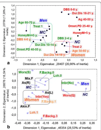
predominat-ing in the highest age groups (60–70 years), and women in the intermediate age groups (50–60 years). This was highlighted and confirmed by the MCA (Fig. 1a).
Age at examination and age at onset of PD
There was no connection between age at onset and presence of the G2019S mutation (χ2 = 0.90, p = 0.34).
However, there was a tendency towards a link between so-called honeymoon period (up to 5 years and more than 5 years) and the presence of the mutation (χ2 = 4.30,
p = 0.04 < 0.05), there were significantly more G2019S MC patients with shorter honeymoon period than the NC patients. The results also showed that 70.4 % of the patients had a honeymoon period shorter than 5 years when taking L-dopa.
Psychological assessment
The χ2 test was not significant (p = 0.60 ≫ 0.05), so there
was no significant difference of MMSE score between the G2019S MC and NC patients (25.3 ± 3.1 and 26.2 ± 2.7 respectively) (Table 2).
In our series, 74.1 % had a normal MMSE (25–30). In the remaining 25.9 %, there were five female and two male patients with a low sociocultural level and one patient with aphasia.
Lead location and DBS complications
Of the 54 contacts used for bilateral STN-DBS (for 27 patients), 53 contacts were localized in or near the STN. We registered one contact in the right side of brain to be localized outside of the STN. In three cases, we encoun-tered DBS complications (one MC patient and two NM patients) with moderate pneumocephalus in the frontal region and three other cases (one MC patient and two NM patients) had stimulator infections.
Comparative evaluations of the optimal stimula-tion parameters are: pulse width 60 μs, frequency 130 Hz, amplitude means are 2.32 ± 0.50 V at left and 2.35 ± 0.54 V at right side STN.
Genetic study
The genetic study revealed the presence of the G2019S mutation in 55.6 % of PD patients who had under-gone DBS stimulation with an average age at examina-tion of 56.2 ± 8.8 years and an average age at the onset of the disease of 40.1 ± 9.4 years. The NC patients had the same average age at examination and average age at onset (54.5 ± 7.1 and 40.3 ± 8.2 years respectively). The average duration of the honeymoon period (period dur-ing which there is a sustained response to dopaminergic treatment and the patient does not have a dyskinesia) was slightly shorter in patients with the MC (3.8 ± 1.7 years) than those with NC (5.4 ± 2.6 years) (Table 2).
Among all the NC patients, we found only two cases (16.66 %) of Parkin mutation (heterozygous c. 1204C>T in exon 11 and c. 458C>G in exon 4) with an age at onset of PD of 48 years, different initial symptoms and duration honeymoon (Table 3).
Clinical UPDRS‑III, S and E scales and H and Y stages The UPDRS-III of MC patients in Off–Off medica-tion-stimulation situations had an average score of 55.8 ± 16.4, which was significantly higher than the scores in three other cases (average score <28; Table 4). Table 1 Clinical characteristics of 27 STN-DBS patients in relation to gender
PD parkinson’s disease, STN subthalamic nucleus, Min minimum, Max maximum
Means Min Max Men (n = 17) Women (n = 10) p
Age groups (years) 55.4 ± 8.0 41 69 56.4 ± 9.5 53.8 ± 4.3 0.34
Age at onset of PD (years) 40.2 ± 8.7 25 55 40.9 ± 10.7 39.0 ± 3.9 0.34
Duration of disease (years) 15.3 ± 3.0 10 20 15.5 ± 3.3 14.9 ± 2.5 0.60
Duration of honeymoon (years) 4.5 ± 2.3 0.25 10 4.5 ± 1.8 4.6 ± 3.0 0.91
Duration of STN-stimulation (years) 4.7 ± 1.4 2.6 8 4.9 ± 1.5 4.2 ± 1.2 0.18
Fig. 1 Multiple correspondence analysis (MCA) between clinical and genetic variables. a On the left side: MC mutation carriers, NC non-carriers, Age: age classes (years), Onset.PD: Age at onset of PD (years), Dur.Dis duration of disease (years), Treat Treatment (Treat 1 <=500 mg/day, Treat 2 > 500 mg/day), DBS duration of deep brain Stimulation (years), HoneyM duration of honeymoon (years). b On the right side: MC mutation carriers, NC non-carriers, F.Backg family background of the disease, Wors Worsening (Q quick, S slow), Inbr inbreeding, asymmetry: As(R) right asymmetry, As(L) left asymmetry, disease form: Mix.F mixed form, Akin.F akinetic rigid form, Lohman: Loh0, Loh1, Loh2, several deaths. The green, orange and red colors were respectively for high, intermediate and high modalities of the variables
Page 5 of 10 Sayad et al. BMC Neurosci (2016) 17:6
Indeed, the mean values of UPDR-III decreased from 55.8 ± 16.4 (in Off–Off situation) to 27.3 ± 20.6 (in Off–On medication-stimulation), therefore, this is an UPDRS-III improvement of 51.1 % compared to the value Off–Off (very highly significant p = 0.000046 ≪ 0.001).
For NC patients, the test was very highly significant too (p < 0.001). The UPDRS-III in Off–Off medication-stim-ulation had an average score of 51.7 ± 14.4 again, and was significantly higher than the other three situations (average score <39) (Table 4). The mean values of UPDR-III decreased by 51.7 ± 14.4 (in Off–Off situations) to 38.5 ± 16.6 (in Off–On medication-stimulation situa-tions). So, this is an UPDRS-III improvement of 25.5 % (non significant p = 0.09). The best scores of UPDRS-III of Parkin mutation patients were observed in a situation of medication (On–Off situation) compared with a post-operative situation (Table 3).
The comparison of the four UPDRS-III situations showed more significance in MC patients (Fridman’s ANOVA p = 0.00005 ≪ 0.001) than in NC patients (p = 0.0003) (Table 4).
Similar results were found for the four S and E scale sit-uations, which showed more significant variations in MC patients (p = 0.00001 ≪ 0.001) compared to NC patients (p = 0.001) (Table 4). The S and E scale for MC patients in Off–On medication-stimulation had an average score of 0.6 ± 0.21, while the Off–Off situation had the low-est at 0.43 ± 0.18 (slightly significant p = 0.036). Similar results were found for the NC patients, with an average score of 0.6 ± 0.26 for the Off–On medication-stimula-tion, while the lowest was at 0.53 ± 0.14 for the Off–Off (non significant p = 0.41).
The comparison of the four H and Y stage situations showed more significance variations in MC patients (p = 0.00002 ≪ 0.001) compared to NC patients Table 2 Clinical comparative study of 27 STN-DBS patients
in relation with presence/absence of G2019S mutations
PD parkinson’s disease, MMSE Mini Mental State Examination, STN subthalamic
nucleus, MC mutation carriers, NC non mutation carriers
MC (n = 15) NC (n = 12) p Age groups (years) 56.2 ± 8.8 54.5 ± 7.1 0.59 Age at onset of PD (years) 40.1 ± 9.4 40.3 ± 8.2 0.97 Duration of disease (years) 16.1 ± 3.0 14.3 ± 2.7 0.10 MMSE (maximum score 30) 25.3 ± 3.1 26.2 ± 2.7 0.60 Duration of honeymoon (years) 3.8 ± 1.7 5.4 ± 2.6 0.08 Duration of STN-stimulation (years) 5.1 ± 1.4 4.1 ± 1.3 0.09
Table 3 Case reports of patients with Parkin mutation
Case 1 Case 2
Gene and Parkin Parkin
Exon with mutation Exon 4 Exon 11 Mutation type Heterozygous Heterozygous
c. 458C>G c. 1204C>T
Protein change P153R p. Arg402Cys
Gender Male Male
Age at onset 48 years 48 years
Disease duration 10 years 13 years Family history of
Parkinson-ism Negative Negative
UPDRS-III Off–Off scores 46/108 49/108 UPDR-III On–Off scores 28/108 32/108 UPDRS-III Off–On scores 51/108 51/108 UPDRS-III On–On scores 30/108 47/108 Initial symptoms Resting tremor in left
hand, dyskinesias 4 years after initiation of treatment. Muscle stiffness in both upper limbs, dyskinesias 7 years after initiation of treat-ment.
Table 4 Clinical UPDRS-III, S and E scales and H and Y stages scores of PD MC and NC patients undergoing STN-DBS (mean ± SD)
Unified parkinson’s disease rating scale III (UPDRS-III); Hoehn and Yahr scale (H and Y scale); Schwab and England’s activities of daily living scale (S and E scale)
MC mutation carriers, NC non mutation carriers
Medication stimulation UPDRS‑III S and E scale H and Y scale
situations MC NC MC NC MC NC Off–Off 55.8 ± 16.4 51.7 ± 14.4 0.43 ± 0.18 0.52 ± 0.14 3.50 ± 0.86 3.54 ± 0.65 On–Off 25.0 ± 13.2 30.6 ± 16.7 0.70 ± 0.13 0.69 ± 0.16 2.36 ± 0.66 2.87 ± 0.90 Off–On 27.3 ± 20.6 38.5 ± 16.6 0.60 ± 0.21 0.60 ± 0.26 2.36 ± 0.69 2.16 ± 0.91 On–On 19.7 ± 18.8 18.8 ± 12.5 0.85 ± 0.16 0.89 ± 0.10 2.13 ± 0.71 1.91 ± .99 p 0.00005 0.0003 0.00001 0.00106 0.00002 0.00091
(p = 0.0009) (Table 4). The H and Y scale in the Off–Off situation had an average score of 3.50 ± 0.87, which was significantly higher than in the three other cases (aver-age score <2.40). The H and Y st(aver-age for MC patients in Off–On medication-stimulation had an average score of 2.36 ± 0.69, while the Off–Off situation had the highest at 3.50 ± 0.87 (very highly significant p = 0.0003 ≪ 0.001).
For NC patients, the test was very highly significant too (p = 0.0009 < 0.001). The H and Y scale in the Off–Off situation had an average score of 3.54 ± 0.66, which was significantly higher than in the three other cases (aver-age score <2.90). The H and Y st(aver-age for NC patients in Off–On medication-stimulation had an average score of 2.16 ± 0.91, while the Off–Off situation had the highest at 3.54 ± 0.66 (very highly significant p = 0.00005 ≪ 0.001). Multiple correspondence analysis (MCA)
Figure 1a shows lower modalities of the variables on the left side for men and higher modalities of the variables on the right side for women.
The MCA provided added value and shows that NC women, who were in the average age groups (50–60 years old), had a disease duration of between 14 and 18 years, treated with high doses (>500 mg/day).
Conversely, MC men and L-dopa dose was reduced to below 500 mg/day after DBS, were in the 60–70 years age group and had developed the disease later than women (40–55 years old).
The distinction between the two groups (enclosed at left side of Fig. 1b) was fairly clear and shows that MC patients have no family history of the disease and tended to have a mixed form of the disease with right asymme-try predominance. However, the Clustering in the left lower figure is characterized by the presence, in female patients, of an akinetic-rigid form of the disease, and a family history of the disease with consanguinity.
The third group (on the right side of Fig. 1b) was that of the non-G2019S mutation carrying male patients, with no consanguinity and rapid worsening of the disease.
Discussion
The average age at onset of the disease was 40.2 ± 8.7 with 48.1 % in the patients having early onset of the dis-ease (25–40 years old) and 51.9 % with a disdis-ease onset age of 40–70 years old. This is in contrast with the literature, where PD is rarely observed to occur before 40 years of age (early PD onset represents less than 10 % of the cases) and 80 % of the cases start between the ages of 40 and 75 years [13]. This may be explained by the prevalence of a common idiopathic form in our series.
There were more males than females in our series, with a gender ratio of about 1.7. This difference would be justi-fied, according to Moisan and Elbaz [14], who reported
a gender ratio of 1.5 to the more frequent occupational exposure of men to neuro-toxic substances, and the neuro-protective effect of estrogen or a genetic factor linked to the X chromosome on women. Indeed, 70 % of the women in our series did not have a profession, whereas the men all did, be it manual laborers or white collar workers. In our series, female gender was more common amongst MC than NC (60 vs. 40 %). Indeed, it has been shown that PD patients with LRRK2 mutations are more likely to be women, suggesting a high genetic load versus idiopathic [15]. In addition, we should not exclude the fact that these patients were pre-selected for DBS after the processing of their files and that the gender criterion did not appear in the selection criteria.
We experienced a few complications with the DBS pro-cedures. These complications are explained by the dura-tion of surgery and the numbers of electrode penetradura-tion [16, 17]. The poor outcome of STN-DBS in PD is gener-ally related either to incorrect implantation or to hard-ware failure [18]. Incorrect lead placement, which may be due to a number of factors, including stereotactic inac-curacy, poor initial targeting or loss of cerebrospinal fluid during surgery, may have led to brain shift [19].
Monopolar stimulation (amplitude 2.32 ± 0.50 V at left and 2.35 ± 0.54 V at right side STN, width 60 μs, fre-quency 130 Hz) has been used in most patients by most studies [20, 21]. Therapeutic amplitudes for DBS nor-mally range between 1 and 4 V and the pulse width for stimulation of STN is 60 μs. Also, the frequency is 130 Hz in order to reach maximal benefit with minimal battery drain [22].
The presence of the G2019S mutation was observed in 15 patients (i.e. 55.6 % of the cases), including 8 sporadic and 7 familial PD patients, all heterozygous. This num-ber is higher than the one reported by Belarbi and col-laborators [5] for the Algerian population; with their 34 MC patients (i.e. 32.4 %) of which 28 were sporadic and 6 were familial PD patients. It was heterozygous in the familial form and sporadic in almost all cases.
The majority of MC patients had late onset of the dis-ease (after 40 years old) and a relatively short honeymoon period (5 years), while NC patients were mostly found to have early onset (between 20 and 40 years old) and a longer honeymoon period (between 6 and 10 years). According to Belarbi and collaborators [5], the compari-son of the clinical and evolutionary signs in G2019S MC and NC in PD showed a similarity in the clinical signs but the motor complications of the treatment induced by L-dopa were more frequent for the MC patients. Thus, this mutation appeared to be associated with the occur-rence of dyskinesias given the high frequency of its com-plications in MC, suggesting a genetic predisposition to these complications.
Page 7 of 10 Sayad et al. BMC Neurosci (2016) 17:6
Two NC patients (16.66 %) showed Parkin muta-tions, one was an early-onset case (48 years). Indeed, we reported in the literature that the Parkin mutation has been identified in several families with autosomal reces-sive early-onset Parkinsonism [23, 24] and frequency has been estimated at 10–25 % [25]. In contrast, the clini-cal characteristics of some European and North African patients with Parkin mutations were characterized with an age at onset of up to 58 years [24, 26]. The “honey-moon period” and initial symptoms were different in two patients with the Parkin mutations (the heterozygous c.1204C>T mutation in exon 11 and c.458C>G mutation in exon 4 of Parkin). In the literatures, the roles of Parkin heterozygous mutations at risk for PD have not been con-clusively shown [27, 28]. Nevertheless, in a recent study, a disease-associated heterozygous mutation of Parkin was found in one patient with early-onset, slowly progres-sive Parkinson’s disease with Lewy bodies and very late development of dementia [29]. However, the number of patients with Parkin mutation in our series so far is small and the correlations between genotype and phenotype are uncertain.
The MCA revealed the presence of some clinical set-tings common to patients with the LRRK2 G2019S mutation compared to NC. The G2019S patients were probably characterized by the mixed form of the disease with predominance of right side asymmetry.
On the other hand, NC patients may show differ-ences with respect to certain clinical gender parameters. Males with no family consanguinity and in an age group between 60 and 70 years possibly developed the disease later than females (40–55 years old). At the same time, females with family consanguinity and in age group between 50 and 60 years old likely developed the disease earlier (20–40 years old), with an akineto-rigid form with predominance of left side asymmetry.
The literature shows that there is no associated phe-notype related to MC. Although the clinical evaluation resembles that of a typical Parkinsonian syndrome, the age of onset of the disease is remarkably variable, ranging from 35 to 78 years [4]. Moreover, recent study reveals that G2019S MC patients is similar to NC PD patients but is characterized with more frequent lower extremity involvement at onset and postural instability and gait dif-ficulty without the associated cognitive impairment [30].
It is also known that during the pathological neurode-generation process, lesion distribution, initially unbal-anced, occurs at the same time as the occurrence and worsening of clinical symptoms. Thus, this evolution of the pathological process can sometimes lead to the domi-nance of a different subtype of clinical expression of the initial diagnosed subtype [31, 32]. A recent neuropatho-logical study also showed a pleomorphic phenotype in
this MC [33]. This clinical and pathological variability suggests that the G2019S mutation plays a role in several neurodegeneration interactions.
No significant difference was observed in MMSE between MC patients and NC. In fact, Goldwurm and collaborators [34] reported that there were no significant cognitive dysfunctions in MC and NC. Lesage and col-laborators [4] also reported an insignificant difference in MMSE in a French and North African population. On the other hand, Barth and collaborators [5] reported the low-est MMSE in MC patients, which the authors attributed to the small size of the sample in their study.
The UPDRS-III graphics show a marked improve-ment in the clinical spectrum of the PD patients. The percentage of improvement of 51.1 % for the G2019S MC patients in Off–On medication-stimulation situ-ations was above 30 %. It is empirically considered that an improvement of more than 30 % of the UPDRS-III is clinically significant [12]. In NC patients, the percentage of improvement under stimulation without drug therapy was below the required 30 % (25.5 %). The S and E scales as well as the H and Y stages in various situations of med-ication and stimulation also showed a marked improve-ment during Off–On medication-stimulation. This result demonstrates the effectiveness of DBS. It is known that DBS at the STN level largely improves the quality of life of patients because it acts on all aspects of the PD triad: tremor, rigidity and akinesia [35]. According to a study dating from 2011, the improvement in UPDRS-III was 41 % compared with the control group 12 months after surgery [36]. The first beneficial effects occur in the min-utes following the start of the stimulation [35]. In the long run, DBS is more stable than L-dopa treatment, and motor fluctuations and dyskinesia become minor [37].
However, the differences in the UPDRS-III, the S and E scales as well as the H and Y stages in the four situa-tions for MC patients were more significant than for NC patients. Also, the comparison between MC and NC in a situation of stimulation and a situation without medication-stimulation in the UPDRS-III and the S and E scales showed the best response for the MC patients. This means that the G2019S MC patients have a better response than NC patients. In fact, it is known from the literature that patients with LRRK2 mutations are good candidates for STN stimulation [38, 39], but limited series are available so far. In contrast, other studies sug-gested that no influence by the LRRK2 G2019S muta-tion exists on STN-DBS results [38, 40, 41]. However, the series reported in the literature are lower than our series (15 MC patients). It is supposed that this mutation makes PD patients more vulnerable to dyskinesias [5] or to some deleterious reorganization of corticostriatal efferents that would be modulated by the STN-DBS [39]. This leads us
consider that this mutation may have impact on the pro-gression and response to STN-DBS. Further larger stud-ies are needed to confirm these findings.
The patients with the Parkin mutation have the best UPDRS-III scores in the On–Off medication-stimulation situation compared to other situations. According to the literatures, the effect of DBS did not differ between patients with and without Parkin mutations [42, 43].
Conclusion
In our study, MCA revealed the presence of two distinct groups: MC and NC had two different clinical evalua-tions. The MC patients were probably characterized by the mixed form of the disease, with a predominance of right side asymmetry. In the NC group, men from non-consanguineous families and with an age (at examina-tion) ranging between 60 and 70 years may develop the disease later than the women (40–55 years).
Other noteworthy findings were a shorter honeymoon period of MC patients compared to the NC. Moreover, in the later group, we found two patients with Parkin muta-tions who had a different honeymoon period and differ-ent initial symptoms. Also, the results showed that the G2019S mutation was not associated with MMSE scores.
Other significant results were a clear improvement in the UPDRS-III more for MC than for NC patients who underwent stimulation with percentages of improvement over the required 30 % for MC patients only (51.1 and 25.5 % respectively). We found the same result for the S and E scales, which thus demonstrated the effectiveness of DBS for MC patients more than for NC patients. This indicates that, compared to NC patient, MC patients are probably the best candidates for STN-DBS. On the other hand, the best scores of UPDRS-III observed in situa-tions where Parkin mutation patients received medica-tion suggest that STN-DBS probably did not benefit these patients.
Ethics approval and consent to participate
All participants provided written informed consent for the publication of individual clinical details.
The study was approved by the institutional review boards and ethical approval was obtained from Eth-ics board and Academic Deontology of the Ministry of Higher Education and Scientific Research.
Abbreviations
Akin.F: akinetic rigid form; ANOVA: analysis of variance; As(L): left asymmetry; As(R): right asymmetry; CAPSIT-PD: Core Assessment Program for Surgical Interventionnal Therapies in PD; CT-scan: computerized tomography scans; Dur.Dis: duration of disease; F.Backg: family background of the disease; H and Y Scale: Hoehn and Yahr scale; HoneyM: duration of honeymoon; Inbr: inbreed-ing; L-dopa: levodopa; Loh: Lohman; LRRK2: leucine-rich repeat kinase 2; Max: maximum; MC: mutation carrier; MCA: multiple correspondence analysis; Min:
minimum; Mix.F: mixed form; MMSE: Mini Mental State Examination; MRI: mag-netic resonance imaging; NC: non-mutation carriers; Onset.PD: age at onset of PD; PD: parkinson’s disease; S and E scale: Schwab and England’s activities of daily living scale; STN-DBS: subthalamic nucleus deep brain stimulation; Treat: treatment; UPDRS-III: unified parkinson’s disease rating scale III; Wors.Q: worsening quick; Wors. S: worsening slow.
Authors’ contributions
MS and MZ: Conception and execution of the study. MC, FF, MN and MB: Medical experts. SL, AB and BA: Reviewing and editing. MBE: Statistical analysis and interpretation of data. All authors read and approved the final manuscript.
Author details
1 Laboratory of Behavioral and Cognitive Neuroscience, FSB, University
of Science and Technology Houari Boumediene, El Alia, Bab Ezzouar, BP 32, 16111 Algiers, Algeria. 2 Department of BPO, Faculty of SNV, Blida 1 University,
BP 270, 09000 Blida, Algeria. 3 Department of Neurology, CHU Ben Aknoun,
16000 Algiers, Algeria. 4 Department of Neurosurgery, Mohamed-Seghir
Nek-kache Military Hospital, Djasr Kasentina, Algiers, Algeria. 5 Department of
Neu-rology, Mohamed-Seghir Nekkache Military Hospital, Djasr Kasentina, Algiers, Algeria. 6 UMR S 1127 PaInserm U 1127, Sorbonne University, UPMC Univ Paris
06, Paris, France. 7 CNRS UMR 7225, Paris, France. 8 The Brain and Spinal Cord
Institute, 75013 Paris, France. 9 Department of Chemistry, Blida 1 University, BP
270, 09000 Blida, Algeria.
Acknowledgements
We would like to thank B. Abdennebi and F. Ysmail-Dahlouk (Department of Neurosurgery, Salim Zemirli Hospital, Algiers, Algeria); S. Abbas (Departmentof Neurology) and S. Chaib-Mamouzi (Department of Immunology, Mohamed Seghir Nekkache Military Hospital, Algiers, Algeria); M. Tazir, S. Belarbi and N. Hecham (Department of Neurology, Mustapha Pacha Hospital, Algiers, Algeria); K. Bouyoucef (Department of Neurosurgery, Frantz Fanon Hospital, Blida, Algeria) and M. Benmahdjoub (Department of Neurology, Frantz Fanon Hospital, Blida, Algeria); D. Ait-Idir and T. Hamadouche (Molecular Biology Laboratory, M’hamed Bougara University, Boumerdes, Algeria) for help in car-rying out the survey which underpinned this work.
Competing interests
The authors declare that they have no competing interests. Received: 22 April 2015 Accepted: 17 January 2016
References
1. Paisán-Ruíz C, Jain S, Evans EW, Gilks WP, Simón J, Van Der Brug M, de Munain AL, Aparicio S, Gil AM, Khan N, Johnson J, Martinez JR, Nicholl D, Carrera IM, Pena AS, de Silva R, Lees A, Martí-Massó JF, Pérez-Tur J, Wood NW, Singleton AB. Cloning of the gene containing mutations that cause PARK8-linked Parkinson’s disease. Neuron. 2004;44:595–600.
2. Zimprich A, Biskup S, Leitner P, Lichtner P, Farrer M, Lincoln S, Kachergus J, Hulihan M, Uitti RJ, Calne DB, Stoessl AJ, Pfeiffer RF, Patenge N, Carbajal IC, Vieregge P, Asmus F, Müller-Myhsok B, Dickson DW, Meitinger T, Strom TM, Wszolek ZK, Gasser T. Mutations in LRRK2 cause autosomal-dominant parkinsonism with pleomorphic pathology. Neuron. 2004;44:601–7. 3. Kachergus J, Mata IF, Hulihan M, Taylor JF, Lincoln S, Aasly J, Gibson JM,
Ross OA, Lynch T, Wiley J, Payami H, Nutt J, Maraganore DM, Czyzewski K, Styczynska M, Wszolek ZK, Farrer MJ, Toft M. Identification of a novel
LRRK2 mutation linked to autosomal dominant parkinsonism: evidence
of a common founder across european populations. Am J Hum Genet. 2005;76:672–80.
4. Lesage S, Ibanez P, Lohman E, Pollak P, Tison F, Tazir M, Leutenegger AL, Guimaraes J, Bonnet AM, Agid Y, Dürr A, Brice A. G2019S LRRK2 mutation in French and North African families with Parkinson’s disease. Ann Neurol. 2005;58:784–7.
5. Belarbi S, Hecham N, Lesage S, Kediha MI, Smail N, Benhassine T, Ysmail-Dahlouk F, Lohman E, Benhabyles B, Hamadouche T, Assami S, Brice A, Tazir M. LRRK2 G2019S mutation in Parkinson’s disease: a
Page 9 of 10 Sayad et al. BMC Neurosci (2016) 17:6
neuropsychological and neuropsychiatric study in a large Algerian cohort. Parkinsonism Relat Disord. 2010;16:676–9.
6. Koller WC, Tse W. Unmet medical needs in Parkinson’s disease. Neurology. 2004;62:S1–8.
7. Charles PD, Van Blercom N, Krack P, Lee SL, Xie J, Besson G, Benabid AL, Pollak P. Predictors of effective bilateral subthalamic nucleus stimulation for PD. Neurology. 2002;59:932–4.
8. Benabid AL, Chabardes S, Mitrofanis J, Pollak P. Deep brain stimulation of the subthalamic nucleus for the treatment of Parkinson’s disease. Lancet Neurol. 2009;8:67–81.
9. Klawans HL. Individual manifestations of Parkinson’s disease after ten or more years of levodopa. Mov Disord. 1986;1:187–92.
10. Schüpbach WM, Chastan N, Welter ML, Houeto JL, Mesnage V, Bonnet AM, Czernecki V, Maltête D, Hartmann A, Mallet L, Pidoux B, Dormont D, Navarro S, Cornu P, Mallet A, Agid Y. Stimulation of the subthalamic nucleus in Parkinson’s disease: a 5 year follow up. J Neurol Neurosurg Psychiatry. 2005;76:1640–4.
11. Defer GL, Widner H, Marié RM, Rémy P, Levivier M. Core Assessment Program for Surgical Interventional Therapies in Parkinson’s Disease (CAPSIT-PD). Mov Disord. 1999;14:572–84.
12. Fahn S, Elton RL. Members of the UPDRS development committee. Unified parkinson’s disease rating scale. In: Fahn S, Marsden CD, Calne DB, editors. Recent devolopments in parkinson’s disease. Florham Park Macmillan: Macmillan Health Care Information; 1987;2. p. 153–64. 13. Tanner CM. Epidemiology of Parkinson’s disease. Neurol Clin.
1992;10:317–29.
14. Moisan F, Elbaz A. Maladie de Parkinson et exposition aux pesticides. Envi-ron risque santé. 2011;10:372–84.
15. Cilia R, Siri C, Rusconi D, Allegra R, Ghiglietti A, Sacilotto G, Zini M, Zecchinelli AL, Asselta R, Duga S, Paganoni AM, Pezzoli G, Seia M, Goldwurm S. LRRK2 mutations in Parkinson’s disease: confirmation of a gender effect in the Italian population. Parkinsonism Relat Disord. 2014;20:911–4.
16. Hamel W, Schrader B, Weinert D, Herzog J, Müller D, Deuschl G, Volkmann J, Mehdorn HM. Technical complication in deep brain stimulation. Zen-tralbl Neurochir. 2002;63:124–7.
17. Okun MS, Tagliati M, Pourfar M, Fernandez HH, Rodriguez RL, Alterman RL, Foote KD. Management of referred deep brain stimulation failures: a retrospective analysis from 2 movement disorders centers. Arch Neurol. 2005;62:1250–5.
18. Lezcano E, Gomez-Esteban JC, Zarranz JJ, Lambarri I, Madoz P, Bilbao G, Pomposo I, Garibi J. Improvement in quality of life in patients with advanced Parkinson’s disease following bilateral deep-brain stimulation in subthalamic nucleus. Eur J Neurol. 2004;11:451–4.
19. Khan FR, Henderson JM. Deep brain stimulation surgical techniques. Handb Clin Neurol. 2013;116:27–37.
20. Eusebio A, Thevathasan W, Doyle Gaynor L, Pogosyan A, Bye E, Foltynie T, Zrinzo L, Ashkan K, Aziz T, Brown P. Deep brain stimulation can suppress pathological synchronisation in parkinsonian patients. J Neurol Neuro-surg Psychiatry. 2011;82:569–73.
21. Sommer M, Stiksrud EM, Von Eckardstein K, Rohde V, Paulus W. When battery exhaustion lets the lame walk: a case report on the importance of long-term stimulator monitoring in deep brain stimulation. BMC Neurol-ogy. 2015;15:113.
22. Castrioto A, Volkmann J, Krack P. Postoperative management of deep brain stimulation in Parkinson’s disease. Handb Clin Neurol. 2013;116:129–46.
23. Kitada T, Asakawa S, Hattori N, Matsumine H, Yamamura Y, Minoshima S, Yokochi M, Mizuno Y, Shimizu N. Mutations in the parkin gene cause autosomal recessive juvenile parkinsonism. Nature. 1998;392:605–8. 24. Abbas N, Lücking CB, Ricard S, Dürr A, Bonifati V, De Michele G, Bouley S,
Vaughan JR, Gasser T, Marconi R, Broussolle E, Brefel-Courbon C, Harhangi BS, Oostra BA, Fabrizio E, Böhme GA, Pradier L, Wood NW, Filla A, Meco G, Denefle P, Agid Y, Brice A. A wide variety of mutations in the parkin gene are responsible for autosomal recessive parkinsonism in Europe. Hum Mol Genet. 1999;8:567–74.
25. Lücking CB, Dürr A, Bonifati V, Vaughan J, De Michele G, Gasser T, Harhangi BS, Meco G, Denèfle P, Wood NW, Agid Y, Brice A. Association between early onset Parkinson disease and mutations in the Parkin gene. N Engl J Med. 2000;342:1560–7.
26. Lücking CB, Abbas N, Dürr A, Bonifati V, Bonnet AM, de Broucker T, De Michele G, Wood NW, Agid Y, Brice A. Homozygous deletions in parkin gene in European and North African families with autosomal recessive juvenile parkinsonism. Lancet. 1998;352:1355–6.
27. Sun M, Latourelle JC, Wooten GF, Lew MF, Klein C, Shill HA, Golbe LI, Mark MH, Racette BA, Perlmutter JS, Parsian A, Guttman M, Nicholson G, Xu G, Wilk JB, Saint-Hilaire MH, DeStefano AL, Prakash R, Williamson S, Suchowersky O, Labelle N, Growdon JH, Singer C, Watts RL, Goldwurm S, Pezzoli G, Baker KB, Pramstaller PP, Burn DJ, Chinnery PF, Sherman S, Vieregge P, Litvan I, Gillis T, MacDonald ME, Myers RH, Gusella JF. Influence of heterozygosity for parkin mutation on onset age in familial Parkinson disease: the GenePD study. Arch Neurol. 2006;63:826–32.
28. Kay DM, Moran D, Moses L, Poorkaj P, Zabetian CP, Nutt J, Factor SA, Yu CE, Montimurro JS, Keefe RG, Schellenberg GD, Payami H. Heterozygous par-kin point mutations are as common in control subjects as in Parpar-kinson’s patients. Ann Neurol. 2007;61:47–54.
29. Pankratz N, Kissell DK, Pauciulo MW, Halter CA, Rudolph A, Pfeiffer RF, Marder KS, Foroud T, Nichols WC, Parkinson Study Group-PROGENI Inves-tigators. Parkin dosage mutations have greater pathogenicity in familial PD than simple sequence mutations. Neurology. 2009;73:279–86. 30. Alcalay RN, Mirelman A, Saunders-Pullman R, Tang M, Santana HM,
Ray-mond D, Roos E, Orbe-Reilly M, Gurevich T, Shira AB, Weisz MG, Yasinovsky K, Zalis M, Thaler A, Deik A, Barrett MJ, Cabassa J, Groves M, Hunt A, Lubarr N, San Luciano M, Miravite J, Palmese C, Sachdev R, Sarva H, Severt L, Shanker V, Swan MC, Soto-Valencia J, Johannes B, Ortega R, Fahn S, Cote L, Waters C, Mazzoni P, Ford B, Louis E, Levy O, Rosado L, Ruiz D, Dorovski T, Pauciulo M, Nichols W, Orr-Urtreger A, Ozelius L, Clark L, Giladi N, Bress-man S, Marder KS. Parkinson disease phenotype in Ashkenazi Jews with and without LRRK2G2019S mutations. Mov Disord. 2013;28:1966–7. 31. Alves G, Larsen JP, Emre M, Wentzel-Larsen T, Aarsland D. Changes in
motor subtype and risk for incident dementia in Parkinson’s disease. Mov Disord. 2006;21:1123–30.
32. Rajput AH, Voll A, Rajput ML, Robinson CA, Rajput A. Course in Parkinson dis-ease subtypes: a 39-year clinicopathologic study. Neurology. 2009;73:206–12. 33. Wider C, Dickson DW, Wszolek ZK. Leucine-rich repeat kinase 2
gene-associated disease: redefining genotype-phenotype correlation. Neuro-degener Dis. 2010;7:175–9.
34. Goldwurm S, Zini M, Di Fonzo A, De Gaspari D, Siri C, Simons EJ, Van Doeselaar M, Tesei S, Antonini A, Canesi M, Zecchinelli A, Mariani C, Meucci N, Sacilotto G, Cilia R, Isaias IU, Bonetti A, Sironi F, Ricca S, Oostra BA, Bonifati V, Pezzoli G. LRRK2 G2019S mutation and Parkinson’s disease: a clinical, neuropsychological and neuropsychiatric study in a large Italian sample. Parkinsonism Relat Disord. 2006;12:410–9.
35. Benabid AL, Pollak P, Gross C, Hoffmann D, Benazzouz A, Gao DM, Laurent A, Gentil M, Perret J. Acute and long-term effects of subthalamic nucleus stimulation in Parkinson’s disease. Stereotact Funct Neurosurg. 1994;62:76–84.
36. Smeding HM, Speelman JD, Huizenga HM, Schuurman PR, Schmand B. Predictors of cognitive and psychosocial outcome after STN DBS in Parkinson’s Disease. J Neurol Neurosurg Psychiatry. 2011;82:754–60. 37. Krack P, Batir A, Van Blercom N, Chabardes S, Fraix V, Ardouin C, Koudsie
A, Limousin PD, Benazzouz A, Le Bas JF, Benabid AL, Pollak P. Five-year follow-up of bilateral stimulation of the subthalamic nucleus in advanced Parkinson’s disease. N Engl J Med. 2003;349:1925–34.
38. Schüpbach M, Lohmann E, Anheim M, Lesage S, Czernecki V, Yaici S, Worbe Y, Charles P, Welter ML, Pollak P, Dürr A, Agid Y, Brice A. Subthalamic nucleus stimulation is efficacious in patients with Parkinsonism and LRRK2 mutations. Mov Disord. 2007;22:119–22.
39. Stefani A, Marzetti F, Pierantozzi M, Petrucci S, Olivola E, Galati S, Bassi MS, Imbriani P, Valente EM, Pastore FS. Successful subthalamic stimulation, but levodopa-induced dystonia, in a genetic Parkinson’s disease. Neurol Sci. 2013;34:383–6.
40. Johansen KK, Jørgensen JV, White LR, Farrer MJ, Aasly JO. Parkinson-related genetics in patients treated with deep brain stimulation. Acta Neurol Scand. 2011;123:201–6.
41. Greenbaum L, Israeli-Korn SD, Cohen OS, Elincx-Benizri S, Yahalom G, Kozlova E, Strauss H, Molshatzki N, Inzelberg R, Spiegelmann R, Israel Z, Hassin-Baer S. The LRRK2 G2019S mutation status does not affect the outcome of subthalamic stimulation in patients with Parkinson’s disease. Parkinsonism Relat Disord. 2013;19:1053–6.
• We accept pre-submission inquiries
• Our selector tool helps you to find the most relevant journal
• We provide round the clock customer support
• Convenient online submission
• Thorough peer review
• Inclusion in PubMed and all major indexing services
• Maximum visibility for your research Submit your manuscript at
www.biomedcentral.com/submit
Submit your next manuscript to BioMed Central
and we will help you at every step:
42. Moro E, Volkmann J, König IR, Winkler S, Hiller A, Hassin-Baer S, Herzog J, Schnitzler A, Lohmann K, Pinsker MO, Voges J, Djarmatic A, Seibler P, Lozano AM, Rogaeva E, Lang AE, Deuschl G, Klein C. Bilateral sub-thalamic stimulation in Parkin and PINK1 parkinsonism. Neurology. 2008;70:1186–91.
43. Kim HJ, Yun JY, Kim YE, Lee JY, Kim HJ, Kim JY, Park SS, Paek SH, Jeon BS. Parkin mutation and deep brain stimulation outcome. J Clin Neurosci. 2014;21:107–10.