HAL Id: hal-01980537
https://hal.sorbonne-universite.fr/hal-01980537
Submitted on 14 Jan 2019
HAL is a multi-disciplinary open access
archive for the deposit and dissemination of
sci-entific research documents, whether they are
pub-lished or not. The documents may come from
teaching and research institutions in France or
abroad, or from public or private research centers.
L’archive ouverte pluridisciplinaire HAL, est
destinée au dépôt et à la diffusion de documents
scientifiques de niveau recherche, publiés ou non,
émanant des établissements d’enseignement et de
recherche français ou étrangers, des laboratoires
publics ou privés.
oxidase/macrocyclase from lankacidin biosynthesis
Jonathan Dorival, Fanny Risser, Christophe Jacob, Sabrina Collin, Gerald
Dräger, Cedric Paris, Benjamin Chagot, Andreas Kirschning, Arnaud Gruez,
Kira Weissman
To cite this version:
Jonathan Dorival, Fanny Risser, Christophe Jacob, Sabrina Collin, Gerald Dräger, et al.. Insights into
a dual function amide oxidase/macrocyclase from lankacidin biosynthesis. Nature Communications,
Nature Publishing Group, 2018, 9, pp.3998. �10.1038/s41467-018-06323-w�. �hal-01980537�
Insights into a dual function amide oxidase/
macrocyclase from lankacidin biosynthesis
Jonathan Dorival
1,4
, Fanny Risser
1
, Christophe Jacob
1
, Sabrina Collin
1
, Gerald Dräger
2
, Cédric Paris
3
,
Benjamin Chagot
1
, Andreas Kirschning
2
, Arnaud Gruez
1
& Kira J. Weissman
1
Acquisition of new catalytic activity is a relatively rare evolutionary event. A striking example
appears in the pathway to the antibiotic lankacidin, as a monoamine oxidase (MAO) family
member, LkcE, catalyzes both an unusual amide oxidation, and a subsequent intramolecular
Mannich reaction to form the polyketide macrocycle. We report evidence here for the
molecular basis for this dual activity. The reaction sequence involves several essential active
site residues and a conformational change likely comprising an interdomain hinge movement.
These features, which have not previously been described in the MAO family, both depend
on a unique dimerization mode relative to all structurally characterized members. Taken
together, these data add weight to the idea that designing new multifunctional enzymes may
require changes in both architecture and catalytic machinery. Encouragingly, however, our
data also show LkcE to bind alternative substrates, supporting its potential utility as a general
cyclization catalyst in synthetic biology.
DOI: 10.1038/s41467-018-06323-w
OPEN
1UMR 7365, Ingénierie Moléculaire et Physiopathologie Articulaire (IMoPA), CNRS-Université de Lorraine, Biopôle de l’Université de Lorraine, Campus
Biologie Santé, 9 Avenue de la Forêt de Haye, BP 20199, 54505 Vandœuvre-lès-Nancy Cedex, France.2Institut für Organische Chemie, Leibniz Universität Hannover, Schneiderberg 1B, Hannover 30167, Germany.3Laboratoire d’Ingénierie des Biomolécules, Ecole Nationale Supérieure d’Agronomie et des Industries Alimentaires (ENSAIA), Université de Lorraine, 2 Avenue de la Fôret de Haye, BP 172, 54518 Vandœuvre-lès-Nancy Cedex, France.4Present
address: Sorbonne Universités, UPMC Univ. Paris 06, CNRS, UMR 8227, Integrative Biology of Marine Models, Station Biologique de Roscoff, CS 90074 Roscoff, Bretagne, France. These authors contributed equally: Fanny Risser, Christophe Jacob. Correspondence and requests for materials should be addressed to A.G. (email:[email protected]) or to K.J.W. (email:[email protected])
123456789
E
nzyme activity within superfamilies frequently evolves via
changes in substrate specificity, whereas reaction chemistry
is usually retained
1,2. There are also well-documented
examples of a conserved protein fold housing diverse catalytic
activities: the
αβ hydrophobic barrel fold accommodates at
least six different types of acid–base chemistry
3, the
fumar-ylacetoacetate hydrolase superfamily includes decarboxylases,
isomerases, hydratases, and hydrolases
4, whereas the enolase
superfamily has > 30,000 members, which share an interdomain
active site architecture and the mechanistic strategy of proton
abstraction
α to a carboxylate and stabilization of the enolate by a
magnesium ion
5. This evolution of novel activity can occur via
several different mechanisms, including the insertion or deletion
of residues, changes in oligomerization state, and the recruitment
of metal ions and cofactors
1. The enzyme LkcE from the
lanka-cidin polyketide biosynthetic pathway of the bacterium
Strepto-myces rochei represents an interesting test case for understanding
such gain-of-function, as it exhibits a monoamine oxidase (MAO)
protein fold yet also carries out a macrocyclization reaction.
The core of the lankacidin molecule is constructed from simple
acyl-CoA and amino-acid-building blocks by a series of gigantic
multimodular polypeptides, which operate in assembly-line
fashion (polyketide synthases (PKSs) and non-ribosomal
pep-tide synthetases (NRPSs), respectively)
6. The full-length chain is
released from the last multienzyme LkcG by a dedicated
thioes-terase domain
7. Unusually for this type of pathway, the product is
liberated from the multienzyme not as a macrolide, but as a
β-oxo-δ-lactone (Fig.
1
). LkcE then catalyzes post-assembly
mac-rocyclization via formation of a C-2−C-18 carbon-carbon bond
8.
This reaction is proposed to occur on a C-7 acetylated metabolite
LC-KA05 (1), although the timing of the acetylation reaction, and
the enzyme responsible, are unknown. A strain of S. rochei in
which LkcE is inactivated accumulates LC-KA05 (Fig.
1
), which
can be converted into lankacidin C (3) by a strain blocked early in
lankacidin biosynthesis, demonstrating the chemical competence
of LC-KA05 as an intermediate
8. In the
final stages of the
biosynthesis, the C-24 hydroxyl group is oxidized to a ketone and
the C-7 acetyl group is cleaved, yielding lankacidin C, with the
OH HO O O O HN O O H NH S O O OH NH S O O OH OH NH S O O OH OH NH S O O OH OH NH S O O OH OH OH NH S O O OH OH OH OH NH S O O O OH OH OH OH NH S O O OH S O HO HO S O O S O HO NH S O O OH TE KS DH MT KR ACP ACP KR KS ACP KS KR ACP KS DH MT KR ACP ACP DH C A PCP MODULE 1 MODULE 2 KS DH MT KR ACP ACP KR KS ACP KS KR ACP MODULE 3 MODULE 4 DH KS ACP MT MODULE 5 MODULE 6
MODULE 7 MODULE 8 MODULE 9 LkcA LkcB LkcC (1st use) LkcF (1st use) LkcB LkcC (2nd use) LkcF (2nd use) LkcC (3rd use) LkcG
Lankacidin C (3) Lankacidinol A (2) LkcE + AT LkcD Lankacidin A 25 OH AcO O H O O HN O HN O H O O OH AcO HO HN OH H O O O OH O O HO O O 18 15 13 7 1 4 19 20 21 ? ? 1,3-bisphospho -glycerate FkbH ORF22 product ACP ORF19 product (DH*) DH* KR* ACP ORF19 product (KR*) DH* KR* ACP ORF19 product (KR*) DH* KR* ORF21 product C-7 acetyl transferase (LkcH?) LC-KA05 (1) 24 5 7 1 18 25 18 15 13 7 1 4 19 20 21 26 27 23 2 KS of LkcC?
Fig. 1 Proposed biosynthetic pathway to lankacidin C. The gene cluster, which has been shown to be sufficient for lankacidin biosynthesis44, encodesfive
assembly-line proteins, LkcA (a hybrid NRPS/PKS subunit), LkcB (a discrete DH), LkcC, LkcF, and LkcG, together containing a total of only four KS domains, although eight KS-mediated extension cycles are required. One possibility that agrees with phylogenetic analysis of KS substrate specificity23is shown
here, in which the assembly-line incorporates multiple copies of the proteins LkcB, LkcC and LkcF (see ref.44for an alternate view). The starter unit may be
either pyruvoyl-ACP or lactoyl-ACP, both derived from 1,3-bisphosphoglycerate45. However, we and others8have found that in an LkcE-deleted mutant,
only the lactoyl form of thefirst free intermediate LC-KA05 accumulates (Supplementary Fig. 8), so we propose that lactoyl-ACP serves as the starter unit. The enzyme responsible for acetyl transfer at C-7 likewise remains to be determined, although a candidate is LkcH, which shows homology to isochorismatases8. The object of this study, LkcE, catalyzes the critical macrocyclization reaction of LC-KA05 (1), resulting in lankacidinol A (2). The
carbons implicated in the ring closure are indicated in red. Domain abbreviations: C, condensation; A, adenylation; PCP, peptidyl carrier protein; KS, ketosynthase; DH, dehydratase; KR, ketoreductase; MT,C-methyltransferase; ACP, acyl carrier protein; TE, thioesterase
relative timing of these two transformations also remaining to be
established (Fig.
1
).
The catalytic mechanism proposed for LkcE involves initial
oxidation of the amide function of LC-KA05 to an iminium ion
8.
This is followed by attack of a C-2 enolate anion on the iminium
to give the 17-membered lankacidinol A (2)
8—an enzymatic
intramolecular Mannich reaction. Although numerous
flavin
adenine dinucleotide (FAD)-dependent amine oxidases have been
characterized to date
9, to our knowledge LkcE is only the second
described amide oxidase
10. This observation, coupled with the
fact that no other known enzyme carries out Mannich chemistry
except as a promiscuous activity
11, motivated our interest in
establishing a detailed structure/function relationship for LkcE.
In this study, we have used a combination of X-ray
crystal-lography, structure-guided mutagenesis, and kinetic analysis, to
reveal key architectural differences between LkcE and other
members of the MAO family, which underpin its second,
cycli-zation activity. We also demonstrate the tolerance of LkcE to
certain structural modifications of the substrate. As 1,3-diketones
and amide functional groups are common in PK/NRP hybrid
metabolites, LkcE represents a potentially valuable addition to a
synthetic biology toolbox as a means to access novel macrocyclic
structures of various sizes and functionality.
Results
Structural characterization of holo and ligand-bound LkcE. S.
rochei LkcE was purified to homogeneity as a recombinant
pro-tein (Supplementary Tables 1 and 2) from Escherichia coli and its
redox cofactor identified as FAD by mass spectrometry following
chromatographic separation under denaturing conditions
(Sup-plementary Fig. 1); thus in common with certain MAO family
members
12but distinct from the only other reported amide
oxidase Af12070 (ref.
10), the FAD cofactor is non-covalently
bound. By UV-Vis, 45% of the protein was estimated to contain
FAD (on a par with AF12070 (ref.
10)) (Methods). We crystallized
the enzyme in its holo form, but additionally in the presence of
substrate analogs: ethyl 2-methylacetoacetate (EMAA) (4) that
mimics the
δ-lactone of LC-KA05, and N,N′-diallyl-
L-tartardia-mide (DATD) (5), which resembles the a-tartardia-mide region (Fig.
2
). The
X-ray crystal structure of the holo protein was solved at 3.15 Å
resolution by single-wavelength anomalous dispersion (SAD)
using data collected on seleniated protein (Fig.
3
a). The structure
of LkcE (Fig.
3
b, c) co-crystallized in the presence of the analogs
was then solved at 2.80 Å by molecular replacement (the statistics
for data collection, refinement, and validation of both structures
are presented in Supplementary Table 3). For all four structures,
> 99.7% of the residues were in allowed regions of the
Rama-chandran plot. In all but the SeLkcE structure, one Gly (299 or
300) was an outlier, whereas Glu313 was an outlier in both forms
of the holo enzyme structure. However, for Glu313, clear electron
density is present corresponding to the residue.
Sequence alignment (Supplementary Fig. 2) and comparison
with the structures of the
five closest structural homologs to LkcE
identified by the DALI
13server confirmed that LkcE belongs to
the MAO family. The respective root mean square deviation
(rmsd) of Cα positions was 4.232 Å (212 Cα) for 6-hydroxy-
L-nicotine oxidase (6HDNO) from Paenarthrobacter nicotinovorans
(PDB
3NG7
)
14; 4.720 Å (254 Cα) for human monoamine oxidase
(hMAO) A (
2Z5X
)
15; 5 Å (267 Cα) for hMAO B (
1GOS
)
16;
10.239 (271 Cα) for protoporphyrinogen oxidase (PPOX) of
Bacillus subtilis (
3I6D
)
17; and 14.660 Å (162 Cα) for a
poly-unsaturated fatty acid isomerase of Propionibacterium acnes
(
2BAB
)
18.
The LkcE monomer contains two domains, a FAD-binding
domain and a substrate-binding domain (Fig.
3
a), which are
joined by a substantial number of loop regions. The
cofactor-binding domain (which includes residues 1−45 incorporating a
characteristic
FAD-binding
motif
(xhxhGxGxxGxxxhxxh
(x)
8hxhE(D)
19), 70−83, 206−279, and 368−438), is topologically
similar to other proteins with the
‘3-layer (BBA) sandwich’ fold in
the CATH database
20, and comprises a central four-stranded
antiparallel
β-sheet flanked on one side by five α-helices, and on
the other side by a second three stranded, antiparallel
β-sheet and
three
α-helices. The FAD cofactor is present at 100% occupancy
(Supplementary Fig. 3), and as in other members of the MAO
family, it adopts an elongated conformation. Although the
specific FAD-binding residues differ among LkcE and its closest
structural homologs (Supplementary Fig. 2), the types of
interactions are similar, with the exception of those to the
isoalloxazine ring. In all of the structures except LkcE and PPOX,
the isoalloxazine is
flanked by two bulky aromatic residues (Tyr,
and in 3NG7 only, a Tyr and a Trp). In LkcE, the equivalent
residue positions are Gly364 and Leu398, respectively, whereas in
PPOX, the analogous amino acids are M413 and G449. Thus, it
LC-KA05 (1) 25 HO HN O OH H O O O OH O O O O O O HN HO HO O OH OH H O O HN OH H N 18 15 13 7 1 4 19 20 21 HO HN OH OH H H H O O O OH HN HO HO OH H O O O O HO HN O H O O O 26 27 23 2 8 (Cyclized 7) EMAA (4) DATD (5) Hydrolytic derivative of LC-KA05 (6) Elimination derivative of LC-KA05 (7) Lankacidinol C (9) (Cyclized 6) HO
Fig. 2 Structures of selected compounds investigated in this study. Compound LC-KA05 (1) was shown by NMR to exist almost exclusively in the enol form, and thus its derivatives6 and 7 are also represented as enols. The gray boxes indicate where 6 and 7 differ from 1. The stereochemistry of the C-6−C-7 double bond in 7 and cyclized 7 (8) is unknown, as indicated
appears that the FAD-binding site has been modified in these two
enzymes in order to accommodate the large macrocycles of the
substrates/products.
The substrate-binding domain contains an orthogonal
α-helix
bundle that packs against a
β-sheet. Visual inspection of the
overlaid structures shows that although the cofactor-binding
domain is well-conserved, the substrate-binding domain is
significantly divergent in terms of both α-helical content and
orientation (Supplementary Fig. 4). This is unsurprising given the
pronounced structural differences between LC-KA05 and the
typical small-molecule amine substrates of the MAO family
21.
Although the majority of characterized MAO family members
are homodimeric, there is some disagreement between
crystal-lographic and solution data, and at least one homolog is
monomeric
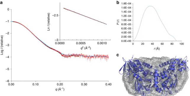
9. LkcE is clearly a homodimeric protein in the
crystal, with an extensive interface (4705 Å
2, representing 15% of
the total surface area). It is also homodimeric in solution, as
shown by small-angle X-ray scattering (Fig.
4
). However, it
dimerizes differently to the human enzymes hMAO A
15and B
16,
as well as to its closest structural homolog 6HDNO
14(Fig.
5
),
giving it a distinctive quaternary organization. In the hMAO B
crystal structure, the dimerization interface is formed by both the
cofactor- and substrate-binding domains, whereas in the case of
6HDNO, stabilization of the homodimer is additionally provided
by two molecules of diacylglycerophospholipid. In contrast, the
dimer interface of LkcE is formed exclusively by the
substrate-binding domain of each monomer. The interface residues include
E68, K69, V87, L106, F108, E114, D121, Q125, N128, Q129, S188,
Y199, R201, H332, and R335. Most of these are well-conserved
between LkcE and its nearest homologs, but not all
(Supplemen-tary Fig. 2). This distinct mode of dimerization, coupled with the
high content of loops at the interdomain interface within each
monomer (Fig.
3
a), would favor movement of the domains
relative to each other, with potentially important implications for
the catalytic mechanism, as discussed below.
Structure-guided site-directed mutagenesis of LkcE. Analysis of
the crystal structures with substrate analogs bound revealed clear
electron density attributable to EMAA and DATD, but only one
of these compounds was present in any given active site, as their
positions overlapped (Fig.
3
b, c). Inspection of the two binding
sites coupled with mechanistic considerations identified E64,
H65, Y168, Y182, R326, and T397 as candidate catalytic residues
(Supplementary Fig. 2). The presence of amino acids in the active
site capable of acting as general acids/bases contrasts with other
flavin-dependent amine oxidase homologs which lack such
residues
14,21.
In particular, E64, Y182, and R326 are strictly conserved in all
genes putatively encoding LkcE-type cyclases (Supplementary
Fig. 2). Site-specific mutagenesis was used to produce five
mutants: E64A, E64Q, Y182F, R326L, and R326Q, which were
purified to homogeneity (FAD content, respectively: E64A, 58%;
E64Q, 53%; Y182F, 42%; R326L, 20%; R326Q, 60%). Analysis by
circular dichroism (CD) confirmed that the mutations had not
dramatically altered the structures (Supplementary Fig. 5).
Isolation of the native substrate of LkcE. To obtain the native
LkcE substrate LC-KA05, we disrupted gene lkcE in S. rochei
by PCR targeting, using a strategy similar to that described
previously
8,22,23(Supplementary Figs. 6 and 7). Following analysis
of the lkcE mutant by high-performance liquid
chromatography-mass spectrometry (HPLC-MS) to verify the presence of LC-KA05
L185 L185
a
b
c
d
L191 T397 Y182 Y168 S186 L191 S186 Q428 Q64 R326 T397 Y182 N179 V372 Y168 L70 H65 W200 L185 L70 W200 S186 L70 T397 L364 Y168 W200 Y182 2.8 3.4 2.9 3.3 3.2 2.9 2.7 2.7 2.8 2.8Fig. 3 Crystal structures of LkcE and its mutants. a Crystal structure of homodimeric, wild-type LkcE. The FAD-binding domain is shown in light blue and light purple for the monomers A and B, respectively, whereas the substrate-binding domain is shown in dark blue and purple. The FAD is colored in yellow. b View of the active site of wild-type LkcE in the presence of bound DATD (pink) with its 2Fo−Fcmap contoured at 1σ. Binding occurs mainly via
hydrophobic interactions and a single hydrogen bond with T397.c View of the active site of wild-type LkcE in the presence of bound EMAA (orange) with the 2Fo−Fcmap contoured at 0.6σ. Comparison with b shows the analog to be binding in the same region of the active site. d View of the active site of the
LkcE E64Q mutant in the presence of bound LC-KA05 (green), which adopts a linear conformation. The 2Fo−Fcmap surrounding the substrate is
and the absence of all downstream metabolites (Supplementary
Fig. 8 and Supplementary Table 4), 1 was obtained in pure form by
preparative HPLC (Supplementary Fig. 9). Unfortunately, LC-KA05
was unstable, undergoing degradation via loss of the C-7
acetate, both by hydrolysis (to give 7-OH 6, Fig.
2
) and
elimination (to give 7 (Fig.
2
)), which co-eluted with LC-KA05.
However, 16 mg of mixed metabolite could be obtained from the
extracts of 20−30 L of culture, with 1 and 7 present in an ~ 2:1 ratio
(chemical data on all three compounds are provided in the
Meth-ods). As HPLC-MS analysis of extracts of both the wild-type strain
a
b
c
d
Fig. 5 Alternate homodimerization mode of LkcE relative to hMAO B and 6HDNO. a Homodimeric structure of LkcE. b Homodimerization mode of 6HDNO. The dimer is superimposed on one monomer of LkcE (in blue ina), in the same orientation. c Homodimerization mode of hMAO B. Again, the dimer has been superimposed on a monomer of LkcE.d Interaction surface between the two monomers in LkcE (yellow), hMAO B (red), and 6HDNO (green), relative to a monomer of LkcE. This analysis clearly shows that the mode of homodimerization of LkcE, as well as its overall quarternary organization, differ from these two homologs
0
a
b
c
–1 –2 –2.5 P ( r) 1.8E–04 1.6E–04 1.4E–04 1.2E–04 1.0E–04 8.0E–05 6.0E–05 4.0E–05 2.0E–05 0.0E+00 0 20 40 60 80 100 Ln I (relativ e) –3 0.0000 0.0005 0.0010 Log I (relativ e) –3 –4 –5 –6 0.00 0.10 0.20 q (Å–1) q2 (Å–2) r (Å) 0.30 0.40Fig. 4 Characterization of recombinant LkcE by SAXS. a Fit between theab initio model computed with GASBOR46(black line) (χ2= 1.44), the theoretical
scattering curve calculated on the basis of the structure of LkcEWTwith CRYSOL47(blue line) (χ2= 3.8) and the experimental SAXS data (red dots). Inset
is the Guinier plot, which yields anRgof 31.0 Å. A molecular weight (MW) of 99.3 kDa was calculated using the SAXS MoW48program (homodimer
calculated MW= 98.6 kDa). b The distance distribution function derived for LkcE calculated with GNOM49, yielding aD
maxof 91 Å.c Averaged ab initio
envelope of LkcE calculated using GASBOR46(gray mesh) with superimposition of the LkcE
and the lkcE mutant did not reveal any evidence for 7, nor, in the
case of the wild type, for its cyclized equivalent 8 (Fig.
2
;
Supple-mentary Fig. 10 and SuppleSupple-mentary Table 4), the cellular
envir-onment must protect against this type of non-productive
degradation. The 7-OH compound 6 was observed in extracts of the
lkcE mutant although not in the wild type (Supplementary Fig. 10
and Supplementary Table 4), presumably because in the presence of
active LkcE it can be productively cyclized to give lankacidinol C (9)
(Fig.
2
) (see below), and from there transformed to lankacidin
C. Interestingly, NMR analysis of purified LC-KA05 revealed that
the
δ-lactone was almost exclusively in the enol form
(Supple-mentary Fig. 9) as recently described for a new lankacidin-derived
metabolite
24, presumably because the six-membered ring forces
overlap in the
π-system.
Kinetic characterization of LkcE and its mutants. Attempts to
determine the kinetics of LkcE-catalyzed conversion of LC-KA05
to lankacidinol A by HPLC-MS were frustrated by the rapid
degradation of 1 under the assay conditions (Supplementary
Fig. 11). Nonetheless, larger scale incubation of 1 in the presence
of LkcE produced authentic lankacidinol A, as judged by
HPLC-MS comparison with extracts of wild-type S. rochei containing
this metabolite, providing conclusive evidence that recombinant
LkcE was active (Supplementary Fig. 12 and Supplementary
Table 4). The enzyme was also found capable of cyclizing the
degradation product 7, as well as purified, deacetylated substrate
6, to give lankacidinol C, as established by HPLC-MS
(Supple-mentary Fig. 12 and Supple(Supple-mentary Table 4), evidence for its
relaxed specificity toward structural variations at the C-6/C-7 ring
positions.
As an alternative and more-sensitive kinetic method, we
employed a coupled enzymatic assay to detect the H
2O
2formed
during each catalytic cycle. For this, we utilized NADH
peroxidase from Streptococcus faecalis, which catalyzes the
NADH-dependent reduction of H
2O
2to water. In this way, LkcE
turnover (production of H
2O
2) was detected via the consumption
of NADH (loss of absorbance at 340 nm) under conditions where
NADH peroxidase was not rate-limiting (see Methods)
25. We
also confirmed that the presence of dimethyl sulfoxide (used to
solubilize the substrates) had no effect on the measured kinetic
parameters (Fig.
6
and Table
1
). However, as 1 was present at
only ca. 66% in the mixture, and degraded spontaneously during
the assays, the kinetic parameters must be considered lower
estimates. Nonetheless, the possibility to rapidly acquire many
data points in the initial stages of the reaction (A
340 nmwas
measured every 0.4 s) meant that effects of the decomposition of 1
were minimized.
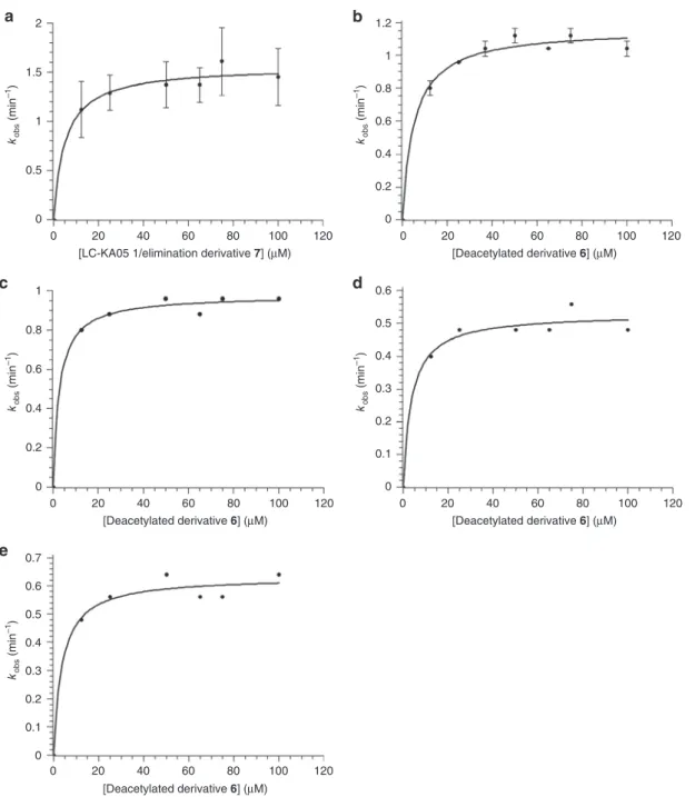
With the ca. 2:1 LC-KA05/eliminated derivative 7 mixture,
this system yielded the following steady-state kinetic parameters
for the wild type (average of measurements in duplicate, and
taking into account the observed proportion of holo LkcE
(45%)): k
cat= 3.4 ± 0.2 min
−1and K
M= 5 ± 2 μM. This k
catis
comparable to both the overall rate of turnover reported for an
intact PKS system in vitro (1.1 min
−1)
26, as well as that for the
amide oxidase AF12070 (ca. 5 min
−1(ref.
10)) (Fig.
6
and
Table
1
). Analysis of the wild-type enzyme with deacetylated
derivative 6 yielded comparable parameters (k
cat= 2.4 ± 0.1 min
−1
, K
M= 5 ± 1 μM) (Fig.
6
and Table
1
). Mutants E64A, E64Q,
and R326L were completely inactive with the 1/7 mixture (as
was boiled protein control), whereas mutants Y182F and R326Q
showed good activity towards the deacetylated derivative 6:
R326Q, k
cat= 0.88 min
−1, K
M= 4 μM; Y182F, k
cat= 1.5 min
−1,
K
M= 4 μM (Fig.
6
and Table
1
) (owing to limited quantities, the
1/7 mixture was not tested with these mutants). Taken together,
these data are consistent with residues E64 and R326 playing
critical roles in the catalytic mechanism (although R can be
substituted by Q without a significant effect on activity), whereas
Y182 is not essential. We also confirmed lack of turnover with
the substrate analogs EMAA and DATD, both separately and
together (using 2 and 10
μM enzyme and up to 400 μM in both
substrates), consistent with their overlapping binding modes
(Fig.
3
b, c).
Crystal structures of LkcE mutants in complex with LC-KA05.
In order to obtain complexes with LC-KA05, mutants E64A,
E64Q, Y182F, R326L, and R326Q were soaked with the 1/7
substrate mixture followed by rapid acquisition of the X-ray
diffraction data (owing both to the low availability of LC-KA05
and its high instability, it was not possible to carry out
co-crystallization experiments). Of the
five mutants, only E64Q and
R326Q yielded co-complexes with 1; 7 was not observed in the
active sites, as the acetate of 1 was clearly visible. The structure of
the LkcE E64Q/LC-KA05 complex was solved at 3.03 Å
resolu-tion and that of the LkcE R326Q/LC-KA05 complex at 2.50 Å
resolution by molecular replacement, using the structure of the
LkcE/EMAA/DATD complex as the search model, and the
pre-sence of LC-KA05 confirmed using a weighted F
o−F
comit map
(Supplementary Fig. 3; Supplementary Table 3).
LC-KA05 lies at the interface between the substrate-binding
domain and the cofactor-binding domain and adopts an extended
conformation. The substrate sits in a deep pocket (∼ 20 Å from
the protein surface and ~ 6−8 Å wide), and is surrounded by
hydrophobic residues (L70, Y168, Y182, L185, M189, W200,
L324, F345, V396, and G398). In addition to these hydrophobic
interactions, binding is likely mediated by seven hydrogen bonds:
two to the lactone ring at the entry point of the pocket (by N179
and T397), one to the acetate at C-7 by the side chain of Q428,
and four to the lactoyl moiety (two with Y168, and one each with
E64 and R326). Notably, in this configuration, the lactoyl portion
of LC-KA05 stacks against the FAD isoalloxazine in a position
appropriate for hydride transfer (the amide proton is situated at
3.2 Å from the FAD N-5). On the other hand, superposition of
the R326Q/LC-KA05 structure and that of the holo wild type
shows that if LC-KA05 bound into the wild type in the same
orientation as in the LkcE R326Q/LC-KA05 complex, its C-24
hydroxyl would sterically clash with R326 (Supplementary
Fig. 13). In the E64Q structure in which R326 is present, the
protein is identical, but the substrate adopts an alternative
orientation, which allows it to interact with R326 (3.1 Å between
the C-23 carbonyl of LC-KA05 and the R326 side chain)
(Supplementary Fig. 13).
In both structures obtained in the presence of 1, the observed
linear configuration of the polyketide chain would not permit
subsequent cyclization involving the C-2 and C-18 centers (they
are separated by 13.2 Å). This observation is consistent with the
idea that LC-KA05 bound into the E64Q and R326Q mutants
represents an on-pathway, pre-cyclization conformation. Indeed,
the fact that the R326Q mutant retains essentially wild-type
activity means that we have not simply captured a
non-productive complex with the enzyme. To create the space
necessary for LC-KA05 to adopt its cyclization-ready state, the
enzyme must undergo a conformational change. Based on our
structural analysis, we propose that this involves a hinge
movement between the cofactor-binding and substrate-binding
domains, made possible by the distinctive dimerization mode of
LkcE. The new substrate configuration may be stabilized by a
second hydrogen bonding interaction with the bifunctional R326.
This new configuration would also position the substrate enol in
proximity to E64. Such a large-scale conformational change of the
enzyme might account for the observation that soaking 1 with the
E64Q and R326Q mutants failed to yield a cyclization-compatible
conformation.
Based on these data, we propose the following mechanism for
LkcE (Fig.
7
). LC-KA05 in its enol form binds into the active site
via interactions that include H-bonding between the amide
portion and R326. The fact that no degradation products of
LC-KA05 are observed in the wild type in vivo strongly suggests that
binding into LkcE is closely coupled to release of the intermediate
from the PKS and its acetylation, perhaps by physical association
of LkcE, the last PKS subunit, LkcG, and the as yet unidentified
acetyltransferase. Hydride transfer
9then occurs to FAD to form
the iminium. Although we initially considered that the conserved
R326 in the active site might facilitate substrate oxidation by
acting as a catalytic general base to deprotonate the amide
27, the
near wild-type activity of the R326Q mutant is instead consistent
with the residue playing a role in maintaining the
cyclization-ready conformation of the substrate established by a subsequent
conformational change. The side chain of E64 would then initiate
ring closure by acting as a general base catalyst to activate the
enol (there is no counterpart of this residue, to our knowledge, in
other MAO family members
9). This key role is supported by the
complete loss of activity seen with the E64A and E64Q mutants.
b
a
2 1.5 kobs (min –1 ) 1 0.5 0 0 20 40[LC-KA05 1/elimination derivative 7] (µM) [Deacetylated derivative 6] (µM)
[Deacetylated derivative 6] (µM) [Deacetylated derivative 6] (µM) [Deacetylated derivative 6] (µM) 60 80 100 120 kobs (min –1 ) 0 0.2 0.4 0.6 0.8 1 0 0.2 0.4 0.6 0.8 1 1.2 0 20 40 60 80 100 120 kobs (min –1) 0 20 40 60 80 100 120 kobs (min –1) 0 20 40 60 80 100 120 kobs (min –1 ) 0 0.1 0.2 0.3 0.4 0.5 0.6 0 0.1 0.2 0.3 0.4 0.5 0.6 0.7 0 20 40 60 80 100 120
d
c
e
Fig. 6 Kinetic analysis by coupled assay of LkcE and its mutants. Data for which errors are shown were obtained by experiments repeated in duplicate (a) (the error bars thus show the two data points) or triplicate (b) (the error bars indicate the standard deviation). a LkcE acting on its native substrate, LC-KA05 (1), as a 2:1 mixture with eliminated product 7. b LkcE acting on deacetylated (7-OH) substrate 6. c LkcE acting on 6 but with each concentration adjusted to contain the maximum amount of DMSO (that present at 100μM 6 in b). As the data in b and c are essentially identical, the concentration of DMSO in this range had no effect on the rate.d LkcE R326Q acting on 6. e LkcE Y182F acting on 6. It must be noted that as we were limited in these assays for reasons of sensitivity to higher concentrations of substrate, it is possible that we missed an earlier sigmoidal dependence on concentration, indicative of cooperative behavior between the two LkcE monomers
This mechanism could also explain the advantage of modifying
the intermediate with an acetyl protecting group
28as this assures
that attack on the iminium occurs only from C-2 and not from
the C-7 hydroxyl.
Discussion
Studying how enzymes naturally gain new functions is a valuable
source of information for attempts to replicate this process in the
laboratory using rational design or combined rational/directed
evolution approaches
29. Here, we have investigated the
bifunc-tional amide oxidase/macrocyclase LkcE to understand how a
subsequent cyclization function might have been acquired by an
MAO scaffold.
Existing structure/function data, although insufficient to
reli-ably trace the evolution of the MAO family, clearly demonstrate
that the shared di-domain structure of the enzymes can support a
common oxidation mechanism of diverse amine substrates,
including primary and secondary amines, polyamines, amino
acids, and methylated lysine side chains in proteins
9. Our results
strongly suggest that the new catalytic activity of LkcE depends,
among other factors, on a change in the mode of dimerization of
the enzyme relative to classical members of the MAO family, such
as hMAOs A and B. With dimerization limited to the
substrate-binding domain in LkcE, the cofactor-substrate-binding domain is
sub-stantially less constrained. This physical decoupling would appear
to permit the two domains to undergo a hinge motion, which we
propose underlies a necessary conformational change of the
initially-bound substrate between an essentially linear and a
cyclization-ready conformation. In addition, the residue at
posi-tion 326 (LkcE numbering), which in the hMAOs is posiposi-tioned at
the dimer interface, is liberated in LkcE, allowing it to serve a
catalytic role in the active site. E64, a second residue that has been
implicated in the reaction mechanism, is located in a position that
favors non-deleterious mutation, on a surface loop whose
sequence is highly divergent between LkcE and hMAOs A and B
(Supplementary Fig. 2).
Evolving specificity for LC-KA05 relative to classical MAO
substrates also evidently required a substantial shift in amino-acid
sequence within the substrate-binding domain (Supplementary
Fig. 2)
1, as well as at least two mutations increasing the space
available adjacent to the FAD cofactor. In this context, the
inherent promiscuity of the MAOs likely provided a favorable
evolutionary starting point for the acquisition of new substrate
specificity
1. We have shown here that LkcE is capable of binding
two substantially smaller substrate analogs, and shows tolerance
toward structural variation at C-6/C-7 in the native substrate.
Although further work will be required to define its substrate
profile in detail, these data encourage the idea that LkcE may find
wider use as a ligation/macrocyclization catalyst in both synthetic
biology and organic synthesis applications. In view of the largely
hydrophobic nature of the binding pocket, it will be particularly
interesting to explore whether minimally functionalized acyl
chains are also substrates.
In conclusion, on the basis of our analysis we propose that the
bifunctional amide oxidase/macrocyclase LkcE acting on a highly
functionalized polyketide chain, arose from an ancestral amine
oxidase not only through substantial modification of the
substrate-binding region and recruitment of two catalytic residues
into the active site, but also by adoption of a different quaternary
organization from modern MAOs. This adds to the emerging
awareness of the wider structural alterations that may be
neces-sary to introduce new reaction chemistry into existing enzyme
active sites
30.
Conformational change Intramolecular Mannich reaction H+ R326 R326 R 326 E64 O HN OH OH O O O AcO AcO OH O H H O– HN HN H FAD OH OH OH OAc H O O O NH2 + H2N + H2N + H2N E64-CO2H + HN HN H O O O O HO HN NH2 NH2 + FADH2Fig. 7 Proposed mechanism of the LkcE-catalyzed reaction. LC-KA05 binds into the active site, and undergoes FAD-catalyzed oxidation to yield the iminium ion. A conformational change then occurs to bring theδ-lactone in proximity to the iminium, with the new LC-KA05 conformation potentially stabilized by interaction with R326. Thefinal step is an intramolecular Mannich reaction, involving general base catalysis by E64 to generate the nucleophilic C-2 enolate
Table 1 Kinetic data obtained for LkcE and its mutants
Enzyme Substrate(s) kcat(s−1) KM(μM)
LkcE wt LC-KA05 (1)/elimination derivative (7) mixture 3.4 ± 0.2 5 ± 2 LkcE wt Deacetylated derivative (6) 2.4 ± 0.1 5 ± 1 LkcE wt Deacetylated derivative (6) complemented with DMSO 2.2 3
LkcE wt EMAA (4) X X
LkcE wt DATD (5) X X
LkcE wt EMAA (4)/DATD (5) X X
LkcE E64A LC-KA05 (1)/elimination derivative (7) mixture X X LkcE E64Q LC-KA05 (1)/elimination derivative (7) mixture X X LkcE R326L LC-KA05 (1)/elimination derivative (7) mixture X X
LkcE R326Q Deacetylated derivative (6) 0.88 4
LkcE Y182F Deacetylated derivative (6) 1.5 4
Methods
Analysis in silico of LkcE. As a starting point for characterizing LkcE, we iden-tified its closest sequence homologs in the NCBI database using BlastP31. For the
top 11 hits, we also determined the genomic context of the genes (Supplementary Fig. 2). This analysis revealed nine LkcE homologs located within complete or partial lankacidin (or the closely related chejuenolide) gene clusters (as determined with reference to that described in S. rochei) (Supplementary Fig. 2), with sequence identity to the S. rochei lkcE gene in the range of 54−100%. To aid in defining features that distinguish LkcE from classical monoamine oxidases, we also included in our alignment the two nearest homologs which are not present in lankacidin clusters. We also used two of the closest structural homologs to LkcE from the PDB, which show amine oxidase activity (as identified using the DALI13server).
Multiple sequence alignment was carried out using ClustalW (https://npsa-prabi. ibcp.fr/cgi-bin/npsa_automat.pl?page=/NPSA/npsa_clustalw.html)32and the
fig-ures created using ESPript33.
Materials and DNA manipulation. Biochemicals and media were purchased from Thermo Fisher Scientific (EDTA), VWR (glycerol, NaPi, NaCl), BD (peptone, yeast extract), Euromedex (isopropylβ-D-1-thiogalactopyranoside; IPTG), and
Sigma-Aldrich (imidazole). The enzymes for genetic manipulation were purchased from Thermo Fisher Scientific and NADH peroxidase from NZYTech. DNA isolation and manipulation were performed using standard methods34,35. Isolation of DNA
fragments from agarose gel and purification of PCR products were carried out using the NucleoSpin Extract II kit (Macherey Nagel, Hoerdt, France). Standard PCR reactions were performed with Phusion high-fidelity DNA polymerase (Thermo Fisher Scientific); reactions were carried out on a Mastercycler Pro (Eppendorf). Synthetic oligonucleotides were purchased from Sigma-Aldrich, and DNA sequencing was carried out by GATC Biotech (Mulhouse, France). All organic solvents used were HPLC grade and purchased from Sigma-Aldrich. Bacterial strains and culture conditions. E. coli strain DH5α was used for cloning (the primer sequences are provided in Supplementary Table 1), E. coli Rosetta 2 and E. coli B834 pRARE2 (DE3) were used respectively for producing unlabeled and seleniated protein, E. coli BW25113 for PCR targeting22,36, and E. coli ET12567
[pUZ8002] for transforming S. rochei var. volubilis ATCC 21250. This strain of S. rochei was a kind gift of Professor P.F. Leadlay (University of Cambridge). E. coli strains were grown on 2TY (16 g L−1tryptone, 5 g L−1yeast extract, 5 g L−1NaCl, adjusted to pH 7.6 with NaOH) for cloning purposes and LB (10 g L−1tryptone, 10 g L−1yeast extract, 5 g L−1NaCl, adjusted to pH 7 with NaOH) for production of unlabeled protein. Seleniated protein was produced in M9 medium (50 mM Na2HPO4, 22 mM KH2PO4, 10 mM NaCl, 20 mM NH4Cl, adjusted to pH 7.2 with
NaOH). After autoclaving, sterile-filtered ingredients were added as follows: 50 mg L−1of thiamine and riboflavin, 4 g L−1glucose, 100μM CaCl2, 2 mM MgSO4, 40
mg L−1selenomethione, and 40 mg L−1of the remaining 19 amino acids). The E. coli cultures also contained the appropriate concentration of antibiotics (50 mg mL
−1kanamycin and 25 mg mL−1chloramphenicol in pre-cultures; 5 and 2.5 mg mL −1, respectively, in protein production cultures). S. rochei was grown on SFM (20 g
L−1mannitol, 20 g L−1soyflour, 20 g L−1agar) in order to produce a spore suspension for conjugation with E. coli, and yeast extract-malt extract-glucose (YMG) medium (4 g L−1glucose, 4 g L−1yeast extract, 10 g L−1malt extract, adjusted to pH 7.2 with KOH) for metabolite production. For conjugation experiments, a spore suspension was mixed with the E. coli 12567[pUZ8002] strain and plated on SFM containing 10 mM MgCl2. For metabolite production, S. rochei
was grown at 30 °C with shaking at 180 rpm in a rotary incubator for 3 days. Sub-cloning and site-directed mutagenesis oflkcE. Plasmid pGEX-LcsJ (a kind gift of Professor P.F. Leadlay; lcsJ is equivalent to lkcE23) was digested with BamHI
and XhoI, and the resulting fragment cloned into the equivalent sites of plasmid pBG-102 (Center for Structural Biology, Vanderbilt University), which codes for a His6-SUMO tag, to yield pBG-102-LkcE (the full list of plasmids used in this study
is provided in Supplementary Table 2). The sequence of lkcE was re-verified by sequencing. Site-directed mutations were introduced into lkcE by PCR using mutagenic oligonucleotides (Supplementary Table 1) and Phusion high-fidelity polymerase, followed by digestion of the parental DNA by 1μL of DpnI Fast digest (Thermo Fischer Scientific). The presence of the correct mutations was confirmed by sequencing.
Expression and purification of recombinant LkcE and mutants. After an over-night pre-culture at 37 °C, E. coli Rosetta 2 containing pBG-102-LkcE was grown in LB medium supplemented with riboflavin (1 g L−1) in order to support FAD
biosynthesis. When the cultures reached an A600of 0.6, the cultures were subjected
to a combined chemical and thermal shock (addition of 3% ethanol, followed by 2 h at 4 °C), and then protein expression induced by the addition of IPTG (final concentration of 0.1 mM). Incubation was then continued at 15 °C overnight. The culture was then centrifuged for 30 min at 3500 g, the resulting cell pellet resus-pended in 30 mL phosphate buffer (100 mM NaPi, 10% glycerol, 10 mM EDTA, pH 7.4), and the cells re-centrifuged, before storage at− 20 °C.
The cells resulting from 1 L of culture were resuspended in lysis buffer (30 mM 4-(2-hydroxyethyl)-1-piperazineethanesulfonic acid (HEPES), 500 mM NaCl, pH
7.5) (10 mL of buffer were used for each A600unit at the end of culturing). In total,
400 units of benzonase were added to each 100 mL of culture, as well as 6 mM MgSO4, in order to eliminate nucleic acids. The cells were then lysed using a cell
disruptor (Basic Z, Constant Systems Ltd.) at 15 kPsi (1000 bars) at 4 °C, and the cellular debris removed by centrifugation (35,000 g for 40 min). After sterile filtration (0.22 μm filter) and addition of 70 mM imidazole, the supernatant was injected at 3 mL min−1onto a Ni-Sepharose column (GE Healthcare Life Sciences) pre-equilibrated in lysis buffer containing 70 mM imidazole, using an Äkta Avant system (GE Healthcare Life Sciences). This was followed by a wash step using equilibration buffer until the OD stabilized. The protein was then eluted using the same buffer but containing 250 mM imidazole, at 5 mL min−1. The LkcE-containing fractions were identified by 12.5% sodium dodecyl
sulfate–polyacrylamide gel electrophoresis (SDS-PAGE), and pooled. The His6
-SUMO tag was removed by incubation with 150μg human rhinovirus 3 C protease (HRV3C protease), and dialyzed against lysis buffer overnight to eliminate the imidazole. The sample was then reinjected onto the Ni-Sepharose column (2 mL min−1), which had been pre-equilibrated with lysis buffer containing 20 mM imidazole, and theflow-through containing LkcE collected. After this reverse nickel step, the protein was diluted three times into reduced ionic strength buffer (30 mM HEPES, 1 mM EDTA, pH 7.5), in order to allow for purification by anion exchange.
For this, the sample was injected (5 mL min−1) onto a Q-sepharose column (trimethylammonium on 6% agarose) equilibrated in buffer (30 mM HEPES, 100 mM NaCl, 1 mM EDTA, pH 7.5). LkcE was then eluted using a NaCl gradient (from 100 mM to 1 M) at 5 mL min−1. The LkcE-containing fractions were identified by 12.5% SDS-PAGE, and pooled. In order to eliminate the remaining contaminants and any aggregates, the protein was subjected to afinal gel filtration step. For this, it was concentrated using an Amicon Ultracel-30 (Merck Millipore) by centrifugation at 4000 g, to obtain a volume of less than 8 mL. This was then injected via a 10 mL loop onto a Superdex 200 26/60 prep grade column (GE), which had been pre-equilibrated in buffer (30 mM HEPES, 150 mM NaCl, 1 mM EDTA, pH 7.5). The protein was eluted in the same buffer at 2 mL min−1. Fractions containing pure LkcE were identified by 12.5% SDS-PAGE, and pooled. The protein was then concentrated to 20 mg mL−1by centrifugation at 4000 g using an Amicon Ultracel-30, and 50μL aliquots frozen in liquid nitrogen prior to storage at−80 °C.
Analysis by CD of LkcE and its mutants. CD was carried out on a Chirascan CD (Applied Photophysics). Data were collected at 0.5 nm intervals in the wavelength range of 200−260 nm at 20 °C, using a temperature-controlled chamber. A 0.01 cm cuvette containing 30μL of protein sample at 50 μM was used for all the mea-surements. Each spectrum represents the average of three scans, and sample spectra were corrected for buffer background by subtracting the average spectrum of buffer alone.
Identification of LkcE FAD cofactor. The LkcE cofactor was identified as FAD by passing the enzyme over a reverse phase C8 column (Grace) using an HPLC (Åkta Explorer, GE Healthcare) in a gradient of 0−80% acetonitrile containing 0.1% trifluoroacetic acid. The peaks corresponding to the protein and to the released cofactor were then analyzed by mass spectrometry (F. Dupire, Mass Spectrometry Service of the Faculty of Sciences and Technologies, Université de Lorraine). The FAD content of LkcE was then estimated by UV-Vis by measuring its absorbance (450 nm,ε = 11500) using a SAFAS spectrometer (UVmc2), as well as that of the
protein (280 nm,ε = 56840). For this, LkcE was prepared at three different con-centrations, and the OD measured at 280 and 540 nm. The value at 280 nm was divided by 56,840 to obtain the protein concentration and that at 540 nm by 11,500 to obtain the FAD concentration, and then the ratio of the two values determined. The % FAD values reported represent the average of the three calculated concentrations.
Creation of thelckE knockout strain of S. rochei. The gene lkcE was inactivated in S. rochei var volubilis ATCC 21250 by PCR targeting, using a method similar to that described previously8. For this, oligonucleotides (Supplementary Table 2) were
designed to amplify an apramycin resistance cassette,flanked on both sides with 39 bp of homology to the genomic regions up and downstream of lkcE. These primers were used in a PCR reaction with plasmid pIJ773 (ref.22) encoding for ApraR. The
resulting PCR product was then used to replace the lkcE region in cosmid Lc2B12 (kind gift of Professor P.F. Leadlay), a derivative of SuperCos1 (ref.23). For this, E.
coli BW25113 was co-transformed with cosmid Lc2B12, plasmid pIJ790 (tem-perature-sensitiveλRed recombination helper plasmid22), and the PCR fragment.
Recombinants were selected by growth on LB supplemented with apramycin, and the presence and correct location of the deletion confirmed by PCR screening and sequencing. E. coli ET12567[pUZ8002] was then transformed with the mutant cosmid, and used for conjugation with spores of S. rochei. Recombinants were selected for apramycin resistance, and the inactivation of lkcE on plasmid pSRV (this plasmid contains the lankacidin cluster8) was confirmed by sequencing. The
presence of the intermediate (and the absence of later-stage metabolites) in the lkcE mutant was confirmed by HPLC-MS analysis, by comparison with the wild-type strain (Supplementary Fig. 8 and Supplementary Table 4).
Isolation of LC-KA05. The lkcE mutant of S. rochei was grown in YMG medium (3 × 10 L of culture). After removal of the cells by centrifugation (4550 g, 30 min), the supernatant was extracted with ethyl acetate (3 × equivalent volume). The organic phase was then evaporated, yielding 100−200 mg of material. Approx. 200 mg crude extract was dissolved in 2 mL methanol and cleared by centrifugation (2 min, 9610 g). The clear supernatant was purified by preparative HPLC on a Nucleodur C18Isis column (5 µm, 250 × 21 mm; Macherey Nagel) using a linear
gradient (0 min: 80% A, 20% B; 100 min: 55% A, 45% B;flow = 15 mL min−1) of water (A) and acetonitrile (B). The chromatography was monitored by electrospray ionization (ESI)-MS using a 1:500 static splitter (methanol was used to achieve splitting at aflow rate of 500 µL min−1) coupled to a ZQ mass spectrometer (capillary voltage 3 kV, 650 L h−1nitrogen, 250 °C desolvation temperature; Waters). LC-KA05 showed a typical retention time of 39−48 min. The fractions were pooled and freeze dried yielding 20 mg as a 2:1 mixture with an elimination product (C25H35NO6) 7, corresponding to loss of the C-7 acetate group. It should
be noted that traces of formic acid in the chromatography solvents lead to quan-titative formation of the elimination product upon freeze drying. An additional hydrolysis product 6 was detected (free C-7-OH group) at a retention time of 23 −27 min. Assignment of the identities of 6 and 7 was based on HR-ESI-MS data (QToF Premier (Waters)), as well as their fragmentation patterns (ESI positive), which show characteristic peaks in common with each other and with 1 (Sup-plementary Table 4). The1H-NMR spectrum of 6 showed the absence of H-27,
while the difference NMR spectrum of 7 also revealed the absence of signals corresponding to H-6, H-6′ and the C-7 acetate (H-27), as expected (Supple-mentary Fig. 9).
Spectral data for LC-KA05 (1) (and see Supplementary Fig. 9):1H-NMR (400
MHz, CD3OD)δ: 6.35 (d, J = 15.4 Hz, 1H, H-9), 6.25 (d, J = 15.6 Hz, 1H, H-15), 5.71 (dd, J= 6.6 and 15.6 Hz, 1H, 14), 5.64−5.45 (m, 4H, 7, 8, 11 and H-17), 4.21−4.16 (m, 2H, H-5 and H-13), 4.12 (q, J = 7.0 Hz, 1H, H-24), 4.02−3.92 (m, 2H, H-18 and H-18´), 2.50−2.36 (m, 3H, H-4, H-12 and H-12´), 2.22 (ddd, J = 6.2, 8.9 and 14.5 Hz, 1H, H-6), 2.05 (s, 3H, H-27), 1.91 (ddd, J = 4.4, 6.6 and 14.5 Hz, 1H, H-6´), 1.83 (s, 3H, H-22), 1.76 (s, 3H, H-21), 1.73 (s, 3H, H-19), 1.35 (d, J= 7.0 Hz, 3H, H-25), 1.27 (d, J = 7.2 Hz, 3H, H-20) ppm.13C-NMR (100 MHz, CD3OD)δ: 176.2 (C-23), 170.7 (C-26), 169.6 (C-3), 168.8 (C-1), 138.3 (C-9), 135.5 (C-16), 134.3 (C-10), 133.9 (C-15), 130.8 (C-14), 129.7 (C-11), 127.2 (C-17), 123.6 8), 96.7 2), 77.7 5), 72.4 7), 71.8 13), 67.7 24), 37.9 (C-18), 36.8 (C-4), 36.5 (C-6), 36.2 (C-12), 19.9* (C-25/C-27), 19.8* (C-25/C-27), 15.4 (C-20), 11.3 (C-22, C-21), 7.2 (C-19). *: These signals cannot conclusively be assigned. HR-ESI-MS: 528.2575 (calc. for C27H39NO8Na+: 528.2573).
Spectral data for the elimination product 7: HR-ESI-MS: 468.2356 (calc. for C25H35NO6Na+: 468.2362) and see Supplementary Fig. 9 and Supplementary
Table 4.
Spectral data for the purified C-7 hydrolysis product 6: HR-ESI-MS: 486.2462 (calc. for C25H37NO7Na+: 486.2468) and see Supplementary Fig. 9 and
Supplementary Table 4.
Crystallization and X-ray data collection. For protein crystallization, LkcE was purified and stored in buffer (30 mM HEPES, 100 mM NaCl, 1 mM EDTA, pH 7.5) at afinal concentration of 20 mg mL−1. Homogeneity was checked by dynamic
light scattering using a Zetasizer NanoS (Malverne). Native LkcE crystals were produced with the JCSG+ kit (Molecular Dimensions), under conditions of 15% PEG 8000, 160 mM calcium acetate, 20% glycerol, 80 mM sodium cacodylate, pH 6.5. The 3 µL drops contained a 2:1 mixture of protein solution (5 mg mL−1, 10 mM DATD, 10 mM EMAA, and 1 mM TCEP). Crystals of SeLkcE were obtained using the hanging drop method in Linbro® plates under conditions of 28% PEG 3350, 200 mM ammonium acetate, 100 mM Bis Tris, pH 6.5. This condition was identified using the INDEX screen (Hampton Research). Drops were formed by mixing 2 µL of a protein solution (8 mg mL−1SeLkcE, DATD 5 mM) with 1 µL of crystallization buffer. Crystals grew in 2−3 days. Crystals of mutants E64Q and R326Q were obtained by microseeding using a Seed Bead kit (Hampton Research). Drops contained 1 µL of crystal shred in 16% PEG 8000, 160 mM calcium acetate, 20% glycerol, 80 mM sodium cacodylate, pH 6.6, and 2 µL of protein solution (6 mg mL−1). Crystals were soaked in crystallization buffer with 30% ethylene glycol before being frozen in liquid nitrogen. The crystals of mutants were additionally soaked in cryoprotection buffer containing 30 mM LC-KA05, before cryo-cooling. X-ray diffraction data on SeLkcE and native/mutant LkcE were collected at the SOLEIL synchrotron on the Proxima2 and Proxima1 beamlines, respectively. The images were integrated using X-ray diffraction spectroscopy in the space group P41212.
Structure determination and refinement. As LkcE shares only 25% sequence identity with the closest homolog in the PDB (6HDNO (PDB 3NG7)14) we
acquired a complete multiple wavelength anomalous diffraction data set on SeLkcE. Initial phases were ultimately generated using SAD using the peak wavelength (λ = 0.9793 nm). Sixteen selenium positions (out of a possible 18) were located with the SHELX37package. An initial model was built automatically using BUCANEER38,39
with several cycles of manual rebuilding in Coot40and refinement with Buster41.
The structures of native LkcE and mutants were solved by molecular replacement with Phaser MR42using the structure of SeLkcE as the search model, manually
rebuilt in Coot and refined with Buster. All the structures were validated using the program MolProbity43. All protein structurefigures were prepared using the
program PyMol (Schrödinger, LLC). Data collection, refinement, and validation statistics are presented in Supplementary Table 3.
LkcE activity tests in vitro with substrate analogs. LkcE (50 µM) was incubated with 1, 5, or 50 mM of the two substrate analogs (in combination or separately) at 25 °C for 1 h in buffer (30 mM HEPES, 150 mM NaCl, 1 mM EDTA, pH 7.5), and the reaction was stopped by adding 2 × the equivalent volume of ethyl acetate. The organic phase was evaporated overnight at room temperature and then the residue analyzed by HPLC-MS (see section 2.1.3), after resuspension in H2O/acetonitrile
(80:20 v/v).
LkcE activity tests in vitro with the native substrate. Steady-state kinetics parameters were determined at 25 °C in gelfiltration buffer (30 mM HEPES, 150 mM NaCl, 1 mM EDTA, pH 7.5) with variable concentrations of substrate, either the LC-KA05 (1)/elimination derivative product 7 mixture, the deacety-lated derivative 6, or the substrate analogs EMAA (4) and DATD (5) (in com-bination and separately). Reactions were carried out with 2μM LkcE (and additionally with 4μM enzyme in experiments with the substrate analogs 4 and 5) in the presence of NADH peroxidase from S. faecalis (0.5 U mL−1) and NADH (0.3 mM). Initial rate measurements were obtained on a SAFAS UVmc2
spectrophotometer by following the oxidation of NADH at 340 nm. Where appropriate, data werefitted to the Michaelis–Menten equation using least-squares regression analysis to determine kcatand KM. We confirmed that the
NADH peroxidase was not rate-limiting in the reaction by showing that dou-bling its concentration (from 3 to 6 U mL−1) had no effect on the rate, whereas increasing the concentration of LkcE by a factor offive (from 5 to 250 μM) produced the expectedfivefold increase in velocity.
Large-scale assays for analysis by HPLC-MS were carried out with 20μM active LkcE or denatured enzyme (obtained via incubation for 10 min at 95 °C) in buffer (30 mM HEPES, 150 mM NaCl, 1 mM EDTA, pH 7.5). In total, 100μM substrate (LC-KA05 1/elimination derivative 7 mixture or deacetylated derivative 6) was added every 10 min, until afinal concentration of 1 mM substrate was reached. Enzymatic reactions were carried out overnight at 25 °C. One milliliter of ethyl acetate was then added and the mixture was thoroughly vortexed. The organic phase was separated from the aqueous phase, and the extraction repeated twice. The combined organic layers were evaporated to dryness on a SpeedVac concentrator.
HPLC-MS analysis. HPLC-MS analysis was performed using an HPLC (Dionex, Ultimate 3000) coupled to a LTQ Orbitrap XL hybrid mass spectrometer (Thermo Scientific) fitted with an ESI source. HPLC-MS data were processed using Xcalibur (v. 2.1) (Thermo Scientific). For analysis of the compounds of interest, the HPLC wasfitted with an Alltima 5μ C18 column (150 mm × 2.1 mm, Grace Alltech) column. A solvent system of acetonitrile and water both containing 0.1% formic acid (v/v) was used. Samples werefirst eluted with a linear gradient from 20−35% acetonitrile over 20 min, followed by a second linear gradient from 35−90% acetonitrile over 15 min, at aflow rate 0.2 mL min−1. The mass spectrometer was
run in either positive or negative ionization modes scanning from m/z 100−1000. Mass spectrometric conditions were as follows for ESI+mode: spray voltage was set at+ 4.5 kV; source gases were set (in arbitrary units min−1) for sheath gas, aux-iliary gas, and sweep gas at 30, 10, and 10 respectively; capillary temperature was set at 275 °C; capillary voltage at+ 4 V; and tube lens, split lens, and front lens voltages at+155 V, −28 V, and −6 V, respectively. The MS parameters for the ESI
−mode were directly transposed from the ESI+mode. To confirm some structures,
MS2fragmentations in the ion trap and exact mass scans using the Orbitrap
analyzer were carried out.
Data availability
Protein Data Bank coordinates for seleniated holo LkcE wild type, LkcE wild type in complex with EMAA (4) and DATD (5), and the LkcE E64Q and R326Q mutants in complex with LC-KA05 (1), have been deposited under accession codes6FJH,6F32,
6F7V, and6F7L, respectively. All data presented in the manuscript are available upon reasonable request from the corresponding authors.
Received: 26 March 2018 Accepted: 20 August 2018
References
1. Tyzack, J. D., Furnham, N., Sillitoe, I., Orengo, C. M. & Thornton, J. M. Understanding enzyme function evolution from a computational perspective. Curr. Opin. Struct. Biol. 47, 131–139 (2017).
2. Furnham, N., Dawson, N. L., Rahman, S. A., Thornton, J. M. & Orengo, C. A. Large-scale analysis exploring evolution of catalytic machineries and mechanisms in enzyme superfamilies. J. Mol. Biol. 428, 253–267 (2016). 3. Eberhardt, R. Y. et al. Filling out the structural map of the NTF2-like
superfamily. BMC Bioinformatics 14, 327 (2013).
4. Weiss, A. K. H., Loeffler, J. R., Liedl, K. R., Gstach, H. & Jansen-Dürr, P. The fumarylacetoacetate hydrolase (FAH) superfamily of enzymes: multifunctional enzymes from microbes to mitochondria. Biochem. Soc. Trans. 46, 295–309 (2018).
5. Gerlt, J. A., Babbitt, P. C., Jacobson, M. P. & Almo, S. C. Divergent evolution in enolase superfamily: strategies for assigning functions. J. Biol. Chem. 287, 29–34 (2012).
6. Staunton, J. & Weissman, K. J. Polyketide biosynthesis: a millennium review. Nat. Prod. Rep. 18, 380–416 (2001).
7. Horsman, M. E., Hari, T. P. A. & Boddy, C. N. Polyketide synthase and non-ribosomal peptide synthetase thioesterase selectivity: logic gate or a victim of fate? Nat. Prod. Rep. 33, 183–202 (2016).
8. Arakawa, K., Sugino, F., Kodama, K., Ishii, T. & Kinashi, H. Cyclization mechanism for the synthesis of macrocyclic antibiotic lankacidin in Streptomyces rochei. Chem. Biol. 12, 249–256 (2005).
9. Gaweska, H. & Fitzpatrick, P. F. Structures and mechanism of the monoamine oxidase family. Biomol. Concepts 2, 365–377 (2011).
10. Ames, B. D. et al. Complexity generation in fungal peptidyl alkaloid biosynthesis: oxidation of fumiquinazoline A to the heptacyclic hemiaminal fumiquinazoline C by theflavoenzyme Af12070 from Aspergillus fumigatus. Biochemistry 50, 8756–8769 (2011).
11. Wu, L.-L., Xiang, Y., Yang, D.-C., Guan, Z. & He, Y.-H. Biocatalytic asymmetric Mannich reaction of ketimines using wheat germ lipase. Catal. Sci. Technol. 6, 3963–3970 (2016).
12. Dym, O. & Eisenberg, D. Sequence-structure analysis of FAD-containing proteins. Protein Sci. 10, 1712–1728 (2001).
13. Holm, L. & Rosenström, P. Dali server: conservation mapping in 3D. Nucleic Acids Res. 38, W545–W549 (2010).
14. Kachalova, G., Decker, K., Holt, A. & Bartunik, H. D. Crystallographic snapshots of the complete reaction cycle of nicotine degradation by an amine oxidase of the monoamine oxidase (MAO) family. Proc. Natl. Acad. Sci. USA 108, 4800–4805 (2011).
15. Son, S.-Y. et al. Structure of human monoamine oxidase A at 2.2-Å resolution: the control of opening the entry for substrates/inhibitors. Proc. Natl. Acad. Sci. USA 105, 5739–5744 (2008).
16. Binda, C., Newton-Vinson, P., Hubálek, F., Edmondson, D. E. & Mattevi, A. Structure of human monoamine oxidase B, a drug target for the treatment of neurological disorders. Nat. Struct. Biol. 9, 22–26 (2002).
17. Qin, X. et al. Structural insight into unique properties of protoporphyrinogen oxidase from Bacillus subtilis. J. Struct. Biol. 170, 76–82 (2010).
18. Liavonchanka, A., Hornung, E., Feussner, I. & Rudolph, M. G. Structure and mechanism of the Propionibacterium acnes polyunsaturated fatty acid isomerase. Proc. Natl. Acad. Sci. USA 103, 2576–2581 (2006).
19. Sillitoe, I. et al. CATH: comprehensive structural and functional annotations for genome sequences. Nucleic Acids Res. 43, D376–D381 (2015).
20. Fagan, R. L. & Palfrey, B. A. In Comprehensive Natural Products II: Chemistry and Biology (eds Liu, H.-W. & Mander, L.) 37–113 (Elsevier, 2010). 21. Gust, B., Challis, G. L., Fowler, K., Kieser, T. & Chater, K. F. PCR-targeted
Streptomyces gene replacement identifies a protein domain needed for biosynthesis of the sesquiterpene soil odor geosmin. Proc. Natl. Acad. Sci. USA. 100, 1541–1546 (2003).
22. Dickschat, J. S. et al. An additional dehydratase-like activity is required for lankacidin antibiotic biosynthesis. Chembiochem 12, 2408–2412 (2011). 23. Lu, C. et al. Two new lankacidin-related metabolites from Streptomyces sp.
HS-NF-1178. J. Antibiot. 71, 397–401 (2018).
24. Parsonage, D., Miller, H., Ross, R. P. & Claiborne, A. Purification and analysis of streptococcal NADH peroxidase expressed in Escherichia coli. J. Biol. Chem. 268, 3161–3167 (1993).
25. Lowry, B. et al. In vitro reconstitution and analysis of the
6-deoxyerythronolide B synthase. J. Am. Chem. Soc. 135, 16809–16812 (2013). 26. Guillén Schlippe, Y. V. & Hedstrom, L. A twisted base? The role of arginine in enzyme-catalyzed proton abstractions. Arch. Biochem. Biophys. 433, 266–278 (2005).
27. Reimer, D. & Bode, H. B. A natural prodrug activation mechanism in the biosynthesis of nonribosomal peptides. Nat. Prod. Rep. 31, 154–159 (2014). 28. Chen, R. Enzyme engineering: rational redesign versus directed evolution.
Trends Biotechnol. 19, 13–14 (2001).
29. Steiner, K. & Schwab, H. Recent advances in rational approaches for enzyme engineering. Comput. Struct. Biotechnol. J. 2, e201209010 (2012).
30. Altschul, S. F., Gish, W., Miller, W., Myers, E. W. & Lipman, D. J. Basic local alignment search tool. J. Mol. Biol. 215, 403–410 (1990).
31. Sambrook, J. & Russell, D. Molecular Cloning: A Laboratory Manual 3rd edn (Cold Spring Harbor Laboratory Press, New York, 2000).
32. Kieser, T., Bibb, M. J., Buttner, M. J., Chater, K. F. & Hopwood, D. A. Practical Streptomyces Genetics (The John Innes Foundation, Norwich, UK, 2000). 33. Datsenko, K. A. & Wanner, B. L. One-step inactivation of chromosomal genes
in Escherichia coli K-12 using PCR products. Proc. Natl. Acad. Sci. USA 97, 6640–6645 (2000).
34. Thorn, A. Experimental phasing: substructure solution and density modification as implemented in SHELX. Methods Mol. Biol. 1607, 357–376 (2017).
35. Cowtan, K. The Buccaneer software for automated model building. 1. Tracing protein chains. Acta Crystallogr. D. Biol. Crystallogr. 62, 1002–1011 (2006). 36. Cowtan, K. Fitting molecular fragments into electron density. Acta Crystallogr.
D. Biol. Crystallogr. 64, 83–89 (2008).
37. Emsley, P. & Cowtan, K. Coot: model-building tools for molecular graphics. Acta Crystallogr. D. Biol. Crystallogr. 60, 2126–2132 (2004).
38. Bricogne, G. et al. BUSTER version 2.10.2.(Global Phasing Ltd., Cambridge, United Kingdom, 2017).
39. McCoy, A. J. et al. Phaser crystallographic software. J. Appl. Crystallogr. 40, 658–674 (2007).
40. Chen, V. B. et al. MolProbity: all-atom structure validation for macromolecular crystallography. Acta Crystallogr. D. Biol. Crystallogr. 66, 12–21 (2010).
41. Tatsuno, S., Arakawa, K. & Kinashi, H. Analysis of modular-iterative mixed biosynthesis of lankacidin by heterologous expression and gene fusion. J. Antibiot. 60, 700–708 (2007).
42. He, H.-Y., Yuan, H., Tang, M.-C. & Tang, G.-L. An unusual dehydratase acting on glycerate and a ketoreductase stereoselectively reducingα-ketone in polyketide starter unit biosynthesis. Angew. Chem. Int. Ed. Engl. 53, 11315–11319 (2014).
43. Svergun, D. I., Petoukhov, M. V. & Koch, M. H. Determination of domain structure of proteins from X-ray solution scattering. Biophys. J. 80, 2946–2953 (2001).
44. Thompson, J. D., Higgins, D. G. & Gibson, T. J. CLUSTAL W: improving the sensitivity of progressive multiple sequence alignment through sequence weighting, position-specific gap penalties and weight matrix choice. Nucleic Acids Res. 22, 4673–4680 (1994).
45. Gouet, P., Robert, X. & Courcelle, E. ESPript/ENDscript: Extracting and rendering sequence and 3D information from atomic structures of proteins. Nucleic Acids Res. 31, 3320–3323 (2003).
46. Svergun, D., Barberato, C. & Koch, M. H. J. CRYSOL– a program to evaluate X-ray solution scattering of biological macromolecules from atomic coordinates. J. Appl. Crystallogr. 28, 768–773 (1995).
47. Fischer, H., de Oliveira Neto, M., Napolitano, H. B., Polikarpov, I. & Craievich, A. F. Determination of the molecular weight of proteins in solution from a single small-angle X-ray scattering measurement on a relative scale. J. Appl. Cryst. 43, 101–109 (2010).
48. Svergun, D. I. Determination of the regularization parameter in indirect-transform methods using perceptual criteria. J. Appl. Crystallogr. 25, 495–503 (1992).
49. Kozin, M. B. & Svergun, D. I. Automated matching of high- and low-resolution structural models. J. Appl. Crystallogr. 34, 33–41 (2001).
Acknowledgements
Funding for this work was provided by the Agence Nationale de la Recherche (ANR-11-JSV8-003-01 and ANR-16-CE92-0006-01 to K.J.W.), the Centre National de la Recherche Scientifique (CNRS), the ‘IMPACT Biomolecules’ project of the Lorraine Université d’Excellence (Investissements d’avenir—ANR 15-004), the Lorraine Region (Bonus Qualité Recherche (BQR) grants to K.J.W. and B.C.) and the Deutsche For-schungsgemeinschaft (Cluster of Excellence REBIRTH,“From Regenerative Biology to Reconstructive Therapy” EXC 62 (to G.D. and A.K.), and grant Ki 397/20-1 (to A.K.)). Jean-Christophe Lec is thanked for invaluable assistance with the kinetics analysis, Russell Cox for helpful discussions, and Peter Leadlay for suggesting the use of EMAA and DATD and for editing of the manuscript. We also acknowledge the following for help in data acquisition: Pierre Legrand and Andrew Thompson (Soleil Synchrotron, Beamline Proxima1), William Shepard and Martin Savko (Soleil Synchrotron, Prox-ima2), and Javier Perez and Aurelien Thureau (Soleil Synchrotron, Swing). A portion of the NMR data was recorded on the NMR spectrometer of the Plateforme de Bio-physique et Biologie Structurale (B2S) (IBSLor, UMS2008, CNRS-UL-INSERM).
Author contributions
J.D. cloned, expressed and purified LkcE, crystallized LkcE, solved the structures, and analyzed data (along with A.G.). F.R. carried out the kinetic analysis of wild-type LkcE and mutants. C.J. generated the LkcE knockout and cultured and extracted the resulting mutant. S.C. contributed to isolation of LC-KA05 (along with B.C., J.D., and A.G.) and the crystallography. G.D. and A.K. purified and structurally characterized LC-KA05. C.P. carried out analysis of the lankacidin metabolites by HPLC-MS, and helped with data interpretation by K.J.W. A.G. and K.J.W. designed the study,