HAL Id: hal-03094103
https://hal.archives-ouvertes.fr/hal-03094103
Submitted on 4 Jan 2021HAL is a multi-disciplinary open access
archive for the deposit and dissemination of sci-entific research documents, whether they are pub-lished or not. The documents may come from teaching and research institutions in France or abroad, or from public or private research centers.
L’archive ouverte pluridisciplinaire HAL, est destinée au dépôt et à la diffusion de documents scientifiques de niveau recherche, publiés ou non, émanant des établissements d’enseignement et de recherche français ou étrangers, des laboratoires publics ou privés.
A proteomic study of resistance to Brown Ring disease
in the Manila clam, Ruditapes philippinarum
M. Smits, Sébastien Artigaud, B. Bernay, Vianney Pichereau, L. Bargelloni,
Christine Paillard
To cite this version:
M. Smits, Sébastien Artigaud, B. Bernay, Vianney Pichereau, L. Bargelloni, et al.. A proteomic study of resistance to Brown Ring disease in the Manila clam, Ruditapes philippinarum. Fish and Shellfish Immunology, Elsevier, 2020, 99, pp.641-653. �10.1016/j.fsi.2020.02.002�. �hal-03094103�
Journal Pre-proof
A proteomic study of resistance to Brown Ring disease in the Manila clam, Ruditapes philippinarum
M. Smits, S. Artigaud, B. Bernay, V. Pichereau, L. Bargelloni, C. Paillard PII: S1050-4648(20)30073-5
DOI: https://doi.org/10.1016/j.fsi.2020.02.002 Reference: YFSIM 6801
To appear in: Fish and Shellfish Immunology Received Date: 28 October 2019
Revised Date: 24 January 2020 Accepted Date: 1 February 2020
Please cite this article as: Smits M, Artigaud S, Bernay B, Pichereau V, Bargelloni L, Paillard C, A proteomic study of resistance to Brown Ring disease in the Manila clam, Ruditapes philippinarum, Fish and Shellfish Immunology (2020), doi: https://doi.org/10.1016/j.fsi.2020.02.002.
This is a PDF file of an article that has undergone enhancements after acceptance, such as the addition of a cover page and metadata, and formatting for readability, but it is not yet the definitive version of record. This version will undergo additional copyediting, typesetting and review before it is published in its final form, but we are providing this version to give early visibility of the article. Please note that, during the production process, errors may be discovered which could affect the content, and all legal disclaimers that apply to the journal pertain.
Credit Author Statement
Morgan Smits : Conceptualization, Methodology, Formal analysis, Writing – original draft Sébastien Artigaud : Formal analysis, Data curation, Validation, Writing – original draft Benoit Bernay : Resources, Software, Formal analysis
Vianney Pichereau : Data curation, Validation , Supervision, Writing – review & editing Luca Bargelloni : Supervision, Funding acquisition, Resources
Christine Paillard : Supervision, Funding acquisition, Project administration, Writing – review & editing
1 A PROTEOMIC STUDY OF RESISTANCE TO BROWN RING DISEASE IN THE MANILA
1
CLAM, Ruditapes philippinarum. 2
3
M. Smits1, 2, S. Artigaud1, B, Bernay3, V. Pichereau1, L. Bargelloni2, C. Paillard1 4
1
Université de Brest, CNRS, IRD, Ifremer, UMR 6539 LEMAR, F-29280 Plouzané - France. 5
2
Department of Comparative Biomedicine and Food Science, University of Padova, Agripolis Campus, Viale 6
dell'Universita', 16, 35020 Legnaro (PD) - Italy. 7
3
Plateforme Proteogen, SFR ICORE 4206, Université de Caen Basse-Normandie, Esplanade de la paix, 14032 Caen 8 cedex - France. 9 10 ABSTRACT 11
Marine mollusk aquaculture has more than doubled over the past twenty years, accounting for over 12
15% of total aquaculture production in 2016. Infectious disease is one of the main limiting factors to 13
the development of mollusk aquaculture, and the difficulties inherent to combating pathogens through 14
antibiotic therapies or disinfection have led to extensive research on host defense mechanisms and host-15
pathogen relationships. It has become increasingly clear that characterizing the functional profiles of 16
response to a disease is an essential step in understanding resistance mechanisms and moving towards 17
more effective disease control. The Manila clam, Ruditapes philippinarum, is a main cultured bivalve 18
species of economic subject to Brown Ring Disease (BRD), an infection induced by the bacterium 19
Vibrio tapetis. 20
In this study, juvenile Manila clams were subjected to a 28-day controlled challenge with Vibrio 21
tapetis, and visual and molecular diagnoses were carried out to distinguish two extreme phenotypes 22
within the experimental clams: uninfected (“RES”, resistant) and infected (“DIS”, diseased) post-23
challenge. Total protein extractions were carried out for resistant and diseased clams, and proteins were 24
identified using LC-MS/MS. Protein sequences were matched against a reference transcriptome of the 25
Manila clam, and protein intensities based on label-free quantification were compared to reveal 49 26
significantly accumulated proteins in resistant and diseased clams. Proteins with known roles in 27
pathogen recognition, lysosome trafficking, and various aspects of the energy metabolism were more 28
abundant in diseased clams, whereas those with roles in redox homeostasis and protein recycling were 29
more abundant in resistant clams. 30
Overall, the comparison of the proteomic profiles of resistant and diseased clams after a month-long 31
controlled challenge to induce the onset of Brown Ring disease suggests that redox homeostasis and 32
maintenance of protein structure by chaperone proteins may play important and interrelated roles in 33
resistance to infection by Vibrio tapetis in the Manila clam.
2 1. INTRODUCTION
35
Mollusks represent over a fifth of the global aquaculture market, accounting for USD 29.2 billion 36
in 2016 of which the most heavily traded species are oysters, clams, scallops, and mussels. 37
Originating from the Asian Pacific coast, the Manila clam, Ruditapes philippinarum has become 38
the second major cultured bivalve in the world, with over 4.4 million tons per year produced 39
worldwide [1]. This species was introduced to the French Atlantic coast for aquaculture 40
diversification in the 1970s [2], and following a rapid increase in clam production, mortality 41
events became increasingly frequent and severe, eventually leading to the closure of many clam 42
production parks [3]. The mass mortality episodes were subsequently associated to Brown Ring 43
disease (BRD) [4,5], a chronic extra-pallial infection caused by Vibrio tapetis. After initial 44
proliferation of the bacteria in the extra-pallial compartment, diseased clams manifest abnormal 45
conchiolin deposits along the inner surface of the shell. In severe infections, the pathogen may 46
cause lesions in the mantle and penetrate the hemolymph, in which case septicemia and death 47
occur within 4-5 days [6,7]. 48
In France, BRD continues to negatively impact production and prevalence can reach 80 - 100 % 49
[8] along the Northern Atlantic coast. While the severity of the disease and the virulence of its 50
etiological agent are known to be largely dependent on a number of environmental factors, 51
namely temperature and salinity, bivalves have a number of sophisticated stress and immune 52
response mechanisms, as well as a highly specific innate immune system on which they rely to 53
directly combat infection [9,10]. As marine bivalves lack an adaptive immune system, the innate 54
genomic component of their immune system plays an essential role in mitigating the host 55
response to pathogens. 56
57
Invertebrate innate immunity relies on a number of pathogen recognition factors that trigger 58
signaling pathways involved in hemocyte recruitment, phagocytosis, and the production of a wide 59
range of antimicrobial compounds for host defense. While resistance to infection in bivalves 60
initially depends on the ability of mucosal interfaces to impede pathogen entrance into the host, 61
circulating hemocytes from fluids (such as hemolymph and extra-pallial fluids) and bioactive 62
molecules in the plasma are responsible for mediating the secondary host response through 63
phagocytosis and direct bacterial neutralization by antimicrobial effectors [11,12]. Interestingly, 64
clams have been shown to recover from BRD through shell repair processes, leading to the 65
3 investigation of this resistant phenotype in several populations [13]. Previous gene expression 66
and transcriptomic studies on the Manila clam have led to the assembly of a transcriptome and 67
have shed light on several factors such as pathogen recognition and killing, modulation of 68
hemocyte cytoskeleton, regulation of apoptosis, and bio-mineralization that are likely to play key 69
roles in the innate immune response against bacterial infections [4–8,14–19]. The factors 70
influencing virulence and highlighting the dynamics of the infection process leading to Brown 71
Ring disease in the Manila clam are increasingly well described [8,20–23], and it is has become 72
clear that the interactions between host and pathogen during infection leads to a complex 73
remodeling of the molecular framework of both organisms, highlighting the importance of 74
understanding the changes in gene expression as well as those occurring on the proteomic level. 75
In addition, several studies focusing on Vibrio-induced expression of immune-related genes in R. 76
philippinarum suggest a tendency towards a downregulation of the inflammatory response and an 77
upregulation of genes related to homeostasis in this species, insisting on the importance of 78
investigating the molecular mechanisms at play to identify markers of resistance [24,25]. The 79
growing number of genomic and transcriptomic resources available for this host species have 80
unveiled particularly high levels of polymorphism, a factor that may mitigate the observed 81
functional variability in the immune response to pathogens [26,27], though to date there remains 82
a significant knowledge gap surrounding the functional response of the Manila clam to infection 83
with Vibrio tapetis, particularly in disease-resistant clams. 84
The present study aims to characterize the proteomic profiles of resistant and susceptible Manila 85
clams following infection with Vibrio tapetis. By comparing the functional response in these two 86
extreme phenotypes, we seek to shed light on the factors responsible for resistance to pathogens 87
in this invertebrate species. 88
4
2. MATERIALS AND METHODS
90
2.1.Experimental design and sampling 91
Juvenile R. philippinarum (average shell length 12.37 mm) from a cohort of mixed families 92
produced at the SATMAR hatchery (Marennes, France) were acclimated for 12 days in aerated 93
seawater tanks at 14ºC. An experimental group (n=1200) was exposed to air for 8 hours at 14ºC 94
to facilitate valve opening, then placed in a shallow tank and injected into the pallial cavity 95
(without damaging the mantle epithelium) with 50 µL of V. tapetis suspension (strain 96
CECT4600T) prepared in filter-sterilized sea water (FSSW) containing 8.2 x10⁶ bacteria.mL-1 97
(4.1 x 10⁵ bacteria injected per clam). A first control group (C1; n=300) was injected with 50 µL 98
of FSSW, and a second control group (C2; n=300) received no treatment. All clams (injected as 99
well as not injected) were then kept in dry conditions for at least 6 hours to ensure that the 100
injected clams retained the fluid, then returned to separate tanks in a thermoregulated room at 101
14ºC with no food and constant aeration for four weeks, according to a standardized protocol 102
established by Paillard & Maes [28]. As BRD is a chronic infection localized in the extra-pallial 103
compartment, injection into the pallial cavity mimics the natural infection process, whereas 104
injecting tissues results in rapid septicemia for V. tapetis injections, or tissue disruption in the 105
case of sterile filtered sea water injection into the tissues. Dead clams, when they occurred, were 106
recorded daily and removed from the tanks. The seawater remained unchanged throughout the 107
duration of the experiment. After the four-week incubation period, all clams were sampled for 108
whole weight, shell weight, shell length, and each clam was individually opened with a cleaned 109
scalpel over a tube, allowing whole tissues and fluids to be collected together, after which the 110
sample was flash frozen in liquid nitrogen and stored at -80ºC. 111
112
2.2.Diagnostic methods 113
For visual diagnosis, shells were observed under a binocular magnifier to identify and quantify 114
the extent index of Brown Ring disease according to the classification system described by 115
Paillard & Maes (1994). For molecular diagnosis, DNA from whole-body tissue and fluid 116
samples was analyzed. Briefly, the samples were homogenized in a volume of phosphate buffer 117
saline (PBS; pH = 7) based on tissue weight, for a final concentration of 0.25 mg/µL tissue in the 118
buffer. Ceramic beads were added to each sample and mechanical tissue disruption was done 119
using 2 cycles of 20 seconds beating (10 sec. pause) at room temperature at 6.5 m/sec on a 120
5 FastPrep-24 benchtop homogenizer (MP-Bio). Total DNA extractions were carried out using 80 121
µ L (eq. 20 mg) of the homogenate and the Nucleospin 96 Tissue Kit (Macherey-Nagel) 122
according to the manufacturer’s protocol with minor adaptations (see detailed protocol in 123
supplementary file 1). The remaining homogenate was flash frozen and stored at -80ºC for 124
subsequent protein extractions. PCR mix was prepared with 1 µL template DNA, 5 µL GoTaq G2 125
Flexi buffer, 0.15 µL GoTaq polymerase enzyme (1 U/µL), 0.5 µL dNTP mix (each 10 mM), 126
17.35 µ L H20, and 0.5 µ L of forward and reverse primers specific to the virB4 gene of the V.
127
tapetis strain CECT4600T (final volume 25 µ L, adapted from Bidault et al. [28]). Initial 128
denaturation was done at 94ºC for 5 min, followed by 40 cycles of denaturation (94ºC), annealing 129
(54ºC), and extension (72ºC) for 30 seconds each, and a final extension step at 72ºC for 3 min. 130
PCR products were deposited on 1% agarose gel and electrophoresis was carried out at 110 V for 131
45 min. In total DNA samples containing V. tapetis DNA, a 173 bp amplicon was then visible by 132
fluorescence. 133
134
2.3.Total protein extraction and digestion 135
Based on both visual and molecular diagnoses, samples were assigned to one of four categories 136
representative of the disease kinetics: BRD-/PCR- (0); BRD-/PCR+ (1); BRD+/PCR- (2); 137
BRD+/PCR+ (3). Total proteins were extracted from three samples from category 0 and three 138
samples from category 3 (presenting the same extent index of conchiolin deposit according to 139
Paillard & Maes [29]), representing the extreme phenotypes hereafter referred to as “RES” 140
(Category 0) and “DIS” (Category 3). Sample homogenates were defrosted on ice and 10 µL 141
protease inhibitor mix (GE Healthcare) was added. After mixing by vortex, the samples were 142
centrifuged at 15 000 x g for 10 min at 4ºC. The supernatant was transferred to a clean tube and 143
proteins were quantified according to the Bradford method [30]. Based on protein concentration, 144
the volume necessary for 50 µg of total proteins was transferred to a clean tube and volume was 145
adjusted to 50 µL with an ammonium bicarbonate (AmBic) solution (100 mM). Samples were 146
reduced with 5 µL dithiothreitol (10 mM) for 40 minutes at 56ºC, then alkylated with 10 µL 147
iodoacetamide (20 mM) for 30 min in the dark. Protein digestion was carried out at 37ºC 148
overnight with 10 µ L trypsin buffer (0.1 µg/µ L). After digestion, 5% formic acid was added and 149
peptide samples were dehydrated using a SpeedVac™ concentrator. 150
6 152
2.4.LC-MS/MS analyses 153
Peptide quantification and identification was carried out through nano-LC MS/MS to allow for 154
the comparison of the proteomic profiles of resistance to Brown Ring disease in the Manila clam. 155
156
2.4.1. Sample Preparation for Mass Spectrometry Analysis 157
For nano-LC fragmentation, peptide samples were first desalted and concentrated onto a µC18 158
Omix (Agilent) before analysis. The chromatography step was performed on a NanoElute 159
(Bruker Daltonics) ultra-high pressure nano flow chromatography system. Peptides were 160
concentrated onto a C18 pepmap 100 (5 mm x 300 µm i.d.) precolumn (Thermo Scientific) and 161
separated at 50°C onto a Aurora reversed phase Reprosil column (25 cm x 75 µm i.d.) packed 162
with 1.6 µm C18 coated porous silica beads (Ionopticks). Mobile phases consisted of 0.1% 163
formic acid, 99.9% water (v/v) (A) and 0.1% formic acid in 99.9% ACN (v/v) (B). The nanoflow 164
rate was set at 400 nl/min, and the gradient profile was as follows: from 2 to 15% B within 60 165
min, followed by an increase to 25% B within 30 min and further to 37% within 10 min, followed 166
by a washing step at 95% B and reequilibration. 167
168
2.4.2. Mass Spectrometry Analysis 169
MS experiments were carried out on a TIMS-TOF pro mass spectrometer (Bruker Daltonics) with 170
a modified nano electrospray ion source (CaptiveSpray, Bruker Daltonics). The system was 171
calibrated each week and mass precision was greater than 1 ppm. A 1400 spray voltage with a 172
capillary temperature of 180°C was typically employed for ionizing. MS spectra were acquired in 173
the positive mode in the mass range from 100 to 1700 m/z. In the experiments described here, the 174
mass spectrometer was operated in PASEF mode with exclusion of single charged peptides. A 175
number of 10 PASEF MS/MS scans was performed during 1.25 seconds from charge range 2-5. 176
177
2.4.3. Peptide Sequencing and Protein Precursor Identification 178
The fragmentation pattern was used to determine the sequence of the peptide. Database searching 179
was performed using the Peaks X software. A custom database was used, consisting in the 180
translated sequences of loci from the digestive gland transcriptome of the Manila clam 181
(unpublished data; a FASTA file containing loci sequences for significantly differentially 182
7 accumulated proteins can be sound in supplementary file 3, with the corresponding annotations in 183
supplementary file 2). The variable modifications allowed were as follows: C-Carbamidomethyl, 184
K-acetylation, methionine oxidation, and Deamidation (NQ). “Trypsin” was selected as 185
Semispecific. Mass accuracy was set to 30 ppm and 0.05 Da for MS and MS/MS mode 186
respectively. Data were filtering according to a FDR of 0.5% and the elimination of protein 187
redundancy on the basis of proteins being evidenced by the same set or a subset of peptides. 188
189
2.5.Data analysis 190
Label-free quantitative data from Peaks X software were imported into Perseus in which 191
statistical analyses were performed [31]. Data were log2-transformed and only proteins 192
identified in every sample of at least one of the conditions tested were kept for further analysis. 193
Data were then compared using a t-test between conditions “RES” and “DIS”, a threshold of 194
significance of 0.05 was applied, below which proteins were considered as statistically 195
differentially accumulated. 196
8 3. RESULTS AND DISCUSSION
198 199
3.1.Identification of RES vs DIS individuals following experimental infection 200
A major goal of this study was to compare the proteomic changes in R. philippinarum 201
individuals, from a single population, showing contrasted susceptibilities to BRD. Overall, a low 202
mortality of 2.4 % was observed during the four-week incubation period, as it often observed 203
when V. tapetis is injected into the pallial fluids. Moreover, this low mortality occurred mostly 204
on days 5-6, as was previously described by authors which suggested this is due to septicemia 205
following accidental injections of V. tapetis in the tissues [32]. The highest mortality per group 206
(5.5 %) occurred in group C1 (inoculated with FSSW), whereas group C2 (no treatment) and the 207
experimental group (inoculated with V. tapetis) showed 1.8 % and 1.9 % total mortality, 208
respectively, comparable to the results obtained in other BRD studies [13]. Shell length 209
(12.37±0.14 mm) and total weight (0.411±0.011 g) were measured for all individuals, and dual 210
diagnosis was carried out for 430 experimental clams. Dual diagnosis of the experimental clams 211
showed that 59 % showed varying degrees of conchiolin deposits, and 42 % were PCR-positive 212
for the strain-specific virB4 173 bp amplicon. Overall, the experimental population was relatively 213
evenly distributed in the four categories (Table 1). Control groups C1 (n=66) and C2 (n=49) 214
showed conchiolin deposits in 1.5 % and 2 % of individuals, respectively, and none of the control 215
samples were positive for molecular diagnosis. 216
The four diagnostic categories in which the experimental clams were placed represent four stages 217
in the infection process as it occurs within the extra pallial compartment, summarized in Figure 1. 218
Clams from the two extreme phenotypes, i.e. category 0 (BRD-/PCR-) and category 3 219
(BRD+/PCR+), were chosen for the following proteomic study. 220
221 222
3.2.Differential shotgun proteomics of resistant (RES) vs diseased (DIS) clams 223
In all, we could identify 2093 proteins, of which 2021 were present in at least 2 out of 3 samples 224
in one or both condition(s) (termed “RES” for category 0/resistant and “DIS” for category 225
3/diseased). Only proteins identified in both RES and DIS samples were retained for downstream 226
analyses. Spectral counts were used to calculate the relative abundance of proteins. A Student T-227
test was used to identify proteins for which abundance was significantly modified in either of the 228
9 two categories, yielding a list of 102 proteins significantly more abundant in either RES or DIS 229
clams (p-val < 0.05; a complete list of these proteins can be found in the supplementary file 2, 230
and a FASTA file containing the corresponding sequences of the loci can be found in 231
supplementary file 3). Of these, 49 proteins had a fold-change of at least 1.5: seventeen proteins 232
were accumulated in the RES group and thirty-two proteins in the DIS group, four of which could 233
not be characterized (C. gigas protein IDs: EKC23703, EKC34161, EKC41442, EKC37917). 234
These 49 proteins are presented in Figure 2 and their annotations are further detailed in Table 2; 235
they were functionally annotated by examining their associated COG categories, biological 236
process GO terms, and literature review, and discussed below according to their potential roles in 237
different aspects of Brown Ring disease, i.e. the “Immune response”, “Energy production” and 238
“Protein metabolism”. 239
240
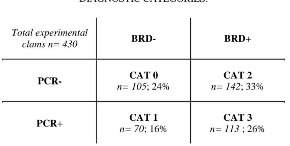
Table 1: Dual diagnosis through visual inspection of the inner surface of both valves (BRD+ or BRD-) and PCR amplification of the virB4 gene region of 173 bp (PCR+ or PCR-) allows for distinction between 4 categories, ranging from CAT 0 (uninfected post-challenge, termed “RES”) to CAT 3 (infected according to both diagnostic methods, termed “DIS”). The 430 samples tested show that 24 % and 26 % of samples fall in CAT 0 and CAT 3, respectively. The link between these categories and the kinetics of infection in the extra pallial compartment with
Vibrio tapetis is described in Figure 1.
DIAGNOSTIC CATEGORIES:
Total experimental
clams n= 430 BRD- BRD+
PCR- n= 105; 24% CAT 0 n= 142; 33% CAT 2
10 241
Figure 1: Schematic representation of the kinetics of Brown ring disease development during 30 days post injection (DPI) in a controlled challenge, adapted from Bidault et al. 2015, showing concentration of V. tapetis in extrapallial fluids (cells.mL-1) and limit of detection (LOD) at 1x101 cells. "RES" clams are negative for both visual and molecular diagnosis. Bacteria enter the extrapallial compartment and become quantifiable at point (a), then proliferate rapidly (clams at this stage are termed “CAT 1” in our
study), with highest concentrations generally observed around 7 DPI. The host then begins to trap the bacteria within the conchiolin deposits characteristic of Brown ring disease, thus leading to a decrease in the concentration of bacteria in the extrapallial compartment (clams at this stage are termed “CAT 2” in our study), represented by point (b). Clams can thus be positive for the visual diagnosis and negative for molecular diagnosis whilst the bacteria remain trapped against the inner surface of the shell, a process during which the host attempts to recalcify over the bacterial biofilm. In the case of “DIS” clams, conchiolin deposits are present but insufficient in limiting the pathogen,
11 242
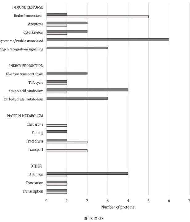
Figure 2: Number of proteins per functional group (i.e. “immune response”, “energy production”, “protein metabolism”) that were more abundant in DIS clams (dark bars) and in RES clams (light bars).
12
Table 2: Proteins significantly abundant (p-val < 0.05) in DIS and RES clams, with fold-change > 1.5 ("group ratio"). Based on Clusters of Orthogous Groups (COG), Biological Process GO terms, and literature review regarding the roles of these proteins in the context of disease, the proteins are grouped into three main functional roles described in the
discussion; “Immune response”, “Energy production”, and “Protein metabolism”.
DIS Intensity RES Intensity Group Ratio Student's T-test p-value Unique peptides R. philippinarum
locus ID C. gigas protein ID
Protein
name COG C. gigas gene description
Biological process GO term
Functional role discussed
1440 948 1.51:1.00 0.002 3 Locus 6868673 EKC40501 LGALS9 W Galectin-9 GO:0006954
inflammatory response
Immune response -
pathogen recognition 1250 421 2.97:1.00 0.041 1 Locus 5288240 EKC31577 BGBP G Beta-1,3-glucan-binding
protein 1
GO:0002752 cell surface pattern recognition receptor signaling pathway Immune response - pathogen recognition
384 244 1.57:1.00 0.026 1 Locus 4334179 EKC24393 C3 O Complement C3 GO:0006955 immune
response
Immune response - signalling 647 1770 2.73:1.00 0.005 3 Locus 615620 EKC23268 IQGAP1 Z Ras
GTPase-activating-like protein GO:1903829 positive regulation of cellular protein localization Immune response - cytoskeleton 1270 832 1.53:1.00 0.025 3 Locus 2955238 EKC27178 DCTN2 Z Dynactin subunit 2
GO:0006888 endoplasmic reticulum to Golgi
vesicle-mediated transport
Immune response - cytoskeleton 14900 4540 3.29:1.00 0.018 9 Locus 7235177 EKC29122 SCP S Sarcoplasmic
calcium-binding protein
GO:0051480 regulation of cytosolic calcium ion
concentration
Immune response - cytoskeleton 3200 2010 1.59:1.00 0.025 4 Locus 2215912 EKC27269 CPVL O Putative serine
carboxypeptidase CPVL
GO:0051603 proteolysis involved in cellular protein catabolic process
Immune response - lysosomal
910 486 1.87:1.00 0.005 1 Locus 2384090 EKC25290 EPDR1 S ependymin related 1 GO:0007160 cell-matrix
adhesion
Immune response - lysosomal
903 446 2.03:1.00 0.045 1 Locus 2964637 EKC26355 OVCH1 O Ovochymase-1 GO:0006508 proteolysis
Immune response - lysosomal 2660 531 5.02:1.00 0.029 2 Locus 3062641 ENSDARP00000139466* PPT1 S palmitoyl-protein
thioesterase 1 GO:0007042 lysosomal lumen acidification Immune response - lysosomal
680 374 1.82:1.00 0.014 1 Locus 665720 EKC31469 RAB43 U Ras-related protein
Rab-43 GO:0090382 phagosome maturation Immune response - vesicles
13 1780 716 2.49:1.00 0.027 6 Locus 2853765 EKC32573 VPS35 U Vacuolar protein
sorting-associated protein 35 GO:0007040 lysosome organization Immune response - vesicles 296 2550 8.61:1.00 0.003 3 Locus 6888709 EKC26119 ACE C Angiotensin-converting
enzyme
GO:0001817 regulation of cytokine production
Immune response - redox
323 669 2.07:1.00 0.019 1 Locus 2057997 EKC35339 TXNDC5 O Thioredoxin
domain-containing protein 5
GO:0045454cell redox homeostasis
Immune response - redox 4480 8140 1.82:1.00 0.045 3 Locus 355976 EKC36585 ACE E Angiotensin-converting
enzyme
GO:0001817 regulation of cytokine production
Immune response - redox
152 245 1.61:1.00 0.048 1 Locus 4589062 EKC37227 ADH5 Q Alcohol dehydrogenase
class-3
GO:0051775 response to redox state
Immune response - redox
399 638 1.60:1.00 0.036 1 Locus 2688502 EKC36531 XDH F Xanthine
dehydrogenase/oxidase
GO:2000379 positive regulation of reactive oxygen species metabolic
process
Immune response - redox
2250 783 2.87:1.00 0.006 2 Locus 6590720 EKC20036 GPX3 O Glutathione peroxidase
GO:0034599 cellular response to oxidative
stress
Immune response - redox
448 753 1.68:1.00 0.003 2 Locus 3201607 EKC33267 SH3GLB1 T Endophilin-B1 GO:0006915 apoptotic
process
Immune response -
apoptosis 1030 593 1.73:1.00 0.007 1 Locus 1202748 EKC29685 PPP3CC T Serine/threonine-protein
phosphatase GO:0035970 peptidyl-threonine dephosphorylation Immune response - apoptosis
749 351 2.13:1.00 0.002 2 Locus 1979842 EKC21473 CECR1 F Adenosine deaminase
CECR1 GO:0006154 adenosine catabolic process Immune response - apoptosis 3500 2120 1.65:1.00 0.036 1 Locus 1236277 EKC20480 AGL G Glycogen debranching
enzyme GO:0005980 glycogen catabolic process Energy production – carbohydrate metabolism 2860 1720 1.67:1.00 0.007 3 Locus 8380444 EKC18570 PGM1 G Phosphoglucomutase-1 GO:0006006 glucose
metabolic process
Energy production - carbohydrate metabolism 11100 6930 1.60:1.00 0.037 7 Locus 381335 EKC27095 PCK2 C Phosphoenolpyruvate
carboxykinase [GTP] GO:0006094 gluconeogenesis Energy production - carbohydrate metabolism 1130 1770 1.57:1.00 0.031 2 Locus 2128190 EKC32958 AUH I Methylglutaconyl-CoA
hydratase GO:0006552 leucine catabolic process Energy production - AAs
581 298 1.95:1.00 0.013 1 Locus 2982362 EKC42273 SLC1A1 E Excitatory amino acid
transporter 1 GO:0006537 glutamate biosynthetic process Energy production - AAs
14 6150 1990 3.09:1.00 0.041 10 Locus 8587681 EKC33186 GPT2 E Alanine aminotransferase
2 GO:0042851 L-alanine metabolic process Energy production - AAs
21400 6060 3.54:1.00 0 19 Locus 2044774 EKC40669 GOT1 E
Aspartate aminotransferase, cytoplasmic GO:0006107 oxaloacetate metabolic process Energy production - AAs 4660 2820 1.65:1.00 0.034 1 Locus 7730822 EKC43060 PCCB EI Propionyl-CoA
carboxylase beta chain
GO:0006552 leucine catabolic process
Energy production -
AAs 9940 24000 2.41:1.00 0.038 8 Locus 4823168 EKC21276 PDHA1 C Pyruvate dehydrogenase
E1 subunit alpha
GO:0006086 acetyl-CoA biosynthetic process from
pyruvate
Energy production -
TCA 38700 24600 1.57:1.00 0.001 26 Locus 2348137 EKC25158 MDH1 C Malate dehydrogenase GO:0006099 tricarboxylic
acid cycle
Energy production -
TCA 1120 662 1.69:1.00 0.026 1 Locus 5269440 EKC19854 REBM I 3-demethylubiquinone-9
3-methyltransferase GO:0006744 ubiquinone biosynthetic process Energy production - ETC 42700 24200 1.77:1.00 0.041 34 Locus 4313121 EKC39329 ATP5A1 C ATP synthase subunit
alpha GO:0006754 ATP biosynthetic process Energy production - ETC
1190 2290 1.92:1.00 0.049 3 Locus 1397283 EKC35325 TTN T Titin GO:0007155 cell adhesion
Protein metabolism -
transport 1940 2980 1.54:1.00 0.041 3 Locus 5145028 EKC29146 YWHAE O 14-3-3 protein epsilon GO:0035556 intracellular
signal transduction
Protein metabolism -
transport
1600 3590 2.24:1.00 0.034 10 Locus 3208485 EKC19309 CAND1 S
Cullin-associated NEDD8-dissociated protein 1 GO:0016567 protein ubiquitination Protein metabolism – proteolysis 498 1630 3.27:1.00 0.044 2 Locus 4231109 EKC28114 PSMC1 O 26S proteasome non-ATPase regulatory subunit 7 GO:0000209 protein polyubiquitination Protein metabolism - proteolysis
440 235 1.88:1.00 0.035 1 Locus 4900110 EKC29780 PSMD7 O 26S protease regulatory
subunit 4 GO:0000209 protein polyubiquitination Protein metabolism - proteolysis 4860 2510 1.94:1.00 0.012 4 Locus 2922613 EKC25378 FKBP14 O FK506 binding protein
14
GO:0046716 muscle cell cellular homeostasis
Protein metabolism - protein folding 2550 4140 1.62:1.00 0.014 16 Locus 2939806 EKC25687 HSP90AB1 O Heat shock protein HSP
90-alpha 1 GO:0050821 protein stabilization Protein metabolism - chaperone
15 633 1200 1.90:1.00 0.048 2 Locus 2389731 EKC42074 PCBP3 A Poly(RC)-binding protein
3 GO:0000122 negative regulation of transcription by RNA polymerase II Other - transcription
1300 563 2.31:1.00 0.023 1 Locus 995640 EKC39351 PURA K Transcriptional activator protein Pur-alpha GO:0006268 DNA unwinding involved in DNA replication Other - transcription 1860 2940 1.58:1.00 0.003 1 Locus 453724 EKC20816 NARS J Asparaginyl-tRNA
synthetase
GO:0006421 asparaginyl-tRNA aminoacylation
Other - translation 1300 752 1.73:1.00 0.024 2 Locus 454248 EKC31246 RPL27A J 60S ribosomal protein
L27a GO:0006412 translation
Other - translation
818 1560 1.91:1.00 0.021 2 Locus 3238632 EKC23703 - - Uncharacterized -
Calycin superfamily - - - Other - unknown
584 357 1.64:1.00 0.018 1 Locus 4959443 EKC41849 ABHD14A S Abhydrolase
domain-containing protein 14A
GO:0006656 phosphatidylcholine biosynthetic process
Other - unknown
670 396 1.69:1.00 0.033 1 Locus 1004421 EKC34161 - - si:ch73-250a16.5 - - - Other - unknown
3520 1910 1.85:1.00 0.042 6 Locus 6596965 EKC41442 - - Uncharacterized -
SH3-like domain - - - Other - unknown
1060 350 3.03:1.00 0.028 3 Locus 4498780 EKC37917 -
-Uncharacterized - Carbohydrate esterase 4
(CE4) family
- - - Other - unknown
Ratios are presented as RES:DIS for those more abundant in RES clams, and as DIS:RES for those more abundant in DIS clams. * Danio rerio protein ID and protein description
16 3.3.IMMUNE RESPONSE-ASSOCIATED PROTEINS
243 244
3.3.1. Pathogen recognition and immune-pathway activation 245
The ongoing infection process in clams from the DIS group is supported by the presence of a 246
number of proteins whose primary functions are associated with pathogen recognition and the 247
subsequent triggering of signaling pathways, such as Galectin-9 (Gal9), 1-3-β-glucan-binding 248
protein (BGBP), and Complement component C3 (C3). Proteins such as Galectins, which have 249
previously been demonstrated as upregulated in the extra pallial fluids of V. tapetis-infected 250
clams, and BGBP act as pattern recognition receptors (PRRs) by recognizing β-galactoside and 251
lipopolysaccharide residues found on bacterial membranes [18,33,34]. This initiates the immune 252
response by activating signaling pathways for chemotaxis, phagocytosis, and opsonization and 253
induces antimicrobial peptide (AMP) synthesis through the prophenoloxidase and complement 254
cascade systems [35,36]. Interestingly, selective breeding for parasite resistance in the Sydney 255
rock oyster found that resistance was directly related to the loss of a specific form of 256
phenoloxidase enzymes, supporting the presence of this particular enzyme as a marker of 257
susceptibility [37,38]. The C3 protein identified in our dataset belongs to the complement cascade 258
system, an essential component of the invertebrate immune response leading to the opsonization 259
and lysis of pathogens [39–42]. Upregulation of proteins associated to the complement system 260
have previously been described in R. philippinarum through several transcriptomic studies 261
investigating response to disease, highlighting the importance of this pathway in host response to 262
Vibrio pathogens [18,24]. The significantly high abundance of proteins specifically involved in 263
pathogen recognition in the DIS group supports a strong acute response to the ongoing infection 264
with V. tapetis in these animals. 265
Interestingly, the RES clam group showed high abundance of Ras GTPase-activating-like protein 266
1 (IQGAP1), a pathway-activating protein that is implicated in a number of immune-associated 267
functions. Namely, during infection by microbial pathogens that target the host microtubule 268
network, similar to the way in which V. tapetis inhibits pseudopod formation in the hemocytes of 269
R. philippinarum, IQGAP1 has been shown to bind to and modulate the activity of proteins 270
involved in bacterial invasion, ultimately interacting with the actin cytoskeleton [19,43]. 271
272 273
17 3.3.2. Pathogen-associated lysosomal activity
274
Other proteins highly abundant in the DIS group are involved in immune response through 275
antimicrobial and lysosomal activity. The putative serine carboxypeptidase (CPLV) and the 276
serine protease Ovochymase (OVCH1) identified in the DIS group are known to have 277
antibacterial activity and can be involved in proteolytic digestion of lysosomal components 278
[44,45]. As lysosomes contain the hydrolytic enzymes necessary for degradation of cellular 279
components as well as encapsulated pathogenic agents, it is also interesting to note the higher 280
abundance of Ependymin-related protein 1 (EPDR1) in DIS samples, which has been suggested 281
to function as a lysosomal activator protein, and Palmitoyl-protein thioesterase-1 (PPT1), which 282
is associated with lysosomal degradation of proteins [46,47]. An uncharacterized protein in the 283
DIS clam group belonging to the carbohydrate esterase 4 (CE4) family may also play a role in 284
degrading phagocytosed bacteria as well as inhibiting biofilm formation, as certain enzymes of 285
the CE4 family, whose main function is to de-acetylate polysaccharides, specifically degrade the 286
essential peptidoglycan polymers of bacterial cell walls [48]. In addition, active intracellular 287
membrane trafficking (ex. phagosomes) in DIS clams is suggested by the high abundance of 288
proteins such as the Ras-related protein RAB43, which regulates vesicular movement following 289
immune system activation by microbial infections, and a vacuolar-sorting protein VPS35, which 290
directs transmembrane cargo proteins to the lysosomal degradation pathway [49,50]. 291
292
3.3.3. Cytoskeleton-associated immune response 293
The cytoskeleton is a network of filaments that plays an essential role in certain aspects of 294
immunity through cell structure maintenance, transport, phagocytosis, and communication 295
between cellular components. As such, it is also known to be one of the targets of invading 296
pathogens such as Perkinsus olseni and V. tapetis in the Manila clam [19,51,52]. Interestingly, a 297
dynactin subunit (DCTN2) and a sarcoplasmic calcium-binding protein (SCP) were highly 298
abundant in DIS clams, both of which play a role in cytoskeleton function. DCTN2 is part of a 299
dynein/dynactin complex which coordinates the microtubule movement of vacuoles towards 300
lysosomes and plays an important role in the biogenesis and transportation of pathogen-301
containing vacuoles in rabbit cells infected by obligate intracellular bacteria [53,54]. This may 302
reflect the internalization of V. tapetis by hemocytes in the case of DIS clams, activating 303
microtubule motors such as the dynein/dynactin complex in an attempt to fuse bacteria-304
18 containing vacuoles with lysosomal membranes. SCPs, which are known to interact with the 305
cytoskeleton by regulating the calcium balance, have been shown to be upregulated in Manila 306
clam hemocytes in response to both parasitic and bacterial infections [18,22,51]. Bacteria of the 307
genus Vibrio have been suggested to inhibit intracellular trafficking and the fusion of bacteria-308
containing phagosomes with lysosomes in order to avoid neutralization, a relatively common 309
immune-evasion technique seen in a number of pathologies [55]. In this light, the elevated 310
abundance of lysosome-associated proteins in clams of the DIS group may reflect an attempt to 311
counter the bacteria’s inhibitory processes. 312
313
3.3.4. Apoptotic processes 314
Apoptosis of host immune cells during an infection can represent one of the ultimate defense 315
strategies against invading pathogens, whereby the host cell is sacrificed so as to eliminate the 316
internalized pathogenic agent [56]. Induction of apoptosis has been suggested to be one of the 317
mechanisms of survival put in place by resistant oysters, Ostrea edulis, perhaps in response to 318
anti-apoptotic mechanisms that are a known survival strategy for some intracellular pathogens, 319
such as the protozoan parasite Bonamia ostreae [57]. Clams from the RES group in our study 320
demonstrated higher abundances of Endophilin B1 (Bif-1), a protein associated with the 321
formation of pores in the outer mitochondrial membrane that leads to apoptosis through the 322
caspase pathway [57–59]. In contrast, DIS clams showed high abundance of adenosine 323
deaminase (CECR1) a protein that, while primarily associated with the purine metabolism, also 324
plays an important role in reducing the concentration of the toxic derivatives of adenosine and 325
deoxyadenosine to protect cells from apoptosis [60,61]. A serine/threonine protein phosphatase 326
PPP3CC, also more abundant in DIS clams, bears close resemblance with the protein phosphatase 327
3 catalytic subunit beta, PPP3CB, which was recently suggested to promote cell-proliferation and 328
may play an anti-apoptotic role in tumorous human kidney cells [62]. While apoptosis is a 329
complex process that can benefit immune defense, anti-apoptosis is also a mechanism by which 330
the host may maintain cellular functions and continue combatting infection, though seemingly at 331
a cost given the relatively high abundance of proteins involved in energy metabolism in the DIS 332
clam group (see part 3.4). 333
334
3.3.5. Redox homoeostasis 335
19 Host production of reactive oxygen species (ROS) and reactive nitrogen species (RNS) is a 336
known mechanism of defense and pathogen neutralization, though this process requires a delicate 337
balance as the accumulation of ROS/RNS can also lead to oxidative damage to the host [63]. 338
Glutathione peroxidase (GPX3), a member of the cellular antioxidant system, was more abundant 339
in the DIS clam group. Interestingly, a previous studies on BRD-infected Manila clams reported a 340
decrease in enzymatic activity of GPX at 7 dpi, though the study did not measure enzyme 341
abundance nor activity at 30 dpi [21]. Oysters subjected to bacterial infection, however, have 342
shown an upregulation of glutathione peroxidase gene expression in pathogen-challenged larvae, 343
suggesting that GPX may be an indicator of oxidative stress that may have been occurring in DIS 344
clams from our study as a result of the ongoing antibacterial processes discussed in part 3.3.2 345
[21,64]. 346
The RES clam group was characterized by a higher abundance of proteins more or less directly 347
associated with ROS/RNS production and scavenging. Thioredoxin domain-containing protein 5 348
(TXNDC5) is a member of the protein disulfide isomerase family whose expression has been 349
associated with oxidative stress and cellular pathology [65,66]. At least two shorter-term (max 7 350
days) studies have investigated the role of thioredoxin in response to V. tapetis, pointing towards 351
an increase in activity of thioredoxin in V. tapetis-challenged clams compared to controls or to 352
resistant clams, thus the higher abundance of thioredoxin observed in RES clams of this study 353
may reflect a possible reversal of this mechanism in the case of long-term resistance (30 days) 354
[21,67]. Similarly, xanthine oxidoreductase (XOR), an enzyme implicated in the purine 355
metabolism, has previously been associated with a number of innate immunity processes 356
including redox homeostasis through the production of ROS and RNS by xanthine oxidase (XO), 357
as well as indirect free radical sequestration by xanthine dehydrogenase (XD) through the 358
synthesis the antioxidant uric acid [63,68–70]. RES clams also presented a significantly higher 359
abundance of two Angiotensin-converting enzymes (ACE), which were recently demonstrated to 360
induce superoxide production and thereby enhance bactericidal activity in mouse neutrophils 361
[71], and class-III alcohol dehydrogenase (ADH3). ADH3 uses the reducing power glutathione to 362
eliminate the organic compounds formaldehyde and S-nitrosothiols (SNO), ultimately limiting 363
their decomposition into the RNS nitric oxide [72,73]. Nitric oxide is a known toxic agent 364
produced by immune cells of invertebrates for pathogen defense, and NO production by R. 365
philippinarum in response to V. tapetis has been correlated to hemocyte rounding and pseudopod 366
20 loss [74], highlighting the possible importance of NO-sequestration by proteins such as ADH3 in 367
order to counter the negative effects of this compound on the host. 368
These findings support previous hypotheses suggesting that ROS/RNS production by hemocytes 369
may represent an alternative anti-bacterial response against pathogens such as V. tapetis, which is 370
known to evade host immune responses such as phagocytosis by inhibiting pseudopod formation 371
[74,75]. In addition, the higher abundance of the redox homeostasis-associated enzymes RES 372
clams supports findings from previous studies indicating that resistance may be associated with a 373
greater ability to balance ROS/RNS production and scavenging, allowing them to rapidly 374
neutralize pathogens before they are able to colonize the host while simultaneously protecting 375
host cells from oxidative damage, a dual process previously suggested using enzyme activity 376
assays in the Manila clam exposed to V. tapetis [21]. 377
378
3.4.PROCESSES ASSOCIATED WITH ENERGY PRODUCTION 379
380
Proteins potentially associated with energy production through carbohydrate and amino acid 381
catabolism represent the second largest group in our dataset, most of which (10 out of 12) are 382
more abundant in the DIS clam group. This section groups together the proteins implicated in the 383
degradation of glycogen and amino acids that generate essential metabolites for the tricarboxylic 384
acid (TCA) cycle, leading to the production of high-energy electron donors such as NADH which 385
can integrate the electron transport chain (Figure 3). 386
387
3.4.1. Amino acid degradation for energy production 388
Propionyl-CoA carboxylase beta chain (PCCB) and Methylglutaconyl-CoA hydratase (AUH), 389
enzymes involved in the degradation of branched-chain amino acids (BCAAs) such as leucine, 390
were significantly more abundant in DIS and RES clams, respectively. Demand for BCAAs as 391
energy metabolites or for the synthesis of immune-related molecules has been demonstrated to 392
increase during disease [76,77]. Decreased levels of a number of other amino acids during Vibrio 393
infection in mussels have also been reported, suggesting that the significantly higher abundance 394
of Alanine aminotransferase (GPT2), Aspartate aminotransferase (GOT1), and Excitatory amino-395
acid transporter-1 (SLC1A1) in DIS clams may also be associated with the degradation and/or 396
21 conversion of amino acids into metabolites such as pyruvate, oxaloacetate, and α-ketoglutarate 397
for the TCA cycle [78–81]. 398
399
3.4.2. Carbohydrate metabolism and TCA cycle 400
Our dataset showed a high abundance of proteins linking glycolysis with the TCA cycle for the 401
aerobic production of ATP such as Pyruvate dehydrogenase (PDH) E1 subunit (alpha type III) in 402
RES clams, as well as Glycogen debranching enzyme (AGL) and Phosphoglucomutase-1 403
(PGM1) in DIS clams, which may reflect the degradation of carbohydrates for the production of 404
pyruvate, namely in the DIS clam group [82]. This group is also characterized by a greater 405
number of proteins associated with various aspects of the immune response, a process known to 406
be energetically demanding [83]. More importantly, weight loss, decreased glycogen reserves, 407
and condition index, which are indicative of energy imbalance and poor health, have previously 408
been attributed to the negative impact of BRD on energy balance in the Manila clam [8,84,85]. 409
Malate dehydrogenase (MDH1), which participates in the TCA cycle by oxidizing malate to form 410
oxaloacetate, was also more abundant in DIS clams, supporting the hypothesis of increased 411
carbohydrate degradation for energy production. Our dataset also indicated a significantly higher 412
abundance of the mitochondrial Phosphoenolpyruvate carboxykinase (PEPCK-M) in the DIS 413
group, which is generally associated with catalyzing the irreversible conversion of oxaloacetate to 414
phosphoenolpyruvate (PEP) for gluconeogenesis. While this appears to contradict the hypothesis 415
of glycogen and glucose degradation, recent studies have demonstrated that overexpression of 416
PEPCK-M (as opposed to the cytosolic form PEPCK-C) may play a role in antiviral immunity in 417
insects, and the accumulation of PEP in infected ticks was suggested to be an antibacterial 418
mechanism against the bacterial pathogen Anaplasma phagocytophilum [86,87]. 419
420
3.4.3. Electron transport chain 421
The end products of the TCA cycle ultimately convey their electrons to the electron transport 422
chain (ETC), composed of molecules within the inner membrane of the mitochondria, including 423
the essential proteins ubiquinone (coenzyme Q10) and ATP synthase. In the DIS group, the high 424
abundance of 3-demethylubiquinone-9 3-O-methyltransferase, which participates in the final step 425
of ubiquinone synthesis, suggests that there is a high demand for electron acceptor molecules 426
which increase the proton gradient in the inter membrane space of the mitochondria [88]. 427
2222 428
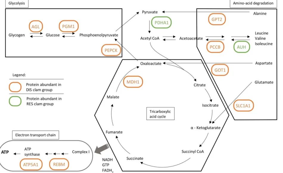
Figure 3: Schematic representation of energy production and the carbohydrate and amino acid metabolisms, including glycolysis, the TCA cycle, amino-acid degradation pathways, and the electron transport chain. The processes in which highly abundant proteins in DIS (orange) and RES (green) clams are implicated are annotated with the abbreviated protein name.
Glycogen debranching enzyme (AGL), Phosphoglucomutase-1 (PGM1), and Phosphoenolpyruvate carboxykinase (PEPCK) are associated to the carbohydrate metabolism (both glycolysis and gluconeogenesis). Alanine aminotransferase (GPT2), Propionyl-CoA carboxylase beta chain (PCCB), Methylglutaconyl-CoA hydratase (AUH), Aspartate aminotransferase (GOT1), and Excitatory amino-acid transporter-1 (SLC1A1) all participate in the degradation of amino acids that can play a role in replenishing metabolites of the TCA
cycle. Malate dehydrogenase 1 (MDH1) is an essential enzyme of the TCA cycle, the high-energy products of which are then shuttles to the electron transport chain where proteins such as 3-demethylubiquinone-9 3-methyltransferase (REBM) and ATP synthase subunit alpha (ATP5A1) participate in the production of ATP.
23 Overall, proteins associated with the energy metabolism, including ATP synthase subunits and ETC- 429
associated proteins, as well as proteins associated with energy production from both amino acids and 430
sugars, are more abundant in the DIS group than in the RES group, reflecting a high energy demand 431
likely as a result of the active immune response described previously. 432
433
3.5. PROTEIN METABOLISM 434
Seven highly abundant proteins in the dataset were associated with various aspects of protein 435
metabolism, namely proteolysis, transport, and chaperones. 436
3.5.1. Proteolysis 437
Proteolytic processes are represented in our dataset by the proteins cullin-associated NEDD8-438
dissociated protein 1 (CAND1) and subunit 4 of the 26S proteasome complex (PSMC1) in the RES 439
group, and by the 26S proteasome non-ATPase regulatory subunit 7 (PSMD7) in the DIS group (Table 440
2). In eukaryotic cells, proteolysis, or the degradation of proteins and recycling of their components, is 441
a process mediated by the conjugation of polyubiquitin chains to proteins which are then recognized by 442
the 26S proteasome complex, a multi-subunit enzyme responsible for proteolysis [89]. CAND1 binds 443
to unneddylated CUL1, one of the three major components of an E3 ubiquitin ligase playing an 444
essential role in protein degradation by regulating ligase ubiquitination [90]. Interestingly, ubiquitin 445
ligase complexes were found to be one of the targets of pathogenic bacteria during infection, whereby 446
inhibiting factors produced by pathogens may be able to effectively bind to CUL1, preventing it from 447
correctly forming the ligase complex [91]. PSMC1 coordinates substrate recruitment and translocation 448
into the proteolytic chamber of the proteasome, and is essential for rapid proteolysis [92]. Similarly, 449
PSMD7 is another component of the proteasome important in mediating the recognition of 450
polyubiquitin chains and cleavage of ubiquitin from degraded proteins [93]. In a previous gene-451
expression study of P. olseni-infected Manila clams, proteasome subunits were found to be 452
downregulated in diseased animals, indicating decreased proteolytic activity [41]. In that respect, the 453
elevated abundance of CAND1 and PSMC1 in RES clams may indicate a trend towards ubiquitin 454
tagging of damaged proteins and more active protein degradation in RES clams than in DIS clams. 455
High proteolytic activity ion RES clams may be linked to the digestion of phagocytosed and 456
neutralized bacteria, and may also reflect the presence of a pro-apoptotic protein in this group. In 457
addition, it may be possible that RES clams were able to sustain the cellular functions necessary for 458
effective elimination of the pathogen, after which proteins damaged by oxidative stress during the 459
immune response are degraded. 460
24 3.5.2. Transport, folding, and chaperone functions
461
Our dataset also contained proteins associated with various aspects of protein synthesis, including 462
protein transport (Table 2). Titin (TTN), a large structural protein thought to function as a scaffold 463
protein, has been shown to interact with actin and filamin, proteins of the cytoskeleton implicated in the 464
movement of cellular components and proteins [94]. In vertebrate striated muscle, TTN was 465
specifically shown to recruit E3 ubiquitin-ligase [95], thus its high abundance in RES clams may reflect 466
transport associated with proteolysis, consistent with the fact that this group also showed higher 467
abundance of CAND1 and a proteasome subunit. Another protein implicated in protein transport is 14-468
3-3 epsilon (YWHAE), a binding protein suggested to play a role in protein transport to the secretory 469
pathway, namely through interaction with GTPase-activating proteins such as IQGAP1 (see 3.3.3 470
Cytoskeleton-associated immune response), also found highly abundant in RES clams [96]. 471
DIS clams showed a high abundance of FK506 binding protein 14 (FKBP14), which is thought to 472
accelerate protein folding as it belongs to a family of peptidyl-prolyl cis-trans isomerases that play a 473
role in the folding of newly synthesized proteins [97]. The higher abundance of this protein in the DIS 474
group may reflect an increase in the synthesis of immune-related proteins, which were also more 475
abundant in DIS clams. Furthermore, it is also interesting to note that this family of proteins (also 476
called immunophilins) has been shown to inhibit early establishment and intracellular infection by 477
bacteria [98]. Though not directly implicated in protein metabolism, the DIS group also presented high 478
abundance of an Abhydrolase domain-containing protein associated with biosynthesis of 479
phosphatidylcholine, an essential class of membrane phospholipids, possibly reflecting an increase in 480
membrane synthesis due to the internalization of bacteria in phagosomes [99]. 481
Heat-shock protein 90 (HSP90), a chaperone protein that plays a crucial role in protecting protein 482
structure in response to stress conditions, was highly abundant in RES clams. Due to its interaction 483
with the major histocompatibility complex (MHC) and the antigen processing pathways in vertebrates 484
[100], HSP90 has been suggested to play a role in the innate immune system response and resistance to 485
infection in invertebrates [101]. As HSP synthesis is promoted by protein denaturation, the trend 486
towards proteolytic activity in RES clams discussed in part 3.5.1 may be partially responsible for 487
activating HSP synthesis. That said, the higher abundance of HSP90 in RES clams may also indicate 488
that resistant clams have a lower threshold for protein denaturation and thus perhaps more rapidly 489
activate the synthesis of protective chaperone proteins, granting them an advantage over DIS clams 490
when it comes to cellular protection. 491
25 3.6. OTHER
493
The transcriptional activator protein Pur-alpha (PURA), a protein involved in controlling DNA 494
replication and gene transcription processes [102,103], was highly abundant in the DIS group, while a 495
Poly (rc)-binding protein 3 (PCBP3) associated with negative regulation of transcription [104] was 496
highly abundant in the RES group [104]. 497
Proteins associated with translation include RPL27A, a structural component of the 60S ribosome 498
subunit whose upregulation has previously been reported in white-spot infected shrimp and in hypoxia-499
stressed oysters [105,106], and asparaginyl-tRNA synthetase (NARS), whose primary function in 500
translation is to catalyze the attachment of asparagine to its corresponding tRNA. 501
Finally, four significantly abundant proteins were uncharacterized; three were highly abundant in the 502
DIS group and one in the RES group. 503
504
4. CONCLUSION 505
Despite being one of the fastest-growing sectors of aquaculture worldwide, mollusk production 506
continues to suffer significant losses due in part to the impact of infectious diseases. The study of 507
proteomic profiles offers the possibility of better understanding the complex functional mechanisms at 508
play during host response to disease, and may shed light on factors associated with resistance to 509
disease. 510
The aim of the present study was to investigate the proteomic profiles of resistance to controlled 511
infection with Vibrio tapetis, the etiological agent of Brown Ring Disease, in the Manila clam, 512
Ruditapes philippinarum. The comparison of proteomic profiles of two extreme phenotypes (RES and 513
DIS) observed in juvenile Manila clams shows a number of functional differences in highly abundant 514
proteins implicated in the immune response-associated processes, energy production, and protein 515
metabolism. Twice as many significantly abundant proteins associated with the immune response were 516
accumulated in the DIS group compared to the RES group, reflecting the ongoing infection as 517
established by the presence of both visual and molecular signs of disease. That said, the function of 518
immune-associated proteins in the RES group was almost consistently associated with redox 519
homeostasis, whereas in DIS group the abundant proteins were mostly involved in pathogen 520
recognition, signaling, and neutralization. This may suggest that disease resistant clams are better 521
equipped to manage ROS production and scavenging in order to simultaneously eliminate the pathogen 522
and protect host cellular components from oxidative stress. Protein degradation as well as protection by 523
chaperones were another process highly represented in resistant clams, with degradation possibly as a 524
26 result of successful elimination of the pathogen which may nonetheless have left a number of cellular 525
components damaged by oxidative stress. The fact that only resistant clams showed significantly high 526
abundance of a chaperone-associated protein suggests that this may be an important factor of resistance 527
to disease. In contrast, diseased clams showed a higher abundance of proteins involved in protein 528
synthesis and functional modifications, possibly in response to activation by the immune system in 529
order to continue fighting infection. Both immune response and protein synthesis are energy 530
demanding processes, which is further supported by the presence of proteins involved in glycolysis, 531
TCA cycle, and the electron transport chain. Overall, the comparison of the proteomic profiles of 532
resistant and sick clams suggests that redox homeostasis and maintenance of protein structure by 533
chaperone proteins may play important and interrelated roles in resistance to infection by Vibrio tapetis 534
in the Manila clam. 535
536
ACKNOWLEDGMENTS 537
The authors would like to acknowledge the “investment for the future” programs LabexMER (ANR-538
10-LABX-19) and ISblue (ANR-17-EURE-0015) for funding international joint PhD agreements and 539
proteomic analyses, the EU Horizon2020 project VIVALDI (grant agreement N°678589) for providing 540
funding and biological material. The authors also would like to thank Fabrizio Ghiselli and Mariangela 541
Ianello from the University of Bologna, and Massimo Milan and Luca Bargelloni from the University 542
of Padova, for providing the full annotated and assembled transcriptome of the Manila clam digestive 543
gland used for protein identification. 544
27 REFERENCES
545
[1] FAO, The State of World Fisheries and Aquaculture 2018: Meeting the sustainable development goals, 546
2018. doi:doi:10.18356/8d6ea4b6-en. 547
[2] J.. Flassch, Y. Leborgne, Introduction in Europe, from 1972 to 1980, of the Japanese Manila clam, ICES 548
Mar. Sci. Symp. 194 (1992) 92–96. 549
[3] C. Zannella, F. Mosca, F. Mariani, G. Franci, V. Folliero, M. Galdiero, P.G. Tiscar, M. Galdiero, 550
Microbial diseases of bivalve mollusks: Infections, immunology and antimicrobial defense, Mar. Drugs. 551
15 (2017). doi:10.3390/md15060182. 552
[4] C. Paillard, P. Maes, R. Oubella, Brown ring disease in clams, Annu. Rev. Fish Dis. 4 (1994) 219–240. 553
doi:10.1016/0959-8030(94)90030-2. 554
[5] C. Paillard, P. Maes, Etiologie de la maladie de l’anneau brun chez Tapes philippinarum: pathogénicité 555
d’un Vibrio sp., Comptes Rendus l’Académie Des Sci. Série 3, Sci. La Vie. 310 (1990) 15–20. 556
[6] B. Allam, C. Paillard, M. Auffret, Alterations in hemolymph and extrapallial fluid parameters in the 557
Manila Clam, Ruditapes philippinarum, challenged with the pathogen Vibrio tapetis, J. Invertebr. Pathol. 558
76 (2000) 63–69. doi:10.1006/jipa.2000.4940. 559
[7] C. Paillard, F. Le Roux, J.J. Borrego, Bacterial disease in marine bivalves, a review of recent studies: 560
Trends and evolution, Aquat. Living Resour. 17 (2004) 477–498. doi:10.1051/alr:2004054. 561
[8] C. Paillard, F. Jean, S.E. Ford, E.N. Powell, J.M. Klinck, E.E. Hofmann, J. Flye-Sainte-Marie, A 562
theoretical individual-based model of Brown Ring Disease in Manila clams, Venerupis philippinarum, J. 563
Sea Res. 91 (2014) 15–34. doi:10.1016/j.seares.2014.03.005. 564
[9] C. Paillard, B. Allam, R. Oubella, Effect of temperature on defense parameters in Manila clam Ruditapes 565
philippinarum challenged with Vibrio tapetis., Dis. Aquat. Organ. 59 (2004) 249–262. 566
doi:10.3354/dao059249. 567
[10] H.I. Reid, P. Soudant, C. Lambert, C. Paillard, T.H. Birkbeck, Salinity effects on immune parameters of 568
Ruditapes philippinarum challenged with Vibrio tapetis, Dis. Aquat. Organ. 56 (2003) 249–258. 569
doi:10.3354/dao056249. 570
[11] B. Allam, E. Pales Espinosa, Bivalve immunity and response to infections: Are we looking at the right 571
place?, Fish Shellfish Immunol. 53 (2016) 4–12. doi:10.1016/j.fsi.2016.03.037. 572
[12] B. Allam, C. Paillard, Defense factors in clam extrapallial fluids, Dis. Aquat. Organ. 33 (1998) 123–128. 573
doi:10.3354/dao033123. 574
[13] N. Trinkler, G. Sinquin, J. Querne, C. Paillard, Resistance to Brown Ring Disease in the Manila clam, 575
Ruditapes philippinarum: A study of selected stocks showing a recovery process by shell repair, J. 576
Invertebr. Pathol. 104 (2010) 8–16. doi:10.1016/j.jip.2009.12.007. 577
[14] F. Jeffroy, F. Brulle, C. Paillard, Differential expression of genes involved in immunity and 578