DOI 10.1007/s10237-012-0395-6 O R I G I NA L PA P E R
Double-network acrylamide hydrogel compositions adapted
to achieve cartilage-like dynamic stiffness
S. Ronken · D. Wirz · A. U. Daniels · T. Kurokawa · J. P. Gong · M. P. Arnold
Received: 19 October 2011 / Accepted: 6 April 2012 / Published online: 21 April 2012 © Springer-Verlag 2012
Abstract Since articular cartilage has a limited potential for spontaneous healing, various techniques are employed to repair cartilage lesions. Acrylate-based double-network (DN) hydrogels containing∼90% water have shown prom-ising properties as repair materials for skeletal system soft tissues. Although their mechanical properties approach those of native cartilage, the critical factor—stiffness—of DN-gels does not equal the stiffness of articular cartilage. This study investigated whether revised PAMPS/PAAm composi-tions with lower water content result in stiffness parameters closer to cartilage. DN-gels containing 61, 86 and 90 % water were evaluated using two non-destructive, mm-scale inden-tation test modes: fast-impact (FI) and slow-sinusoidal (SS) deformation. Deformation resistance (dynamic modulus) and energy handling (loss angle) were determined. The dynamic modulus increased with decreasing water content in both testing modes. In the 61 % water DN-gel, the modulus resem-bled that of cartilage (FI-mode: DN-gel = 12, cartilage = 17; SS-mode: DN-gel = 4, cartilage = 1.7 MPa). Loss angle increased with decreasing water content in fast-S. Ronken (
B
)· D. Wirz · A. U. DanielsLaboratory of Biomechanics and Biocalorimetry (LOB2), Biozentrum/Pharmazentrum, Faculty of Medicine, University of Basel, Klingelbergstrasse 50-70, 4056 Basel, Switzerland e-mail: [email protected]
D. Wirz· M. P. Arnold
Department of Orthopedic Surgery and Skeletal Traumatology, Kantonsspital Bruderholz, 4101 Bruderholz, Switzerland T. Kurokawa· J. P. Gong
Department of Biological Sciences, Graduate School of Science, Hokkaido University, Sapporo, Japan
T. Kurokawa
Creative Research Institution “Sousei”, Hokkaido University, Sapporo, Japan
impact, but not in slow-sinusoidal deformation. However, loss angle was still much lower than cartilage (FI: DN-gel= 5, cartilage = 11; SS: DN-gel = 10, cartilage = 32◦), indi-cating somewhat less ability to dissipate energy. Overall, results show that it is possible to adapt DN-gel composi-tion to produce dynamic stiffness properties close to normal articular cartilage.
Keywords Double-network hydrogels· Modulus · Loss angle· Cartilage repair · Water content
1 Introduction
Articular cartilage is an extraordinary tissue because of its ability to tolerate a tremendous amount of intensive and repetitive physical stress and to frequently do so for a life-time (Newman 1998). However, the greatest limitation of articular cartilage is its poor capacity to heal spontaneously (Mandelbaum et al. 1998; Hunter 1743). Consequently, unhealed damage to articular cartilage in the knee is a com-mon clinical problem. Widuchowski et al. (2007) exam-ined 25,124 knees from patients with acute knee injuries or unexplained knee pain and dysfunction from 1989 to 2004. Arthroscopy revealed that 60 % of the patients had chondral lesions. Also, after anterior cruciate ligament injuries, the risk of cartilage lesions is very high (Slauterbeck et al. 2009). These unhealed injuries can lead to chronic pain, markedly restricted mobility and further degeneration of the articular cartilage, such as secondary osteoarthritis. In many cases, the situation must finally be ameliorated by major surgery—that is, total joint arthroplasty.
In recent years, several techniques have been devised and used to repair cartilage lesions and stave off fur-ther degeneration. These include microfracture, autologous
chondrocyte transplantation, mosaicplasty and most recently tissue-engineered constructs (Negrin et al. 2011;Peterson et al. 2010; Robert 2011; Santoro et al. 2010). These repair techniques focus on repairing the cartilage structure. Unfor-tunately, most of these methods do not result in cartilage with initial local mechanical properties (e.g., stiffness, strength) at the time of implantation that are even remotely similar to normal articular cartilage. Another drawback of most of these methods is that the rehabilitation period takes several months up to 1–2 years in order to establish repaired tissue capable of bearing cyclic impact loads of the knee of the mag-nitude and frequency associated with normal daily activity (Mandelbaum et al. 1998;Minas 1999;Mithoefer et al. 2007; Saris et al. 2008). From a patient’s point of view, these repair-ing techniques still need improvement.
Therefore, our research focuses on the replacement of cartilage function, so energy distribution and dissipation in the surrounding cartilage will remain similar and further degeneration of cartilage is avoided. The repair material studied here is an example of a double-network hydro-gel (DN-hydro-gels) developed by Gong et al. (Azuma et al. 2006; Gong et al. 2003;Nakajima et al. 2009; Nakayama et al. 2004; Yasuda et al. 2005). It is based on a highly crosslinked first polymer, PAMPS—poly(2-acrylamido-2-methylpropane sulphonic acid), with a second slightly cross-linked polymer, PAAm—poly(acrylamide), then created within the first structure. PAMPS/PAAm is a tough, tear-resistant, non-cytotoxic, non-absorbable DN-gel. The sur-prising result is a material with impact and tear resistance approaching an elastomer in spite of the high water content. Structurally, they also resemble cartilage and other skele-tal system soft tissues, which are also high–water content materials or structures with a double-network strategy to achieve their mechanical properties. Cartilage for instance can be seen as a high–water content double-network mate-rial or structure comprised of highly crosslinked collagen fibres interspersed with proteoglycan gel.
Importantly, a closely related formulation, PAMPS/ PDMAAm DN-gel, has previously been shown to support cartilage formation. In a rabbit study (Yasuda et al. 2009), a DN-gel plug inserted in a large osteochondral defect induced substantial spontaneous cartilage formation in vivo by 4 weeks. In contrast, this was rarely found for empty defects or defects filled with either a polyvinylacrylate gel or an ultra-high molecular weight polyethylene plug.
It also has been previously shown (Arnold et al. 2011) that a 90 % water PAMPS/PAAm DN-gel approaches the stiffness of articular cartilage. It also has attractive surgery-related attachment properties—for example, it can be sutured or can be attached to tissue with cyanoacrylate tissue adhe-sives. However, this 90 % water PAMPS/PAAm DN-gel is still not as stiff as native articular cartilage. Accordingly, to improve the possibility of using DN-gels as a cartilage repair
material, PAMPS/PAAm DN-gel water content was adapted in this study to approach dynamic stiffness properties of nor-mal articular cartilage.
2 Materials and methods 2.1 Materials
PAMPS/PAAm DN-gels with lower water content: In order to fine-tune the biomechanical properties of the DN-gels, the molecular ratios of both the first and the second net-work had to be varied accordingly in order to produce gels with different water contents. The three PAMPS/PAAm DN-gels produced were determined to have water contents of 90.9, 86.5 and 61.3 % (see Table1). The dimensions of the DN-gel specimens were 20×10×3mm. The method used to produce these various DN-gels is described elsewhere (Gong et al. 2003). After producing the DN-gels, they were shipped to Basel in normal saline and stored at 4–6◦C before testing at room temperature.
Swine cartilage specimens: Cylindrical osteochondral plugs of 7.6 mm in diameter were harvested from the knee of 10-month-old swine using a standard diamond core-drill designed for mosaicplasty (Synthes, Oberdorf, Switzerland). Plugs were harvested from the lateral condyles and kept wet with phosphate-buffered saline (PBS) prior to and during testing.
2.2 Mechanical testing
Two micro-indentation methods were used as previously described (Arnold et al. 2011;Ronken et al. 2011) to deter-mine the dynamic stiffness parameters (dynamic modulus E* and loss angleδ) of cartilage, meniscus and possible implant materials. The dynamic modulus is a measure of the defor-mation resistance of a material. The loss angle is a measure of the energy dissipation. If a cyclic load is applied to a vis-coelastic material, the time to maximum strain will lag the time to maximum stress (Lakes 1999).
Articular cartilage is a structure, but as a first approxima-tion it can be treated as a material for purposes of assess-ing mechanical properties and comparassess-ing them with those of true materials. The dynamic stiffness parameters of poro-viscoelastic materials, for example, cartilage, are even more strain rate dependent since the extent to which water is forced from the material also depends on strain rate and time. This is seen in the behaviour of cartilage under two different load-ing regimes—(a) the sudden transient deformations which occur during gait and are too brief to force water out of the tissue due to its low permeability, and (b) the slow quasi-cyclic deformations which cause fluid to move in and out of cartilage and thus provide a means for nutrition. Therefore,
Table 1 Preparing ratios and water content of the three double-network (DN) hydrogels
DN-gel Components first network Components second network Water (%)
Monomer (mol/l) Crosslinker (mol%) UVI (mol%) Monomer (mol/l) Crosslinker (mol%) UVI (mol%)
PAMPS/PAAm 90 % AMPS 1 MBAA 4 0.1 AAm 2 MBAA 0.01 0.03 90.9
PAMPS/PAAm 86 % AMPS 1 MBAA 4 0.6 AAm 2 – 0.01 86.5
PAMPS/PAAm 61 % AMPS 3 MBAA 3 0.1 AAm 7.9 MBAA 0.02 0.1 61.3
AMPS 2-acrylamido-2-methylpropanesulphonic acid, AAM acrylamide, MBBA N, N‘-methylenebisacrylamide, UVI ultraviolet light initiator
both a Fast-Impact Mode and a Slow-Sinusoidal Mode test method were developed and are used by the authors working in Basel. For both test modes, the dynamic modulus can be calculated as described byWirz et al.(2008) andKren et al. (2005). The loss angle can be calculated directly from the time lag of the displacement curve relative to the load curve. 2.2.1 Fast-impact (FI) mode
To simulate the impact velocity in normal human gait, a fast-impact micro-indentation instrument was used. This is a modified version of an instrument developed at the Minsk Institute of Physics (Kren et al. 2005). A pendulum-mounted spherical indenter (diameter 1.0 mm; 1.9g) falls down on the specimen under gravitational force. The motion of the inden-ter is captured electromagnetically during indentation and rebound. The duration of impact was 1–2 ms and the initial impact velocity∼0.3m/s. On each specimen, 10 replicate FI measurements were performed on the same spot at∼20s time intervals. Resultant E* andδ were calculated for each impact and then each set was averaged to get one set of specimen values.
2.2.2 Slow-sinusoidal (SS) mode
To simulate more static loading patterns of human carti-lage, a Synergie 100 MTS mechanical testing instrument was used to perform slow-sinusoidal micro-indentations. A spherical indenter (diameter 1.0 mm) was moved sinu-soidally with a frequency of 0.1 Hz under computer soft-ware control of displacement. Indentation was performed to a depth of∼0.05mm (SS-0.05) and ∼0.1mm (SS-0.1), with a maximum speed of∼0.015 and ∼0.03m/s. The same speci-mens were measured as in FI-mode. On each specimen, three replicate SS measurements were performed at intervals of ∼1min on the same spot. Resultant E* and δ were averaged to get one set of specimen values.
2.3 Statistics
A Lilliefors test was used to determine whether the E* and δ data sets were normally distributed. If data were normally distributed, a two-sample t test was performed withα = 0.05,
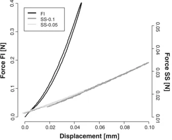
0.0 0.02 0.04 0.06 0.08 0.10 0.0 0.1 0.2 0.3 0.4 Displacement [mm] FI SS-0.1 SS-0.05 ] N[ I F e cr o F 0 .01 0.02 0.03 0.04 0.05 Force SS [N]
Fig. 1 Typical force–displacement curves of the PAMPS/PAAm 90.9 % in FI-mode (black), in SS-0.1-mode (dark grey) and in SS-0.05-mode (light grey)
otherwise a Wilcoxon rank sum test would be performed. Sta-tistical analysis was accomplished using R (R Development Core Team (2010). R: A language and environment for sta-tistical computing. R Foundation for Stasta-tistical Computing, Vienna, Austria).
3 Results
In FI-mode, force rose more rapidly with displacement than in SS-mode (Fig. 1). This indicates a higher stiffness of the DN-gels in FI-mode. The force-displacement slopes in SS-0.05 and SS-0.1 modes were comparable, which indi-cates a similar stiffness in these test methods.
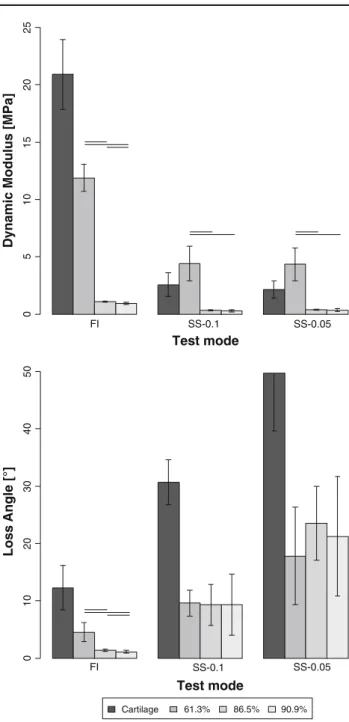
The data sets were all normally distributed and there-fore a two-sample t test was used. The calculated E* was significantly higher in FI-mode compared to SS-mode for all three DN-gels tested. PAMPS/PAAm 61 % had a higher E* in all test modes compared to PAMPS/PAAm 86 % and PAMPS/PAAm 90 %. In FI-mode, PAMPS/PAAm 86 % had a higher E* than PAMPS/PAAm 90 % (Fig.2).
The δ was significantly higher in SS-mode in all gels compared to FI-mode. In SS-0.1-mode, the δ was lower
Test mode FI SS-0.1 SS-0.05 ] a P M[ s ul u d o M ci m a n y D ]° [ el g n A s s o L SS-0.1 Test mode 0 5 10 15 20 25 61.3% 86.5% 90.9% Cartilage 0 1 02 03 0 4 05 0 FI SS-0.05
Fig. 2 Calculated dynamic modulus (E*; up) and loss angle (δ; bot-tom) of swine cartilage, PAMPS/PAAm 61 %, PAMPS/PAAm 87 % and PAMPS/PAAm 91 % in the different test modes (FI, SS-0.1 and SS-0.05). Solid horizontal lines indicate a significant difference with p < 0.05. E* is significantly higher in FI-mode compared to both SS-modes in all gels. In all gels, there is a significant difference between all test modes inδ
than in SS-0.05-mode in all gels. In SS-mode, no difference was found inδ among the three different gels. However, in FI-mode theδ was higher in PAMPS/PAAm 61% compared to PAMPS/PAAm 86 % and PAMPS/PAAm 90 %, and higher in PAMPS/PAAm 86 % than in PAMPS/PAAm 90 %.
Cartilage had a higher E* in FI-mode compared to SS-mode. In FI-mode, E* values of cartilage were higher
compared to all DN-gels (Fig.2). In SS-mode, E* of cartilage was significantly higher compared to PAMPS/PAAm 86 % and PAMPS/PAAm 90 % and lower than PAMPS/PAAm 61 %.
The δ of cartilage was lower in FI-mode compared to SS-mode. In SS-0.1-mode, theδ was lower than in SS-0.05-mode. In all test modes, theδ of cartilage was higher than all DN-gel tested.
4 Discussion
4.1 DN-gel water content effects
The dynamic modulus, E*, increased as the water content decreased in all test modes. A possible explanation is that if the concentration of polymer is higher, there is more struc-ture per unit volume to resist deformation. Conversely, the DN-gelδ did not change as a function of water content in SS-mode. In our previous work, we suggest that the δ in SS-mode is mainly due to water movement within the struc-ture (Ronken et al. 2011). The results presented here imply that the water movement is similar in all PAMPS/PAAm DN-gels and the polymer/water ratio does not change the ability of a given deformation to move water within the struc-ture. But in addition, in FI-mode, theδ increases with decreas-ing water content. This means that an increase in polymer concentration increases theδ at high deformation rates. Since the cross-linked polymer structures are themselves viscoelas-tic, a higher concentration of polymer could be expected to dissipate more energy. However, the ratio between the two polymers was necessarily different among the three DN-gels, and this perhaps makes trying to explain the results on a basis of water content alone too simplistic.
4.2 Dynamic stiffness of DN-gels compared to cartilage The results show that it was possible to bring the dynamic stiffness of the PAMPS/PAAm DN-gels closer to normal car-tilage by modifying the structures in a way which allowed lower water content. As shown in Fig.2, the PAMPS/PAAm 61 % was about 1.5–2 times stiffer than cartilage in SS-mode, but ∼30% less stiff in FI-mode. Compared to tissue-engineered constructs, which are only up to 10 % of cartilage stiffness (Santoro et al. 2010), and autologous chondrocyte transplantation, which is about 60 % of cartilage stiffness a year after surgery (Peterson et al. 2002), initial repair stiff-ness is closer to native cartilage. On the other hand, theδ of PAMPS/PAAm 61 % in FI-mode was∼60% lower than that of cartilage and∼70% lower in SS-mode. PAMPS/PAAm 86 and 90 % had a lower E* and a lowerδ compared to cartilage in all test modes.
The crucial dynamic mechanical difference between all three of the PAMPS/PAAm DN-gels and normal cartilage is that all three had much lower loss angles(δ) than cartilage in both test modes. This means that compared to cartilage, these gels are less able to dissipate energy. Also, due to its higherδ, the E* of cartilage is more strain rate dependent than that of DN-gels. Therefore, by adjusting water content in the man-ner done here, the low loss angles of PAMPS/PAAm DN-gels mean that their E* values could not be made similar to carti-lage at both strain rates—that is, during both fast-impact (FI) and slow-sinusoidal (SS) testing. For example, if one “tunes” the DN-gel value of E* to be similar to cartilage in FI, one is left with a gel which has a higher E* in SS-mode. The con-sequence of this difference in mechanical properties with the surrounding tissue can only be speculated upon. However, the difference would be much lower compared to the same properties produced using tissue repair techniques already in use.
One possible structural reason that the δ of all the PAMPS/PAAm DN-gels is low compared to cartilage may be because both components of the polymer structure are highly chemically crosslinked. This crosslinking reduces the possi-bility of sliding between the polymer chains during defor-mation and thus reduces the frictional dissipation of energy. Another possible cause of lower energy dissipation compared to cartilage might be less movement of water either within the DN-gel structure or out of the structure during deformation. Water can be forced out of cartilage by static loads (Mankin et al. 2000) but similar loads do not result in forcing water out of PAMPS/PAAm DN-gels.Gong et al.(2003) showed that after deformation to ∼20% of the original thickness, still no water is squeezed out of the structure. In other words, the water within the DN-gel structure is more highly trapped compared to cartilage.
These results show that cartilage-like dynamic stiffness can be achieved by these compositional changes; however, it is unknown how it affects other important mechanical proper-ties. Therefore, the authors plan to investigate other mechan-ical properties, such as strength, fatigue and tear resistance in further study. These DN-gels have already been shown to be superior to conventional gels in simulated use friction and wear tests (Yasuda et al. 2005).
These DN-gels have potential for clinical use. They are easy to sterilise since autoclaving has been shown not to affect their structures. They can be trimmed or produced in desired shapes and the surgical fixation, for example, with sutures or tissue adhesive, capability has proven to be substantial. Besides this, if the gel is created with large enough pore size, cell infiltration is likely possible, to assure integration to the surrounding tissue. Also, as previously mentioned (Yasuda et al. 2009) non-porous plugs of a highly similar DN-gel have been shown to foster cartilage formation in a rabbit osteo-chondral defect model.
Although these DN-gels look promising as a cartilage repair material, in this study only their dynamic stiffness was investigated. Before these DN-gels can be used in clinic, other aspects should be investigated mainly focussed on the biocompatibility, such as immunological reactions, absorp-tion and integraabsorp-tion to the surrounding tissues.
5 Conclusion
In all three of the PAMPS/PAAm DN-gels, theδ increases with decreasing deformation rate in SS-mode compared to FI-mode. This is what is expected for viscoelastic materi-als (Lakes 1999; Park et al. 2004). Although the DN-gels thus show normal viscoelastic behaviour with respect strain rate andδ, they do not do so with respect to E*. For normal viscoelastic materials, E* increases with increasing strain rate (Lakes 1999; Park et al. 2004)—and is thus higher in FI-mode compared to SS-mode. However, for the DN-gels no difference in E* was found between the two SS-modes, even though the deformation rate was doubled for SS-0.1 compared to SS-0.05 (∼0.03 vs. 0.015m/s).
Biomechanically these DN-gels look promising as poten-tial cartilage repair materials. However, other properties, such as fixation stability and mechanical performance in vivo, have to be explored.
Acknowledgments Hardy and Otto Frey-Zünd Stiftung for the sup-port and H.J. Wyss for the funds donated to University Basel.
References
Arnold MP, Daniels AU, Ronken S, Ardura Garcia H, Friederich NF, Kurokawa T, Gong JP, Wirz D (2011) Acrylamide polymer double-network hydrogels: candidate cartilage repair materials with cartilage-like dynamic stiffness and attractive surgery-related attachment mechanics. Cartilage. doi:10.1177/194760351 1402320
Azuma C, Yasuda K, Tanabe Y, Taniguro H, Kanaya F, Nakayama A, Chen YM, Gong JP, Osada Y (2006) Biodegradation of high-toughness double network hydrogels as potential materials for arti-ficial cartilage. J Biomed Mater Res A. doi:10.1002/jbm.a
Gong JP, Katsuyama Y, Kurokawa T, Osada Y (2003) Double-network hydrogels with extremely high mechanical strength. Adv Mater 15(14):1155–1158. doi:10.1002/adma.200304907
Hunter W (1743) On the structure and diseases of articular cartilage. Philos T R Soc Lond 42:514–521
Kren AP, Rudnitskii VA, Deikun IG (2005) Determining the viscoelas-tic parameters of vulcanisates by the dynamic indentation method using a non-linear deformation model. Int Polym Sci Technol 32(7):19–23
Lakes RS (1999) Viscoelastic solids. CRC Press, Boca Raton Mandelbaum BR, Browne JE, Fu F, Micheli L, Mosely JB, Erggelet C,
Minas T, Peterson L (1998) Articular cartilage lesions of the knee. Am J Sports Med 26(6):853–861
Mankin H, Mow V, Buckwalter JA, Iannotti J, Ratcliffe A (2000) Artic-ular cartilage structure, composition, and function. In: Buckwalter
JA (ed) Orthopaedic basic science: biology and biomechanics of the musculoskeletal system. American Academy of Orthopaedic Surgeons, Rosemont pp 443–470
Minas T (1999) The role of cartilage repair techniques, including chon-drocyte transplantation, in focal chondral knee damage. AAOS Instr Cours Lect 48:629–643
Mithoefer K, Scopp JM, Mandelbaum BR (2007) Articular cartilage repair in athletes. AAOS Instr Cours Lect 56:457–468
Nakajima T, Furukawa H, Tanaka Y, Kurokawa T, Osada Y, Gong JP (2009) True chemical structure of double network hydrogels. Macromolecules 42(6):2184–2189. doi:10.1021/ma802148p
Nakayama A, Kakugo A, Gong JP, Osada Y, Takai M, Erata T, Kawano S (2004) High mechanical strength double-network hydrogel with bacterial cellulose. Adv Funct Mater 14(11):1124–1128. doi:10. 1002/adfm.200305197
Negrin L, Kutscha-Lissberg F, Gartlehner G, Vecsei V (2011) Clini-cal outcome after microfracture of the knee: a meta-analysis of before/after-data of controlled studies. Int Orthop. doi:10.1007/ s00264-011-1364-x
Newman AP (1998) Articular cartilage repair. Am J Sport Med 26(2):309–324
Park S, Hung C, Ateshian G (2004) Mechanical response of bovine articular cartilage under dynamic unconfined compression loading at physiological stress levels. Osteoarthr Cartil 12:65–73. doi:10. 1016/j.joca.2003.08.005
Peterson L, Brittberg M, Kiviranta I, Åkerlund EL, Lindahl A (2002) Autologous chondrocyte transplantation: biomechanics and long-term durability. Am J Sport Med 30(1):2–12. doi:10. 1097/BLO.0b013e3180e79c6a
Peterson L, Vasiliadis HS, Brittberg M, Lindahl A (2010) Autologous chondrocyte implantation: a long-term follow-up. Am J Sport Med 38(6):1117–1124. doi:10.1177/0363546509357915
Robert H (2011) Chondral repair of the knee joint using mosaicplasty. Orthop Traumatol Surg Res 97(4):418–429. doi:10.1016/j.otsr. 2011.04.001
Ronken S, Arnold MP, Ardura García H, Jeger A, Daniels AU, Wirz D (2011) A comparison of healthy human and swine articular
carti-lage dynamic indentation mechanics. Biomech Model Mechano-biol. doi:10.1007/s10237-011-0338-7
Santoro R, Olivares AL, Brans G, Wirz D, Longinotti C, Lacroix D, Martin I, Wendt D (2010) Bioreactor based engineering of large-scale human cartilage grafts for joint resurfacing. Biomaterials 31(34):8946–8952. doi:10.1016/j.biomaterials.2010.08.009
Saris DBF, Vanlauwe J, Victor J, Haspl M, Bohnsack M, Fortems Y, Vandekerckhove B, Almqvist KF, Claes T, Handelberg F, Lagae K, van der Bauwhede J, Vandenneucker H, Yang KG, Jelic M, Verdonk R, Veulemans N, Bellemans J, Luyten FP (2008) Charac-terized chondrocyte implantation results in better structural repair when treating symptomatic cartilage defects of the knee in a ran-domized controlled trial versus microfracture. Am J Sports Med 36(2):235–246. doi:10.1177/0363546507311095
Slauterbeck JR, Kousa P, Clifton BC, Naud S, Tourville TW, Johnson RJ, Beynnon BD (2009) Geographic mapping of menis-cus and cartilage lesions associated with anterior cruciate ligament injuries. J Bone Joint Surg Am 91:2094–2103. doi:10.2106/JBJS. H.00888
Widuchowski W, Widuchowski J, Trzaska T (2007) Articular cartilage defects: study of 25,124 knee arthroscopies. Knee 14(3):177–182. doi:10.1016/j.knee.2007.02.001
Wirz D, Kohler K, Keller B, Göpfert B, Hudetz D, Daniels AU (2008) Dynamic stiffness of articular cartilage by single impact micro-indentation (SIMI). J Biomech 41(Suppl 1):S172 Yasuda K, Gong JP, Katsuyama Y, Nakayama A, Tanabe Y, Kondo E,
Ueno M, Osada Y (2005) Biomechanical properties of high-tough-ness double network hydrogels. Biomaterials 26(21):4468–4475. doi:10.1016/j.biomaterials.2004.11.021
Yasuda K, Kitamura N, Gong JP, Arakaki K, Kwon HJ, Onodera S, Chen YM, Kurokawa T, Kanaya F, Ohmiya Y, Osada Y (2009) A novel double-network hydrogel induces spontaneous articular cartilage regeneration in vivo in a large osteochondral defect. Macromol Biosci 9(4):307–316. doi:10.1002/mabi.200800223