HAL Id: hal-02954468
https://hal.archives-ouvertes.fr/hal-02954468
Submitted on 1 Oct 2020HAL is a multi-disciplinary open access archive for the deposit and dissemination of sci-entific research documents, whether they are pub-lished or not. The documents may come from teaching and research institutions in France or abroad, or from public or private research centers.
L’archive ouverte pluridisciplinaire HAL, est destinée au dépôt et à la diffusion de documents scientifiques de niveau recherche, publiés ou non, émanant des établissements d’enseignement et de recherche français ou étrangers, des laboratoires publics ou privés.
Analysis of mPygo2 mutant mice suggests a requirement
for mesenchymal Wnt signaling in pancreatic growth and
differentiation
Nicolas Jonckheere, Erin Mayes, Hung-Ping Shih, Boan Li, Oleg Lioubinski,
Xing Dai, Maike Sander
To cite this version:
Nicolas Jonckheere, Erin Mayes, Hung-Ping Shih, Boan Li, Oleg Lioubinski, et al.. Analysis of mPygo2 mutant mice suggests a requirement for mesenchymal Wnt signaling in pancreatic growth and differ-entiation. Developmental Biology, Elsevier, 2008, 318 (2), pp.224-235. �10.1016/j.ydbio.2008.03.014�. �hal-02954468�
Analysis of mPygo2 mutant mice suggests a requirement for
mesenchymal Wnt signaling in pancreatic growth and
differentiation
Nicolas Jonckheere1,5, Erin Mayes1,5, Hung-Ping Shih1, Boan Li2,4, Oleg Lioubinski1,3, Xing Dai2, and Maike Sander1,*
1Department of Developmental and Cell Biology, University of California Irvine, Irvine, CA 92697-2300,
U.S.A.
2Department of Biological Chemistry, University of California Irvine, Irvine, CA 92697-1700, U.S.A.
Summary
Pygopus has recently been identified in Drosophila as an essential component of the nuclear complex
required for canonical Wnt signaling. Here, we have investigated the role of the mammalian
pygopus ortholog, mPygo2, in pancreas development. We show that a null mutation of mPygo2 in
mice causes pancreas hypoplasia due to decreased progenitor cell proliferation after embryonic day (e) 12.5. During the same time window, mPygo2-deficient embryos begin to display a reduction in endocrine progenitors and consequently a decrease in islet endocrine cell mass. Consistent with its function after e12.5, late-developing endocrine cell types, such as beta, delta and PP cells, are specifically reduced, while the earlier-forming alpha cells develop normally. We find canonical Wnt signaling to be predominantly active in the mesenchyme at the time when mPygo2 is required and demonstrate the dependence of Wnt signal transduction on mPygo2. Furthermore, conditional deletion of mPygo2flox allele in the pancreatic epithelium does not phenocopy the defects in
mPygo2-null mutants. Since mPygo2 is expressed in the pancreatic mesenchyme and the role of the
mesenchyme in epithelial progenitor cell expansion is well documented, our findings suggest an indirect role for mPygo2 in epithelial growth and differentiation through regulation of mesenchymal signals. Together, our data suggest a previously unappreciated role for mesenchymal Wnt signaling in regulating pancreatic organ growth and cell differentiation.
Keywords
pygopus; Wnt; pancreas development; mesenchyme; islet; endocrine; proliferation; differentiation; mouse
*author for correspondence: Tel: 949-824-4952, Fax: 949-824-4709, Email: msander@uci.edu.
3Present address: Max-Delbrück Center for Molecular Medicine, Robert-Rössle-Str. 10, 13092 Berlin, Germany 4Present address: School of Life Sciences, Xiamen University, Xiamen, Fujian 361005, P. R. China
5these authors contributed equally
Publisher's Disclaimer: This is a PDF file of an unedited manuscript that has been accepted for publication. As a service to our customers
we are providing this early version of the manuscript. The manuscript will undergo copyediting, typesetting, and review of the resulting
NIH Public Access
Author Manuscript
Dev Biol. Author manuscript; available in PMC 2009 June 15. Published in final edited form as:
Dev Biol. 2008 June 15; 318(2): 224–235. doi:10.1016/j.ydbio.2008.03.014.
NIH-PA Author Manuscript
NIH-PA Author Manuscript
Introduction
In mammals, the pancreas develops as separate ventral and dorsal evaginations from the gut endoderm (Slack, 1995). As the dorsal and ventral pancreatic buds appear at embryonic day (e) 9.5 and e10.5, respectively, they each become surrounded by a cap of dense mesenchyme. Subsequent proliferation of the epithelial progenitors rapidly leads to formation of a branched epithelium that gives rise to an endocrine and an exocrine tissue compartment. The pancreatic endocrine compartment is comprised of glucagon-producing alpha-, the insulin-producing beta-, the somatostatin-producing delta- and pancreatic polypeptide (PP)-producing cells that form sequentially during development and cluster in so-called islets of Langerhans shortly before birth. The exocrine acinar cells and ducts become visible as distinct structures by about e14.5.
All pancreatic cells arise from a common pool of pancreatic epithelial progenitor cells that are marked by the transcription factor Pdx1 (Gu et al., 2002). Expression of the transcription factors Ngn3 and after e13.5, Ptf1a, subsequently restrict the developmental potential of these pluripotent progenitors to an endocrine and exocrine fate, respectively (Gu et al., 2002; Zhou et al., 2007). Since the pancreas has limited capacity for adaptive growth, the final size of the endocrine and exocrine cell compartments at birth is largely determined by the size of the highly proliferative pluripotent progenitor cell pool at the pancreatic bud stage (Stanger et al., 2007). Mutations that affect progenitor cell expansion early in development therefore commonly result in peri- and postnatal pancreatic hypoplasia. Proliferation and branching of the epithelial buds is controlled by both autonomous cues in the epithelium as well as non-autonomous signals from the surrounding mesenchyme (Murtaugh, 2007). When cultured in the absence of mesenchyme, epithelial buds show minimal growth and branching, which is restored upon recombination with mesenchyme (Golosow and Grobstein, 1962; Wessells and Cohen, 1967). The mesenchyme not only controls progenitor cell expansion, but also promotes both endocrine and exocrine cell differentiation (Attali et al., 2007; Li et al., 2004b;
Scharfmann, 2000).
Previous studies have provided evidence that the Wnt signaling pathway controls several aspects of pancreatic development. Early in development, the level of endodermal Wnt activity appears to control the specification of intestinal, liver and pancreatic fates in foregut endoderm (Heller et al., 2002; McLin et al., 2007; Ober et al., 2006). Gain- and loss-of-function studies in mice further indicate that during pancreas morphogenesis, Wnt signaling is required for growth and differentiation of the epithelium (Dessimoz et al., 2005; Heiser et al., 2006; Murtaugh et al., 2005; Papadopoulou and Edlund, 2005; Wells et al., 2007). Additional roles for Wnt signaling have been further suggested during the perinatal and postnatal periods in islet formation, islet cell proliferation and beta cell function (Fujino et al., 2003; Kim et al., 2005; Rulifson et al., 2007).
While these studies have established that Wnt signaling is important in the pancreas, the spatial and temporal pattern of Wnt signal transduction and the specific requirements for Wnt activity in the epithelium and mesenchyme have remained elusive. Notably, Wnt signals can be intracellularly transduced through a so-called canonical (Wnt/β-catenin) pathway or non-canonical (Wnt/Ca+ and Wnt/planar cell polarity) pathways (Miller, 2002). The existence of parallel pathways leaves ambiguity as to which intracellular signals control the phenotype when Wnt signaling is perturbed at the Wnt ligand or frizzled receptor level. As conditional deletion of the Wnt-effector β-catenin in pancreatic epithelial progenitor cells causes defects in progenitor cell expansion and exocrine cell differentiation, it has been recently suggested that normal pancreas morphogenesis requires canonical Wnt signaling autonomously in the epithelium (Murtaugh et al., 2005; Wells et al., 2007).However, interpretation of the mechanisms that underlie these phenotypic changes is complicated by the fact that β-catenin
NIH-PA Author Manuscript
NIH-PA Author Manuscript
also has Wnt-independent functions in cell adhesion (Nelson and Nusse, 2004). To better understand the role of canonical Wnt signaling in pancreas development, it is therefore necessary to compare the effects of perturbing signal transduction at several levels of the pathway.
In response to Wnt signaling, cytoplasmic β-catenin becomes stabilized and enters the nucleus, where it functions in a complex with Tcf/Lef transcription factors to activate Wnt target genes (Nelson and Nusse, 2004). Recently, pygopus has been identified as an additional core component of this complex in Drosophila and appears to act as a nuclear anchor as well as coactivator for the β-catenin/Tcf/Lef complex (Kramps et al., 2002; Krieghoff et al., 2006; Stadeli and Basler, 2005; Thompson, 2004; Townsley et al., 2004a; Townsley et al., 2004b). Analysis of loss-of-function phenotypes in Drosophila and Xenopus have shown that
pygopus is an essential regulator of canonical Wnt pathway responses (Belenkaya et al.,
2002; Lake and Kao, 2003; Parker et al., 2002; Thompson et al., 2002). The murine genome contains two pygopus orthologs, pygopus 1 (mPygo1) and pygopus 2 (mPygo2). Consistent with the broader expression of mPygo2 than mPygo1 during development and in adult tissues (Li et al., 2004a), loss of mPygo2 function causes severe developmental defects in various Wnt-dependent tissues, while mPygo1-deficient mice exhibit no apparent abnormalities (Li et al., 2007; Schwab et al., 2007; Song et al., 2007). Therefore, mPygo2 appears to be the functionally important ortholog in mice.
We show here that germline mutation of mPygo2 in mice causes pancreatic hypoplasia and a specific defect in endocrine cell differentiation. We find that these defects result from decreased proliferation of undifferentiated pancreatic progenitors and reduced formation of endocrine progenitors after e13. Since the pancreas develops normally in mice with a pancreatic epithelial-specific deletion of mPygo2, our data suggest that mPygo2 controls mesenchymal signals required for pancreatic growth and differentiation. Accordingly, we find that canonical Wnt signaling is active in the pancreatic mesenchyme at the time mPygo2 mutant mice first show a phenotype and demonstrate dependence of this signal on mPygo2 function. Therefore, our results suggest that expansion and differentiation of pancreatic epithelial progenitors requires canonical Wnt signaling in the mesenchyme.
Material and Methods
Mice
Mice carrying the null allele for mPygo2 were generated by Cre-mediated recombination in ES cells (Li et al., 2007). For pancreas-specific deletion of mPygo2, mice with a floxed
mPygo2 allele (mPygo2flox) (Li et al., 2007) were crossed with Pdx1-Cre mice (Gu et al., 2002). In these experiments, mPygo2flox /+;Pdx1-Cre or mPygo2+/−;Pdx1-Cre littermates served as controls. PCR detection of the mPygo2flox allele was performed using primers forward 5’-AGC GTG TCT AAG GTC AGC CAG AGG TTT G-3’ and reverse 5’-GTA AAG CGT TGG GGG AGA GGA GGA GGA C-3’. Axin2+/lacZ and BatGAL mice have been previously described (Lustig et al., 2002; Maretto et al., 2003). All strains were maintained on a C57BL/6J background. Experiments were performed with the approval of the UC Irvine Institutional Animal Care and Use Committee protocol 2002–2420. Noon of the vaginal plug day was considered as e0.5. To label S-phase nuclei, some mice were injected with 50µg/g body weight of BrdU i.p. 45 min prior to sacrifice.
Tissue processing, immunohistochemistry and microscopy
Pancreata were fixed in 4% paraformaldehyde in PBS for 4 hours and immunofluorescence was performed on 10 µm cryopreserved or 7 µm paraffin sections. Staining with hematoxylin/ eosin (Fisher) was performed according to the manufacturer’s instructions. For
NIH-PA Author Manuscript
NIH-PA Author Manuscript
immunostaining we followed a previously described protocol (Sander et al., 1997). The following antibodies were used: rabbit mPygo2 (1:500; (Li et al., 2007), guinea pig anti-insulin (1:5000; Linco), mouse anti-glucagon (1:50; Sigma), mouse anti-somatostatin (1:3000; gift from Palle Serup, Hagedorn Institute), rabbit anti-pancreatic polypeptide (1:2000; Dako), rabbit anti-amylase (1:500; Sigma), armenian hamster anti-MUC1 (1:200; Lab Vision Corporation), mouse anti-β-catenin (1:100; Sigma), guinea pig anti-Pdx1 (1:5000; gift from Chris Wright, Vanderbilt University), guinea pig anti-Ngn3 (1:1000; (Henseleit et al., 2005)), rabbit anti-Ptf1a (1:2000; gift from Helena Edlund, Umea University), mouse anti-BrdU (1:100; Chemicon), rat anti-E-cadherin (1:2000; Sigma), rabbit anti-mPygo1 (1:3000; Schwab et al., 2007), guinea pig anti-Pbx1 (1:400; Kim et al., 2002), guinea pig anti-Isl1 (1:5000; gift from Johan Ericson, Karolinska, Stockholm), rabbit anti-Sox9 (1:2000; Chemicon). For immunohistochemistry, biotinylated anti-mouse or anti-guinea pig IgG antibodies were used (1:200; Vector Laboratories). For immunofluorescence, antigen-antibody complexes were detected with Cy3- (1:2000; Jackson Laboratory) or Alexa488- conjugated (1:2000; Molecular Probes) goat-raised secondary antibodies. For detection of Pdx1, Ptf1a, Ngn3 and BrdU, antigen retrieval was performed by incubation in pH 6.0 citrate buffer at 37°C for 1h. Glucagon and BrdU were detected using the M.O.M. Immunodetection Kit (Vector Laboratories). TUNEL assays were performed using the ApopTag® Plus Peroxidase In Situ Apoptosis Detection Kit (Chemicon). Using 5-bromo-4-chloro-3-indolyl-beta-D-galactopyranoside (X-gal) as a substrate, whole-mount X-gal staining was performed on either whole embryos or isolated abdominal organs as described (Mombaerts et al., 1996). Images were collected on a Zeiss Axioplan2 microscope with a Zeiss AxioCam driven by Zeiss AxioVision v. 4.0 software. Morphometric quantification of cell numbers and area
For morphometric analysis, all pancreata were embedded in the same orientation and serial sections were collected from the entire organ. Following immunostaining, images from an entire pancreas section were acquired and if necessary assembled in Adobe Photoshop. Cell counting and morphometric analysis were performed on every fifth section. E-cadherin was used to visualize the entire pancreatic epithelial area and DAPI staining for detection of individual nuclei. Mesenchymal cell area was measured by subtracting the epithelial cell area from the total pancreatic area, including the mesenchyme. Morphometry was conducted using Image-Pro Plus v. 5.0.1 (Media Cybernetics). The data are displayed as averages ± standard error of the mean. Statistical significance was determined using Student’s t-test (Minitab v. 14.20).
RNA preparation and RT-PCR and real-time quantitative PCR
Total RNA from dissected pancreatic anlagen was extracted with the RNeasy kit (Qiagen) and treated with DNAse. cDNA was prepared by in vitro transcription using the iScript cDNA synthesis kit (Biorad). The following primers were used for PCR: for detection of mPygo2 forward 5’-GGA GCG AAG AAA GTC CAA TAC-3’ and reverse 5’-GTT AGA AGC GAC CAG ATG ATC-3’ (75 bp product); for detection of mPygo1 forward 5’-GTG GTG ACA GTG GAC TGG ATG-3’ and reverse 5’-CTG AGT GAG TAA GGA CCA CAG-3’ (323 bp product); for detection of β-actin as an internal control forward 5’-TGT TAC CAA CTG GGA CGA CA-3’ and reverse 5’-GGG GTG TTG AAG GTC TCA AA-3’. Amplification was performed for 35 PCR cycles.
For quantitative RT-PCR analysis, all transcripts were amplified with 1x SYBR Green PCR master mix (Applied Biosystems), as described previously (Kioussi et al., 2006). Listed 5’ to 3’, primer sequences were as follows: mPygo1 forward: CCATCGCGCTGAAGAGAGTTA; mPygo1 reverse: GTGTGTTGGCCTTGCGTTTT; mPygo2 forward:
AAGGCCGGTCTGCAAATGAA; mPygo2 reverse: GTTAGAAGCGACCAGATGATCC;
NIH-PA Author Manuscript
NIH-PA Author Manuscript
Axin2 forward: TGACTCTCCTTCCAGATCCCA; Axin2 reverse: TGCCCACACTAGGCTGACA.
Results
Expression pattern of mPygo2 in the developing pancreas
To determine the domains of mPygo2 expression, we performed co-immunofluorescence analyses with an anti-mPygo2 specific antiserum in conjunction with antibodies against various pancreatic markers on embryonic pancreas sections. Specificity of the anti-mPygo2 antibody was demonstrated by absence of immunostaining in pancreas from mPygo2-null mutant embryos (Supplementary Fig. 1D–F). Similar to previously analyzed tissues (Li et al., 2007; Schwab et al., 2007), pancreatic mPygo2 expression was exclusively nuclear at all stages (Fig. 1). Between e10.5 and e12.5, when the pancreatic epithelium is still largely undifferentiated, mPygo2 broadly co-localized with the pancreatic epithelial progenitor cell marker Pdx1 throughout the ventral and dorsal pancreatic anlage (Fig. 1A–F, data not shown). The level of mPygo2 expression varied between cells in the epithelium, with some cells expressing high and others low levels of mPygo2. Strong mPygo2 immunoreactivity was also found in mesenchymal cells surrounding the ventral and the dorsal pancreatic anlage at e10.5 and e12.5 (Fig. 1A–F, data not shown). Similar to mPygo2, the Wnt effector β-catenin was also expressed in both the pancreatic epithelium and surrounding mesenchyme (Fig. 1G–I). Consistent with previous reports (Heiser et al., 2006), nuclear localization of β-catenin was not observed. At e15.5, mPygo2 expression in E-cadherin+ cells suggested persistence of the epithelial mPygo2 expression (Fig. 1J–L). In addition to its epithelial expression, the presence of scattered E-cadherin-negative/mPygo2+ interstitial cells indicated persistent expression of mPygo2 also in the mesenchyme (Fig. 1L, arrowheads). The staining intensity of mPygo2 in the epithelium varied between different cell types. While newly differentiated beta cells, characterized by high levels of Pdx1 expression, showed weak mPygo2 immunoreactivity (Fig. 1O), strong expression was seen in the forming acinar compartment (Fig. 1M–O). Scattered mPygo2+ mesenchymal cells were also detected in late gestation (Fig. 1P–R). At e18.5, mPygo2 was no longer found in the acinar and endocrine compartments (Fig. 1Q,R), but persisted in a subset of ductal cells (Fig. 1P,R). In contrast to mPygo2, mPygo1 was predominantly localized to the pancreatic mesenchyme, but scattered Pdx1+ epithelial cells expressing mPygo1 were also found (Supplementary Fig. 1J–H). In the absence of mPygo2 activity, the overall expression pattern (Supplementary Fig. 1K–M) and mRNA levels (Supplementary Fig. 1O) of mPygo1 were unchanged. However, the number of epithelial cells expressing mPygo1 appeared to be reduced in mPygo2-deficient mice (Supplementary Fig. 1M). Different from mPygo2,
mPygo1 expression was only transiently detected in the pancreas and no longer found after
e15.5 (Supplementary Fig. 1G,N). Reduced pancreas size in mPygo2−/− embryos
To determine whether pancreas development is dependent on the function of mPgyo2, we analyzed pancreata from mPygo2−/− embryos at e18.5. The perinatal lethality associated with
mPygo2-null mutations precluded studies in postnatal mice (Li et al., 2007; Schwab et al.,
2007). Inspection of pancreas morphology revealed a marked reduction in organ size in mPygo2-deficient embryos (Fig. 2A,B). The reduction was evident in both the duodenal portion of the pancreas, which is derived from the ventral pancreatic anlage, as well as the splenic portion, which originates from the dorsal anlage. To exclude the possibility that the reduced pancreatic size is a mere consequence of the 15–20% reduction in body weight of neonatal mPygo2−/− embryos (Li et al., 2007), we normalized pancreatic weight to the total body weight of the embryo. The ratio of pancreatic weight to body weight showed a statistically significant ~20% reduction in mPygo2−/− embryos compared to their wild type littermates (Fig. 2C). Histological analysis of pancreas sections revealed grossly normal exocrine and endocrine
NIH-PA Author Manuscript
NIH-PA Author Manuscript
cell compartments (Fig. 2D,E). To determine the origin of pancreas hypoplasia in mPygo2-deficient embryos, we examined whether a reduction in pancreatic size is already present at earlier developmental time points. At e15.5, when the overall size and weight of mPygo2−/− embryos is indistinguishable from wild type littermates (data not shown), the ventral and dorsal pancreas showed a similar size reduction as observed at e18.5 (Fig. 2F–H). Prior to e15.5, we assessed pancreatic size by separate quantitative morphometric measurements of the ventral and dorsal pancreatic epithelial area. At e13.5, the pancreatic epithelium in mPygo2−/− embryos showed less branching and a significant reduction in size (Fig. 2I–K). At e12.5, however, no difference was found between mPygo2−/− and wild type embryos (Fig. 2L–N). Thus,
mPygo2 is first required for growth and branching of the pancreatic epithelium after e12.5.
To reveal the mechanism that underlies impaired pancreatic growth in mPygo2−/− embryos, we studied whether mPygo2 controls the timing of cell differentiation, cell proliferation or cell survival. During pancreas development, major cell differentiation of both the exocrine and endocrine compartments does not occur until after e13.5. With the exception of occasional insulin-producing cells, glucagon-expressing cells are the only cell type that differentiates between e10.5 and e13.5. To assess whether the diminished size of the pancreas in mPygo2-deficient embryos results from depletion of the pancreatic progenitor cell pool due to premature cell cycle exit and differentiation, we quantified the number of glucagon+ cells in relation to the number of Pdx1+ pancreatic progenitors between e10.5 and e13.5. The relative number of glucagon+ cells was similar in mPygo2−/− embryos and wild type littermates (Fig. 3A–C, data not shown), suggesting that absence of mPygo2 is not associated with premature cell differentiation. This conclusion was further supported by the finding that insulin+ or exocrine cells also do not differentiate prematurely (data not shown). To determine whether absence of mPygo2 affects pancreatic cell proliferation, we next compared the number of BrdU-incorporating cells in mPygo2−/− and control embryos at e13.5, which is the time point when the epithelial size reduction is first evident in mPygo2−/− embryos. mPygo2-null mutants showed a consistent 20–25% reduction in the proliferative rate of Sox9+ progenitor cells in both the ventral and the dorsal pancreatic anlage (Fig. 3D–F). Significantly, no difference was observed at e12.5, when the epithelium is still normal in size (data not shown). Since apoptotic cells were rarely seen in the pancreatic epithelium or mesenchyme and no difference was found between mPygo2−/− and control embryos (Supplementary Fig. 2A,B), our findings suggest that mPygo2 controls pancreatic growth by ensuring proliferation of undifferentiated pancreatic progenitor cells after e13. As the size of the pancreatic progenitor cell pool has recently been shown to dictate final organ size (Stanger et al., 2007), this early function of mPygo2 in progenitor cell proliferation most likely also accounts for the reduced pancreas size in neonatal
mPygo2−/− mice.
mPygo2 controls endocrine but not exocrine cell formation
To assess a potential role for mPygo2 in pancreatic cell differentiation, we compared the size of the endocrine and exocrine cell compartments in mPygo2-null mutant embryos to their littermate controls. Morphometric analysis of the insulin- and glucagon-expressing cell areas in relation to total pancreatic size revealed a reduction in beta cells, but not in alpha cells in
mPygo2−/− mutants at e15.5 and at birth (Fig. 4A–F, data not shown). A similar reduction as was observed for beta cells was also seen for somatostatin- and PP-producing cells (Fig. 4G– L). Unlike the endocrine cell compartment, the amylase+ acinar cell compartment was not affected by mPygo2-deficiency at both ages (Fig. 4M–O, data not shown). As assessed by histology and immunofluorescence for DBA and mucin, the ductal cell compartment developed normally in mPygo2−/− mice (data not shown).
The numbers of Ptf1a+ progenitors, which become restricted to an exocrine fate around e13.5 (Zhou et al., 2007), appeared slightly increased in mPygo2−/− embryos (Fig. 5A–C). However,
NIH-PA Author Manuscript
NIH-PA Author Manuscript
this difference did not reach statistical significance (Fig. 5C). To determine whether the reduction in endocrine cells can be attributed to a lower rate of cell differentiation, we analyzed the number of Ngn3+ endocrine progenitors. The 50% reduction in beta cells in mPygo2−/− embryos (Fig. 4C) was indeed preceded by a 40–50% reduction in endocrine progenitors in the ventral and dorsal pancreatic anlage at e13.5 (Fig. 5D–F). Notably, Ngn3+ cell numbers were normal in mPygo2−/− mutants at e12.5 (data not shown). As a recent study has demonstrated that beta, delta and PP cells are the major endocrine cell types to arise from Ngn3+ progenitors after e13 (Johansson et al., 2007), the selective reduction in these cell types in Pygo2−/− mutants may be explained by a specific requirement for mPygo2 in the generation of endocrine progenitors during this time window. Combined, our data show that mPygo2 has a critical role for progenitor cell proliferation and endocrine cell genesis after e12.5.
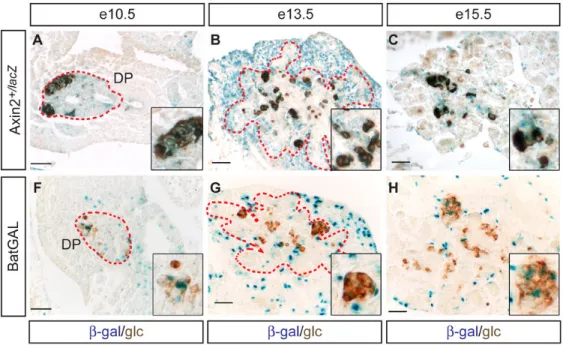
Wnt signaling is mainly active in the pancreatic mesenchyme when mPygo2 is required In Drosophila, pygopus has been shown to be an obligatory regulator of Wnt signal transduction (Belenkaya et al., 2002; Kramps et al., 2002; Parker et al., 2002; Stadeli and Basler, 2005; Thompson et al., 2002; Thompson, 2004; Townsley et al., 2004a). To determine whether a similar genetic interaction exists between mPygo2 and Wnt signaling in the mammalian pancreas, we analyzed the spatial and temporal profile of Wnt signal transduction during pancreas development. A similar analysis has been previously performed in pancreas from e14.5 embryos and older, but not at earlier embryonic stages (Dessimoz et al., 2005). To identify sites of active Wnt signaling in mouse tissue, a number of different lacZ-based reporter strains have been developed. One of these strains is based on the expression of Axin2, which is exclusively expressed at sites of Wnt signal transduction (Jho et al., 2002; Lustig et al., 2002). In embryos that express lacZ under control of the Axin2 locus, β-galactosidase (β-gal) activity was mainly confined to the undifferentiated pancreatic epithelium and early
differentiated glucagon+ cells, but was also detected in scattered cells in the surrounding mesenchyme at e10.5 (Fig. 6A). At e13.5, the pattern was reversed, evidenced by abundant
lacZ expression in the mesenchyme and only occasional lacZ+ cells in the epithelium (Fig. 6B). The predominant localization of β-gal activity to the mesenchyme persisted at e15.5 (Fig. 6C). Consistent with the previous study (Dessimoz et al., 2005), few glucagon+ cells also showed detectable lacZ expression (Fig. 6B and C, inset). At e18.5, lacZ+ cells were found in the pancreatic ducts, but were largely absent from the endocrine and exocrine compartments (Fig. 6D,E). Since reporter mouse lines for Wnt activity have been shown to not necessarily monitor all sites of Wnt signal transduction, we confirmed our results in BatGAL mice, in which expression of a lacZ transgene is controlled by tandem repeats of Wnt-dependent Tcf/Lef transcription factor binding sites (Maretto et al., 2003). The sites of β-gal activity in the developing pancreas of BatGAL embryos largely mirrored those identified in Axin2+/lacZ mice (Fig. 6F–J). The overall number of lacZ+ pancreatic cells, however, was smaller in BatGAL mice at all stages. In the adult pancreas, β-gal activity was not detected in either Wnt reporter strain (data not shown).
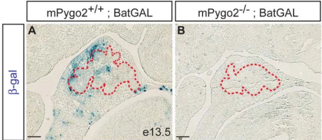
To address whether loss of mPygo2 affects Wnt signaling, we crossed the BatGAL transgene into the mPygo2-null mutant background and assayed for β-gal activity in the pancreas. Previous studies have shown that in many tissues deletion of mPygo2 results in a reduction but not complete absence of BatGAL transgene expression (Li et al., 2007; Schwab et al., 2007). After a four-hour incubation in X-Gal staining solution, we consistently observed lacZ+ cells in wild type pancreas, but no signal was detected in pancreas of mPygo2−/− embryos (Fig. 7A,B). However, after more than 12 hours in X-Gal staining solution, we occasionally observed
lacZ+ cells also in mPygo2-null mutants (data not shown). These findings indicate that loss of pancreatic mPygo2 activity results in a drastic reduction, but not complete block of Wnt signal transduction. This was further supported by the observation that the mRNA of Axin2 was reduced, but not absent in mPygo2-deficient pancreata (Supplementary Fig. 1O). Consistent
NIH-PA Author Manuscript
NIH-PA Author Manuscript
with observations in other tissues, mPygo2 therefore appears to function as a modulator of Wnt signaling in the developing pancreas.
Pancreatic growth and cell differentiation depends on mPygo2 activity in the mesenchyme Since the mesenchyme is known to stimulate proliferation of pancreatic progenitor cells, we considered the possibility that the effects of germline mPygo2 deletion on epithelial growth and differentiation are non-autonomous and mediated by mesenchymal mPygo2 function. To distinguish between autonomous functions for mPygo2 in the epithelium and non-autonomous functions in the mesenchyme, we selectively ablated mPygo2 in pancreatic epithelial precursors. Previous studies have shown that Pdx1-Cre transgenic mice efficiently target ventral and dorsal pancreas precursors at the time of pancreatic bud formation (Gu et al., 2002; Heiser et al., 2006; Seymour et al., 2007). Consistent with these reports, we observed complete recombination in progenitors of both pancreatic anlagen by e9.5, when Pdx1-Cre mice were crossed to the R26R (Soriano, 1999) reporter mouse strain (data not shown). To ablate mPygo2 in pancreatic epithelial precursors, we used the Pdx1-Cre transgene to delete a floxed mPygo2 (mPygo2flox) allele (Fig. 8A). Analysis of pancreata from mPygo2flox/− ;Pdx1-Cre (mPygo2Δpan/−) embryos revealed efficient pancreatic recombination of the mPygo2flox allele (Fig. 8B). mPygo2 immunoreactivity was almost completely absent from the pancreatic epithelium, but maintained in the surrounding mesenchyme (Fig. 8C–H). A few mPygo2+ cells, however, were occasionally observed in the epithelium (Fig. 8H). Unlike mPygo2−/− mice,
mPygo2Δpan/− embryos showed a pancreas of normal size and weight at both e15.5 and e18.5 (Fig. 9A–C; data not shown). Similarly, at e13.5 the overall size and branching pattern of the ventral and dorsal pancreatic epithelium was indistinguishable between mPygo2Δpan/− embryos and control littermates (Fig. 9D–F). Thus, epithelial-specific deletion of mPygo2 does not phenocopy the pancreatic hypoplasia observed in mPygo2-null mutant mice. Likewise, in contrast to mPygo2-null mutants (Fig. 4A–C), beta cell mass in mPygo2Δpan/− embryos was equivalent to control littermates (Fig. 9G–I). We considered the possibility that mPygo2 is required for expansion and maintenance of the pancreatic mesenchyme, which would explain the lack of epithelial growth. However, the mesenchymal compartment was of normal size in
mPygo2−/− embryos (Supplementary Fig. 2C–F), suggesting that mPygo2 is not required for formation of the pancreatic mesenchyme. Consistent with this notion, the expression of Isl1 and Pbx1 was not affected by mPygo2 deletion (Supplementary Fig. 3). Together, these findings suggest that mPygo2 controls expansion of pancreatic progenitors and endocrine cell formation by a non-autonomous mechanism, possibly by regulating signals from the
mesenchyme.
Discussion
In this study, we have defined the role of pygopus, a nuclear component of the canonical Wnt signaling pathway, in pancreas development. We show that mPygo2 is required for pancreatic progenitor cell proliferation and endocrine cell neogensis. While mPygo2 is expressed in both the pancreatic epithelium and mesenchyme, we identify the mesenchyme as a predominant site of canonical Wnt signaling around e13 and show that absence of mPygo2 impairs transduction of this signal. Our finding that conditional deletion of mPygo2 in just the epithelium fails to phenocopy the null mutant defects suggests a role for mesenchymal Wnt signaling in pancreas organogenesis.
Does mPygo2 function in the Wnt signaling pathway?
Genetic studies in Drosophila have identified pygopus as an obligatory component of the wingless signaling pathway (Belenkaya et al., 2002; Kramps et al., 2002; Parker et al., 2002; Stadeli and Basler, 2005; Thompson et al., 2002; Thompson, 2004; Townsley et al., 2004a). Recent studies of pygopus function in mammals, however, suggest that complete loss of
NIH-PA Author Manuscript
NIH-PA Author Manuscript
pygopus activity reduces, but does not completely abrogate Wnt signaling (Li et al., 2007; Schwab et al., 2007). Though deletion of mPygo1 and mPygo2 affects the development of many Wnt-dependent tissues, the developmental defects caused by absence of pygopus activity in mice are notably milder than those observed when Wnt signal transduction is completely blocked (Li et al., 2007; Schwab et al., 2007; Song et al., 2007). Some Wnt-dependent phenotypes, such as the defect in early embryonic axis formation as a result of β-catenin deficiency or the defect in intestinal stem cell proliferation caused by loss of Tcf4 activity (Haegel et al., 1995; Huelsken et al., 2000; Korinek et al., 1998), are not phenocopied by the combined deletion of both murine pygopus orthologs (Li et al., 2007; Schwab et al., 2007; Song et al., 2007). These findings have led to the suggestion that Pygo1 and Pygo2 are not obligatory for canonical Wnt signaling in mammals, but rather function as modulators of Wnt signaling intensity. Our observation that even though significantly reduced, BatGAL reporter activity and Axin2 mRNA were still detectable in mPygo2-deficient pancreas supports the idea that mPygo2 also functions as a Wnt modulator during pancreas development. Remaining Wnt activity in the absence of mPygo2 may also explain why mPygo2Δpan/− mice do not display pancreatic hypoplasia and defects in acinar cell development, as observed in mice after
Pdx1-Cre-mediated β-catenin ablation (Dessimoz et al., 2005; Murtaugh et al., 2005; Wells et al.,
2007). Alternatively, compensation of mPygo1 for mPygo2 function could account for the difference in phenotype caused by pancreatic β-catenin and mPygo2 ablation. We consider this possibility highly unlikely, as we detected mPygo2 protein predominantly in the mesenchyme and only in scattered epithelial cells. Moreover, mPygo1-deficient mice are indistinguishable from wild type mice and no apparent genetic cooperativity has been observed between
Pygo1 and Pygo2 in mice (Schwab et al., 2007; Song et al., 2007). Notably, since β-catenin is
known to have Wnt-independent functions in cell adhesion (Nelson and Nusse, 2004), it is also possible that the reported defects in β-catenin-deficient pancreata are not dependent on Wnt signaling. To obtain a more complete understanding of Wnt function in the pancreas, it will be necessary to study the effects of inactivation of additional Wnt signaling components on pancreas development.
mPygo2 function in pancreas development
Starting at e13.5, we found that mPygo2-deficient pancreata are significantly smaller than wild type controls. Our data show that a reduced proliferation rate of pancreatic progenitors and not increased apoptosis or premature differentiation accounts for the observed hypoplasia of mPygo2-deficient pancreata. Surprisingly, these defects were not detected after a specific deletion of mPygo2 in the epithelium. Since mPygo2 protein was almost completely absent from the epithelium of mPygo2Δpan/− embryos at e13.5, we consider it unlikely that competition by residual wild type cells accounts for the difference in phenotype. Because the phenotype of
mPygo2−/− mutants is strikingly similar to the defects associated with perturbations in mesenchymal signaling, we favor the explanation that mPygo2 controls pancreatic growth indirectly through functions in the pancreatic mesenchyme. Similar to mPygo2-deficiency, lack of mesenchymal signals results in reduced proliferation of undifferentiated Pdx1+ pancreatic progenitors (Attali et al., 2007; Bhushan et al., 2001; Pictet and Rutter, 1972). A mesenchymal function for mPygo2 is also consistent with the observed reduction in endocrine cells in mPygo2−/− mutants, as signals from the mesenchyme have been shown to positively influence the numbers of Ngn3+ precursors and final beta cell numbers (Attali et al., 2007). The specific effect of mPygo2 deletion on the beta, delta and PP cell lineages, but not on alpha cells can be explained by the fact that the specification of glucagon+ cells precedes the time window, during which mPygo2 is functionally important in the mesenchyme. Consistent with this notion, it has been shown that Pdx1+ pancreatic progenitors acquire competence to differentiate into alpha cells as early as e9.5, while competence to differentiate into the other endocrine cell types is not acquired until e11.5 (Johansson et al., 2007). Notably, our study does not entirely exclude the possibility that mPygo2 affects pancreatic development through
NIH-PA Author Manuscript
NIH-PA Author Manuscript
functions in endothelial cells. However, we consider this explanation unlikely, as mPygo2−/− mutants are viable until birth and vascular defects are associated with early embryonic lethality (Li et al., 2007) (Edsbagge et al., 2005; Esni et al., 2001). Direct demonstration of a
mesenchymal role for mPygo2 will require a Cre-expressing mouse that specifically targets the pancreatic mesenchyme, which is currently not available.
Previous studies have shown that the development of the pancreatic mesenchyme critically depends on the transcription factors Isl1 or Pbx1. In the absence of either Isl1 or Pbx1 the pancreatic mesenchyme is severely hypoplastic, which in turn results in impaired epithelial growth and differentiation (Ahlgren et al., 1997; Kim et al., 2002). This phenotype is strikingly different from mPygo2-deficient embryos, which do not show mesenchymal hypoplasia. Accordingly, we found that mPygo2 does not control the expression of either Isl1 or Pbx1. More likely, mesenchymal mPygo2 is required to regulate key mesenchymal signals that mediate cross talk between mesenchyme and epithelium. Fibroblast growth factors (FGFs) and epidermal growth factor (EGF) are two signaling molecules that have been shown to positively affect progenitor cell proliferation in pancreas explants in vitro (Cras-Meneur et al., 2001; Elghazi et al., 2002; Miralles et al., 1999). While analysis of receptor mutants have confirmed the importance of these signaling pathways for pancreatic growth in vivo (Miettinen et al., 2000; Pulkkinen et al., 2003), little is known about which specific mesenchymal ligand(s) mediate(s) the response at different developmental stages. One more recently identified mesenchymal signal is FGF10, which is specifically expressed in the pancreatic mesenchyme between e9.5 and e12.5 (Bhushan et al., 2001). Fgf10 mutant embryos display a severe reduction in the Pdx1+ progenitor cell pool and hypoplastic dorsal and ventral pancreata (Bhushan et al., 2001). We therefore considered the possibility that deletion of mPygo2 affects mesenchymal Fgf10 expression, but did not detect a difference in pancreatic Fgf10 mRNA expression between wild type and mPygo2−/− embryos (data not shown). It is important to note that the peak of mesenchymal Fgf10 expression precedes the occurrence of pancreatic hypoplasia in mPygo2-null mutant embryos (Bhushan et al., 2001). Future work will address which FGFs regulate pancreatic growth at later time points and whether mPygo2 is involved in regulating their expression.
An important question to consider is whether the pancreatic defects in mPygo2-deficient embryos are a consequence of perturbed Wnt signaling or whether mPygo2 possibly has Wnt-independent functions. Together with previous reports that several Wnt genes, frizzled receptors and Tcf/Lef transcription factors are expressed in the pancreatic mesenchyme (Heller et al., 2002; Kim et al., 2005; Lin et al., 2001), our current findings in Wnt-reporter mice underscore the notion that Wnt signaling is active in the mesenchyme. The idea that mPygo2 may exert its effects by regulating mesenchymal Wnt activity is consistent with our observation that Wnt-reporter gene activity is dependent on mPygo2 and that the occurrence of the pancreatic phenotype in mPygo2−/− mutants coincides with the time point at which Wnt signaling is most active in the mesenchyme. However, as the role of mesenchymal Wnt signaling in pancreatic development is still entirely unknown, understanding of the relationship between mPygo2 and Wnt signaling will require a better knowledge of mesenchymal Wnt function. Notably, Wnt-independent functions for mPygo2 have recently been described in lens development and have also been noted for pygopus in Drosophila (Belenkaya et al., 2002; de la Roche and Bienz, 2007; Parker et al., 2002; Song et al., 2007). The broader expression of mPygo2 compared to the locations of Wnt signaling activity may be an indication that mPygo2 also has Wnt-independent functions in the pancreas.
In summary, our study demonstrates that Wnt signaling is active in the pancreatic mesenchyme during the phase of major pancreatic growth and differentiation. The combined evidence that this Wnt activity depends on mPygo2 function and that global, but not pancreatic epithelial-specific deletion of mPygo2 impairs pancreatic development, points to a possible role of
NIH-PA Author Manuscript
NIH-PA Author Manuscript
mesenchymal Wnt signaling in pancreas organogenesis. Since our study is the first to identify a potential function for Wnt in the mesenchyme, additional studies will be required to fully understand how Wnt signaling mediates the cross talk between the pancreatic mesenchyme and epithelium.
Supplementary Material
Refer to Web version on PubMed Central for supplementary material.
Acknowledgements
We are grateful to Jessica Wu for experimental contributions, Philip Seymour for expert advice on antibody staining, and to members of the Sander laboratory for critical reading of the manuscript. We thank Stefano Piccolo, Walter Birchmeier, and Douglas Melton for the generous contribution of mice and Chris Wright, Palle Serup, Helena Edlund, Johan Ericson, Michael Cleary and Xinhua Lin for antibodies. This work was supported by grants from the American Diabetes Association (35306), the National Institutes of Health (NIH) (R01-DK068471 and R01-DK078803), and a Career Development Award from JDRF to M.S., and grants from the Department of Defense (W81XWH-04-1-0516), and the NIH (R01-AR47320 and K02-AR51482) to X.D.
References
Ahlgren U, et al. Independent requirement for ISL1 in formation of pancreatic mesenchyme and islet cells. Nature 1997;385:257–260. [PubMed: 9000074]
Attali M, et al. Control of beta-cell differentiation by the pancreatic mesenchyme. Diabetes 2007;56:1248–1258. [PubMed: 17322477]
Belenkaya TY, et al. pygopus Encodes a nuclear protein essential for wingless/Wnt signaling. Development 2002;129:4089–4101. [PubMed: 12163411]
Bhushan A, et al. Fgf10 is essential for maintaining the proliferative capacity of epithelial progenitor cells during early pancreatic organogenesis. Development 2001;128:5109–5117. [PubMed: 11748146]
Cras-Meneur C, et al. Epidermal growth factor increases undifferentiated pancreatic embryonic cells in vitro: a balance between proliferation and differentiation. Diabetes 2001;50:1571–1579. [PubMed: 11423478]
de la Roche M, Bienz M. Wingless-independent association of Pygopus with dTCF target genes. Curr Biol 2007;17:556–561. [PubMed: 17320388]
Dessimoz J, et al. Pancreas-specific deletion of beta-catenin reveals Wnt-dependent and Wnt-independent functions during development. Curr Biol 2005;15:1677–1683. [PubMed: 16169491]
Edsbagge J, et al. Vascular function and sphingosine-1-phosphate regulate development of the dorsal pancreatic mesenchyme. Development 2005;132:1085–1092. [PubMed: 15689381]
Elghazi L, et al. Role for FGFR2IIIb-mediated signals in controlling pancreatic endocrine progenitor cell proliferation. Proc Natl Acad Sci U S A 2002;99:3884–3889. [PubMed: 11891329]
Esni F, et al. Dorsal pancreas agenesis in N-cadherin- deficient mice. Dev Biol 2001;238:202–212. [PubMed: 11784004]
Fujino T, et al. Low-density lipoprotein receptor-related protein 5 (LRP5) is essential for normal cholesterol metabolism and glucose-induced insulin secretion. Proc Natl Acad Sci U S A 2003;100:229–234. [PubMed: 12509515]
Golosow N, Grobstein C. Epitheliomesenchymal interaction in pancreatic morphogenesis. Dev. Biol 1962;4:242–255. [PubMed: 13899987]
Gu G, et al. Direct evidence for the pancreatic lineage: NGN3+ cells are islet progenitors and are distinct from duct progenitors. Development 2002;129:2447–2457. [PubMed: 11973276]
Haegel H, et al. Lack of beta-catenin affects mouse development at gastrulation. Development 1995;121:3529–3537. [PubMed: 8582267]
Heiser PW, et al. Stabilization of beta-catenin impacts pancreas growth. Development 2006;133:2023– 2032. [PubMed: 16611688]
NIH-PA Author Manuscript
NIH-PA Author Manuscript
Heller RS, et al. Expression patterns of Wnts, Frizzleds, sFRPs, and misexpression in transgenic mice suggesting a role for Wnts in pancreas and foregut pattern formation. Dev Dyn 2002;225:260–270. [PubMed: 12412008]
Henseleit KD, et al. NKX6 transcription factor activity is required for alpha- and beta-cell development in the pancreas. Development 2005;132:3139–3149. [PubMed: 15944193]
Huelsken J, et al. Requirement for beta-catenin in anterior-posterior axis formation in mice. J Cell Biol 2000;148:567–578. [PubMed: 10662781]
Jho EH, et al. Wnt/beta-catenin/Tcf signaling induces the transcription of Axin2, a negative regulator of the signaling pathway. Mol Cell Biol 2002;22:1172–1183. [PubMed: 11809808]
Johansson KA, et al. Temporal control of neurogenin3 activity in pancreas progenitors reveals competence windows for the generation of different endocrine cell types. Dev Cell 2007;12:457– 465. [PubMed: 17336910]
Kim HJ, et al. Wnt5 signaling in vertebrate pancreas development. BMC Biol 2005;3:23. [PubMed: 16246260]
Kim SK, et al. Pbx1 inactivation disrupts pancreas development and in Ipf1- deficient mice promotes diabetes mellitus. Nat Genet 2002;30:430–435. [PubMed: 11912494]
Kioussi C, et al. Prediction of active nodes in the transcriptional network of neural tube patterning. Proc Natl Acad Sci U S A 2006;103:18621–18626. [PubMed: 17132738]
Korinek V, et al. Depletion of epithelial stem-cell compartments in the small intestine of mice lacking Tcf-4. Nat Genet 1998;19:379–383. [PubMed: 9697701]
Kramps T, et al. Wnt/wingless signaling requires BCL9/legless-mediated recruitment of pygopus to the nuclear beta-catenin-TCF complex. Cell 2002;109:47–60. [PubMed: 11955446]
Krieghoff E, et al. Nucleo-cytoplasmic distribution of beta-catenin is regulated by retention. J Cell Sci 2006;119:1453–1463. [PubMed: 16554443]
Lake BB, Kao KR. Pygopus is required for embryonic brain patterning in Xenopus. Dev Biol 2003;261:132–148. [PubMed: 12941625]
Li B, et al. Cloning and developmental expression of mouse pygopus 2, a putative Wnt signaling component. Genomics 2004a;84:398–405. [PubMed: 15234002]
Li B, et al. Developmental phenotypes and reduced Wnt signaling in mice deficient for pygopus 2. Genesis 2007;45:318–325. [PubMed: 17458864]
Li Z, et al. Multifaceted pancreatic mesenchymal control of epithelial lineage selection. Dev Biol 2004b; 269:252–263. [PubMed: 15081371]
Lin Y, et al. Induction of ureter branching as a response to Wnt-2b signaling during early kidney organogenesis. Dev Dyn 2001;222:26–39. [PubMed: 11507767]
Lustig B, et al. Negative feedback loop of Wnt signaling through upregulation of conductin/axin2 in colorectal and liver tumors. Mol Cell Biol 2002;22:1184–1193. [PubMed: 11809809]
Maretto S, et al. Mapping Wnt/beta-catenin signaling during mouse development and in colorectal tumors. Proc Natl Acad Sci U S A 2003;100:3299–3304. [PubMed: 12626757]
McLin VA, et al. Repression of Wnt/beta-catenin signaling in the anterior endoderm is essential for liver and pancreas development. Development 2007;134:2207–2217. [PubMed: 17507400]
Miettinen PJ, et al. Impaired migration and delayed differentiation of pancreatic islet cells in mice lacking EGF-receptors. Development 2000;127:2617–2627. [PubMed: 10821760]
Miller JR. The Wnts. Genome Biol 2002;3REVIEWS3001
Miralles F, et al. Signaling through fibroblast growth factor receptor 2b plays a key role in the development of the exocrine pancreas. Proc Natl Acad Sci U S A 1999;96:6267–6272. [PubMed: 10339576] Mombaerts P, et al. Visualizing an olfactory sensory map. Cell 1996;87:675–686. [PubMed: 8929536] Murtaugh LC. Pancreas and beta-cell development: from the actual to the possible. Development
2007;134:427–438. [PubMed: 17185316]
Murtaugh LC, et al. Beta-catenin is essential for pancreatic acinar but not islet development. Development 2005;132:4663–4674. [PubMed: 16192304]
Nelson WJ, Nusse R. Convergence of Wnt, beta-catenin, and cadherin pathways. Science 2004;303:1483–1487. [PubMed: 15001769]
NIH-PA Author Manuscript
NIH-PA Author Manuscript
Ober EA, et al. Mesodermal Wnt2b signalling positively regulates liver specification. Nature 2006;442:688–691. [PubMed: 16799568]
Papadopoulou S, Edlund H. Attenuated Wnt signaling perturbs pancreatic growth but not pancreatic function. Diabetes 2005;54:2844–2851. [PubMed: 16186384]
Parker DS, et al. Pygopus, a nuclear PHD-finger protein required for Wingless signaling in Drosophila. Development 2002;129:2565–2576. [PubMed: 12015286]
Pictet, R.; Rutter, WJ. Development of the embryonic endocrine pancreas. In: Society, AP., et al., editors. Handbook of Physiology. Vol. section 7, volume 1. Washington DC: Williams and Wilkins; 1972. p. 25-66.
Pulkkinen MA, et al. The IIIb isoform of fibroblast growth factor receptor 2 is required for proper growth and branching of pancreatic ductal epithelium but not for differentiation of exocrine or endocrine cells. Mech Dev 2003;120:167–175. [PubMed: 12559489]
Rulifson IC, et al. Wnt signaling regulates pancreatic beta cell proliferation. Proc Natl Acad Sci U S A 2007;104:6247–6252. [PubMed: 17404238]
Sander M, et al. Genetic analysis reveals that PAX6 is required for normal transcription of pancreatic hormone genes and islet development. Genes Dev 1997;11:1662–1673. [PubMed: 9224716] Scharfmann R. Control of early development of the pancreas in rodents and humans: implications of
signals from the mesenchyme [In Process Citation]. Diabetologia 2000;43:1083–1092. [PubMed: 11043853]
Schwab KR, et al. Pygo1 and Pygo2 roles in Wnt signaling in mammalian kidney development. BMC Biol 2007;5:15. [PubMed: 17425782]
Seymour PA, et al. SOX9 is required for maintenance of the pancreatic progenitor cell pool. Proc Natl Acad Sci U S A 2007;104:1865–1870. [PubMed: 17267606]
Slack JM. Developmental biology of the pancreas. Development 1995;121:1569–1580. [PubMed: 7600975]
Song N, et al. pygopus 2 has a crucial, Wnt pathway-independent function in lens induction. Development 2007;134:1873–1885. [PubMed: 17428831]
Soriano P. Generalized lacZ expression with the ROSA26 Cre reporter strain. Nat Genet 1999;21:70–71. [PubMed: 9916792]
Stadeli R, Basler K. Dissecting nuclear Wingless signalling: recruitment of the transcriptional co-activator Pygopus by a chain of adaptor proteins. Mech Dev 2005;122:1171–1182. [PubMed: 16169192]
Stanger BZ, et al. Organ size is limited by the number of embryonic progenitor cells in the pancreas but not the liver. Nature 2007;445:886–891. [PubMed: 17259975]
Thompson B, et al. A new nuclear component of the Wnt signalling pathway. Nat Cell Biol 2002;4:367– 373. [PubMed: 11988739]
Thompson BJ. A complex of Armadillo, Legless, and Pygopus coactivates dTCF to activate wingless target genes. Curr Biol 2004;14:458–466. [PubMed: 15043810]
Townsley FM, et al. Pygopus and Legless target Armadillo/beta-catenin to the nucleus to enable its transcriptional co-activator function. Nat Cell Biol 2004a;6:626–633. [PubMed: 15208637] Townsley FM, et al. Pygopus residues required for its binding to Legless are critical for transcription and
development. J Biol Chem 2004b;279:5177–5183. [PubMed: 14612447]
Wells JM, et al. Wnt/beta-catenin signaling is required for development of the exocrine pancreas. BMC Dev Biol 2007;7:4. [PubMed: 17222338]
Wessells NK, Cohen JH. Early pancreas organogenesis: morphogenesis, tissue interactions and mass effects. Dev. Biol 1967;15:237–270.
Zhou Q, et al. A multipotent progenitor domain guides pancreatic organogenesis. Dev Cell 2007;13:103– 114. [PubMed: 17609113]
NIH-PA Author Manuscript
NIH-PA Author Manuscript
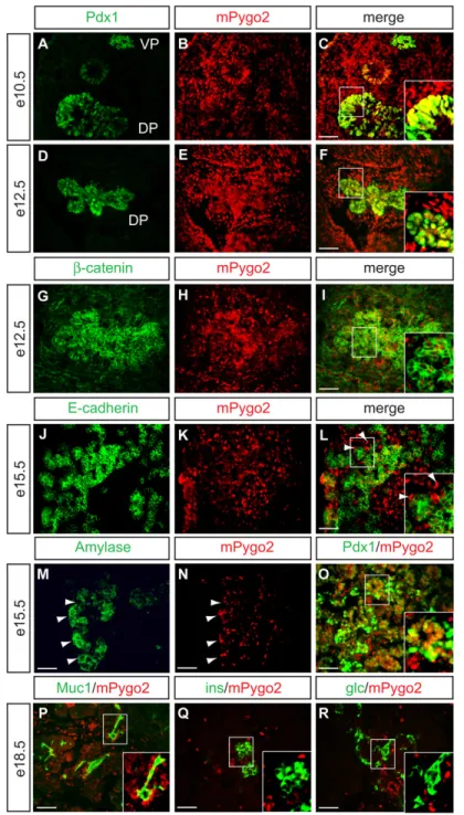
Figure 1. mPygo2 is expressed in the pancreatic epithelium and mesenchyme
Immunofluorescence detection of mPygo2 protein (red) at e10.5 (A–C), e12.5 (D–I), e15.5 (J– O) and e18.5 (P–R). (A–C) At e10.5, mPygo2 (B and C) colocalizes with the progenitor cell marker Pdx1 (green in A and C) in both dorsal and ventral pancreatic anlage. (D–F) At e12.5, colocalization of mPygo2 (E,H) with Pdx1 (green in D,G) persists in pancreatic epithelial cells. mPygo2 is also abundantly detected in the mesenchyme surrounding both pancreatic anlagen at e10.5 (B) and e12.5 (E). (G–I) β-catenin (green in G and I) is coexpressed with mPygo2 (H and I) in the epithelium and the mesenchyme. (J–L) At e15.5, most E-cadherin+ epithelial cells (green in J and L) still express mPygo2 (K and L). The interstitial mPygo2+, but E-cadherin-negative cells (arrowheads in L) suggest persistent mesenchymal expression of mPygo2 at
NIH-PA Author Manuscript
NIH-PA Author Manuscript
e15.5. (M,N) Immunostaining for amylase (green in M) and mPygo2 (red in N) on adjacent sections demonstrates localization of mPygo2 to the same cluster of cells that expresses amylase (arrowheads in M and N). (O) mPygo2 (red) and Pdx1 (green) are co-expressed in epithelial cell clusters that resemble forming exocrine acini (inset in O). (P–R) At e18.5, mPygo2 expression is restricted to mucin 1 (MUC1)+ ductal cells (inset in P) and interstitial cells, but absent from insulin+ (ins, inset in Q) or glucagon+ (glc, inset in R) cells. VP, ventral pancreas; DP, dorsal pancreas; scale bars = 50 µm.
NIH-PA Author Manuscript
NIH-PA Author Manuscript
Figure 2. Deletion of mPygo2 in mice results in pancreas hypoplasia
(A,B) Examination of gross pancreas morphology at e18.5 reveals a reduction of both the dorsal pancreas (DP) and the ventral pancreas (VP) in mPygo2-null mutants. The dashed yellow line indicates the separation between dorsal and ventral pancreas. (C) The morphological reduction in pancreas size in mPygo2−/− embryos correlates with a significant reduction in the ratio between pancreas weight and total body weight at e18.5 (p<0.05; n=7). (D,E) Hematoxylin and eosin staining of paraffin sections from control (D) or mPygo2−/− (E) pancreas reveals no alteration of global pancreas histology and normally developed endocrine and exocrine cell compartments. (F–H) At e15.5, mPygo2-deficient embryos show a similar reduction in pancreas size (F,G) and in pancreas weight to body weight ratio (H) as seen at e18.5 (p<0.05;
NIH-PA Author Manuscript
NIH-PA Author Manuscript
n=6). (I–N) The pancreatic epithelium was visualized by Pdx1 immunofluorescence staining (red in I,J and L,M) and the immunopositive area morphometrically quantified (K,N). The dorsal and ventral pancreatic epithelial area in mPygo2-null mutants is reduced at e13.5 (K; p<0.02 for ventral pancreas; not statistically significant for dorsal pancreas; n=5), but normal at e12.5 (N; n=3). Scale bar = 50 µm.
NIH-PA Author Manuscript
NIH-PA Author Manuscript
Figure 3. Pancreatic hypoplasia in mPygo2−/− mice correlates with decreased epithelial cell proliferation
(A–C) Immunofluorescence staining for glucagon (glc, red in A and B) and Pdx1 (green in A and B). Morphometric quantification of the number of glucagon+ cells in relation to the total area occupied by Pdx1+ pancreatic progenitor cells reveals no premature differentiation of glucagon+ cells in mPygo2−/− pancreas at e13.5 (C; n=3). (D–F) By contrast, quantification of the number of cells co-immunopositive for BrdU (red in D and E) and the progenitor cell marker Sox9 (green in D and E) reveals a ~20% decrease in the proliferative rate of ventral and dorsal pancreatic progenitor cells in mPygo2−/− mutants at e13.5 (F; p<0.01; n=5). Scale bar = 50 µm.
NIH-PA Author Manuscript
NIH-PA Author Manuscript
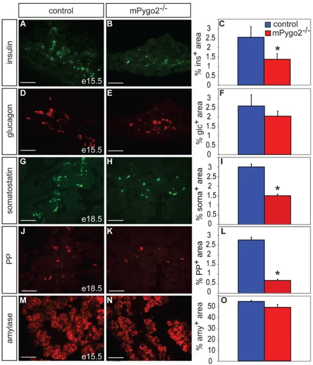
Figure 4. Loss of mPygo2 activity affects formation of late-born endocrine cells
(A–C) Insulin immunofluorescence staining (green in A and B) and morphometric
quantification of the insulin+ area over total pancreatic epithelial area at e15.5 reveals a 50% reduction of beta cells in mPygo2-deficient pancreas compared to control littermates (C; p<0.05; n=3). (D–F) Similar analysis for the glucagon+ (red in D and E) cell area at e15.5 shows no reduction in alpha cells (F; n=3). (G–L) The somatostatin+ (green in G and H) and pancreatic polypeptide+ areas (red in J and K) at e18.5 are similarly reduced as the insulin+ area (I, L; p<0.01; n=3). (M–O) The relative area occupied by amylase+ cells (red in M and N) is similar in e15.5 control and mPygo2-null mutant embryos (O; n=3). Scale bar = 50 µm.
NIH-PA Author Manuscript
NIH-PA Author Manuscript
Figure 5. Loss of mPygo2 activity affects formation of endocrine progenitors after e13
(A–C) Consistent with normal development of the exocrine compartment, mPygo2-null mutant embryos show no reduction in Ptf1a+ progenitor cells (red in A and B) at e13.5 (C; n=4). (D– F) By contrast, the relative number of Ngn3+ endocrine progenitor cells (red in D and E) is 50% reduced in mPygo2−/− embryos at e13.5 (F; p<0.01; n=5). Scale bar = 50 µm.
NIH-PA Author Manuscript
NIH-PA Author Manuscript
Figure 6. The canonical Wnt signaling pathway is predominantly active in the pancreatic mesenchyme at the time mPygo2 activity is required in pancreas development
X-gal staining of pancreata from Axin2+/lacZ (A–E) or BatGAL embryos (F–J) and subsequent immunoperoxidase detection of glucagon (glc, A–D and F–I) or insulin (ins, E and J) on paraffin sections. The dashed red line in A,B,F,G demarcates the outer margin of the pancreatic epithelium. At e10.5, β-galactosidase (β-gal) activity is predominantly found in the pancreatic epithelium, but occasional β-gal+ cells are also detected in the surrounding mesenchyme of both Axin2+/lacZ (A) and BatGAL embryos (F). Some overlap between β-gal and glucagon expression is observed (inset in A,B). At e13.5, mesenchymal cells are uniformly β-gal+ in
Axin2+/lacZ mice (B), while scattered β-gal activity is detected throughout the mesenchyme of
NIH-PA Author Manuscript
NIH-PA Author Manuscript
BatGAL mice (G). In both Axin2+/lacZ and BatGAL embryos, pancreatic epithelial cells are rarely β-gal+ at e13.5, and those that are occasionally express glucagons (inset in B,G). With the exception of occasional epithelial cells, some of which are glucagon/β-gal co-positive in
Axin2+/lacZ mice (inset in C,H), the predominantly mesenchymal location of β-gal activity persists at e15.5 in both reporter strains (C,H). At e18.5, β-gal activity is largely absent from glucagon+ (D,I) and insulin+ cells (E,J) and predominantly localized to the pancreatic ducts in both Axin2+/lacZ and BatGAL embryos (arrowheads in D,I). DP, dorsal pancreatic epithelium; i, islet. Scale bar = 50 µm.
NIH-PA Author Manuscript
NIH-PA Author Manuscript
Figure 7. mPygo2 deletion reduces transduction of canonical Wnt signals in the pancreas
Pancreatic sections from X-gal stained mPygo2+/+;BatGAL (A) or mPygo2−/− ;BatGAL (B) embryos at e12.5. mPygo2-deficiency results in absence of β-galactosidase (β-gal)+ cells in both the pancreatic epithelium and mesenchyme. The dashed red lines demarcate the outer margin of the pancreatic epithelium. Scale bar = 50 µm.
NIH-PA Author Manuscript
NIH-PA Author Manuscript
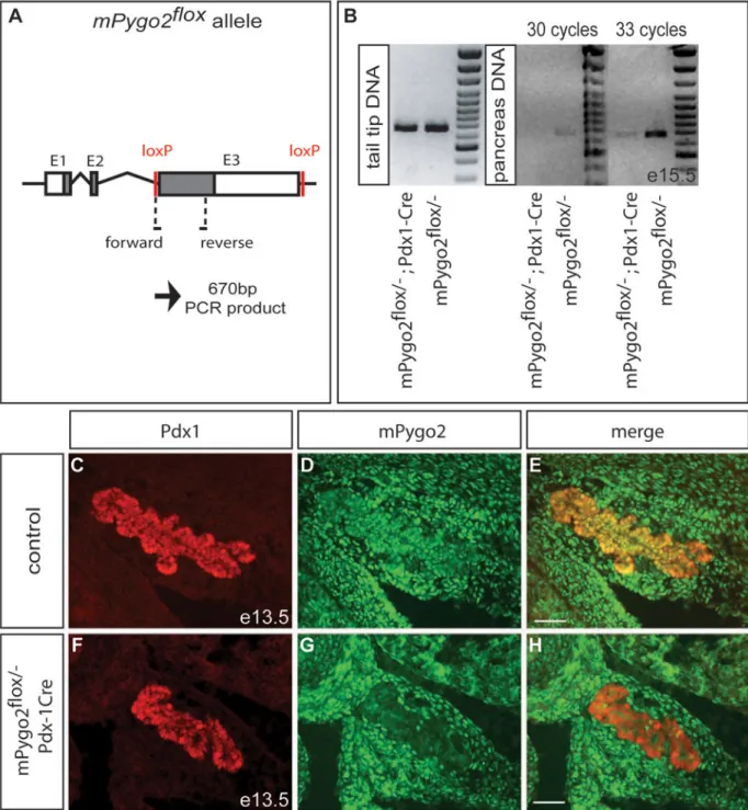
Figure 8. Almost complete absence of epithelial mPygo2 after Pdx1-Cre-mediated recombination of the mPygo2flox allele
(A) In mPygo2flox mice, exon 3 is flanked by loxP sites. Positions of the forward and reverse primers for detection of the unrecombined mPygo2flox allele are indicated. (B) The
unrecombined mPygo2flox allele is equally detected in tail tip DNA from mPygo2flox/− ;Pdx1-Cre and mPygo2flox/− mice. While also readily detectable in DNA from mPygo2flox/− pancreas after 30 PCR cycles, the unrecombined mPygo2flox allele is not amplified from
mPygo2flox/−;Pdx1-Cre pancreas at e15.5. The faint band amplified from mPygo2flox/− ;Pdx1-Cre pancreas after 33 PCR cycles most likely results from the presence of mesenchymal cells
in e15.5 pancreas that are not targeted by the Pdx1-Cre transgene, but can also be explained
NIH-PA Author Manuscript
NIH-PA Author Manuscript
by residual unrecombined epithelial cells. (C–H) Immunofluorescence detection of mPygo2 (green) and Pdx1 (red) on pancreas sections of mPygo2flox/−;Pdx1-Cre and control littermates
at e13.5. While mPygo2 is expressed in the epithelium and mesenchyme of control embryos, mPygo2 is selectively absent from the epithelium in mPygo2flox/−;Pdx1-Cre embryos.
Residual mPygo2+ cells are rarely observed in the epithelium. Scale bar = 50 µm.
NIH-PA Author Manuscript
NIH-PA Author Manuscript
Figure 9. Normal pancreas development in mPygo2flox/−;Pdx1-Cre mice
(A,B) Examination of gross pancreas morphology at e18.5 reveals a normal sized dorsal pancreas (DP) and ventral pancreas (VP) in mPygo2flox/−;Pdx1-Cre (mPygo2Δpan/−) embryos.
The dashed yellow line indicates the separation between dorsal and ventral pancreas. (C) Likewise, the ratio between pancreas weight and total body weight is similar in
mPygo2Δpan/− and control embryos at e15.5 (n=5). (D–F) The pancreatic epithelium was visualized by Pdx1 immunofluorescence staining (red in D,E). Morphometric quantification shows normal dorsal and ventral pancreatic epithelial areas in mPygo2Δpan/− embryos at e13.5 (F; n=4). (G–I) Insulin immunofluorescence staining (green in G and H) and morphometric quantification of the insulin (ins)+ area over total pancreatic epithelial area at e15.5 reveals normal beta cell formation in mPygo2Δpan/− embryos (I; n=6). Scale bar = 50 µm.