HAL Id: hal-01824317
https://hal.archives-ouvertes.fr/hal-01824317
Submitted on 13 Apr 2020
HAL is a multi-disciplinary open access
archive for the deposit and dissemination of
sci-entific research documents, whether they are
pub-lished or not. The documents may come from
teaching and research institutions in France or
abroad, or from public or private research centers.
L’archive ouverte pluridisciplinaire HAL, est
destinée au dépôt et à la diffusion de documents
scientifiques de niveau recherche, publiés ou non,
émanant des établissements d’enseignement et de
recherche français ou étrangers, des laboratoires
publics ou privés.
Exercise-induced cardioprotection: a role for eNOS
uncoupling and NO metabolites
C. Farah, A. Kleindienst, G. Bolea, G. Meyer, S. Gayrard, B. Geny, P. Obert,
Olivier Cazorla, T. Sandré, Cyril Reboul
To cite this version:
C. Farah, A. Kleindienst, G. Bolea, G. Meyer, S. Gayrard, et al.. Exercise-induced cardioprotection:
a role for eNOS uncoupling and NO metabolites. Basic Research in Cardiology, Springer Verlag, 2013,
108 (6), �10.1007/s00395-013-0389-2�. �hal-01824317�
O R I G I N A L C O N T R I B U T I O N
Exercise-induced cardioprotection: a role for eNOS uncoupling
and NO metabolites
C. Farah•A. Kleindienst•G. Bolea •
G. Meyer•S. Gayrard•B. Geny•P. Obert•
O. Cazorla•S. Tanguy•Cyril Reboul
Received: 10 January 2013 / Revised: 8 July 2013 / Accepted: 20 September 2013 / Published online: 9 October 2013 ! Springer-Verlag Berlin Heidelberg 2013
Abstract Exercise is an efficient strategy for myocardial
protection against ischemia–reperfusion (IR) injury.
Although endothelial nitric oxide synthase (eNOS) is phosphorylated and activated during exercise, its role in exercise-induced cardioprotection remains unknown. This study investigated whether modulation of eNOS activation during IR could participate in the exercise-induced car-dioprotection against IR injury. Hearts isolated from sed-entary or exercised rats (5 weeks training) were perfused with a Langendorff apparatus and IR performed in the
presence or absence of NOS inhibitors [N-nitro-L-arginine
methyl ester, L-NAME or N5-(1-iminoethyl)-L-ornithine,
L-NIO] or tetrahydrobiopterin (BH4). Exercise training
protected hearts against IR injury and this effect was
abolished byL-NAME or by L-NIO treatment, indicating
that exercise-induced cardioprotection is eNOS dependent. However, a strong reduction of eNOS phosphorylation at
Ser1177 (eNOS-PSer1177) and of eNOS coupling during
early reperfusion was observed in hearts from exercised
rats (which showed higher eNOS-PSer1177 and eNOS
dimerization at baseline) in comparison to sedentary rats. Despite eNOS uncoupling, exercised hearts had more S-nitrosylated proteins after early reperfusion and also less nitro-oxidative stress, indexed by lower malondialdehyde content and protein nitrotyrosination compared to seden-tary hearts. Moreover, in exercised hearts, stabilization of
eNOS dimers by BH4treatment increased nitro-oxidative
stress and then abolished the exercise-induced cardiopro-tection, indicating that eNOS uncoupling during IR is required for exercise-induced myocardial cardioprotection. Based on these results, we hypothesize that in the hearts of exercised animals, eNOS uncoupling associated with the
improved myocardial antioxidant capacity prevents
excessive NO synthesis and limits the reaction between NO
and O2!- to form peroxynitrite (ONOO-), which is
cytotoxic.
Keywords Nitro-oxidative stress! Training !
Myocardial infarction! eNOS monomerization
Introduction
Exercise training protects the heart against ischemia–
reperfusion (IR) injuries [10,17,18,39]. The mechanisms
responsible for this cardioprotective effect remain unclear, although they could provide new targets to reduce heart vulnerability to IR injury. To date, exercise-induced car-dioprotection to IR injury is mainly explained by
improvement of the myocardial antioxidant status [23,39,
Electronic supplementary material The online version of this article (doi:10.1007/s00395-013-0389-2) contains supplementary material, which is available to authorized users.
C. Farah! A. Kleindienst ! G. Bolea ! G. Meyer ! S. Gayrard ! P. Obert! C. Reboul (&)
Laboratoire de Pharm-Ecologie Cardiovasculaire (EA4278), Faculty of Sciences, Avignon University, 33 rue Louis Pasteur, 84000 Avignon, France
e-mail: [email protected] G. Meyer! B. Geny
E.A. 3072, Fe´de´ration de Me´decine Translationelle, Faculty of Medicine, University of Strasbourg, Strasbourg, France O. Cazorla
INSERM U1046, Universite´ Montpellier 1, Universite´ Montpellier 2, 34295 Montpellier, France
S. Tanguy
TIMC PRETA, CNRS UMR 5525, Laboratoire Coeur and Nutrition, Grenoble University Joseph Fourrier, 38041 Grenoble, France
41] and calcium homeostasis [13, 17, 18, 50]. Chronic exercise also increases nitric oxide (NO) bioavailability by enhancing endothelial nitric oxide synthase (eNOS or
NOS3) expression [33,43] and/or its phosphorylation [21,
53, 54]. Different strategies that improve NO
bioavail-ability in the heart markedly reduced heart vulnerbioavail-ability to
IR [20, 29, 36]. More particularly, the eNOS signaling
pathway protects myocardium against IR injury [16,24,25,
42, 46]; however, the link between eNOS signaling and
exercise-induced cardioprotection during IR is underex-plored, although it may play an essential role as recently
suggested [5].
eNOS is a critical homodimeric enzyme that, in physiological conditions, consumes NADPH to synthesize
NO and L-citrulline from L-arginine and oxygen. eNOS
dimers are stabilized by the cofactor tetrahydrobiopterin
(BH4). The activity of eNOS is also modulated by
phosphorylation at multiple sites, particularly at Ser1177,
which is considered to be the main activation site of this
enzyme [15] and its phosphorylation is promoted by
exercise [3, 5]. Depletion or oxidation of BH4can result
in eNOS uncoupling, which generates superoxide anion
(O2!-) instead of NO. In addition, O2!-can react with local
NO to produce peroxynitrite (ONOO-) that has strong
cytotoxic activity [37]. Thus, eNOS uncoupling clearly
may play a major role in myocardial IR injury by increasing reactive oxygen species (ROS) production and
limiting NO availability [27]. In addition, many studies
have demonstrated attenuation of heart sensitivity to IR when the eNOS–NO pathway is stimulated, for instance,
through eNOS overexpression [4, 12], NO administration
[47] or BH4 supplementation [49, 52]. The pathway by
which NO could modulate heart sensitivity to IR remains controversial. Indeed, depending on the model used to protect the heart, some reported that NO-dependent
car-dioprotection is mainly related to NO/cGMP [22],
whereas others have shown that protein S-nitrosylation (SNO) is the key trigger of NO-dependent
cardioprotec-tion [45].
We thus investigated the role of eNOS activation in exercise-induced cardioprotection by evaluating the effects of exercise training on eNOS phosphorylation and dimer-ization during cardiac IR. Unexpectedly, we found that eNOS was markedly uncoupled during early reperfusion in hearts from exercised rats but without consequence on NO bioavailability. Especially, we reported here that pharma-cological stabilization of eNOS dimers during early reperfusion was not associated with improved NO
bio-availability, but increased the formation of ONOO- and
then abolished exercise-induced cardioprotection. The major result of the present work is that exercise-induced cardioprotection is mainly dependent on eNOS uncoupling during early reperfusion.
Methods
Detailed information on the methodology is available in the online data supplement.
Animals and myocardial IR protocol
All investigations complied with the Guide for the Care and Use of Laboratory Animals published by the US National Institutes of Health (NIH Publications No. 85-23, revised 1996) and with the regulations of the French Ministry of Agriculture. All experiments were approved by the local research ethics committee (Comite´ Re´gional d’Ethique, no: 84.004).
Wistar male rats (Charles River Laboratories, France) (12-week old; 382 ± 38 g) were randomly assigned to the sedentary (Sed) or exercised (Ex) group. Rats in the exer-cised group were trained on a motor driven treadmill at 70 % of the maximal aerobic velocity (25 m/min; 45 min/ day), 5 days/week for 5 weeks. Such a training program has previously been reported by our team to increase citrate synthase activity of the soleus muscle (35.2 ± 4.7 IU/g wet weight in sedentary rats and 62.4 ± 7.9 IU/g wet
weight in exercised rats; p\ 0.01) [19]. In addition,
Doppler echocardiography was performed to evaluate the effects of exercise training on cardiac adaptations. Exercise training increase left ventricular end diastolic diameters
(LVEDd, Sed 5.84 mm, Ex 6.36 mm, p\ 0.05) and peak
velocities of early left ventricular inflow (Peak E, Sed
59.5 cm/s, Ex 68.3 cm/s, p\ 0.05), suggesting that such
training program improves diastolic function. This corre-sponds to a common feature of adaptations to exercise. 24 h after the end of the training period, hearts were iso-lated and frozen in liquid nitrogen for biochemical mea-surements or mounted on a Langendorff apparatus as
previously described [13]. Briefly, animals were
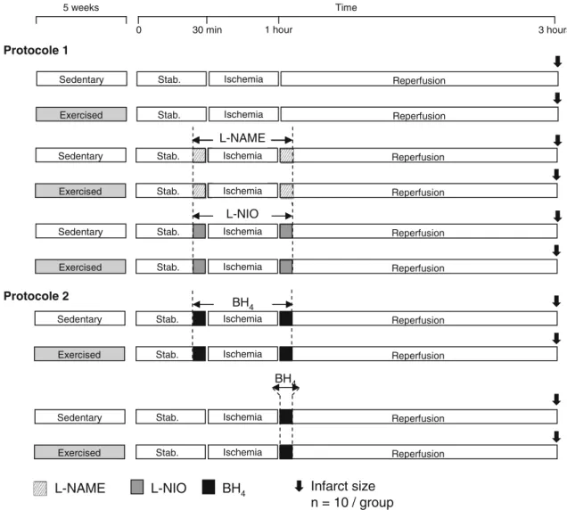
anesthe-tized (sodium pentobarbital, 100 mg/kg, i.p.), a thoracot-omy was performed and hearts were retrogradely perfused using a Langendorff apparatus. Hearts were paced at a rate of 300 beats/min (low voltage stimulator, BSL MP35 SS58L, 3 V) and a non-compliant balloon was inserted into the left ventricle (LV) to monitor the LV pressure. Hearts were stabilized for 30 min and then subjected to global no-flow ischemia for 30 min followed by 120 min of reper-fusion. During the entire IR procedure, cardiac functional parameters, such as LV developed pressure (LVDP), were recorded (MP35, BioPac System Inc) and coronary efflu-ents were collected to evaluate coronary blood flow. At the end of the experiments, the infarct size was assessed by triphenyltetrazolium chloride (TTC) staining. In protocol 1
(Fig.1), to assess the involvement of eNOS in
exercise-induced cardioprotection, hearts were perfused with
inhibitor, or with 10lM N5-(1-iminoethyl)-L-ornithine
(L-NIO), a more specific inhibitor of eNOS, 5 min before
and after ischemia. In protocol 2 (Fig.1), to evaluate the
role of eNOS uncoupling in exercise-induced
cardiopro-tection, hearts were perfused with 50lM BH4, an eNOS
coupling agent, 5 min before and after ischemia or only during the first 5 min of post-ischemic reperfusion. At the end of the ischemic period (ischemia) or after 10 min of
post-ischemic reperfusion (reperfusion), hearts were
quickly removed, the right ventricle cleaned and LV samples frozen in liquid nitrogen for biochemical analysis.
Biochemical assays
Measurement of nitrite
Quantification of nitrite in LV tissue lysates was carried out with the high-sensitivity nitrite assay kit
(Measure-iTTM High-Sensitivity Nitrite Assay Kit; InvitrogenTM)
and was performed according to the manufacturer’s instructions.
Measurement of cGMP
Quantification of total cGMP in LV tissue lysates (prepared in 0.1 M HCl) was carried out by enzyme immunoassay kit (Enzo Life Sciences) and was performed according to the manufacturer’s instructions.
Western blot analysis
Proteins from LV homogenates were separated on poly-acrylamide-SDS gels and transferred onto PVDF
mem-branes. Membranes were incubated with primary
antibodies at 4"C in 10 % milk or 3 % bovine serum
albumin in Tris-buffered saline containing 0.05 %
Stab. Ischemia Reperfusion
5 weeks Time
0 30 min 1 hour 3 hours
L-NAME Infarct size
n = 10 / group
BH4
Sedentary
Exercised Stab. Ischemia Reperfusion
Stab. Ischemia Reperfusion Sedentary
Exercised Stab. Ischemia Reperfusion
Stab. Ischemia Reperfusion Sedentary
Exercised Stab. Ischemia Reperfusion Stab. Ischemia Reperfusion Sedentary
Exercised Stab. Ischemia Reperfusion
L-NAME
Protocole 1
Protocole 2 BH
4
BH4
Stab. Ischemia Reperfusion Sedentary
Exercised Stab. Ischemia Reperfusion
L-NIO
L-NIO
Fig. 1 Schematic illustration of the experimental protocol.L-NAME, N-nitro-L-arginine methyl ester; L-NIO, N5-(1-iminoethyl)-L -orni-thine; BH4, tetrahydrobiopterin. Hatched (L-NAME), gray (L-NIO)
or black (BH4) boxes indicate when isolated hearts were perfused with L-NAME,L-NIO or BH4(for 5 min before and/or after the ischemic period) during ischemia–reperfusion using a Langendorff apparatus
Tween-20 overnight. The primary antibodies used in this study were: anti-mouse eNOS (1:1,000; BD Transduction
Laboratory), anti-mouse eNOS-PSer1177 (1:1,000; BD
Transduction Laboratory), anti-rabbit GAPDH (1:3,000; Santa Cruz) and anti-rabbit nitrotyrosine (1:20,000; Millipore Corporation). Immunodetection was carried out
using ECL or ECL Plus system (SuperSignal# West Pico
Chemiluminescence Substrate, Thermo Scientific;
Lumi-nata TM Forte Western HRP substrate, Millipore
Corpo-ration, respectively) and membranes were then exposed to X-ray films for revelation. Protein content was expressed relative to the GAPDH content. To evaluate the eNOS dimer/monomer ratio, SDS-resistant eNOS dimers were detected using non-denaturing conditions and low-temperature SDS-PAGE.
Measurement of protein S-nitrosylation
S-nitrosylation was determined by the modified S-nitro-sylation switch assay that uses a non-biological iodoTMT Reagent for labeling (Pierce S-nitrosylation Western blot kit, Thermo Fischer Scientific). Briefly, unmodified cys-teines were first blocked with a sulfhydryl-reactive com-pound. S-nitrosylated cysteines were then selectively reduced with ascorbate in HENS Buffer for specific labeling with iodoTMTzero Reagents, which irreversibly bind to the cysteine thiol that has been S-nitrosylated. Proteins from LV homogenates were then separated on polyacrylamide-SDS gels and transferred onto PVDF membranes. Detection of the TMT reagent-modified pro-teins was then performed using an anti-TMT antibody (Thermo Fischer Scientific). Immunodetection was carried out using ECL Plus system (Thermo Scientific; Luminata
TM
Forte Western HRP substrate, Millipore Corporation) and membranes were then exposed to X-ray films for revelation.
Measurement of total and NOS-dependent ROS generation
ROS production was measured by electron paramagnetic resonance (EPR) in fresh frozen LV homogenates as
described [35]. Briefly, homogenates were treated with
1 mM 1-hydroxy-3-methoxycarbonyl-2,2,5,5-tetramethyl-pyrrolidine solution (1:1 v/v), put in the EPR glass capillary tube (Noxygen Science Transfer & Diagnostics, Germany), and were placed inside the e-scan spectrom-eter (Bruker, Germany) for data acquisition. The proce-dure was then repeated on the same samples but in the
presence of L-NAME (3.10-4M). The difference
between the two conditions was the NOS-dependent ROS production. ROS production was normalized to the protein content of each sample and then expressed in lmol/min/mg.
Measurement of lipid peroxidation
Malondialdehyde (MDA), an end product of lipid peroxi-dation, was measured by dot blot analysis as previously
described [1]. Briefly, homogenized tissues were spotted
onto nitrocellulose membranes and incubated with the anti-rabbit MDA (1:500; Millipore Corporation) antibody at
4 "C overnight. Signal was revealed as described for
Western blotting. MDA content was expressed relative to Ponceau S staining.
Measurement of myocardial total antioxidant capacity
To evaluate the heart total antioxidant capacity, tissues were homogenized in PBS (137 mM NaCl, 24 mM KCl,
19 mM Na2HPO4, 18 mM KH2OP4; pH 7) at 4 "C.
Homogenates were centrifuged at 3,000g at 4"C for
10 min and the total antioxidant capacity was determined with the Total Antioxidant Power Kit (TAO2, Oxford Biomedical Research) and was expressed in mM of Trolox.
Statistical analysis
Data were expressed as the mean ± SEM. For comparison of multiple experimental conditions, analysis of variance (ANOVA) or repeated ANOVA were used, followed by the Bonferroni adjusted t test. For assessing the difference between values, the Student’s t test was used. A value of
p\ 0.05 was considered statistically significant.
Results
Implication of eNOS in exercise-induced cardioprotection against IR injury
Exercise training is classically reported to affect myocardial
NO synthesis, mainly by increasing eNOS expression [28] or
phosphorylation at Ser1177 [5,21]. In our model, exercise
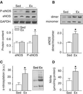
training had no effect on eNOS expression, but significantly
increased the baseline level of eNOS-PSer1177 (Fig.2a). In
addition, exercise training increased the eNOS dimer/mono-mer ratio, indicating an increase of eNOS coupling in the hearts from exercised rats in comparison to sedentary animals
(Fig.2b). Chronic exercise did not affect the expression
pat-tern of neuronal NOS and inducible NOS (online Fig. 1) in
accordance with previous studies [5,9]. In addition, exercise
increased the level of NO metabolites storage as indicated by the higher protein S-nitrosylation forming S-nitrosothiols
(SNO) (Fig.2c) and the amount of nitrite (Fig.2d) in
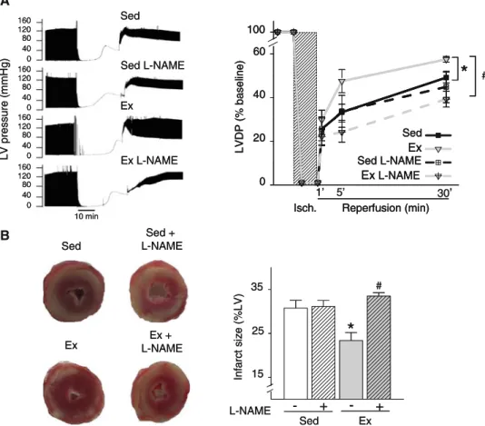
exer-cised hearts. As expected, chronic exercise protected the heart against IR injury as indicated by the higher recovery of
in exercised than in sedentary rat hearts. To investigate the involvement of the NOS signaling pathway in the cardiopro-tective effects of exercise, hearts were perfused with either the
NOS inhibitorL-NAME or the more specific eNOS inhibitor
L-NIO before and after ischemia (Fig.1, protocol 1). In the
presence of L-NAME, post-ischemic LVDP recovery and
infarct size in hearts from exercised animals were comparable to those observed in hearts from sedentary rats, which were not
affected by exposure toL-NAME (Fig.3a, b). Interestingly,
the same results were obtained with the use ofL-NIO (online
Fig. 2). Thus, these results indicate that the cardioprotective effects of exercise against IR are NOS dependent and may be mediated by eNOS, whose activation was altered by exercise.
eNOS role in exercise-induced cardioprotection
Time course of eNOS phosphorylation and coupling during IR
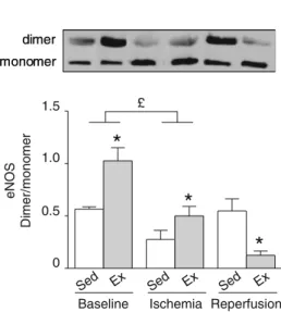
We next evaluated eNOS activation during IR in sedentary and exercised animals. Ischemia (30 min) reduced the level
of eNOS-PSer1177in both exercised (-88 %) and sedentary
(-57 %) hearts (Fig. 4a). However, after 10 min of
reperfusion, eNOS-PSer1177 was restored to the baseline
level in sedentary animals, but not in exercised hearts.
Thus, while the level of eNOS-PSer1177 was higher in
exercised hearts before ischemia, it was strongly reduced
during IR in comparison to sedentary animals (Fig.4a). No
modification of eNOS expression was observed in both
sedentary and exercised hearts (Fig. 4a).
Many studies have suggested that eNOS uncoupling has a major role in IR injuries because uncoupled eNOS
pro-duces O2!- instead of NO [8, 31]. We thus investigated
whether exercise could protect myocardium during IR by limiting eNOS uncoupling. To this aim, we evaluated eNOS coupling by measuring the eNOS dimer/monomer ratio during IR. Before ischemia, the eNOS dimer/mono-mer ratio was higher in exercised hearts than in sedentary
hearts (Fig.4b). Ischemia decreased the eNOS dimer/
monomer ratio in both groups by about 51 %, although the ratio values remained higher in exercised than in sedentary
hearts (Fig.4b). After 10 min of reperfusion, eNOS dimer/
monomer ratio was restored to baseline level in sedentary hearts, whereas it was further reduced in exercised hearts and then was significantly lower than that in sedentary
hearts (Fig.4b).
Altogether, these findings indicate that exercise-induced
cardioprotection is associated with reduced eNOS-PSer1177
and increased eNOS uncoupling and consequently reduced eNOS activation during early reperfusion. This may affect the amount of NO and ROS produced.
NO and nitro-oxidative stress during reperfusion
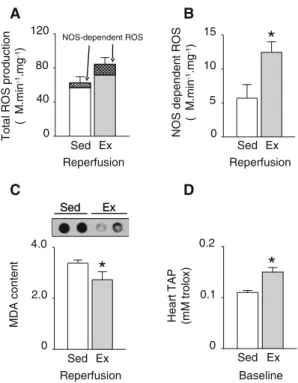
As eNOS uncoupling increases ROS production [51], we
first tested whether this phenomenon can modulate oxida-tive stress in exercised hearts during early reperfusion. After 10 min of reperfusion, despite no difference of total ROS production between sedentary and exercised hearts
(Fig.5a), the contribution of NOS-dependent ROS
pro-duction was increased in exercised hearts (Fig.5a, b). This
result further suggests eNOS uncoupling after early reperfusion in exercised hearts. Potential oxidative stress damages were assessed by measuring the content of MDA, an end product of lipid peroxidation. Despite eNOS uncoupling, exercised hearts had lower MDA content than
sedentary hearts after reperfusion (Fig.5c). This lower
myocardial oxidative stress was associated with increased myocardial total antioxidant power (TAP) in exercised
hearts (Fig.5d).
Considering that eNOS uncoupling is also associated with reduced NO production, we next evaluated the bio-availability of NO during IR in the presence of a recoupling
agent (BH4). For this purpose, some hearts were perfused
*
Sed Ex 0 20 40 60 N itr ite (µ m ol /m g pr ot ei n)*
)*
SedEx s-ni trosy lation (a .u .) 10 20 30 0 Sed Ex*
SedEx A*
Protein conten t 0 0.3 0.9 Ex Sed eNOS P-eNOS 0.6 Ex Sed P-eNOS eNOS GAPDH Ex Sed*
Ex Sed 0 1.0 2.0 eN OS di mer /mono mer dimer monomer*
B D CFig. 2 Exercise training increases eNOS–NO pathway. a Effects of exercise on eNOS expression and eNOS phosphorylation at Ser1177 (P-eNOS) analyzed by Western blotting in hearts from sedentary (Sed) and exercised (Ex) rats. eNOS and P-eNOS levels were relative to the expression values of GAPDH and total eNOS, respectively. b Effect of exercise on eNOS dimer/monomer ratio. eNOS dimers were detected by SDS-PAGE at low temperature and in non-denaturing conditions. c Effects of exercise on left ventricle protein S-nitrosylation. d Effects of exercise on nitrite concentration in the left ventricle
with BH4before ischemia and/or during early reperfusion
(protocol 2, Fig.1). To ensure that this pharmacological
strategy could not be considered as a preconditioning strategy, we freeze clamped control rat hearts at the end of
the BH4perfusion procedure (5 min, 50lM) and measured
nitrite concentration in LV homogenates. No effect of BH4
perfusion on nitrite concentration was observed (online Fig. 3). However, this treatment induced eNOS coupling as indicated by the higher eNOS dimer/monomer ratio in treated exercised hearts at the end of the ischemic period
(Fig.6a) and during early reperfusion (Fig.6b). In
addi-tion, the eNOS dimer/monomer ratio in BH4-treated
exer-cised hearts was restored to the level of sedentary hearts
during early reperfusion (Fig.6b). We next tested whether
eNOS uncoupling and recoupling modulate NO availability during early reperfusion. In the heart, NO acts either by activation of soluble guanylyl cyclase, to increase the conversion of GTP to cGMP, or directly by protein
S-nitrosylation to generate SNO. We measured the level of cGMP, nitrite and SNO in heart homogenates frozen after 10 min of reperfusion. Despite eNOS uncoupling, cGMP
(Fig.6c) was not altered and nitrite tended to be lower
(p = 0.075, Fig.6d) in exercised hearts when compared to
sedentary hearts. However, SNO remained significantly
higher when compared to sedentary hearts (Fig.6e). In
addition, we observed that L-NAME infusion during
ischemia–reperfusion markedly reduced nitrite and nitrate
(NOx) in coronary effluents of only sedentary hearts (online
Fig. 4). The result strongly suggests that despite eNOS uncoupling, NO availability is not altered and that NO synthesis during early reperfusion in exercised hearts is mainly eNOS independent. Then, we tested whether the
recoupling agent BH4 could improve NO availability in
exercised hearts during early reperfusion. Interestingly, in
exercised hearts, the levels of cGMP (Fig.6c), nitrite
(Fig.6d) and SNO (Fig.6e) during early reperfusion were
LV pres sure ( m mHg) 160 120 40 0 80 160 120 40 0 80 160 120 40 0 80 160 120 40 0 80 10 min Ex Sed Sed L-NAME Ex L-NAME LV pres sure ( m mHg) 160 120 40 0 80 160 120 40 0 80 160 120 40 0 80 160 120 40 0 80 10 min Ex Sed Sed L-NAME Ex L-NAME Ex
Sed L-NAMESed +
Ex + L-NAME Ex
Sed L-NAMESed +
Ex + L-NAME In fa rc ts iz e ( % LV ) Sed Ex L-NAME - + - + 35 25 15
*
# In fa rc ts iz e ( % LV ) Sed Ex L-NAME - + - + 35 25 15*
# Sed Ex Sed L-NAME Ex L-NAME 0 20 40 60 100 LV DP ( % basel ine ) Isch. 1’ 5’ 30’ Reperfusion (min)*
# Sed Ex Sed L-NAME Ex L-NAME 0 20 40 60 100 LV DP ( % basel ine ) Isch. 1’ 5’ 30’ Reperfusion (min)*
# Sed Ex Sed L-NAME Ex L-NAME 0 20 40 60 100 LV DP ( % basel ine )Isch. Reperfusion (min)
*
#A
B
Fig. 3 Implication of eNOS in exercise-induced cardioprotection. a Left panel Left ventricular (LV) pressure traces in hearts mounted on a Langendorff apparatus, perfused or not withL-NAME during the last 5 min of stabilization and the first 5 min of reperfusion after 30 min of total global ischemia. Right panel Left ventricular developed pressure (LVDP) in hearts from sedentary and exercised rats, perfused or not withL-NAME. LVDP is expressed as percentage of the baseline value. b Left panel Heart sections stained with
triphenyltetrazolium chloride (TTC) to show the infarct size after 30 min of total global ischemia and 120 min of reperfusion. Right panel Infarct size in hearts from exercised and sedentary rats, treated or not with L-NAME as described in a, following ischemia– reperfusion. Infarct size is expressed as percentage of the left ventricular section area. Values are the mean ± SEM. *p\ 0.05 vs. sedentary group;#p\ 0.05 vs. homolog group treated with
not altered in the presence of BH4, indicative of unchanged
NO availability. In pro-oxidant conditions such as ische-mia–reperfusion, NO can react with oxidants to form potent nitroso-oxidant species such as peroxynitrite
(ONOO-). Then, we measured the amount of protein
ni-trotyrosination, which is an index of peroxynitrite
forma-tion, derived from the reaction between NO and O2!-, and is
considered an index of nitro-oxidative stress. The amount of protein nitrotyrosination at early reperfusion was lower
in exercised than in sedentary hearts (Fig.6f), suggesting
that in exercised hearts, the formation of peroxynitrite is
reduced during early reperfusion. In the presence of BH4,
protein nitrotyrosination was significantly increased in exercised hearts and was then normalized to sedentary
hearts level (Fig.6f).
Role of eNOS uncoupling in exercise-induced cardioprotection
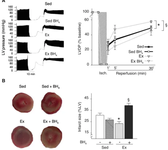
Finally, we tested whether eNOS uncoupling was required for the cardioprotective effects of chronic exercise.
Main-tenance of eNOS coupling during reperfusion in BH4
-treated exercised hearts caused the total loss of the car-dioprotective effects of exercise, as indicated by their
reduced post-ischemia LVDP recovery (Fig.7a) and
increased infarct size (by 60 %) (Fig.7b). Similar results
were obtained when hearts were treated with BH4 only
during reperfusion (online Fig. 6). Conversely, the infarct
size tended to be lower in BH4-treated than in untreated
sedentary hearts, but the difference did not reach statistical significance. These data suggest that the exercise-induced cardioprotective effects require eNOS uncoupling during IR injury.
Discussion
Recently eNOS activation has been linked to the
cardio-protective effects of voluntary exercise [5]. In the present
study, we confirm that eNOS is a major player in the mechanism of exercise-induced cardioprotection and show that, during IR, such cardioprotective effects requires eNOS uncoupling.
In accordance with previous works [5, 21, 53], we
confirmed that exercise training increases baseline eNOS-PSer1177and eNOS dimerization. Both modifications lead to eNOS activation and this might, in part, explain the finding that exercise training increases NO metabolites storage,
such as nitrite and SNO [5], which is also obvious in our
work. Modulation of eNOS activity is also dependent on
L-arginine availability. However, exercise was previously
reported to have no effect neither on L-arginine
concen-tration [44] nor on its influx into the cells [32]. Increased
eNOS-PSer1177 is an efficient way to protect the
myocar-dium [2] and is an essential trigger of exercise-induced
cardioprotection [5]. Accordingly, here we show that eNOS
plays a major role in exercise-induced cardioprotection
because eNOS inhibition (byL-NAME or more specifically
Reperfusion Ischemia
Baseline
Sed Ex Sed Ex Sed Ex
P-eNOS/eNO S 1.0 0 0.5 1.5
*
*
*
& £ Reperfusion Ischemia BaselineSed Ex Sed Ex Sed Ex
eNOS Di mer/mono me r 1.0 0 0.5 1.5
*
*
*
£ P-eNOS eNOS GAPDH dimer monomer P-A B eNOS eNOS GAPDH dimer monomerFig. 4 Reduced eNOS phosphorylation at Ser1177
and eNOS dimer-ization during ischemia–reperfusion in exercised hearts. a Time course of eNOS phosphorylation at Ser1177(P-eNOS) in hearts from sedentary (Sed) and exercised (Ex) rats during ischemia–reperfusion (baseline = stabilization, ischemia = after 30 min of ischemia, reperfusion = after 10 min of post-ischemic reperfusion) analyzed by Western blotting. eNOS is expressed relative to GAPDH content
and P-eNOS relative to eNOS total content. b Time course of eNOS dimer/monomer ratio in hearts from sedentary and exercised rats during ischemia–reperfusion analyzed by detecting SDS-resistant eNOS dimers using low-temperature SDS-PAGE. Values are the mean ± SEM. *p\ 0.05 vs. sedentary group.£p\ 0.05 vs. baseline, and p\ 0.05 sedentary ischemia vs. sedentary reperfusion
by L-NIO) during IR abolished the protective effects of exercise training on heart sensitivity to IR, whereas no effect of eNOS inhibition was observed in sedentary hearts. However, the role of eNOS activation modulation (i.e., phosphorylation and coupling) during IR in such cardio-protective effect was never investigated.
Here, we show that eNOS-PSer1177 and eNOS coupling
strongly decrease in exercised heart during ischemia and also during early reperfusion. As increased eNOS coupling is considered to have beneficial consequences in
patho-logical states [31,34,48] and that exercise is an efficient
strategy to protect the myocardium [13,39,40,50], eNOS
uncoupling in exercised myocardium during reperfusion was unexpected. Then, to evaluate whether this phenome-non could play a role in exercise-induced cardioprotection or was only a collateral damage of reperfusion without consequences on heart vulnerability to IR, we perfused
hearts with BH4. An essential point of this work is that
pharmacological maintenance of eNOS coupling in
exer-cised hearts perfused with BH4before and after ischemia or
during reperfusion only (online Fig. 5) was associated with total loss of exercise-induced cardioprotection, as indicated by the increased infarct size and reduced myocardial function. This result clearly shows that eNOS uncoupling is required for exercise-induced lower heart sensitivity to IR. In our work, ischemia induced eNOS uncoupling in both sedentary and exercised hearts. This may be due to the fact
that ischemia strongly reduces BH4 content and this
reduction is proportional to the duration of the blood flow
restriction [11]. In addition, Chen et al. [6] proposed
recently that increased eNOS phosphorylation at Ser1177
could favor eNOS uncoupling during stress, which could notably be obvious in the post-ischemic heart. To test this hypothesis in our model, sedentary rats received a single bolus of epinephrine prior IR (online Fig. 6A) to rapidly
increase, in a b-adrenergic receptors-dependent manner,
eNOS-PSer1177(online Fig. 6B), mimicking then the effects
of exercise on eNOS pathway [5]. Epinephrine increased
the level of eNOS-PSer1177, but had no effect on eNOS
coupling during early reperfusion (online Fig. 6D). This suggests that eNOS uncoupling at reperfusion is an exer-cise-dependent mechanism, in which the increase of eNOS-PSer1177 before IR is not directly involved. Then, how exercise decreased eNOS coupling at early reperfusion
remains to be investigated. In addition, b-adrenergic
receptors-dependent increase of eNOS-PSer1177 performed
on sedentary animals was previously reported to reduce
heart sensitivity to IR [2,24], which is also obvious in our
work (online Fig. 6C). Nonetheless, we can note that
increased eNOS-PSer1177 induced by epinephrine was
maintained at early reperfusion (online Fig. 6E), whereas it was clearly reduced in exercised hearts. Taken together, these results mainly suggest that eNOS uncoupling and
decreased eNOS-PSer1177 at early reperfusion constitute
specific exercise-dependent mechanisms, and that
cate-cholamine-dependent increase of eNOS-PSer1177 is not
sufficient to mimic them.
Classically, eNOS uncoupling results in NO decrease
[27]. In the present study, eNOS uncoupling increased after
reperfusion without affecting NO bioavailability as indi-cated by the higher SNO and unchanged cGMP content in exercised hearts. Considering that during myocardial
ischemia, NO originates mostly from NO metabolites [30],
we propose that the higher NO metabolites content, pro-duced in exercised hearts and measured in the present study, constitutes a storage for NO that will be available during ischemia and reperfusion. A previous report using genetic mouse model reached similar conclusions that increased NO metabolites storage by exercise in baseline plays a major role in the exercise-induced cardioprotection against IR, without exploring NO homeostasis during IR
To ta lR O S pr od uc tio n (M .m in -1.m g -1) Sed Ex NOS-dependent ROS NOS dependen tROS ( M .m in -1.m g -1) Sed Ex MDA co nt en t Sed Ex Ex Sed Ex Sed Heart T AP (m M trolo x) Sed Ex 40 80 120 0 5 10 15
*
0*
0 2.0 4.0*
0 0.1 0.2 Reperfusion Reperfusion Reperfusion Baseline A B C DFig. 5 eNOS uncoupling is associated with increased NOS-depen-dent ROS production, but reduced ROS-induced cell damages during early reperfusion in exercised rat hearts. a Total ROS production measured by electron paramagnetic resonance (EPR) in fresh frozen LV homogenates of sedentary (Sed) and exercised (Ex) hearts after 10 min of post-ischemic reperfusion. b NOS-dependent ROS pro-duction measured by electron paramagnetic resonance (EPR) in fresh frozen LV homogenates. Values represented correspond to the difference between ROS measurements obtained in the presence or absence ofL-NAME. c Malondialdehyde (MDA) content, used as an index of lipid peroxidation, measured by dot blot after 10 min of post-ischemic reperfusion in hearts from sedentary and exercised rats. d Heart total antioxidant power (TAP) measured in homogenates of sedentary and exercised hearts. TAP is expressed in mM of Trolox equivalent antioxidant capacity. Values are the mean ± SEM. *p\ 0.05 vs. sedentary group
[5]. In addition, such NO metabolites may contribute, during ischemia and early reperfusion, to S-nitrosylation of specific targets, which constitute essential triggers for
cardioprotection [7].
In our model, despite eNOS uncoupling and increased NOS-dependent ROS production, total ROS production slightly increased in exercised hearts without reaching statistical significance. In addition, oxidative stress dam-ages were markedly reduced in exercised hearts as indi-cated by lower lipid peroxidation after reperfusion compared with sedentary hearts. This could be explained by the previously described beneficial effect of exercise on
the antioxidant capacity [17,23] that was confirmed in the
present study (higher total antioxidant capacity in exercised than in sedentary hearts). In addition, increased eNOS-PSer1177, which normally enhances eNOS-derived NO production, increases eNOS-derived ROS production when
eNOS is uncoupled [6]. Therefore, the decrease in
eNOS-PSer1177observed during IR in exercised rat hearts could be beneficial by limiting the amount of eNOS-dependent ROS production. During IR, the protective or detrimental effect of NO may largely depend on the concentration NO and
O2!- and their ratio, promoting a pathological
concentra-tions of ONOO- or not [14]. In our work, eNOS
uncou-pling, by limiting excessive amount of NO synthesis during early reperfusion could contribute, in pro-oxidant
C F E § 0 1 2 eNOS dimer/mono mer Ex Ex +BH4 3 Ischemia dimer monomer Sed Ex Ex+B H4 Sed § 0 1 2 eNOS dimer/mono mer Ex Ex +BH4 3 Ischemia dimer monomer Sed Ex Ex+B H4 Sed § 0 1 2 eNOS dimer/mono mer 3 Reperfusion dimer monomer Ex Ex+B H4 Sed
*
Ex Ex +BH4 Sed § 0 1 2 eNOS dimer/mono mer 3 Reperfusion dimer monomer Ex Ex+B H4 Sed*
Ex Ex +BH4 Sed Protein conten t(a .u .) 170 26 S-nitrosylation 10 0*
20 M.W. (kDa) Ex Ex +BH4 Sed Ex Ex +BH4 Sed Protein conten t(a .u .) 170 26 S-nitrosylation 10 0*
20 M.W. (kDa) Ex Ex +BH4 Sed Ex Ex +BH4 Sed § 170 72 Protei n content (a .u .) Nitrotyrosination Reperfusion Reperfusion 5 0*
10 Ex Ex +BH4 Sed Ex Ex +BH4 Sed M.W. (kDa) § 170 72 Protei n content (a .u .) Nitrotyrosination 5 0*
10 Ex Ex +BH4 Sed Ex Ex +BH4 Sed M.W. (kDa) cG M P (p m ol /m g pr ot ei n) 2 0 4 Ex Ex +BH4 Sed Reperfusion cG M P (p m ol /m g pr ot ei n) 2 0 4 Ex Ex +BH4 Sed Reperfusion 0 Ex Ex +BH4 Sed Reperfusion Ni trit e (µ mol/mg pr ot ein) 50 100 150 200 0 Ex Ex +BH4 Sed Reperfusion Ni trit e (µ mol/mg pr ot ein) 50 100 150 200 D A BFig. 6 S-nitrosylation remains higher in exercised rat hearts during early reperfusion despite eNOS uncoupling. eNOS recoupling with BH4is not able to improve NO bioavailability, but significantly increases protein nitrotyrosination. a Effect of BH4on eNOS dimer/ monomer ratio in hearts from exercised rats, measured at the end of the ischemic period. BH4 was perfused during the last 5 min of stabilization. b Effects of BH4on eNOS dimer/ monomer ratio in hearts from exercised, measured after 10 min of post-ischemic reperfusion. BH4was perfused during the last 5 min of stabilization and the first 5 min of reperfusion. c Effects of exercise on cGMP
concentration measured after 10 min of post-ischemic reperfusion in hearts perfused or not with BH4. d Effects of exercise on nitrite concentration measured after 10 min of post-ischemic reperfusion in hearts perfused or not with BH4. e Effects of exercise on protein S-nitrosylation measured by Western blot, using the switch assay TMT method, after 10 min of post-ischemic reperfusion in hearts perfused or not with BH4. f Effects of exercise on protein
nitrotyrosination measured by Western blot after 10 min of post-ischemic reperfusion in hearts perfused or not with BH4. Values are the mean ± SEM. *p\ 0.05 vs. sedentary group; §p\ 0.05 vs. homolog group treated with BH4
conditions such as post-ischemic reperfusion, to prevent
the toxic reaction between NO and O2!-to form the potent
cytotoxic ONOO-. Indeed, such cardiac phenotype was
associated with lower protein nitrotyrosination, suggesting
that ONOO- formation was reduced in exercised hearts
during IR. Interestingly, the recoupling of eNOS with BH4
had no beneficial or detrimental effects on NO bioavail-ability, since both SNO and cGMP were not altered in exercised hearts by this pharmacological strategy. How-ever, a main point of this work is that in the presence of
BH4, protein nitrotyrosination was markedly increased in
exercised hearts and reached the level observed in seden-tary rats. This could also contribute to maintain a high level
of SNO in exercised hearts perfused with BH4, since
ONOO-also reacts with thiols to form SNO [14].
Alto-gether, the results strongly suggest that, during early reperfusion, excessive level of NO produced by eNOS
rapidly reacts with O2!- to form ONOO- and then
aggravates nitro-oxidative stress. In summary, in exercise-induced cardioprotection, eNOS uncoupling and eNOS
dephosphorylation at Ser1177 during early reperfusion in
association with increased antioxidant capacity to buffer ROS may contribute to prevent the reaction between NO
and O2!-to form ONOO-.
Finally, because eNOS inhibition by L-NAME or by
L-NIO abolished the exercise cardioprotective effects, we
cannot ignore the potential contribution of eNOS-depen-dent ROS production in the activation of classical
ROS-dependent anti-apoptotic pathways [38]; however, further
studies will be needed to investigate this aspect.
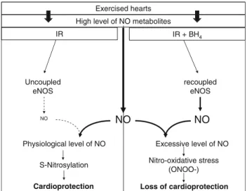
In conclusion, we show here that heart preconditioning by exercise is not the result of a simple increase of eNOS-PSer1177 by a catecholamine-dependent pathway, but of a complex interplay of NO metabolites storage, heart anti-oxidant capacity and eNOS uncoupling at the early stage of
reperfusion (Fig.8). Indeed, despite that eNOS recoupling
A
*
§ Sed Ex Sed BH4 ExBH4 0 20 40 60 100 LVDP (% baseline) Isch. 1’ 5’ 30’ Reperfusion (min) In fa rc ts iz e (%LV) Sed Ex BH4 - + - + 35 25 15*
§ 45 10 min LV pr es sur e (m m H g) 160 120 40 0 80 160 120 40 0 80 160 120 40 0 80 160 120 40 0 80 Ex Sed Sed BH4 Ex BH4 10 min LV pr es sur e (m m H g) 160 120 40 0 80 160 120 40 0 80 160 120 40 0 80 160 120 40 0 80 Ex Sed Sed BH4 Ex BH4 Sed Sed + BH4 Ex + BH4 Ex Sed Sed + BH4 Ex + BH4 Ex BFig. 7 Exercise training-induced cardioprotection requires eNOS uncoupling at early reperfusion. a Left panel left ventricular (LV) pressure traces in hearts mounted on a Langendorff apparatus, perfused or not with BH4during the last 5 min of stabilization and the first 5 min of reperfusion after 30 min of total global ischemia. Right panel left ventricular developed pressure (LVDP) in hearts from sedentary and exercised rats, perfused or not with BH4. LVDP is expressed as percentage of the baseline value. b Left panel heart
sections stained with triphenyltetrazolium chloride (TTC) to show the infarct size after 30 min of total global ischemia and 120 min of reperfusion. Right panel infarct size in hearts from exercised and sedentary rats, treated or not with BH4as described in a, following ischemia–reperfusion. Infarct size is expressed as percentage of the left ventricular section area. Values are the mean ± SEM. *p\ 0.05 vs. sedentary group;§p\ 0.05 vs. homolog group treated with BH4
by BH4 supplementation during ischemia has been
pro-posed as a potential therapeutic approach to improve post-ischemic functional recovery, in the present study we show
that BH4perfusion has no beneficial effects on LV
func-tional recovery in sedentary animals and only a very modest, non-significant effect on the infarct size.
More-over, we demonstrate that BH4 supplementation could
dramatically increase nitro-oxidative stress and then heart vulnerability to IR in exercised hearts as keeping eNOS uncoupling during ischemia–reperfusion could be benefi-cial in some populations. Although more research is needed to better understand how eNOS uncoupling occurs during ischemia–reperfusion, this finding opens new perspectives on the role of the eNOS–NO pathway and puts into
per-spective the potential therapeutic effects of BH4on cardiac
health. Our work might offer a new explanation on how exercise protects the myocardium against IR.
Study limitations
Endothelial NOS contributes to coronary blood flow reg-ulation and is highly expressed in coronary endothelial
cells. However, whether this single layer of cells contrib-utes to exercise-induced cardioprotection is not clear. In the present work, no effect of exercise on the recovery of coronary blood flow during IR was observed (online Table 1), suggesting that coronary endothelial function during IR is not altered by exercise. However, the method used by collecting of coronary effluents may not be sen-sitive enough to detect minor or moderate coronary blood flow changes. In addition, in this work, we did not take into
account the potential antioxidant properties of BH4 [26].
However, the lack of cardioprotective effect of BH4
per-fusion in sedentary rat hearts mainly suggests that, at concentration used in the present work, these effects are insignificant.
Conflict of interest On behalf of all authors, the corresponding author states that there is no conflict of interest.
References
1. Andre´ L, Gouzi F, Thireau J, Meyer G, Boissiere J, Delage M, Abdellaoui A, Feillet-Coudray C, Fouret G, Cristol JP, Lacam-pagne A, Obert P, Reboul C, Fauconnier J, Hayot M, Richard S, Cazorla O (2011) Carbon monoxide exposure enhances arrhyth-mia after cardiac stress: involvement of oxidative stress. Basic Res Cardiol 106:1235–1246. doi:10.1007/s00395-011-0211-y
2. Aragon JP, Condit ME, Bhushan S, Predmore BL, Patel SS, Grinsfelder DB, Gundewar S, Jha S, Calvert JW, Barouch LA, Lavu M, Wright HM, Lefer DJ (2011) Beta3-adrenoreceptor stimulation ameliorates myocardial ischemia–reperfusion injury via endothelial nitric oxide synthase and neuronal nitric oxide synthase activation. J Am Coll Cardiol 58:2683–2691. doi:10. 1016/j.jacc.2011.09.033
3. Balligand JL, Feron O, Dessy C (2009) eNOS activation by physical forces: from short-term regulation of contraction to chronic remodeling of cardiovascular tissues. Physiol Rev 89:481–534. doi:10.1152/physrev.00042.2007
4. Brunner F, Maier R, Andrew P, Wolkart G, Zechner R, Mayer B (2003) Attenuation of myocardial ischemia/reperfusion injury in mice with myocyte-specific overexpression of endothelial nitric oxide synthase. Cardiovasc Res 57:55–62. doi: 10.1016/S0008-6363(02)00649-1
5. Calvert JW, Condit ME, Aragon JP, Nicholson CK, Moody BF, Hood RL, Sindler AL, Gundewar S, Seals DR, Barouch LA, Lefer DJ (2011) Exercise protects against myocardial ischemia– reperfusion injury via stimulation of beta(3)-adrenergic receptors and increased nitric oxide signaling: role of nitrite and nitroso-thiols. Circ Res 108:1448–1458. doi:10.1161/CIRCRESAHA. 111.241117
6. Chen CA, Druhan LJ, Varadharaj S, Chen YR, Zweier JL (2008) Phosphorylation of endothelial nitric-oxide synthase regulates superoxide generation from the enzyme. J Biol Chem 283:27038–27047. doi:10.1074/jbc.M802269200
7. Chouchani ET, Methner C, Nadtochiy SM, Logan A, Pell VR, Ding S, James AM, Cocheme HM, Reinhold J, Lilley KS, Par-tridge L, Fearnley IM, Robinson AJ, Hartley RC, Smith RAJ, Krieg T, Brookes PS, Murphy MP (2013) Cardioprotection by S-nitrosation of a cysteine switch on mitochondrial complex I. Nat Med 19:753–759. doi:10.1038/nm.3212
Uncoupled eNOS recoupled eNOS IR Excessive level of NO Physiological level of NO S-Nitrosylation Cardioprotection Nitro-oxidative stress (ONOO-) Loss of cardioprotection Exercised hearts NO NO
High level of NO metabolites IR + BH4
NO
Fig. 8 Proposed mechanism for the role of eNOS uncoupling in exercise-induced cardioprotection. Exercise increased NO metabo-lites (SNO and nitrites) storage in hearts. During early post-ischemic reperfusion, eNOS of exercised hearts is uncoupled, but NO availability is maintained at high level (increased SNO and no alteration of cGMP concentration), suggesting that NO synthesis is mainly dependent of NO metabolites and is independent of NOS activity. This, associated with the higher capacity of exercised hearts to buffer ROS, results in lower nitro-oxidative stress, higher protein S-nitrosylation, and then cardioprotection. When a recoupling agent (BH4) is perfused in exercised hearts during ischemia and reperfusion, eNOS is recoupled, which could result in excessive production of NO. However, in pro-oxidant conditions such as ischemia–reperfusion, this excessive NO level does not contribute to increase NO availability (since SNO as well as cGMP are not increased in the presence of BH4), but increase nitro-oxidative stress, suggesting that excessive NO rapidly reacts with O2!-to form ONOO-and thus the cardioprotection is lost
8. Crabtree MJ, Tatham AL, Al-Wakeel Y, Warrick N, Hale AB, Cai S, Channon KM, Alp NJ (2009) Quantitative regulation of intracellular endothelial nitric-oxide synthase (eNOS) coupling by both tetrahydrobiopterin-eNOS stoichiometry and biopterin redox status: insights from cells with tet-regulated GTP cyclo-hydrolase I expression. J Biol Chem 284:1136–1144. doi:10. 1074/jbc.M805403200
9. de Waard MC, van Haperen R, Soullie T, Tempel D, de Crom R, Duncker DJ (2010) Beneficial effects of exercise training after myocardial infarction require full eNOS expression. J Mol Cell Cardiol 48:1041–1049. doi:10.1016/j.yjmcc.2010.02.005
10. Demirel HA, Powers SK, Zergeroglu MA, Shanely RA, Hamilton K, Coombes J, Naito H (2001) Short-term exercise improves myocardial tolerance to in vivo ischemia–reperfusion in the rat. J Appl Physiol 91:2205–2212
11. Dumitrescu C, Biondi R, Xia Y, Cardounel AJ, Druhan LJ, Ambrosio G, Zweier JL (2007) Myocardial ischemia results in tetrahydrobiopterin (BH4) oxidation with impaired endothelial function ameliorated by BH4. Proc Natl Acad Sci USA 104:15081–15086. doi:10.1073/pnas.0702986104
12. Elrod JW, Greer JJ, Bryan NS, Langston W, Szot JF, Gebregz-labher H, Janssens S, Feelisch M, Lefer DJ (2006) Cardiomyo-cyte-specific overexpression of NO synthase-3 protects against myocardial ischemia–reperfusion injury. Arterioscler Thromb Vasc Biol 26:1517–1523. doi:10.1161/01.ATV.0000224324. 52466.e6
13. Farah C, Meyer G, Andre L, Boissiere J, Gayrard S, Cazorla O, Richard S, Boucher F, Tanguy S, Obert P, Reboul C (2010) Moderate exercise prevents impaired Ca2?handling in heart of CO-exposed rat: implication for sensitivity to ischemia–reperfu-sion. Am J Physiol Heart Circ Physiol 299:H2076–H2081. doi:10.1152/ajpheart.00835.2010
14. Ferdinandy P, Schulz R (2003) Nitric oxide, superoxide, and peroxynitrite in myocardial ischaemia–reperfusion injury and preconditioning. Br J Pharmacol 138:532–543. doi:10.1038/sj. bjp.0705080
15. Fleming I (2010) Molecular mechanisms underlying the activa-tion of eNOS. Pflugers Arch 459:793–806. doi: 10.1007/s00424-009-0767-7
16. Frantz S, Adamek A, Fraccarollo D, Tillmanns J, Widder JD, Dienesch C, Schafer A, Podolskaya A, Held M, Ruetten H, Ertl G, Bauersachs J (2009) The eNOS enhancer AVE 9488: a novel cardioprotectant against ischemia reperfusion injury. Basic Res Cardiol 104:773–779. doi:10.1007/s00395-009-0041-3
17. French JP, Hamilton KL, Quindry JC, Lee Y, Upchurch PA, Powers SK (2008) Exercise-induced protection against myocar-dial apoptosis and necrosis: MnSOD, calcium-handling proteins, and calpain. FASEB J 22:2862–2871. doi:10.1096/fj.07-102541
18. French JP, Quindry JC, Falk DJ, Staib JL, Lee Y, Wang KK, Powers SK (2006) Ischemia–reperfusion-induced calpain activa-tion and SERCA-2a degradaactiva-tion are attenuated by exercise training and calpain inhibition. Am J Physiol Heart Circ Physiol 290:H128–H136. doi:10.1152/ajpheart.00739.2005
19. Goret L, Reboul C, Tanguy S, Dauzat M, Obert P (2005) Training does not affect the alteration in pulmonary artery vasoreactivity in pulmonary hypertensive rats. Eur J Pharmacol 527:121–128. doi:10.1016/j.ejphar.2005.10.013
20. Guo Y, Sanganalmath SK, Wu W, Zhu X, Huang Y, Tan W, Ildstad ST, Li Q, Bolli R (2012) Identification of inducible nitric oxide synthase in peripheral blood cells as a mediator of myo-cardial ischemia/reperfusion injury. Basic Res Cardiol 107:253. doi:10.1007/s00395-012-0253-9
21. Hambrecht R, Adams V, Erbs S, Linke A, Krankel N, Shu Y, Baither Y, Gielen S, Thiele H, Gummert JF, Mohr FW, Schuler G (2003) Regular physical activity improves endothelial function in patients with coronary artery disease by increasing
phosphorylation of endothelial nitric oxide synthase. Circulation 107:3152–3158. doi:10.1161/01.CIR.0000074229.93804.5C
22. Hamid SA, Totzeck M, Drexhage C, Thompson I, Fowkes RC, Rassaf T, Baxter GF (2010) Nitric oxide/cGMP signalling mediates the cardioprotective action of adrenomedullin in rep-erfused myocardium. Basic Res Cardiol 105:257–266. doi:10. 1007/s00395-009-0058-7
23. Hamilton KL, Staib JL, Phillips T, Hess A, Lennon SL, Powers SK (2003) Exercise, antioxidants, and HSP72: protection against myocardial ischemia/reperfusion. Free Radic Biol Med 34:800–809. doi:10.1016/S0891-5849(02)01431-4
24. Heusch G (2011) Beta3-adrenoceptor activation just says NO to myocardial reperfusion injury. J Am Coll Cardiol 58:2692–2694. doi:10.1016/j.jacc.2011.09.034
25. Heusch G, Boengler K, Schulz R (2008) Cardioprotection: nitric oxide, protein kinases, and mitochondria. Circulation 118:1915–1919. doi:10.1161/CIRCULATIONAHA.108.805242
26. Kojima S, Ona S, Iizuka I, Arai T, Mori H, Kubota K (1995) Antioxidative activity of 5,6,7,8-tetrahydrobiopterin and its inhibitory effect on paraquat-induced cell toxicity in cultured rat hepatocytes. Free Radic Res 23:419–430. doi:10.3109/1071576 9509065263
27. Landmesser U, Dikalov S, Price SR, McCann L, Fukai T, Hol-land SM, Mitch WE, Harrison DG (2003) Oxidation of tetrahy-drobiopterin leads to uncoupling of endothelial cell nitric oxide synthase in hypertension. J Clin Invest 111:1201–1209. doi:10. 1172/JCI200314172
28. Lauer N, Suvorava T, Ruther U, Jacob R, Meyer W, Harrison DG, Kojda G (2005) Critical involvement of hydrogen peroxide in exercise-induced up-regulation of endothelial NO synthase. Cardiovasc Res 65:254–262. doi:10.1016/j.cardiores.2004.09.010
29. Li Q, Guo Y, Wu WJ, Ou Q, Zhu X, Tan W, Yuan F, Chen N, Dawn B, Luo L, O’Brien E, Bolli R (2011) Gene transfer as a strategy to achieve permanent cardioprotection I: rAAV-mediated gene therapy with inducible nitric oxide synthase limits infarct size 1 year later without adverse functional consequences. Basic Res Cardiol 106:1355–1366. doi:10.1007/s00395-011-0207-7
30. Martin C, Schulz R, Post H, Boengler K, Kelm M, Kleinbongard P, Gres P, Skyschally A, Konietzka I, Heusch G (2007) Micro-dialysis-based analysis of interstitial NO in situ: NO synthase-independent NO formation during myocardial ischemia. Cardio-vasc Res 74:46–55. doi:10.1016/j.cardiores.2006.12.020
31. Masano T, Kawashima S, Toh R, Satomi-Kobayashi S, Shinohara M, Takaya T, Sasaki N, Takeda M, Tawa H, Yamashita T, Yo-koyama M, Hirata K (2008) Beneficial effects of exogenous tetrahydrobiopterin on left ventricular remodeling after myocar-dial infarction in rats: the possible role of oxidative stress caused by uncoupled endothelial nitric oxide synthase. Circ J 72:1512– 1519. doi:10.1253/circj.CJ-08-0072
32. Matsuura C, Brunini TM, Carvalho LC, Resende AC, Carvalho JJ, de Castro JP, Mendes-Ribeiro AC (2010) Exercise training in doxorubicin-induced heart failure: effects on theL-arginine-NO pathway and vascular reactivity. J Am Soc Hypertens 4:7–13. doi:10.1016/j.jash.2009.10.005
33. McAllister RM, Newcomer SC, Laughlin MH (2008) Vascular nitric oxide: effects of exercise training in animals. Appl Physiol Nutr Metab 33:173–178. doi:10.1139/H07-146
34. Moens AL, Kass DA (2006) Tetrahydrobiopterin and cardiovas-cular disease. Arterioscler Thromb Vasc Biol 26:2439–2444. doi:10.1161/01.ATV.0000243924.00970.cb
35. Niu X, Watts VL, Cingolani OH, Sivakumaran V, Leyton-Mange JS, Ellis CL, Miller KL, Vandegaer K, Bedja D, Gabrielson KL, Paolocci N, Kass DA, Barouch LA (2012) Cardioprotective effect of beta-3 adrenergic receptor agonism: role of neuronal nitric oxide synthase. J Am Coll Cardiol 59:1979–1987. doi:10.1016/j. jacc.2011.12.046
36. Obal D, Dai S, Keith R, Dimova N, Kingery J, Zheng YT, Zweier J, Velayutham M, Prabhu SD, Li Q, Conklin D, Yang D, Bhatnagar A, Bolli R, Rokosh G (2012) Cardiomyocyte-restricted overexpression of extracellular superoxide dismutase increases nitric oxide bioavailability and reduces infarct size after ische-mia/reperfusion. Basic Res Cardiol 107:305. doi: 10.1007/s00395-012-0305-1
37. Pacher P, Beckman JS, Liaudet L (2007) Nitric oxide and per-oxynitrite in health and disease. Physiol Rev 87:315–424. doi:10. 1152/physrev.00029.2006
38. Pagliaro P, Moro F, Tullio F, Perrelli MG, Penna C (2011) Cardioprotective pathways during reperfusion: focus on redox signaling and other modalities of cell signaling. Antioxid Redox Signal 14:833–850. doi:10.1089/ars2010.3245
39. Powers SK, Demirel HA, Vincent HK, Coombes JS, Naito H, Hamilton KL, Shanely RA, Jessup J (1998) Exercise training improves myocardial tolerance to in vivo ischemia–reperfusion in the rat. Am J Physiol 275:R1468–R1477
40. Powers SK, Quindry JC, Kavazis AN (2008) Exercise-induced cardioprotection against myocardial ischemia–reperfusion injury. Free Radic Biol Med 44:193–201. doi:10.1016/j.freeradbiomed. 2007.02.006
41. Ramires PR, Ji LL (2001) Glutathione supplementation and training increases myocardial resistance to ischemia–reperfusion in vivo. Am J Physiol Heart Circ Physiol 281:H679–H688 42. Schulz R, Kelm M, Heusch G (2004) Nitric oxide in myocardial
ischemia/reperfusion injury. Cardiovasc Res 61:402–413. doi:10. 1016/j.cardiores.2003.09.019
43. Sessa WC, Pritchard K, Seyedi N, Wang J, Hintze TH (1994) Chronic exercise in dogs increases coronary vascular nitric oxide production and endothelial cell nitric oxide synthase gene expression. Circ Res 74:349–353. doi:10.1161/01.RES.74.2.349
44. Sindler AL, Delp MD, Reyes R, Wu G, Muller-Delp JM (2009) Effects of ageing and exercise training on eNOS uncoupling in skeletal muscle resistance arterioles. J Physiol 587:3885–3897. doi:10.1113/jphysiol.2009.172221
45. Sun J, Aponte AM, Kohr MJ, Tong G, Steenbergen C, Murphy E (2013) Essential role of nitric oxide in acute ischemic precondi-tioning: S-nitros(yl)ation versus sGC/cGMP/PKG signaling? Free Radic Biol Med 54:105–112. doi:10.1016/j.freeradbiomed.2012. 09.005
46. Szelid Z, Pokreisz P, Liu X, Vermeersch P, Marsboom G, Gillijns H, Pellens M, Verbeken E, Van de Werf F, Collen D, Janssens SP
(2010) Cardioselective nitric oxide synthase 3 gene transfer protects against myocardial reperfusion injury. Basic Res Cardiol 105:169–179. doi:10.1007/s00395-009-0077-4
47. Takano H, Tang XL, Qiu Y, Guo Y, French BA, Bolli R (1998) Nitric oxide donors induce late preconditioning against myocar-dial stunning and infarction in conscious rabbits via an antioxi-dant-sensitive mechanism. Circ Res 83:73–84. doi:10.1161/01. RES.83.1.73
48. Tiefenbacher CP, Bleeke T, Vahl C, Amann K, Vogt A, Kubler W (2000) Endothelial dysfunction of coronary resistance arteries is improved by tetrahydrobiopterin in atherosclerosis. Circulation 102:2172–2179. doi:10.1161/01.CIR.102.18.2172
49. Verma S, Maitland A, Weisel RD, Fedak PW, Pomroy NC, Li SH, Mickle DA, Li RK, Rao V (2002) Novel cardioprotective effects of tetrahydrobiopterin after anoxia and reoxygenation: identifying cellular targets for pharmacologic manipulation. J Thorac Cardiovasc Surg 123:1074–1083. doi:10.1067/mtc. 2002.121687
50. Wisloff U, Loennechen JP, Currie S, Smith GL, Ellingsen O (2002) Aerobic exercise reduces cardiomyocyte hypertrophy and increases contractility, Ca2?sensitivity and SERCA-2 in rat after myocardial infarction. Cardiovasc Res 54:162–174. doi:10.1016/ S0008-6363(01)00565-X
51. Xia Y (2007) Superoxide generation from nitric oxide synthases. Antioxid Redox Signal 9:1773–1778. doi:10.1089/ars2007.1733
52. Yamashiro S, Noguchi K, Matsuzaki T, Miyagi K, Nakasone J, Sakanashi M, Koja K (2002) Beneficial effect of tetrahydrobi-opterin on ischemia–reperfusion injury in isolated perfused rat hearts. J Thorac Cardiovasc Surg 124:775–784. doi:10.1067/mtc. 2002.124393
53. Zhang QJ, McMillin SL, Tanner JM, Palionyte M, Abel ED, Symons JD (2009) Endothelial nitric oxide synthase phosphory-lation in treadmill-running mice: role of vascular signalling kinases. J Physiol 587:3911–3920. doi:10.1113/jphysiol.2009. 172916
54. Zhou M, Widmer RJ, Xie W, Jimmy Widmer A, Miller MW, Schroeder F, Parker JL, Heaps CL (2010) Effects of exercise training on cellular mechanisms of endothelial nitric oxide syn-thase regulation in coronary arteries after chronic occlusion. Am J Physiol Heart Circ Physiol 298:H1857–H1869. doi:10.1152/ ajpheart.00754.2009