HAL Id: tel-01452845
https://tel.archives-ouvertes.fr/tel-01452845
Submitted on 2 Feb 2017HAL is a multi-disciplinary open access archive for the deposit and dissemination of sci-entific research documents, whether they are pub-lished or not. The documents may come from
L’archive ouverte pluridisciplinaire HAL, est destinée au dépôt et à la diffusion de documents scientifiques de niveau recherche, publiés ou non, émanant des établissements d’enseignement et de
structural basis of kinesin motility
Luyan Cao
To cite this version:
Luyan Cao. structural basis of kinesin motility. Biomolecules [q-bio.BM]. Université Paris-Saclay, 2016. English. �NNT : 2016SACLS267�. �tel-01452845�
Université Paris-Saclay
Espace Technologique / Immeuble Discovery
2016SACLS267
T
HESE DE DOCTORAT
DE
L’U
NIVERSITE
P
ARIS
-S
ACLAY
PREPAREE A
L’
U
NIVERSITE
P
ARIS
-S
UD
E
COLED
OCTORALE N°
569
Innovation thérapeutique : du fondamental à l'appliqué
Spécialité de doctorat : Biochimie et biologie structurale
Par
Mlle LuYan CAO
Structural Basis Of Kinesin Motility
Thèse présentée et soutenue à Gif-sur-Yvette, le 27 Septembre 2016 : Composition du Jury :
M, MINARD, Philippe Professeur, Université Paris-Sud Président Mme, MOORES, Carolyn Professeur, Université de Londres Rapporteur M, PICOT, Daniel Directeur de recherche, Université Paris Diderot Rapporteur M, LIPPENS, Guy Directeur de recherche, LISBP Examinateur M, KNOSSOW, Marcel Directeur de recherche émérite, CNRS Directeur de thèse M, GIGANT, Benoît Chargé de recherche, CNRS Directeur de thèse
Acknowledgement
Many thanks to Prof. Philippe MINARD, Prof. Carolyn MOORES, M. Daniel PICOT, M. Guy LIPPENS, M. Marcel KNOSSOW and M. Benoît GIGANT for presenting as members of my thesis jury.
I would like to express my sincere thanks to M. Marcel KNOSSOW and M. Benoît GIGANT for their expert, sincere and invaluable guidance extended to me. Without their consistent and illuminating instruction, this thesis would not have reached its present form.
I am grateful to Valérie, Soraya, Weiyi, Shoeb and other members in our group. They provided their help and professional advice generously for this thesis. It’s my honor to join the group and work with you.
I also thank Prof. Chunguang WANG. She shares her experience and knowledge which benefits me greatly.
In this thesis, diffraction data were collected at SOLEIL synchrotron (PX1 and PX2 beam lines). I am most grateful to the machine and beam line groups for making these experiments possible.
I sincerely thank Mme. Marie-France CARLIER and M. Julian PERNIER for sharing their Stopped- flow machine and their advices.
Supports on initial crystallization screening were provided by Mme. Armelle VIGOUROUX. DSC experiment has been done with the help of Mme. Magali AUMONT. The IMAGIF platform offered their professional supports to this work as well. I thank the HTX lab in Grenoble sincerely for their technical supports on crystallization screening.
Thank you, my dear friends, Quyen, Loïc, Abbas, Kai, Roman, Pierre for your supports and encouragements.
Thank you, Florian, for being there.
感谢我亲爱的爸爸妈妈,奶奶,姐姐,姑父姑妈,以及其他远在中国的亲人。没有你们的 支持,也就不会有今天的我。
感谢建乔,周晗,毛毛,袁浩,和你们一起度过的那个夏天给了我希望和勇气。
Contents
Acknowledgement ... 1
Abreviations ... 4
Preface ... 6
Introduction ... 7
1. Microtubules and tubulin ... 7
1.1 Microtubules ... 7
1.2 Tubulin ... 9
1.3 DARPin ... 12
2. Kinesin in general ... 15
2.1 Classification of the kinesin superfamily ... 15
2.2 Conserved motor domain ... 17
2.3 Other cytoskeletal motors ... 20
3. Kinesin-1 ... 23
3.1 The kinetic mechanism of kinesin ... 24
3.2 Motility model of kinesin-1 ... 28
3.3 Advancement of kinesin-1 structural studies ... 32
4. Kinesin-14 ... 39
4.1 Kinesin-14 in general ... 39
4.2 Ncd ... 40
Objectives ... 45
Results and Discussion ... 46
1. Structure of apo-kinesin-1 bound to tubulin ... 46
1.1 Context ... 46
1.2 Paper: “The structure of apo-kinesin bound to tubulin links the nucleotide cycle to movement.” ... 48
1.3 Discussion ... 58
2. Structures of isolated apo-kinesin-1 ... 66
2.2 Paper: “The structural switch of nucleotide-free kinesin.” ... 67
2.3 Discussion ... 94
3. Crystallization of kinesin-14 in complex with tubulin-DARPin ... 102
3.1 Objectives ... 102
3.2 Constructs ... 106
3.3 Characterization ... 109
3.4 Crystallization... 114
3.5 Discussion ... 115
Conclusions and Perspectives ... 118
Materials and Methods ... 124
1. Differential Scanning Calorimetry assay on kinesin-1 and its mutants ... 124
2. Purification of Kinesin-14 heterodimer ... 124
3. Production of Kinesin-Tubulin-DARPin complex ... 131
4. Nucleotide state analysis ... 133
5. Spin down assay ... 133
Abreviations
2YT Yeast extract/bacto Tryptone medium LB Lysogeny Broth medium
DTT Dithiothreitol
EGTA Ethylene Glycol-bis(β-aminoethyl ether)-N,N,N',N'-Tetraacetic Acid EDTA EthyleneDiamineTetraacetic Acid
SDS-PAGE Sodium Dodecyl Sulfate-PolyAcrylamide Gel Electrophoresis Mes 2-(N-Morpholino) ethanesulfonic acid
Pipes Piperazine-N,N′-bis(2-ethanesulfonic acid)
Hepes 4-(2-Hydroxyethyl)-1-piperazineethanesulfonic acid PEG PolyEthylene Glycol
ADP Adenosine DiPhosphate
Mant-ADP 2'-(or-3')-O-(N-Methylanthraniloyl) Adenosine 5'-DiPhosphate ATP Adenosine TriPhosphate
AMPPNP Adenylyl-imidodiphosphate Pi Inorganic phosphate GDP Guanosine DiPhosphate GTP Guanosine TriPhosphate GMPCPP Guanosine-5’-[(α,β)-methyleno] triphosphate KD Dissociation constant kon Association rate koff Dissociation rate Ki Inhibitory constant
Km Michaelis constant
Ka Acid dissociation constant
EPR Electron Paramagnetic Resonance
FRET Fluorescence Resonance Energy Transfer
EM Electron Microscopy
P-loop Phosphate-binding loop TEV Tobacco Etch Virus
Preface
Microtubules are one of the main components of the eukaryotic cytoskeleton. Together with the actin-myosin system, and among other functions, they organize the intracellular transport, forming the filaments along which dyneins and kinesins travel. These motor proteins use the energy produced by ATP hydrolysis to walk along microtubule, carrying loads. The hallmark of kinesins is a relatively well-conserved motor domain, which comprises both the binding site for the nucleotide and the binding site for microtubule. Based on the position of the motor domain in the sequence, at the N-terminus, internal or at the terminus, kinesins can be divided into three subfamilies, N-type, internal and C-type, which are further divided into 14 classes according to differences in the motor domain sequence. N-type kinesins, including classes 1 to 12, move towards the plus end of microtubule, i.e. broadly speaking from the center to the periphery of interphase cells; whereas C-type kinesins, which kinesin-14 belongs to, move towards microtubule minus end. Kinesin-13s, internal kinesins, are not motile but depolymerize microtubule, showing that kinesins have roles beyond transport, for instance in the regulation of microtubule dynamics.
Kinesin binding to or dissociating from microtubules is tightly coupled with its nucleotide cycle. ADP-kinesin has a low affinity for microtubules and a low ADP dissociation rate. Binding to microtubules enhances ADP dissociation in kinesin and converts kinesin to a high affinity state for microtubules. Subsequent ATP binding triggers the conformational changes leading to the mechanical step. After ATP hydrolysis and Pi release, kinesin, back to ADP bound state, detaches from microtubules to complete the cycle. Although this general framework is well established, our understanding of several steps of this cycle is still incomplete, including how ADP dissociation is accelerated by microtubules and how ATP binding triggers the movement. Structurally, the conformation of ADP-kinesin detached from microtubule is known from X-ray crystallography whereas both crystal and electron microscopy structures have been determined for kinesin in an ATP state, bound to a microtubule (or to tubulin, which is the microtubule building block). What is missing are high resolution structural data on nucleotide-free kinesin, both isolated and as a complex with microtubules. These data should highlight the mechanism of ADP release from kinesin and complete our view of the kinesin structural cycle related to its mechanochemical cycle. This is the objective of my work.
Introduction
1. Microtubules and tubulin
In eukaryotic cells, combined with microfilaments and intermediate filaments, microtubules serve as cytoskeleton, providing cells with an inner framework, maintaining the shape of cells. Microtubules contribute to the structural formation of mitotic spindle, centrosome, cilia and flagella. Also, microtubules serve as a track for intercellular transport. Motor proteins, as kinesin and dynein, move along microtubules to transport cargoes. Microtubules are observed in all eukaryotes, playing important roles in different cellular functions including cell division, cell transport, morphogenesis etc.
1.1
Microtubules
Structure of microtubules
Microtubules have a cylinder-shaped structure with an external diameter of 24 nm. They are typically formed by 13 linear and parallel protofilaments that interact laterally. In vitro, they can consist of 9 to 16 protofilaments. Each of the protofilaments is composed of α/β tubulin heterodimers arranged head-to-tail (Fig. 1.1a), so in a protofilament, one end is an exposed α tubulin and the other is an exposed β tubulin. Because of the parallel arrangement of the protofilaments, microtubules are polarized: the end with the β subunit exposed is called the plus end; the other end with α tubulin exposed is called the minus end. Microtubules can be elongated at both the plus and minus ends, but it extends more rapidly at its plus end (Walker et al. 1988).
A standard microtubule with 13 protofilaments has a pitch of 3 monomers; consequently microtubule has a “seam”. Laterally, protofilaments contact each other with the same subunit (i.e. α tubulin to α tubulin, β tubulin to β tubulin), except the contacts in the “seam”, where α subunits are adjacent to neighbor β subunits (Fig. 1.1a).
Dynamics of microtubules
Dynamic instability of microtubules is studied as a foundation of cell physiology (Brouhard 2015). Figure 1.1b presents the dynamics of microtubule including several main processes: polymerization, depolymerization, catastrophe and rescue (Conde and Caceres 2009).
Figure 1.1 General information on microtubules. (Conde and Caceres 2009) a) A microtubule is usually composed of 13 protofilaments, templated by the γ-tubulin ring
complex. The straight arrow indicates the microtubule seam (where tubulins of adjacent protofilaments interact with a subunit of the other type).
b) The microtubule cycle.
In a tubulin heterodimer, each subunit binds a nucleotide, one of which is neither hydrolysable nor exchangeable GTP bound to α tubulin. Concerning the nucleotide in β tubulin, if it is a GTP, then the tubulin is called GTP-tubulin; if it is a GDP instead, the tubulin is called GDP-tubulin. That GTP-tubulins keep binding to microtubule ends is responsible for the growth of microtubules. This event is also called “polymerization”. The hydrolysis of the GTP molecule in GTP-tubulin, accompanying microtubule assembly, leads to the paradox that microtubules are composed mainly of GDP-tubulin, which tends to fall
off from the tip of microtubules. The paradox is resolved because of the delay between microtubule assembly and GTP hydrolysis, which leads to the formation of a “GTP-cap”, preventing microtubules from depolymerization at microtubule ends. When the GTP in the cap is hydrolyzed, microtubules will go through rapid shrinkage. The loss of the GTP-cap and its induced disassembly is called a “catastrophe”. It is observed by cryo-electron microscopy that protofilaments peel from the ends into ring-like structures during shrinkage (Mandelkow, Mandelkow, and Milligan 1991). GTP tubulin can also bind to the shrinking end to form a new cap preventing the microtubule from depolymerizing. The process that a microtubule stops shrinking and starts to grow is called a ‘rescue’. Microtubule growth and shrinkage is the basis of its functions.
1.2
Tubulin
Tubulin belongs to a protein family of several members sharing a conserved GTPase domain. There are seven different kinds of tubulin in eukaryotic cells: α, β, , , , and
(Oakley 2000)Among them, , , and tubulin are considered as rare tubulins and do not have a ubiquitous distribution in eukaryotic organisms (Dutcher 2001). The other tubulins are found in all eukaryotes. tubulin, localized primarily in centrosomes and spindle pole bodies, is associated with microtubules nucleation and polar orientation. α and β tubulin are always associated in an heterodimer which is the building block of microtubule in all eukaryotic cells. The tubulin talked about in this thesis refers to the α/β tubulin heterodimer.
Characters of tubulin heterodimers
The sequence of α and β tubulins is highly conserved across species. For example, the comparison of tubulin sequences from human, Drosophila, Chlamydomonas, Trypanosoma and Paramecium indicates that α tubulins are 89-95% similar and that β tubulins are 88–94% similar (Dutcher 2001).
α tubulins and β tubulins, both having a molecular weight about 50 000 Dalton, share 40% amino-acid sequence identity. The isoelectric point of these subunits is between 5.2 and 5.8. Each monomer consists of about 440 amino acids upon which post-translational modifications play an important role in its dynamics and its organizations (Song and Brady 2015).
In cells, α/β tubulin colliding with the end of a protofilament extends a microtubule. The higher the concentration of tubulin is, the more frequently these collisions occur, and therefore the microtubule grows faster. For a single protofilament, α/β tubulin binds to its ends rapidly (at about 4 μM−1 s−1), but it falls off as rapidly when there is no lateral bond
accompanied by GTP hydrolysis, though the hydrolysis of GTP is not obliged. With GMPCPP, a GTP non-hydrolysable analog, tubulin heterodimers can still form microtubules (Nogales and Wang 2006).
In solution, tubulin is instable and tends to form polymers. This process is GTP consuming and sensitive to the temperature. At 4°C, tubulins do not self-assemble and exist as heterodimers in the solution. When incubated at 37 °C with GTP and magnesium ions (Mg2+) in the solution, tubulin heterodimers polymerize to form microtubules. If the temperature is decreased, microtubules start to depolymerize. This behavior of tubulin is used to purify it by several cycles of polymerization and depolymerization but also gives trouble for tubulin crystallization. Protein crystallization usually requires high homogeneity of samples, while tubulin heterodimers tend to polymerize and form various polymers. As a result, tubulin crystallization without any stabilizer is very difficult. Actually, so far there is no tubulin heterodimer alone without stabilizer that has been reported to be crystallized.
Structural studies on tubulin
Tubulin can exist in three major forms: straight protofilaments, as found in microtubule, heterodimers and curved oligomers. Different approaches have been used to characterize them structurally. The first near-atomic model of tubulin was determined by electron crystallography (Nogales, Wolf, and Downing 1998). In the presence of zinc ion, tubulin assembles into two-dimensional sheets made of antiparallel protofilaments. These Zn-sheets have been studied by electron diffraction. More recently, thanks to the progress in Cryo-electron microscopy, the structure of microtubules has been determined at about 3.5 Å resolution (Zhang et al. 2015). The structure of non-microtubular tubulin has also been determined but by X-ray crystallography with different tubulin stabilizers. Our lab published a crystal structure of a complex of GDP tubulin, two tubulins arranged head to tail, with the stathmin-like domain of RB3, a stathmin family protein (Ravelli et al. 2004). Following this, more high resolution crystal structures of tubulin oligomers and heterodimers in complex with stabilizers have been determined (Nawrotek, Knossow, and Gigant 2011).
As expected from their ~40% sequence identity, α tubulin and β tubulin present a similar overall structure (Fig. 1.2). They are globular proteins with a diameter of about 4 nm. Each subunit consists of two interacting β sheets surrounded by α helices. The subunit can be divided into three structural domains, including the nucleotide binding domain, an intermediate domain and the C-terminal domain. The nucleotide binding domain, also referred to as the N-terminal domain, forms a Rossmann fold where parallel β strands alternate with α helices. The Rossmann fold is typical for nucleotide-binding proteins. The nucleotide binding site on α- tubulin is called N-site (Non-exchangeable site); the one on β- tubulin is called E-site (exchangeable site). The intermediate domain, containing the taxol
binding site, also has a β sheet surrounded by α helices. The C-terminal domain, consisting of 2 α helices contacting the previous domains, is exposed to the outside surface of the microtubule and is usually the target of microtubule associated proteins (MAPs) and motor proteins (Downing and Nogales 1998).
Figure 1.2 Structure of a tubulin heterodimer (Amos and Schlieper 2005) A tubulin heterodimer is colored based on different domains. The GTP binding domains (red) are responsible for nucleotide binding. The activation domain colored in blue provides the catalytic α-tubulin Glu 254 residue which participates in the hydrolysis of the GTP in the next β-tubulin along a protofilament. The core helix (yellow) connects the two globular domains in each monomer. The C-terminal domain on the external surface is colored in green.
The last C-terminal residues of each subunit are flexible and are usually not seen in the structures. They are enriched in acidic residues. This part is also where the most
the α subunit and the addition of glutamate chains (Wloga and Gaertig 2010). Nevertheless, these residues are removable by a protease called subtilisin which will reduces the total number of isotypes for tubulin consequently (Lobert and Correia 1992), so it’s likely that subtilisin treatment may improve the possibility of tubulin’s crystallization. For β subunit, there are more than one subtilisin site which have different accessibility to the protease corresponding to the heterodimer or polymer form of the tubulin.
Ligands of tubulin
Tubulin is the target of agents which can perturb microtubule dynamics with either a stabilizing or a destabilizing effect (Cormier et al. 2010). Some of these ligands are used clinically for cancer treatment. Different ligands target various sites in the tubulin heterodimer. Here I will present only two ligands of tubulin that I have used during my work either as a tubulin stabilizer for crystallization purpose, or as a microtubule stabilizer needed for kinetic studies.
Colchicine
Colchicine is originally extracted from plants of the genus Colchicum. It prevents tubulin heterodimer from polymerizing into microtubules. Structural studies identified the colchicine binding site on β tubulin; it is mostly buried in the β tubulin’s intermediate domain (Ravelli et al. 2004). So as a tubulin stabilizer, colchicine has probably no direct impact on kinesin or kinesin-like protein binding. It has been cheeked afterwards by gel filtration chromatography that with or without colchicine a peak of kinesin-tubulin-DARPin complex shows at a similar position.
Taxane
Taxanes are originally found in plants of the genus Taxus. Taxol and Taxotere are used widely as chemotherapy agents because of their ability to interfere with cell division by stabilizing microtubules. These compounds are used to generate stable microtubules, when they are bound to tubulin heterodimers in a one to one ratio. The taxol binding site was identified by electron crystallography (Nogales, Wolf, and Downing 1998) on Zn-sheet tubulin, as a hydrophobic pocket located also in β tubulin.
1.3
DARPin
DARPin is the abbreviation for ‘Designed Ankyrin Repeat ProteIN’ (Fig. 1.3). Anti-tubulin DARPins have been selected in the lab to interfere with microtubule assembly in a unique manner. The ideal DARPins used in this thesis should be able to bind tubulin stably with a ratio of one to one forming a tubulin DARPin trimer, while not preventing kinesins from binding to tubulin.
Figure 1.3 Structure of a DARPin named D1.
D1 is a typical selected DARPin, able to stabilize the tubulin heterodimer. It contains an N-cap (blue) and a C-cap (red), between which there are three internal ankyrin repeats. Its N-terminus also has six His (wheat) to facilitate purification.
Selection of DARPins
The DARPin library initially used for screening consists of DARPins having four or five ankyrin repeats. The N-terminal and the C-terminal ankyrin repeats stabilize the structure, between which there are two (N2C) or three (N3C) variable repeats. Tubulin was fixed on the surface of microtiter plates through its α subunit; consequently, its β subunit was exposed. The library was screened through four rounds of ribosome display. Then the cDNAs of selected DARPins were subcloned and clones producing tubulin binders were identified by ELISA on crude cell extracts (Pecqueur et al. 2012).
Several DARPins generating a strong signal during screening were expressed and purified. Because of the way tubulin was immobilized, the likelihood to obtain β subunit-specific DARPins was enhanced. However, the precise binding site remains unknown until the DARPin-tubulin complex structure is determined.
DARPin-tubulin complex
Several structures of DARPins in complex with tubulin heterodimer have been determined. D1-tubulin structure at 2.2 Å resolution has been determined (Pecqueur et al. 2012). D1, binding a tubulin heterodimer with a one to one ratio, has a high affinity for tubulin regardless the nucleotide state of the tubulin. Its dissociation equilibrium constant from GDP tubulin is about 120 nM; that from GTP tubulin is about 155 nM. The structure shows
tubulin from polymerizing. In D1-tubulin, the kinesin binding site located on the lateral surface of a heterodimer remains uncovered. Hence, D1 is likely a good candidate to be a crystallization chaperone for tubulin-MAP complexes, in particular tubulin-kinesin complexes.
Another anti-tubulin DARPin structure published is D2-tubulin in complex with kinesin and an ATP-analog at 3.2 Å (Gigant et al. 2013). It’s the first successful case where a DARPin is used to help crystallizing a tubulin-kinesin complex, in order to observe the conformational changes of kinesin when bound to tubulin. D2 binds to the β subunit’s longitudinal interface, with a different binding mode compared with D1 (Fig. 1.4), stabilizing tubulin. It’s proved that D2 doesn’t modify the interaction between kinesin and tubulin, because kinesin’s ATPase activities stimulated by tubulin or by D2-tubulin are similar with nearly identical catalytic constants and similar Km. In the structure, kinesin with an ATP analog binding tubulin fits the previous 8 Å Cryo-EM map where a microtubule was decorated with kinesin-AMPPNP. Details of this structure, especially of kinesin, will be presented in the kinesin-1 part of this introduction. This complex structure points to a promising way to generate tubulin-kinesin crystal structures using DARPins as tubulin stabilizers.
Figure 1.4 D1- and D2- tubulin structures. Tubulin heterodimers have been superimposed.
α tubulins (magenta) and β tubulins (blue) in both structures are almost identical with a low r.m.s.d of about 0.4 Å. Although D1 (yellow) and D2 (green) have a similar structure and both contact β tubulin, they don’t bind identically.
2.
Kinesin in general
Cytoskeletal motors play important roles in eukaryotic cells. They convert chemical energy into mechanical work and take nanometer steps along their tracks to transport a variety of cargos in the cytoplasm. Failures of motors can cause severe diseases or be lethal in some cases. Cytoskeletal motors are divided into three families: myosins, using actin filaments as tracks, dyneins and kinesins, both moving on microtubules to travel inside the cell (Schliwa and Woehlke 2003).
Kinesin superfamily proteins (KIFs) are reported to be able to transport organelles, protein complexes and mRNAs to specific destinations, using the energy produced by ATP hydrolysis. Thanks to them, the intracellular transport is spatially and temporally well controlled in cells. They also contribute to chromosomal and spindle movements during mitosis and meiosis (Hirokawa and Noda 2008).
KIFs share a conserved catalytic motor domain, also called head, containing an ATP binding site and a microtubule binding site. This part, where the chemical energy is converted into work, is responsible for ATP hydrolysis and kinesin’s directional motility. Beside from the motor domain, kinesin also have a quiet variable non-motor domain, usually containing a coiled-coil segment, known as “stalk” region and a globular “tail” region. The tail region has a wide variety of functions in different kinesins, including interacting with cargo, regulating the ATPase activity of the motor domain; containing ATP independent MT binding sites and targeting kinesins to different locations (Ovechkina and Wordeman 2003). In some kinesins, the “tail” doesn’t contact cargoes directly, but through light chains or associated protein instead (Hirokawa and Noda 2008).
2.1
Classification of the kinesin superfamily
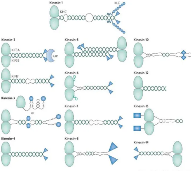
Different KIFs have various structures and functions. Whereas the motor domain sequence of kinesins is relatively conserved and is the hallmark of this protein superfamily, sequence differences have led to the classification of kinesins in 14 sub-families (Fig. 2.1). The organization of their polypeptide chains has also led to the definition of 3 types of kinesins (Hirokawa et al. 2009).
Figure 2.1 Members of the kinesin superfamily (Verhey and Hammond 2009). The motor domains are colored in light green. Kinesin-1-12s, whose motor domain is located at kinesin’s N-terminus, move toward the plus end of microtubule; Kinesin-14s, whose motor domain is located at its C-terminus, undergo minus end-directed motility; Kinesin-13s, whose motor domain is in the middle of the polypeptide sequence, don’t move along microtubule actively; they diffuse instead. Kinesin-6’s motor domain has a unique loop with approximately 65 amino acids insertion, colored in light green.
Many kinesins are oligomerized by their coiled-coil segments, colored in dark green. Most kinesins exist as dimers; some exist as either monomers or dimers (Kinesin-3 for example); some kinesins exist as tetramers (Kinesin-5 for example).
Non-motor domains, colored in dark blue, vary in kinesins. They are responsible for their isoform-specific regulation or functional properties or both. In addition to its-motor containing heavy chain, Kinesin-1 has a light chain contacting the C-terminal non-motor domain of the
dimerized heavy chain. The light chain allows Kinesin-1 to bind its cargoes indirectly. Some Kinesin-2s are heterotrimers, containing two motor subunits (KIF3A and KIF3B) and a Kinesin Associated Protein (KAP3). KAP3, either interacting with cargoes or regulating motor activity, contacts heterodimerized motor subunits through their non-motor tails (Marszalek and Goldstein 2000).
N-type kinesins, constituted of kinesins in classes 1 to 12, whose motor domains are located at the N-terminus of the sequence, have been found either to move towards the microtubule plus end or not to move. Most N-type kinesins, which move towards microtubule plus end, are processive. It means they are capable of taking hundreds of steps before dissociation. During the cycle of translocation, it’s required that kinesin keeps attaching to microtubule. In this case, there is at least one motor domain that always binds to the microtubule. This type of kinesin is believed to move along microtubule in a hand-over-hand manner. The details of its motility will be introduced later.
Kin-Is, for internal kinesins, all belonging to the 13th sub-family, are characterized by a motor domain that is internal to the polypeptide sequence. They hydrolyze ATP and use the energy to disassemble microtubules from both ends. This kind of kinesin doesn’t generate movement; instead, they reach microtubule ends, either by direct binding or by diffusing along microtubules. There, they either induce or stabilize a depolymerizing-favoring conformational change in GTP tubulin dimer. Kinetic analysis also suggests that some Kin-Is can depolymerize microtubules processively, removing approximately 20 tubulin dimers from the end of a protofilament one by one before dissociation (Hunter et al. 2003).
The third type, called type, which coincides with the kinesin-14 sub-family, has a C-terminal motor; these kinesins move towards the microtubule minus end. Some of them contain a non-ATP dependent microtubule binding site in their non-motor region so that they can cross link microtubules. When their motor domain hydrolyzing ATP generates a movement, they are able to slide one microtubule with respect to another. Moreover, some members of kinesin-14 are also reported to have microtubule destabilizing ability (Wordeman et al., 2003).
2.2
Conserved motor domain
Although kinesin superfamily has a large number of members with different functions, they all share a conserved motor domain which is responsible for nucleotide consumption and microtubule binding. Among various KIFs, the motor domains have amino acid sequence similarity of about 30-60%. The sequence alignment of some most studied members is presented in Fig. 2.2 (Sablin et al. 1996).
Four conserved nucleotide binding motifs are marked, named N-1 to N-4. N-1, also called the P-loop (Phosphate binding loop) motif, with the conserved sequence GQTxxGKS/T, interacts with the α and β phosphates of the nucleotide; N-2, known as the Switch1 motif, whose pattern is NxxSSR, can bind the phosphate of ATP; N-3, Switch 2 motif (DxxGxE), is supposed to contact the phosphate as well; N-4, with the conserved sequence RxRP located near the N terminus of the kinesin, interacts with the adenine ring of the nucleotide through hydrophobic interactions. These nucleotide associated motifs are also observed in myosin and some GTPase proteins (Sack, Kull, and Mandelkow 1999).
Figure 2.2 Sequence alignment of the motor domain of some kinesin family members (Sablin et al. 1996).
Residues with absolute conservation are marked in red; those with relatively lower (but still significant) conservative substitutions are marked in yellow. The positions of secondary structure elements are pointed, as well as the four nucleotide binding motifs (N-1 to N-4). Kinesins from different subfamilies and from different species are listed here as examples.
Figure 2.3 Structure of a rat-brain kinesin (Sack, Kull, and Mandelkow 1999). In kinesin, central strands (blue) are surrounded by α helices (pink). α helices associated with microtubule binding are colored in green; the nucleotide binding region (purple) and the ADP are shown.
As the motor domains of various members in the kinesin superfamily are conserved, they share a similar structure. A typical kinesin motor domain structure (pdb id 2kin, (Sack et al. 1997)) is presented in the Figure 2.3. Generally, the kinesin motor domain consists of a series β strands locating in the middle of the structure with three α helices on each side. The nucleotide binding pocket with loops where conserved nucleotide binding motifs embed is clearly identified because in most cases the kinesin is co-crystallized with ADP.
2.3
Other cytoskeletal motors
In addition to kinesin, there are two other cytoskeletal motor families: myosins which move along actin filaments and dyneins which move along microtubules but usually towards the minus end. All of three use ATP as the source of energy. Among them, kinesins and myosins share a structural homology and a similarity in their mechanochemistry. Despite their low sequence identity, the topologies and tertiary structure of kinesin and myosin indicate they may derive from a common ancestor (Rayment 1996). By contrast, dyneins deviate from kinesins and myosins in both energy generation mechanism and in overall structure (Bhabha et al. 2016). In this section, a brief introduction on both myosin and dynein will be given.
Myosin
Myosin, responsible for actin-based motility, plays important roles in muscle contraction and motility processes in eukaryotes. It was found in both striated muscle tissue and smooth muscle tissue by Pollard and Korn (Pollard and Korn 1973). Myosins constitute a large and divergent protein family. Usually, myosins are heteromers consisting of one or two heavy chains and a variable number of light chains. A general structure of myosin V is presented in figure 2.4. Myosin heavy chain includes motor (head), neck and tail domain (Maravillas-Montero and Santos-Argumedo 2012). The motor domain is responsible for both actin filament binding and ATP hydrolysis. The neck domain serves as a linker between the catalytic motor domain and the tail; also, it is able to act as a lever arm for force transduction. The neck domain usually contains the myosin light chain binding site. The tail domain plays various roles in different myosin subfamilies. Most myosins move from the actin pointed (minus) end to its barbed (plus) end, except myosin VI which moves in the reverse direction (Sweeney and Houdusse 2010).
The motor domain structures of various myosins have been determined in different nucleotide state. In general, the myosin motor domain can be divided into four subdomains which are able to generate nucleotide-state associated movement (Coureux et al. 2003). The motor domain in myosin superfamily shares a similar catalytic pocket with that of the kinesin superfamily, including three conserved nucleotide associated motifs: P-loop, Switch 1 and Switch 2. Moreover, the secondary structure elements of kinesin are also found in myosin with a central β sheet surrounded by several α helices (Fig. 2.5).
Figure 2.4 Domain structure of a myosin V (Hammer and Sellers 2012).
The head domain of myosin contains the nucleotide binding site and actin binding site. The neck domain is composed of α helices able to bind a calmodulin molecule. The tail domain consists of a coiled-coil stalk dimerizing the heavy chains, followed by two globular tail domains (GTDs). GTDs are responsible for cargo binding.
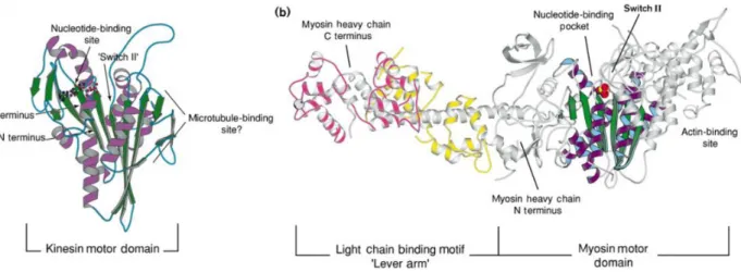
Figure 2.5 Comparison of the motor domains in kinesin and myosin (Rayment 1996). a) The kinesin motor domain. Central β strands are colored in green, with α helices
(purple) surrounding. ADP is shown in balls and sticks.
b) A subfragment of a chicken skeletal myosin in nucleotide-free state. A solvent (space-filling model in red and yellow) replacing ADP locates at the nucleotide binding site. In
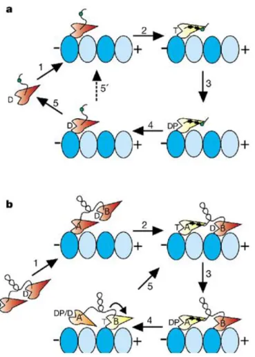
Figure 2.6 Processive movement of myosin Va motor (Hammer and Sellers 2012).
In myosin, movement corresponds to the nucleotide cycle of motor domains (Vale and Milligan 2000). The cycle alternates through high and low affinity for actin. Usually, the myosin motor domain in nucleotide-free state or ADP state is able to bind actin tightly; while in ATP or ADP and inorganic phosphate (Pi) state it dissociates from actin easily. Here, the processive movement of a myosin Va, corresponding to its nucleotide cycle, is presented as an example (Fig. 2.6). Myosin Va dwells in a state with both motor domain binding ADP attaching to the actin filament. The two motors exert intramolecular strain on each other. ADP releases from the trailing motor so that this motor is able to bind ATP.
After ATP binding, the dissociation of this motor occurs and so does ATP hydrolysis; meanwhile, the actin binding motor undergoes a power stroke, leading the dissociated motor, turning to its ADP-Pi state, to its forward binding site. Rebinding to actin filament, the ADP-Pi motor releases its Pi quickly and attaches to actin tightly. The cycle resets to the beginning and restarts all over again with myosin Va moving forward about 36 nm in each cycle.
Dynein
Cytoplasmic dynein belongs to the AAA (ATPases Associated with diverse cellular Activites) family. Although it is able to move along microtubules using the energy produced by hydrolysis of ATP, it is quite different from kinesin and myosin for the following aspects. First, structurally, its catalytic core (AAA ring) is separated from its microtubule binding domain (MTBD) by a long coiled coil. In both myosin and kinesin, the same motor domain is responsible for both ATP hydrolysis and microtubule binding. Second, unlike in kinesin and myosin in which two motor domains cooperate with each other during movement, the two dynein heads step independently of each other. Third, usually the movement of kinesin and myosin is processive; while dynein exihibits a weak directional bias and variable step sizes.
3.
Kinesin-1
Kinesin-1, also called conventional kinesin, has been found in squid giant axon in 1985 (Vale, Reese, and Sheetz 1985). It was regarded as the only kinesin for a long time. It is the most studied kinesin, both through in-depth characterization of kinetic and motile aspects and through structural study.
As a typical N-type kinesin, once attached, kinesin-1 moves toward the (+)-end along the microtubule surface in 8 nm increments as it hydrolyzes one ATP per step (Howard 1996). As mentioned above, kinesin-1 can be described as a homodimer of heterodimers. It comprises two identical polypeptides which dimerize to form a rod-shaped coiled-coil stalk, with a tail at the C-terminal end and twin globular heads at the other end (Fig. 3.1). Each heavy chain contacts cargoes with the help of a partner “light chain”, which binds its C-terminal tail. Each head comprises the binding sites for both nucleotide and microtubule, which allows it to attach to microtubules with nucleotide-dependent affinity (Asbury 2005). Consequently, studies of kinesin-1’s motor domain of its heavy chain (KHC), bring insights into Kinesin’s conserved mechanisms, including those for ADP release and ATP binding and also into the mechanism of N-type kinesin’s motility.
Figure 3.1 An artistic view of Kinesin-1 transporting cargoes along a microtubule (Johnson, G., et al. 2000).
Kinesin-1 light chain (KLC), connecting to the C-terminal tail of kinesin-1 heavy chain (KHC), contributes to cargo binding. Motor domains (magenta), located on the other side of the coiled-coil stalk, are responsible for walking along the microtubule. A neck linker embedded in the neck domain (yellow) of each motor plays an important role in kinesin-1’s motility. Kinesin-1’s motor domain plus its neck linker (orange cycle) is studied in this thesis.
3.1 The kinetic mechanism of kinesin
Kinesin is an ATPase whose motility is driven by its ATP turnover. Study of kinesin’s kinetic mechanism is essential for understanding chemical energy consumption and motile force generation. The kinetic mechanism and relevant constants on kinesin-1 are summarized below (Cross 2004).
In the absence of microtubule
In the absence of microtubule, kinesin-1 binds ATP and hydrolyzes it on its own. When it is detached from the microtubule, kinesin-1 is trapped in an ADP-bound state, characterized by a low ADP dissociation rate (koff about 0.01 s-1 for wild type dimers). ADP dissociation is the rate limiting step for an ATP turnover by isolated kinesin. The ADP dissociation rate is usually determined using the fluorescence variation signal of Mant-ADP which is a fluorescent analog of ADP with the same dissociation rate as ADP (Fig. 3.2).
After ADP dissociation, ATP is able to bind kinesin. For apo-kinesin-1, the rate of ATP binding is about 4 M-1 s-1. For this measurement, kinesin is required. To generate apo-kinesin in the absence of microtubules, EDTA and apyrase are used to accelerate the release of ADP and to hydrolyze it. But it is reported that apo-kinesin is prone to denature in the absence of a stabilizer (Sadhu and Taylor 1992; Huang and Hackney 1994). The KD for ATP dissociation is about 75 M.
The rate of ATP hydrolysis by a microtubule-unbound single motor is about 7 s-1 (Ma and Taylor 1997). After ATP hydrolysis, phosphate releases before ADP dissociation, as ADP is trapped tightly by kinesin.
Figure 3.2 MANT-ADP (2'-(or-3')-O-(N-Methylanthraniloyl) Adenosine 5'-Diphosphate, Na Salt).
In the presence of microtubules
The relation of the nucleotide cycle with the interaction between kinesins and microtubules is summarized in Figure 3.3 (Ma and Taylor 1997; Vale and Milligan 2000; Wang et al. 2015).
0.01 s -1 to about 50 s -1 for a monomer). A key discovery was made by Hackney that ADP release is sequential in a kinesin-1 dimer (Hackney 1994). He found that upon microtubule binding only one of the motors releases ADP directly, while the other head releases its ADP only after ATP binding to the first head (Cross 2004).
After ADP release, the apo-kinesin motor binds microtubules tightly with an unbinding rate constant of about 0.002 s-1. The apo-kinesin in complex with microtubule, often called “rigor” following the equivalent state of myosin, is able to rebind ATP at a rate of 2-4 M-1 s -1. ATP unbinding rate is about 150 s-1. Actually ADP is also able to bind microtubule-attached kinesin with a rate of about 1.5 M-1 s-1. Hence ATP and ADP actually bind to and dissociate from rigor complex with similar rates. So ADP is a competitive inhibitor of ATP binding to kinesin with an inhibition constant Ki of about 150 M. Indeed it has been shown that ADP tends to detach kinesin-1 from microtubules and is able to reduce both the velocity and run length of walking kinesin-1 (Yajima et al. 2002). However, in physiological conditions, the concentration of ATP is at least five times higher than that of ADP (Tantama et al. 2013), so microtubule-bound apo-kinesin binds ATP preferentially and in this way completes its cycle.
Microtubule binding doesn’t only stimulate kinesins’ ADP dissociation but also accelerates ATP hydrolysis in an ATP turnover. The hydrolysis rate increase from 7 s-1 to about 250 s-1 (Ma and Taylor 1997). Then phosphate releases quickly with a rate higher than 100 s-1. The low affinity between microtubule and the ADP-kinesin leads to its detachment from the microtubule.
Figure 3.3 Kinesin’s association with microtubules is coupled with its nucleotide cycle.
Although the frame of kinesin’s nucleotide cycle is clear (Fig. 3.3), detail information on the effects caused by microtubule binding, including for instance how ADP dissociation is accelerated and how ATP hydrolysis is favored, is still missing.
Mutagenesis studies gives insights into kinesin’s ADP release mechanism
Despite the unclearness of how ADP dissociation is accelerated by microtubule binding, some kinesin mutants are reported to have a higher ADP dissociation rate. These mutants may provide insights into kinesin’s ADP release mechanism.
Most of these mutations, leading to a high spontaneous ADP release rate, are associated with its P-loop motif that interacts with the phosphates of the nucleotide. P-loop motif, a conserved nucleotide associated motif present in NTPases, including kinesin, myosin and G protein, has the sequence consensus motif GxxxxGKT/S. For kinesins there are two extra conserved residues in their P-loop motif so that their P-loop motif pattern is GQTxxGKS/T (Sack, Kull, and Mandelkow 1999).
Higuchi et al. mutated the first threonine of the P-loop motif to serine (GQTxxGKS/T) in
Drosophila kinesin-1, because in myosin the equivalent residue is a serine (Higuchi et al.
2004). They found the T>S mutant releases ADP (koff ≈ 0.0128 s-1) faster than wild type (koff ≈ 0.0035s-1) by 3.6 times without microtubules being involved.
A conserved residue of the so-called Switch 2 motif, E236 (in human kinesin-1 numbering) interacts with this P-loop threonine residue T87 (in human kinesin-1 numbering). If this glutamic acid is mutated to an alanine, its connection with the P-loop will be interfered. In the absence of microtubules, the E236A mutation increases the ADP dissociation rate from 0.01 s-1 to 1.1 s-1 (Rice et al. 1999). Interestingly, this mutation doesn’t influence the microtubule-stimulated ADP release rate.
A related mutation is G234A, also located in the Switch 2 motif. This glycine is conserved in myosin and G protein; it forms a hydrogen bond with the γ phosphate of a tri-phosphate nucleotide. The G234A kinesin-1 mutant releases ADP very quickly with a koff of around 10 s-1. In addition, and in contrast to the case of the E236A mutant, in the case of G234A the microtubule-stimulated ADP release is disturbed. Moreover, this mutant is reported to be unable to undergo a conformational change corresponding to ATP binding so that its ADP dissociation cannot be accelerated by microtubules (Rice et al. 1999).
Another interesting mutant is associated with T92, another conserved threonine of the P-loop (GxxxxGKT/S). Nakata et al. found that if this threonine is mutated to an isoleucine or an asparagine, the mutant will bind microtubule strongly without capacity of detaching even in the presence of a high concentration of ATP (Nakata and Hirokawa 1995). Because
100 times) than wild type kinesin-1 (Krylyshkina et al. 2002). Interestingly, if the threonine is mutated to serine, as is the case in some kinesin subfamilies, i.e. kinesin-3, the mutant is still capable of moving along microtubules as wild type (Nakata and Hirokawa 1995). These results highlighted the importance of nucleotide binding motifs on nucleotide trapping in kinesin. Nevertheless, in order to translate these results into microtubule-accelerated ADP release mechanism, more studies, especially structural studies, are required.
3.2 Motility model of kinesin-1
Kinesin-1 moves in a hand-over-hand way
Two main models of kinesins motility have been proposed (Yildiz and Selvin 2005).
One is called the inchworm model. Kinesin dimer walks with a leading motor always leading and always followed by the rear motor. The first motor steps about 8.3 nm once, which is about the length of a tubulin heterodimer; then the second motor steps the same distance while never bypassing the leading one. It suggests only one motor is catalytically active. It also means that both motors revert to the same conformation without requiring the rotation of the stalk.
The other model is called hand-over-hand, in which kinesin moves like people walk. The rear motor bypasses the leading motor to take a step, binding to the next binding site while becoming the leading motor. In this case, if the centroid position of the dimer moves 8.3 nm, a single motor moves twice this distance, 16.6 nm, as a step. In the next step, the former leading motor which is the rear motor now moves 16.6 nm forward, becoming the leading motor again. During this process, the other motor always sticks to its original binding site without moving until it’s its own turn to step.
Evidences presented so far mostly support the hand-over-hand model. For example, Kaseda et al. found the movement of a heterodimeric construct, in which one motor contains a mutation decreasing its ATPase activity (18 times slower than WT) while the other one has the normal catalytic rate, is not uniform. The heterodimer makes one step with long dwell time following one step with short dwell time, because one of the motors takes more time to hydrolyze ATP. The overall moving speed of the heterodimer is 9 times slower than that of a wild type, which matches its average speed of ATP consumption (Kaseda, Higuchi, and Hirose 2003). If kinesin moves in an inchworm way, for each step the dwell time would be constant and the velocity of its movement would either be normal (with a normal motor as a leading head), or 18 times slower (with a mutated motor as a leading head). In another set of experiments, Yildiz et al. labeled one motor of a kinesin dimer with a Cy3 fluorophore, so the movement of the marked individual motor in the
dimer can be visualized (Fig. 3.4). They found that instead of moving about 8 nm for each step, as predicted by the inchworm model, one motor moves 16 nm in a step. This result matches the hand-over-hand model (Yildiz et al. 2004).
Figure 3.4 Different models of kinesin-1’s motility (Yildiz et al. 2004).
In this figure, the motors of kinesin are colored in different colors to indicate the movement of each one. Although in both models, kinesin moves about 8.3 nm for each step; in hand-over-hand model, a single motor moves about 16.6 nm as a step, while in inchworm model, a step of a single motor is about 8.3 nm. Yildiz, et al. labeled one motor with a dye to observe the movement of a single motor.
Kinesin’s motility coupled with its nucleotide cycle
Kinesin-1 walks towards microtubules plus end for hundreds of steps before dissociation. The processivity and unidirectional movement relies on the communication between the two motors. The neck linker model has been proposed and is generally accepted to account for this communication (Fig. 3.5a). The neck linker is a peptide of about 15 residues located at the C-terminus of a kinesin-1 motor domain and connecting each motor to the coiled-coil
flexible. Furthermore, the conformational change of the neck linker is associated with kinesin’s nucleotide state and also with its attachment to microtubule.
EPR spectroscopy and FRET studies on isolated ADP-kinesin suggest the neck linker is not attached to the catalytic core in a uniform rigid way. The neck linker may have a docked (Sindelar et al. 2002) or an undocked (Kull et al. 1996) conformation, both of which were observed in ADP-kinesin crystal structures. Upon kinesin binding to a microtubule coupled to ADP release, the neck linker is mainly in a detached and disordered conformation. However, when ATP binds to kinesin, the neck linker docks on the catalytic core. Neck linker docking requires both ATP (or an ATP analog, as AMP-PNP or ADP-AlF-4,) and microtubule binding. After phosphate release, the neck linker turns back to a mobile state again and the ADP-bound motor dissociates from the microtubule to complete the cycle (Rice et al. 1999).
Taking both motors in a dimer into consideration, the processive and unidirectional movement of kinesin can be summarized as the followings (Fig. 3.5b). In the absence of microtubule, each motor of a kinesin dimer traps an ADP. Once one of the motors (motor A) binds a microtubule, its ADP dissociation is accelerated. Apo-motor A binds to the microtubule tightly, while being able to accept a nucleotide. During this process, the other motor (motor B) is always in its ADP state and keeps unbound from the microtubule, until ATP, whose concentration in vivo is higher than that of ADP, binds motor A. As discussed above, in a microtubule- and ATP-bound motor, the neck linker interacts with (docks on) the catalytic core, pointing towards the microtubule plus end. Neck linker docking on motor A narrows the distance between ADP-bound motor B and its next microtubule binding site, which is closer to microtubules plus end, favoring the attachment of motor B to the next binding site. Then ADP dissociates from motor B and ATP rebinding is allowed on apo-motor B; meanwhile, in motor A, ATP is hydrolyzed and ADP-motor A is able to detach from the microtubule. Afterwards, ATP binding induces neck linker docking on motor B, while ADP-motor A is driven to its next binding site. ADP-motor A rebinds to microtubule and its motile cycle continues, so does the cycle on motor B. Therefore, a kinesin dimer, with two motors going through alternative stages of the same cycle, walks step by step processively in one direction.
Figure 3.5 Kinesin-1 monomers and processive dimers’ motility models (Rice et al. 1999).
The tubulins’ two subunits are colored in light and dark blue. The motor domain is colored either in yellow, when it is in microtubule-bound and nucleotide triphosphate states; or in red, when it is in other states. The D marked on the motor domain refers to ADP binding; the T refers to ATP binding; and the DP refers to ADP-Pi state. A cysteine (Cysteine 333, in the neck linker, shown as a green spot) has been introduced for biophysical probes labeling.
a) Kinesin-1 monomer. The neck linker is disordered when the kinesin is in ADP or nucleotide-free state. Once the motor is bound to the microtubule, ADP release is accelerated. When ATP binds to the apo-kinesin motor, kinesin’s neck linker docks on its catalytic core. The labelled cysteine, located at the tail of the neck linker moves towards the plus end of microtubule. After ATP is hydrolyzed, ADP-kinesin may dissociate from microtubule.
b) Kinesin-1 dimer. Initially, one motor (named motor A) attaches to the microtubule and then releases its ADP. Once ATP binds to this motor A, accompanyed with neck linker docking, the partner motor (named motor B) moves forward with a chance to attach the next binding site,
the neck linker docks on motor B, motor A, now with bound ADP, can detach from microtubule and be displaced about 16 nm to the forward binding site. The cycle continues, allowing Kinesin-1 to move processively towards the plus end of microtubule.
3.3
Advancement of kinesin-1 structural studies
ADP kinesin-1
Since the first kinesin-1 structure was published, several ADP-bound kinesin-1 structures from different species have become available in the PDB. They are either monomers or dimers, from various species, with either a flexible or a rigid docked neck linker.
One of them is shown in figure 3.6 (pdb id 1BG2) (Kull et al. 1996). It consists of a central β sheet with a group of 3 α helices on each side. Most of the helices of kinesin are somehow unusual. One of them is interrupted by an inserted loop. At least two of them are ‘distorted’. The nucleotide-binding pocket is formed by four regions of the polypeptide chain: P-loop, Switch 1, Switch 2 and N-4. The P-loop motif, as presented above, also called N-1 in kinesin, with the conserved sequence GQTxxGKS/T, interacts with α and β phosphates of the nucleotide. Switch1 motif forms a short α helice H3a in this ADP binding kinesin. Switch 2 motif is followed by a long flexible loop and the H4 (also called α4) helix which has 4 turns. N-4, with the conserved sequence RxRP near the N terminus of the kinesin interacts with the adenine ring of the nucleotide through hydrophobic interactions (Sack, Kull, and Mandelkow 1999).
Sindelar et al., using site-specific EPR measurement to detect the conformation of kinesin’s neck linker in the absence of microtubules, found that both docked and undocked neck linker exists in the ADP-kinesin population. Following this, they determined the structure of ADP-kinesin (pdb id 1MKJ) with a docked neck linker (Sindelar et al. 2002). In general, this structure has a similar conformation as 1BG2: P-loop motif binds the ADP phosphates; Switch 1 forms the short H3a; Switch 2 is embedded in L11, most of which is disordered. By contrast, H4 tilts slightly and creates a space for the first residue of the neck linker to dock (Fig. 3.7). ADP-Kinesin-1 structures from different species also show a similar conformation (e.g. pdb id 2KIN).
Kinesin-1 dimers were also successfully crystallized with ADP bound (pdb id 3KIN, 2Y5W) (Kaan, Hackney, and Kozielski 2011). In both cases, the neck linker is rigid, which makes their motors’ structure resemble to that of a monomer with a docked neck linker. For example, the r.m.s.d. of Cα between 1MKJ and 2Y5W is only about 0.4 Å, despite the two kinesins are from different species (human and drosophila).
a.
b.
Figure 3.6 ADP bound human kinesin-1 (KHC) motor domain (pdb id 1BG2, rainbow colored from N-ter to C-ter) (Kull et al. 1996).
a) The neck linker which is located at the C-terminal of KHC motor domain is disordered and is not shown. The nucleotide binding motifs (Switch1, Switch2, P-loop and N-4) are labeled to indicate their locations. They define the environment of the nucleotide. Part of loop 11 (L11) in which Switch2 is embedded is disordered (shown as a dashed line).
b) View of 1BG2 from another side. The flexible neck linker is shown as a dashed line. The nucleotide is marked.
a.
b.
Figure 3.7 ADP bound human kinesin-1 (KHC) motor domain (pdb id 1MKJ, rainbow colored from N-ter to C-ter)(Sindelar et al. 2002).
a) The neck linker in this structure is rigid and interacts with the catalytic core in the same way as observed in microtubule-bound ATP-kinesin (Parke et al. 2010b). The nucleotide binding motifs (Switch1, Switch2, P-loop and N-4), which are labeled in different colors to indicate their locations, have similar conformations as those in the neck linker undocked ADP kinesin-1.
b) View of 1MKJ from another side. The C-terminal neck linker in this structure is ordered and attached to the catalytic core. The nucleotide is marked.
ATP analog kinesin-1
The structure of the kinesin motor domain in an ATP-like state in complex with tubulin (Fig. 3.8) has been determined in the laboratory (Gigant et al. 2013). It explains the mechanism of microtubule-stimulated ATP hydrolysis and also uncovers the connection between ATP, microtubule binding and the conformation of the neck linker. The main findings are summarized here:
- Upon tubulin binding, the Switch2 loop interacts with α tubulin and becomes ordered. This leads to the elongation of the H4 helix at its N-terminus, which in turns reinforces the binding to the microtubule. The analog of the third phosphate and the nucleotide-bound Mg2+ form several hydrogen bonds with conserved residues in Switch1 and Switch2, leading to significant conformational changes, mostly of Switch1.
- The conserved Glu236 residue, which is the C-terminal residue of Switch2, relocates and makes a salt-bridge with a conserved residue, Arg 203, of Switch1. The relocation of Glu236 puts it at the right place to activate the water molecule abstracting a proton from the water that has been proposed to be the nucleophile attacking the - phosphodiester bond. As a consequence, the chemical step of ATP hydrolysis is accelerated. This two water ATP hydrolysis mechanism had been initially proposed based on the isolated Kinesin-5–ATP structure (Parke et al. 2010b).
- The structure also explained how the conformational change of ATP-kinesin allows the interaction of the neck linker with the core of the motor domain, which causes the docking of the neck linker and leads to the power stroke.
-It also defines the tubulin-kinesin interface (Fig. 3.9). Residues of L11, H4 and H6 in the kinesin contact H’3, H’11 and H’12 in α tubulin. On β tubulin, most of the interaction zone is located on H12, which interacts with L8, L12 and S5a on the kinesin.
a.
b.
Figure 3.8 ATP analog bound human kinesin-1 (KHC) motor domain complex with tubulin heterodimer (pdb id 4HNA, rainbow colored from kinesin’s N-ter to C-ter)
(Gigant et al. 2013).
a) Loop L11 in which Switch 2 is embedded becomes ordered, extending in particular the H4 helix at its N-terminal end. The conformation of Switch 1 changes as well to contact the γ phosphate. As expected, the neck linker is docked on the catalytic core.
b) View of microtubule bound ATP analog kinesin from another side. The C-terminal neck linker in this structure is ordered and attached to the catalytic core. The tubulin heterodimer and DARPin in the complex structure are not shown here.
Figure 3.9 Tubulin-kinesin binding interface (pdb id 4HNA) (Gigant et al. 2013). The contacting areas are highlighted on tubulin (left) and on kinesin (right). In the upper figure, the contacting area on α tubulin is colored in blue, on β tubulin it is colored in purple and on kinesin in green. In the lower figure, color code on tubulin remains the same, while in kinesin the interface is colored corresponding to the tubulin subunit contact. Relevant
The missing structures of apo kinesin-1
Cryo-EM data of nucleotide-free kinesin-1 bound to microtubule structure have been obtained at 9 Å resolution (Sindelar and Downing 2007). At this resolution, only a rough orientation of the motor domain as well as the extension of H4 could be inferred. The resolution is too low to determine an atomic model. More detailed information can be offered by an atomic structure of nucleotide-free kinesin bound to a microtubule or to tubulin, especially to reveal the mechanism of ADP release.
Some kinesin subfamily has a relatively high spontaneous ADP release rate. An internal kinesin, pKinI (spontaneous ADP release koff ≈ 8 s-1), whose motor domain is in the middle of the sequence instead of at its N-terminus like most kinesins, was crystallized and reported to have a similar conformation as ADP kinesin (pdb id 1RY6 (Shipley et al. 2004). Its insight on kinesin’s ADP release mechanism in a general case is limited, because this kind of kinesin has a different power stroke mechanism from N-terminus kinesins, including kinesin-1.
There are only two microtubule-unbound apo N-type kinesin crystal structures in the PDB, probably due to the difficulty of generating stable apo-kinesin (Sadhu and Taylor 1992; Huang and Hackney 1994).
One is a structure o Eg5 bound to an inhibitor (pdb id 3WPN). In this case, an allosteric inhibitor able to suppress ATP binding is co-crystallized with Eg5 (Yokoyama et al. 2015). This inhibitor binds to the H4/H6 pocket which is 15 Å from the nucleotide binding pocket, leading also to a conformational change of the neck linker. As a consequence, conformational changes also occur around the nucleotide binding pocket and ATP binding is interfered. Hence, this structure doesn’t represent a natural apo-like conformation. The other apo-kinesin crystal structure (pdb id 3WRD) is generated by soaking ADP-Kif5C crystals with kinesin’s C-terminal peptide (Morikawa et al. 2015). The structure is claimed to have an ATP-like conformation, in which a C-terminal neck linker docks into kinesin itself. In microtubule binding apo-kinesin EM structure, neck linker docking is not observed. The apo-Kif5C structure and ADP-Kif5C structure are very similar with an RMSD equaling 0.36 Å.
Determining the crystal structure of the complex of apo-kinesin-1 with tubulin is one of the objectives of this thesis. It is an intermediate state between ADP binding and ATP binding, and the main missing part in kinesin’s nucleotide cycle coupled with microtubule binding (Fig. 3.10). Compared with ADP kinesin, it can give an insight to the ADP dissociation stimulated by microtubule binding; compared with ATP-analog-kinesin in complex with tubulin, it can help to explain the reason for neck linker docking.
Figure 3.10 Kinesin’s cycle.
The atomic structure of microtubule-unbound ADP kinesin has been determined long time ago. Recently, ATP analog-kinesin in complex with a tubulin heterodimer has been crystallized as well. As the main missing part of kinesin’s cycle, the nucleotide-free kinesin in complex with tubulin can provide more information on i.e. microtubule stimulated ADP release mechanism and ATP associated neck linker docking.
4.
Kinesin-14
4.1 Kinesin-14 in general
Kinesin-14, as presented before, is a subfamily of kinesins whose motor domain locating at the C-terminus of its sequence. They are dimerized through an N-terminal coiled-coil. Unlike most kinesins using an asymmetric hand-over-hand mechanism for a microtubule plus-end-directed processive movement, kinesin-14 subfamily members travel in a reverse direction, going through a different mechanism of motility. The force, allowing kinesin-14